Proteolysis-Targeting Chimeras (PROTACs) have emerged as a transformative therapeutic modality capable of inducing selective, catalytic degradation of disease-relevant proteins. These heterobifunctional molecules consist of a ligand for the protein of interest (POI), an E3 ligase recruiter, and a chemical linker that together facilitate formation of a productive ternary complex. By hijacking the Ubiquitin–Proteasome System (UPS), PROTACs promote K48-linked polyubiquitination of the POI, leading to its subsequent degradation by the 26S proteasome. Unlike traditional occupancy-based inhibitors, PROTACs operate through an event-driven mechanism, enabling a single degrader molecule to eliminate multiple POI molecules and achieve potent activity at sub-stoichiometric concentrations. This technology has broadened the druggable proteome, allowing targeted removal of proteins previously considered inaccessible including transcription factors (e.g., MYC, STAT3) and mutant or overexpressed proteins implicated in drug resistance (e.g., BTK C481S, AR-V7). Several PROTACs have progressed into clinical development, most notably ARV-471, the first degrader to undergo New Drug Application (NDA) submission to the U.S. FDA. However, challenges remain, including suboptimal physicochemical properties that often violate the Rule-of-Five, concentration-dependent hook effects, and dependence on a limited set of E3 ligases such as CRBN and VHL. Emerging strategies spanning AI/ML-driven design, expansion of the E3 ligase toolbox, and integration with next-generation modalities such as Nano-PROTACs, antibody–oligonucleotide conjugates (AOCs), and lysosome-directed degradation platforms (e.g., LYTACs) are poised to further advance this rapidly evolving field. This review summarizes the mechanistic basis, design principles, therapeutic potential, and future directions of PROTAC technology, highlighting its growing promise in precision medicine.
Keywords
PROTACs, Targeted Protein Degradation (TPD), Ubiquitin-Proteasome System (UPS), E3 Ligase, Ternary Complex, Catalytic Protein Degradation, Oncology, Nano-PROTACs, LYTACs, AI/ML.
Introduction
×
Conventional small-molecule inhibitors (SMIs) rely on occupancy-driven pharmacology, necessitating high-affinity binding to well-defined active sites[1]. This approach renders an estimated 70-80% of human proteins “undruggable,” including numerous scaffolding proteins and transcription factors [17]. Targeted Protein Degradation (TPD), implemented via PROTACs, offers a solution by fundamentally shifting the therapeutic strategy from inhibition to elimination.
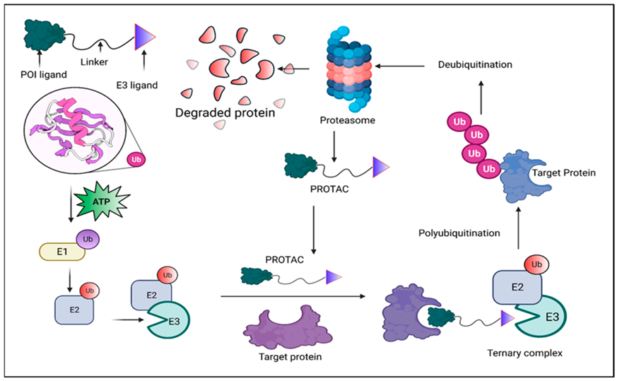
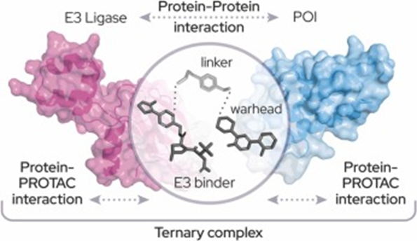
A PROTAC is a heterobifunctional molecule that utilizes a POI ligand and an E3 ligase ligand connected by a linker [1]. The function is dictated by proximity-induced degradation. The PROTAC recruits an E3 ligase (part of the UPS cascade) to the POI, forming a ternary complex. This complex catalyzes the transfer of ubiquitin chains (specifically K48-linked polyubiquitin chains) onto the POI, marking it for irreversible destruction by the 26S proteasome. Crucially, the PROTAC is then released and catalytically recycled. This event-driven mechanism means that PROTACs achieve their functional outcome through the complete removal of the target protein, thereby eliminating all its associated functions, including non-enzymatic roles [26]. The stability and cooperativity of the ternary complex, often mediated by neocontacts, determine efficiency and can promote highly selective degradation even when using a broadly binding warhead.
Figure 1. Schematic Representation of the PROTAC- Proteolysis-targeting chimeras.
Mechanism of Action: Hijacking the Ubiquitin-Proteasome System (UPS)
Molecular Architecture and Components
A PROTAC is a hetero bifunctional molecule composed of three parts:
POI Ligand (Warhead): Binds selectively to the protein of interest (POI) and determines target specificity.
E3 Ligase Ligand (Recruiter): Engages an E3 ubiquitin ligase.
Linker: Connects the two ligands and positions the POI and E3 ligase in the correct orientation to enable ubiquitination [18].
PROTACs often fall into the beyond Rule-of-Five (bRo5) chemical space due to their structural complexity [1].
Importantly, the warhead does not need to bind to the active site of the POI; any binding site with sufficient affinity that allows formation of the POI-PROTAC-E3 ligase ternary complex is enough to induce ubiquitination and subsequent degradation.
The Degradation Cascade
PROTACs induce targeted protein degradation by redirecting the ubiquitinproteasome system (UPS) [1].
Ubiquitination Enzyme Cascade:
E1 activates ubiquitin using ATP.
E2 carries the activated ubiquitin.
E3 ligase transfers ubiquitin to the substrate; more than 600 E3 ligases exist in humans.
Ternary Complex Formation: The PROTAC brings the POI and E3 ligase into proximity, forming a stable POI-PROTAC-E3 ternary complex.
Ubiquitination: The E3 ligase ubiquitinates lysine residues on the POI. Successive additions generate a K48-linked polyubiquitin chain, marking the POI for degradation [22].
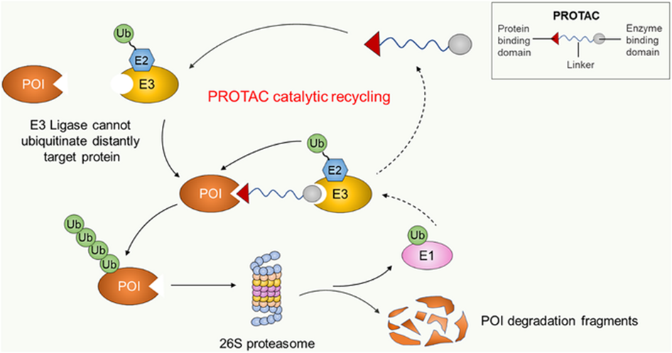
Proteasomal Degradation and Recycling: The 26S proteasome recognises the tagged POI and degrades it. The PROTAC is released intact and can repeatedly initiate new degradation cycles.
Figure 2. PROTAC Mode of Action: Ternary Complex Formation, Ubiquitination, and Catalytic Recycling.
Pharmacological Distinction
PROTACs function through an event-driven mechanism, unlike traditional small-molecule inhibitors (SMIs) that rely on occupancy-driven activity [28].
Table 1. Comparative Overview of Event-Driven PROTACs and Occupancy-Driven Small-Molecule Inhibitors.
Feature
PROTACs (Event-Driven)
Traditional SMIs (Occupancy-Driven)
Mode of Action
Catalytic; a single PROTAC can degrade multiple copies of the target protein.
Often effective at low or sub-stoichiometric concentrations due to catalytic turnover.
Requires higher systemic exposure to maintain target engagement.
Functional Outcome
Eliminates the entire protein, abolishing all associated functions.
Inhibits only the active/functional site while the protein remains intact.
Target Affinity
High affinity not essential; low-affinity warheads can work if a stable ternary complex forms.
High-affinity binding is typically required for sustained inhibition.
Selectivity
Increased selectivity from cooperative ternary complex formation.
Selectivity driven mainly by binary binding; higher off-target risk.
Resistance Potential
Can overcome mutation-driven resistance by degrading resistant variants.
Often ineffective against mutations that reduce inhibitor binding.
Pharmacodynamics
Sustained protein knockdown persists even after drug clearance.
Effect is directly dependent on drug presence and occupancy.
Duration of Effect
Long-lasting due to slow resynthesis of degraded proteins.
Shorter duration; activity declines as the drug is metabolized or dissociates.
Off-Target Effects
Potentially reduced due to requirement for productive ternary complex formation.
Higher likelihood of off-target inhibition from non-specific binding.
Because PROTAC activity depends on inducing ubiquitination rather than sustainedbinding, even transient or low-abundance ternary complexes can drive robust protein degradation.
Structural Basis of Function: Ternary Complex Dynamics and Neocontacts:
The stability and cooperativity of the ternary complex (TC) largely determine PROTAC efficiency, often outweighing the affinities of the individual ligands.
Structural Insights:
The first atomic-level PROTAC TC structure, MZ1, bound to VHL and the BRD4 bromo domain, showed that PROTACs induce new protein-protein and protein-ligand interactions at the interface.
These interfaces exhibit strong surface complementarity (e.g., ~688 Aº) and are stabilized by hydrophobic and electrostatic contacts.
Such “neocontacts” are critical for TC stability and overall degradation efficiency [10].
Cooperativity and Selectivity :
The binding of one protein (POI) can enhance PROTAC affinity for the E3 ligase, creating positive cooperativity. This promotes TC formation and allows highly selectivedegradation, even when using a broadly binding warhead.
Role of the Linker:
The linker controls the orientation, conformation, and dynamics of the TC.
Proper alignment ensures that key lysine residues on the POI are positioned toward the E2-ubiquitin catalytic site [18].
Linker length, rigidity, and topology directly influence ubiquitination efficiency and overall degradation potency.
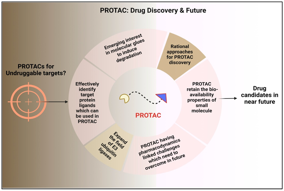
Advantages in Drug Discovery and Efficacy:
Targeting the “Undruggable” Proteome
A major advantage of PROTACs is their ability to dsegrade proteins traditionally considered “undruggable” significantly expanding the druggable proteome.
Figure 3. The Future of Drug Discovery with PROTAC Technology.
Limitations of Traditional SMIs:
SMIs depend on occupancy-driven pharmacology and require well-defined activepockets for high-affinity binding.
Approximately 70-80% of human proteins lack such pockets, making them inaccessible to conventional small molecules [2].
How PROTACs Overcome These Barriers:
PROTACs rely on proximity-induced degradation, not functionalsite inhibition.
They require only any binding site on the POI, not a catalytic pocket.
Even moderateor low-affinity ligands can be effective if they support ternary complex formation.
Degradation results in the complete removal of the protein and all associated functions.
Examples of Previously Undruggable Targets Now Accessible:
Transcription Factors: Proteins like MYC and STAT3 (e.g., SD-36 degrader) can now be selectively degraded [30].
Scaffolding/Regulatory Proteins: Non-enzymatic proteins such as FAK are now tractable targets [2].
Oncogenic Mutants: PROTACs have enabled new strategies against mutant oncoproteins, including KRASG12C[18].
Enhanced Selectivity and Specificity
PROTACs exhibit superior selectivity compared to traditional SMIs, an important advantage for reducing off-target effects typically associated with higher drug exposures.
Dual-Binding Mechanism and Selectivity:
Selectivity arises from the requirement to form a specific POI-PROTAC-E3 ternarycomplex, adding a layer of discrimination beyond warhead binding [21].
Both the POI ligand and E3 ligase ligand can be optimized to favor degradation of a single isoform or paralogue, even when ligand-binding sites are highly conserved.
This mechanism enables conversion of non-selective inhibitors into highly selective degraders.
Examples of Improved Selectivity and Reduced Toxicity:
MZ1 selectively degrades BRD4 over BRD2/BRD3 using the non-selective BET inhibitor JQ1 as the warhead [8].
A PROTAC derived from a pan-HDAC inhibitor selectively degraded HDAC6 [13].
DT2216 selectively degrades BCL-xL without causing thrombocytopenia by exploiting E3 ligases minimally expressed in platelets, unlike traditional inhibitors such as Navitoclax [10].
SD-36 selectively degrades STAT3, sparing closely related STAT1 and STAT4.
Overcoming Drug Resistance
Drug resistance, often due to protein over expression, compensatory pathways, or point mutations, limits the long-term efficacy of conventional inhibitors. PROTACs offer multiple mechanisms to overcome resistance.
Mechanisms for Circumventing Resistance:
Complete Protein Removal: Degrading the entire POI prevents resistance caused by target upregulation or non-enzymatic (scaffolding/regulatory) functions [12].
Mutation Tolerance: PROTACs only require transient binding to initiate degradation, making them less sensitive to point mutations that impair inhibitor occupancy [33].
Use of Non-Inhibitory Ligands: Even loss-of-function ligands or agonists can trigger degradation [12].
Lower Drug Pressure: Catalytic activity and low dosing reduce evolutionary selection pressure for resistant mutations [25].
Examples Demonstrating Resistance Overcome:
BTK Mutants: Degraders such as NX-2127 effectively degrade BTK variants, including the clinically important C481S mutation resistant to ibrutinib.
Hormone Receptors: AR degraders (ARV-110, ARCC-4) reduce AR levels even in mutation-driven resistant cancers, including cases where antagonists convert to agonists [15].
BCR-ABL Mutants: Dasatinib-based PROTACs such as SIAIS056 degrade multiple clinically relevant imatinib and dasatinib-resistant BCR-ABL1 mutants [12].
Viral Resistance: PROTACs have been developed against drug-resistant Hepatitis CVirus (HCV) strains, effectively degrading viral proteins despite mutational escape.
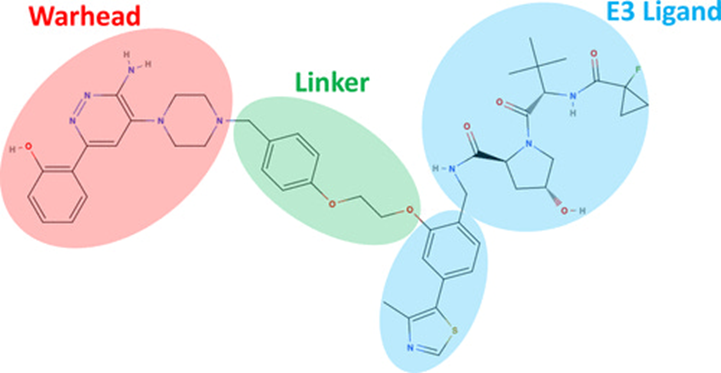
Molecular Components and Rational Design Strategies
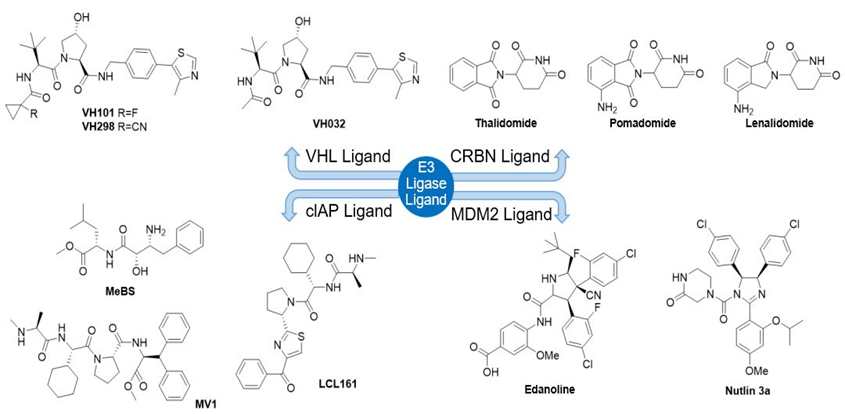
Figure 4. Chemical Architecture of a PROTAC Design Showing: Warhead (Target Binder), Linker and E3 Ligase (Ligand).
Expanding the E3 Ligase Toolbox
The design of PROTACs centers on three components: a POI-binding warhead, an E3 ligase ligand, and a linker that optimally orients both proteins to form a productive ternary complex. CRBN and VHL dominate current PROTAC design due to well characterized ligands and favorable drug-like properties, although expanding the E3 toolbox (e.g., DCAF15, KEAP1, RNF114, RNF43) is a key priority for improving tissue selectivity and overcoming resistance [1, 31].
Warhead affinity need not be high as long as it supports ternary complex formation; even non-inhibitory ligands can be repurposed for degradation. Linkers remain the most empirical design element, influencing orientation, cooperativity, permeability, and PK. PEG and alkyl linkers are common, while rigidified linkers can improve oral exposure. Computational tools including docking, MD, and AI-based linker generators now accelerate rational design by predicting geometries and cooperativity determinants.
Advanced PROTAC Modalities
Next-generation PROTAC modalities aim to improve tissue selectivity, reduce systemic toxicity, and provide external control over degradation activity. Prodrug or “pro-PROTAC” [4] strategies mask active moieties with cleavable groups, enabling activation only under disease-specific conditions, such as hypoxia, high ROS, or tumor-associated enzymes. Light-controlled systems include photocaged PROTACs, which become active upon UV/visible irradiation, and photo switchable degraders that toggle between active and inactive conformations.
Other innovations include radiation-activated RT-PROTACs for spatially restricted degradation during radiotherapy [26], CLIPTACs, which assemble intracellularly from two smaller fragments to bypass permeability limitations; and conjugate-based formats such as antibody-PROTAC conjugates and aptamer-PROTACs that provide cell-type-specific delivery. These modalities significantly expand the therapeutic window and allow greater control over degrader activity in vivo.
PROTAC Warhead Design and POI Ligands
The warhead determines POI recognition and contributes to degradation potency.
Key Principles of Warhead Selection:
High affinity is not essential and may even be counterproductive by stabilizing nonproductive binary complexes.
Warheads need not bind active sites any binding site enabling ternary complex formation is sufficient.
Warheads are interchangeable, allowing rapid expansion across disease targets (>40 proteins reported).
Ligands often originate from known inhibitors or structure-guided design.
Figure 5. E3 ligase ligands used in PROTAC Design.
Computational Support:
Traditionally based on synthesis and SAR screening.
Newer approaches include DNA encoded libraries (DELs) for high-throughput warhead discovery [1].
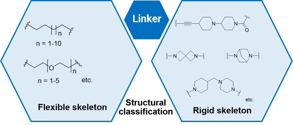
Linker Design Strategies: Geometry and “Linkerology”
The linker influences ternary complex formation, degradation efficiency, and physicochemical properties.
Common Linker Types:
No universal rules; design is largely empirical.
PEG (54%) and alkyl chains (31%) dominate due to tunable length, flexibility, and solubility benefits [25].
Figure 6. Structure of the linkers used in PROTAC Design.
Geometric and Functional Considerations:
Length: Too short leads to steric clash; too long leads to weak PPIs, both can reduce degradation.
Orientation: Must position POI and E3 ligase to facilitate productive PPIs and present accessible lysine residues for ubiquitination.
Attachment Site: Should be at solvent-exposed positions that preserve warhead and E3 ligand affinity.
Computational Acceleration:
Tools like docking, MD simulations, PPI modelling, and conformational sampling guide rational linker design.
AI/ML platforms (PROTAC-RL) use large datasets (e.g., PROTAC-DB, 6,000+ molecules) to generate optimized linker architectures with desired PK profiles [14].
Therapeutic Applications across Major Diseases
PROTACs in Cancer (Oncology)
PROTACs are transforming cancer therapy by selectively degrading oncogenic proteins, overcoming drug resistance, and expanding targeting possibilities beyond traditional inhibition.
Table 2. Selected PROTACs in Cancer Therapy and Their Clinical Status.
SMIs cannot target ~80% of human proteins due to the absence of ligandable pockets; PROTACs bypass this by requiring only transient binding to form a ternary complex [1].
Transcription factors: MYC, STAT3 (e.g., KT-333 in Phase I trials) [22].
Vif: PROTAC L15 degrades Vif and demonstrates antiviral activity [19].
Delivery: Exosome-mediated systems show potential for targeting latent HIV reservoirs.
Coronaviruses (e.g., SARS-CoV-2)
Mpro degraders: Compounds such as HP211206 and MPD2 degrade SARS-CoV-2 Mpro, including resistant variants [24].
Indomethacin-based PROTACs: Enhance antiviral potency via PGES-2 degradation and show broad coronavirus activity.
Targets include Mpro, PLpro, and RdRP[37].
Influenza and Other Viruses:
PROTACs offer novel antiviral strategies for influenza, human cytomegalovirus (HCMV), and other viral infections.
Influenza:
Oseltamivir-PROTACs degrade neuraminidase, effective against resistant strains.
HA degraders (oleanolic acid-based) show broad IAV activity.
Host-directed strategies (e.g., GSPT1 degradation) inhibit multiple viruses including IAV and SARS-CoV-2.
PROTAC-engineered live attenuated influenza virus demonstrates vaccine potential.
HCMV: THAL-SNS-032 shows enhanced anti-HCMV and dual antiviral effects [38].
Overcoming and Predicting Drug Resistance
Overcoming Resistance to Traditional SMIs
PROTACs address resistance that limits small-molecule inhibitors by degrading targets rather than merely inhibiting them.
Complete Protein Elimination: Removes enzymatic and non-enzymatic (scaffolding/ regulatory) functions, counteracting resistance from gain/loss of function mutations and target overexpression.
Mutation Tolerance : Event-driven action requires only transient binding; even lower-affinity ligands can drive degradation.
Key Examples:
AR: ARV-110 / ARCC-4 overcome CRPC resistance from AR overexpression, LBD mutations (e.g., F876L, T877A) and splice variants (e.g., AR-V7).
BTK: BTK degraders eliminate ibrutinib-resistant mutants such as C481S.
Pathway rewiring e.g., ER down regulation and HER/MAPK activation with ARV-471.
Hook effect at high concentrations (excess binary complexes reducing ternary complex formation) [18].
Mitigation Strategies:
Expanding and switching E3 ligases (e.g., from VHL to CRBN) for the same POI.
Genomic/proteomic profiling to identify resistance drivers and biomarkers.
Combination therapies, e.g., with efflux inhibitors or chemo (BRD4 degraders restoring doxorubicin sensitivity) [14].
Computational design/ML to optimize ternary complex stability and minimize resistance risk.
Neurodegenerative Diseases (NDDs)
Neurodegenerative diseases arise from toxic protein buildup. PROTACs can selectively degrade tau, α-synuclein, LRRK2, and mutant huntingtin, offering a promising approach for treating Alzheimer’s, Parkinson’s, and Huntington’s diseases
Promotes clearance of mHTT; potential disease modification
PROTACs are promising for NDDs by clearing aggregation-prone or dysregulated proteins often inaccessible to SMIs.
Alzheimer’s disease (AD):
Tau degraders: Peptide PROTACs (e.g., TH006, Keap1-based constructs) and small molecules (e.g., C004019, QC-01-175) lower tau levels and improve neuronal function in models[38].
GSK-3β degraders (e.g., PG-21): Reduce tau phosphorylation and Aβ production, protecting neurons.
HMG-CoA reductase (HMGCR): Statin-based PROTACs (e.g., P22A, VHL-lovastatin conjugates) degrade HMGCR, potentially avoiding statin-induced compensatory upregulation and achieving stronger cholesterol lowering in vivo [38].
Other targets:
PTP1B: Degradation may improve insulin sensitivity in diabetes/obesity.
Nuclear receptors and transcription factors (e.g., LXR-β, SREBP): Degraders modulate lipid and cholesterol homeostasis and show benefit in atherosclerosis models [18].
6.Applications in Immune and Inflammatory Disorders
PROTACs allow precise modulation of complex immune and inflammatory pathways that are difficult to fully control with classical inhibitors.
A.IRAK4 and Clinical Proof-of-Concept
IRAK4 integrates TLR and IL-1R signalling and has both kinase and scaffolding functions [36].
KT-474 (SAR444656): First IRAK4 degrader in Phase II trials for atopic dermatitis (AD) and hidradenitis suppurativa (HS); shows deep IRAK4 degradation, broad cytokine suppression, and clinical symptom improvement.
KT-413: Dual degrader of IRAK4 and IMiD substrates (Ikaros/Aiolos) for MYD88-mutant tumors [8].
Other molecules (e.g., PROTAC47) achieve >90% IRAK4 degradation in cells.
TNF-α and receptors and CD147 have been targeted with PROTACs in models of autoimmune and inflammatory disease.
B. Skin Diseases and Immuno-Inflammation
PROTACs (particularly peptide PROTACs) are being explored for eczema, dermatitis, psoriasis, and acne, owing to precise targeting and potential topical delivery.
KT-474 provides the leading clinical example for inflammatory skin disease [18].
C. Synergy with Immunotherapy in Cancer
PROTACs can reshape the tumor microenvironment (TME) by degrading immunosuppressive proteins (e.g., NAMPT in myeloid cells) and enhancing responses to checkpoint inhibitors.
Table 6. Key PROTAC Targets and Therapeutic Applications in Inflammatory, Metabolic, and Dermatological Disorders.
Expanding Scope: Metabolic Disorders and Dermatology
Metabolic Disorders
Building on the HMGCR and PTP1B examples:
PROTACs are being developed for diabetes, obesity, and dyslipidemia by degrading:
HMGCR (P22A and related degraders) to improve lipid control.
SREBP and nuclear receptors (e.g., LXR-β) to reprogram metabolic gene expression.
PTP1B to enhance insulin signalling.
Dermatology
Dermatology is a rapidly emerging area, especially for topical PROTACs with reduced systemic exposure.
Androgen-Driven Conditions (Alopecia, Acne)
GT20029: First topical AR-targeting PROTAC in clinical trials for androgenetic alopecia and acne; Phase I showed good safety, and Phase II in male AGA has met its primary endpoint.
Anti-ageing: Peptide PROTACs target proteins involved in collagen loss and matrix degradation, showing anti-wrinkle/skin-barrier benefits in models.
Inflammation: Topical and peptide PROTACs degrading inflammatory mediators (e.g., IRAK4, NF-κB/STAT-related targets) are being explored for eczema, dermatitis, and acne [18].
Dermal Delivery Strategies
Skin-penetrating peptides (SPPs) and other delivery enhancers are key to achieving sufficient skin and follicular penetration for peptide and large-molecule PROTACs.
Challenges, Clinical Status, and Future Directions of PROTACs
1. Physicochemical Properties and Bioavailability
PROTACs typically occupy beyondthe Rule-of-Five (bRo5) space, creating major developability challenges.
Key Issues
High MW (≈700-1,300 Da), high PSA and HBD countlead to low solubility, poor permeability, and limited oral bioavailability [38].
Many clinical PROTACs (e.g., ARV-110) require relatively high doses due to poor PK.
Improvement Strategies
Molecular optimisation: Reduce MW, HBDs, rotatable bonds; use rigidified and H-bondmasked linkers to lower PSA and improve permeability.
E3 choice: CRBN-based PROTACs often show more favourable oral profiles than VHL-based analogues.
Formulation approaches: NanoDDSs and amorphous solid dispersions (ASDs) increase solubility and exposure (e.g., ARCC-4) [7].
2. Pharmacological-Specific Challenges
On-target toxicity: Complete protein removal in normal tissues can cause more severe, irreversible effects than partial inhibition.
Off-target degradation (neosubstrates): Especially with early CRBN ligands, unintended substrates can be recruited and degraded.
Drug resistance: Often driven by genetic alterations in E3 ligase machinery (e.g., CRBN, CUL2) rather than POI mutations.
Limited E3 repertoire: Heavy reliance on CRBN/VHL restricts tissue- and context-specific targeting [8].
PK/PD complexity:Event-driven pharmacology requires new metrics (DC50, Dmax) rather than traditional occupancy-based parameters.
Clinical Status and Future Directions:
PROTAC technology has successfully transitioned from academia to the pharmaceutical industry, marking a paradigm shift in drug discovery.
Current landscape
>50 PROTACs in clinical development, largely for oncology.
Leading examples: ARV-110 (AR, prostate cancer), ARV-471 / vepdegestrant (ER, breast cancer; advanced to Phase III and NDA stage), with additional candidates such as KT-333 (STAT3) and multiple BTK degraders [13].
Future priorities
Expanding E3 ligase space beyond CRBN/VHL to increase target diversity and tissue selectivity.
Advanced delivery: Lipid nanoparticles, antibody-PROTAC conjugates, and prodrug strategies for improved oral or targeted delivery.
CNS targeting: Design of brain-penetrant PROTACs via MW/PSA reduction, transporter hijacking, and nanocarriers.
Next-generation designs: Reversible PROTACs, multi-target degraders, and AI/ML-guided design to optimize ternary complex formation and PK/PD [18].
The Hook Effect
Concept: At high concentrations, PROTACs favor non-productive binary complexes (PROTAC–POI or PROTAC–E3) over the ternary complex, leading to reduced degradation at higher doses [1].
Relevance: Intrinsic to bifunctional design; often used as a mechanistic signature of true PROTAC activity.
Mitigation
Enhance positive cooperativity and formation of stabilizing neocontacts in the ternary complex.
Carefully map concentration–response curves to define a non-hook therapeutic window.
Structural strategies (e.g., macrocyclization) and nanodelivery/split-mix nano-PROTACs to modulate local exposure [26].
Covalent and Reversible PROTACs
Non-covalent PROTACs: Standard format; maintain full catalytic, event-driven degradation and recyclability.
Irreversible covalent PROTACs (IC):
POI-covalent: Often sacrifice catalytic turnover and may impair ubiquitin transfer; some fail to induce degradation.
E3-covalent: Can retain catalytic degradation of multiple POI molecules.
Reversible covalent (RC) PROTACs:
Use reversible covalent motifs (e.g., cyanoacrylamides for BTK).
Combine strong, selective engagement with preserved catalytic cycling.
Can improve intracellular accumulation and target residence time.
Advanced Molecular Strategies for Controllability (Prodrugs)
“Smart” or pro-PROTACs aim to introduce spatiotemporal control and reduce systemic toxicity.
Light-controlled PROTACs
Photocaged PROTACs: Inactive until UV/visible light removes a photolabile group on the POI or E3 ligand site.
Photoswitchable PROTACs (PHOTACs): Reversibly toggled between active/inactive states via isomerization (e.g., azobenzene).
RT-PROTACs: X-ray–triggered release for deep-tissue, radiotherapy-linked degradation [38].
Tumor microenvironment-responsive systems
Hypoxia-activated PROTACs (HAP-TACs): Use bioreductive triggers (e.g., indolequinone) to unmask active PROTACs in hypoxic tumors.
ROS-activated PROTACs: Triggered by elevated reactive oxygen species.
Split the PROTAC into two smaller, permeable fragments that undergo bioorthogonal click chemistry in cells, bypassing poor permeability associated with fully assembled, high-MW PROTACs.
Emerging Modalities and Conjugate Technologies
The development of clinical-grade PROTACs has driven the evolution of peptide-based degraders, biomacromolecule PROTAC conjugates, and nano-PROTACs, all aimed at improving targetability, PK, and safety.
Peptide-Based PROTACs: Design, Advantages, and Scope
Peptide-based PROTACs were the first PROTACs (e.g., Protac-1 recruiting SCFβ-TRCP to degrade MetAP-2) and use peptidic ligands that mimic natural protein–protein interaction motifs.
Key features and advantages
Use peptide POI ligands, E3-binding peptides, linkers, and often cell-penetrating sequences.
Offer high selectivity, biocompatibility, and low toxicity, with good suitability for “undruggable” PPI targets (e.g., transcription factors).
Allow multitarget and modular design by concatenating different peptide segments.
Therapeutic examples
Oncology: Peptide PROTACs against p300 and STAT3; cyclic peptide PROTACs reducing PD-L1 levels [19].
Neurodegeneration: KEAP1-recruiting peptide PROTACs that degrade tau in Alzheimer’s models.
Hematologic/skin disease and membrane proteins are promising areas due to peptide compatibility.
Limitations and solutions
Main issues: high MW and poor permeability, historically requiring microinjection.
New approaches: tumor-penetrating peptides (e.g., iRGD) and nanodelivery systems to improve uptake.
Biomacromolecule-PROTAC Conjugates (AOCs/APCs)
Antibody-PROTAC Conjugates (AOCs/DACs)
AOCs (DACs) couple PROTACs to monoclonal antibodies, paralleling ADCs.
Mechanism: Antibody binds a cell-surface antigen causes endocytosis leading to intracellular release of the PROTAC for target degradation[23].
Advantages: High cell/tissue specificity, reduced off-target exposure, and antibody-like PK (extended half-life).
Examples:
Trastuzumab–PROTAC constructs degrading BRD4 selectively in HER2? cells.
ROR1-directed DACs.
AbTACs recruiting RNF43 to degrade PD-L1 at the cell surface.
Challenges: Very large, complex conjugates with difficult synthesis, formulation, and potential immunogenicity.
AptamerPROTAC Conjugates (APCs)
APCs use nucleic acid aptamers as targeting ligands.
Mechanism: Aptamer binds a specific receptor (e.g., HER2, nucleolin) and mediates receptor-driven uptake of the PROTAC [37].
Advantages: High specificity, low immunogenicity, good tissue penetration; enable tumor-selective degradation.
Examples:
HER2-targeting APCs for selective degradation in HER2? tumors.
Aptamer-PROTAC ZL216, showing good serum stability, solubility, and potent nucleolin degradation.
Nano-Drug Delivery Systems (Nano-PROTACs)
NanoDDS-based PROTACs tackle the core liabilities of PROTACs are high MW, polarity, low solubility, and poor bioavailabilityand help localize activity.
General advantages
Improve solubility, stability, circulation time, and cellular uptake.
Enable passive (EPR) and active targeting, reducing systemic off-target effects and helping modulate the hook effect via controlled local exposure.
Key strategies
Physical encapsulation: Encapsulation in liposomes or polymeric nanoparticles to enhance stability and enable co-delivery with other drugs.
Chemical conjugation: Covalent linkage of PROTACs to nanocarriers with stimuli-responsive linkers (pH, enzymes, redox, radiation) for TME-triggered release.
Carrier-free self-assembly: PROTACs co-assemble with other hydrophobic/π–π stacking molecules (e.g., photosensitizers), achieving very high drug loading and avoiding carrier toxicity.
“Split-and-mix” / CLIPTAC-like platforms: Deliver two smaller ligands (POI and E3 ligands) separately and assemble the active PROTAC in situ, improving permeability and reducing hook-effect–driven self-inhibition [26].
Representative nanoplatforms
Polymeric nanoparticles (PNPs): Stimuli-responsive systems (e.g., PSRNs) delivering CDK4/6 PROTACs that release cargo in acidic, cathepsin B-rich tumor sites.
Lipid-based nanoparticles (LBNPs): Widely used to enhance PROTAC exposure and tumor accumulation; also explored for delivering bioPROTACs.
Inorganic nanoparticles (INPs): Gold or silica NPs for theranostics; e.g., UCNP–MSN systems enabling NIR-triggered release of photocaged BRD4 PROTACs.
Exosome-based carriers: Natural vesicles with excellent biocompatibility and barrier penetration, promising for precise PROTAC delivery (including to hard-to-reach sites).
Clinical Translation, Technology, and Future Directions
PROTACs have rapidly evolved from a conceptual degradation technology into a clinically validated therapeutic modality. Their successespecially in oncologyhas been enabled by improvements in molecular design, computational modeling, and diversification of targeted protein degradation (TPD) systems beyond the proteasome.
Clinical Landscape and Regulatory Challenges
Clinical Status
Since the first PROTAC entered clinical trials in 2019, the pipeline has grown to 30–50 candidates. Most clinically advanced degraders target hormone-driven cancers.
ARV-471 (ER degrader): Completed Phase III, submitted the first-ever PROTAC NDA to the US FDA.
ARV-110 (AR degrader): In Phase II/III for mCRPC [8].
Physicochemical limitations: Large MW (700-1300 Da)[18], high polarity, poor permeability and oral bioavailability (e.g., ARV-110 ~23% in rats).
Safety risks: Complete protein removal increases risk of irreversible toxicity and off-target/neosubstrate degradation.
Regulatory uncertainty: New evaluation paradigms are required, including degradation biomarkers, and quantitative degradation assays.
Computational and AI-Assisted PROTAC Design
Designing optimal linkers and ternary complexes remains labor-intensive. Computational tools now drive PROTAC design by predicting linker geometry, ternary complex formation, and degradation efficiency.
Figure 7. Computer aided drug design in the development of PROTAC.
Key tools and approaches:
Ternary complex modeling: Using PRosettaC, PROTAC-Model, RosettaDock, RDKit.
AI/ML integration:
AlphaFold for high-fidelity 3D protein structures and modeling when crystal structures are unavailable.
DeepPROTACs for degradation prediction.
AIMLinker, ShapeLinker for generative linker design.
These tools significantly accelerate rational design and reduce trial-and-error synthesis [14].
PROTAC Databases and Resources
Bioinformatic resources support rational PROTAC design by providing access to chemical structures, target information, linkers, and biological activity, enabling systematic optimization and prediction of protein degradation efficiency [42].
Table 7. Key Databases and Resources for PROTAC Design and Research.
Database / Resource
URL
Description / Use
PROTAC-DB
https://cadd.zju.edu.cn/protacdb
Comprehensive PROTAC database: structures, targets, E3 ligases, biological activity. Useful for design and SAR studies.
PROTACpedia
https://www.protacpedia.com
Curated PROTAC data: degradation efficiency, cell lines, linkers. Ideal for comparing designs and selecting ligands/linkers.
UbiBrowser
http://ubibrowser.ncpsb.org
Predicts E3 ligase–substrate interactions. Useful for PROTAC target design.
Pharos
https://pharos.nih.gov
Target protein info including druggability. Helps select druggable targets for PROTACs.
Methodological Advances in PROTAC Profiling
Accurate assessment of degradation kinetics and specificity is essential for clinical optimization.
Key innovations:
Live-cell kinetic profiling: CRISPR-tagging and bioluminescence assays (e.g., Promega’s platform) to monitor real-time degradation.
Structural/mechanistic profiling:
Native mass spectrometry for predicting ternary complex stability.
SPR for ternary complex binding/dissociation kinetics.
PK/PD modeling: Techniques like SILAC/pulsed-SILAC quantify protein turnover, guiding dose selection.
ProtacID: Global proteomics to identify all proteins affected by a degrader, including unexpected neosubstrates across multiple E3 ligases [39].
Expansion Beyond the Proteasome: Next-Generation TPD Modalities
Classic PROTACs function via the ubiquitin-proteasome system (UPS), which limits them to intracellular proteins. New TPD platforms extend degradation to extracellular,membrane,and cytosolic targets.
LYTACs (Lysosome-Targeting Chimeras)
Target Scope: Extracellular and membrane proteins (e.g., receptor tyrosine kinases [RTKs], G protein-coupled receptors [GPCRs]).
Mechanism: Recruit lysosome-targeting receptors such as CI-MPR or ASGPR to shuttle proteins to lysosomes for degradation.
Status: No clinical candidates yet; strong potential as a complement to PROTACs.
2. AUTACs (Autophagy-Targeting Chimeras)
Target Scope: Cytosolic proteins, protein aggregates, and organelles.
Mechanism: Use autophagy-mediated degradation by tagging targets for autophagic clearance.
Applications: Particularly relevant to neurodegenerative diseases and clearance of aggregated proteins.
Limitation: Autophagy is a ubiquitous pathway, increasing potential off-target effects.
3. ATTECs (Autophagosome-Tethering Compounds)
Target Scope: Cytosolic proteins and aggregates.
Mechanism: Directly tether targets to autophagosomes, bypassing ubiquitination, for lysosomal degradation.
Advantage: Can degrade proteins or structures not efficiently handled by the UPS.
Relation: Mechanistically closer to LYTACs than classical PROTACs[43].
CONCLUSION
PROTACs have rapidly evolved from a conceptual protein degradation strategy to a clinically validated therapeutic modality, with more than 50 candidates in trials and the first NDA submission for ARV-471. Their catalytic mechanism, ability to eliminate rather than inhibit proteins, and capacity to target previously “undruggable” proteins highlight their strong therapeutic potential. PROTACs also show promise in overcoming mutation-driven resistance, such as the degradation of TKI-resistant BCR-ABL1 variants.
Despite this progress, key challenges including high molecular weight, limited oral bioavailability, the hook effect, and reliance on CRBN and VHL still hinder broad clinical translation. Future advancement will depend on AI/ML-guided molecular design, expansion of the E3 ligase repertoire, and improved delivery platforms such as Nano-PROTACs, AOCs, and pro-PROTACs. Additionally, emerging technologies like LYTACs and AUTACs extend targeted protein degradation beyond the proteasome. Together, these innovations are expected to strengthen the clinical potential of PROTACs and establish them as a major next-generation therapeutic approach.
REFERENCES
Liu. Z, Hu. M, Yang. Y, Du. C, Zhou. H, Liu. C, Chen. Y, Fan. L, Ma. H, Gong. Y, Xie Y., An overview of PROTACs: a promising drug discovery paradigm. Molecular biomedicine, 2022, 3 (1), 46, https://doi.org/10.1186/s43556-022-00112-0.
Vishal. M, Mukund. J, Iqra. Z, M. Mahajan, S. Nazar, S. Ali, A. Ilyas, S. Tanweer, J. Ali, O. Alam., Design and development of PROTACs: A new paradigm in anticancer drug discovery, Medicine in Drug Discovery,2025,27,100221, https://doi.org/10.1016/j.medidd.2025.100221.
Rutherford, K. A, M. Manus, K. J., PROTACs: Current and Future Potential as a Precision Medicine Strategy to Combat Cancer, Molecular cancer therapeutics, 2024, 23 (4), 454-463. https://doi.org/10.1158/1535-7163.MCT-23-0747.
Hakem. F, Abdelwaly. A, Alshaman R, Alattar. A, Alanazi. F, E. Zaitone. S. A, Helal. M. A., Recent Advances in the Development of Pro-PROTAC for Selective Protein Degradation, Pharmaceutics, 2025, 17 (9), 1160, https://doi.org/10.3390/pharmaceutics17091160.
Njoka. M, Kamath. D, Bossmann. S., Anti-apoptotic Proteolysis Targeted Chimeras (PROTACs) in Cancer Therapy, Medical Research Archives, 2025 13 (1), https://doi.org /10.18103/mra.v13i1.6284.
Li. X, Pu, W. Zheng, et al., Proteolysis-targeting chimeras (PROTACs) in cancer therapy, Mol Cancer , 2022, 21, 99, https://doi.org/10.1186/s12943-021-01434-3.
Lihua Liu, Xian Guan and Jiezhen Zhuo et al., PROTACs improve selectivity for targeted proteins, Acta Materia Medica, 2025, 4 (3): 390-412, https://doi.org/10.15212/AMM-2025-0015.
Vetma.V, O’Connor. S, Ciulli. A., Development of PROTAC degrader drugs for cancer, Annual Review of Cancer Biology, 2024, 9 (1), 119-140. https://doi.org/10.1146/annurev-cancerbio-061824-105806.
Casan. J, M. L, Seymour, J. F., Degraders upgraded: the rise of PROTACs in hematological malignancies, Blood, 2024, 143 (13), 1218-1230, https://doi.org/10.1182/blood.2023022993.
Bond. M. J, Crews. C. M., Proteolysis targeting chimeras (PROTACs) come of age: entering the third decade of targeted protein degradation, RSC Chemical Biology, 2021, 2 (3), 725-742, https://doi.org/10.1039/d1cb00011j.
Wu. K. Y, Hung. T. I, Chang. C. A., PROTAC-induced protein structural dynamics in targeted protein degradation, eLife, 2025, 13, https://doi.org/10.7554/elife.101127.3.
Burke. M. R, Smith. A. R, Zheng. G., Overcoming Cancer Drug Resistance Utilizing PROTAC Technology, Frontiers in cell and developmental biology, 2022, 10, 872729. https://doi.org/10.3389/fcell.2022.872729.
Cecchini. C, Pannilunghi. S, Tardy. S, Scapozza. L., From Conception to Development: Investigating PROTACs Features for Improved Cell Permeability and Successful Protein Degradation, Frontiers in chemistry, 2021, 9, 672267. https://doi.org/10.3389/fchem.2021.672267.
Ibrahim. S, Umer Khan. M, Khurram. I, Rehman. R, Rauf. A, Ahmad. Z, Aljohani. A. S. M, Al Abdulmonem. W, Quradha, M. M., Navigating PROTACs in Cancer Therapy: Advancements, Challenges, and Future Horizons, Food science & nutrition, 2025, 13(2), e70011, https://doi.org/10.1002/fsn3.70011.
Qi. S. M, Dong. J, Xu. Z. Y, Cheng. X. D, Zhang. W. D, Qin. J. J., PROTAC: An Effective Targeted Protein Degradation Strategy for Cancer Therapy,Frontiers in pharmacology, 2021, 12, 692574, https://doi.org/10.3389/fphar.2021.692574.
Weng. G, Cai. X, Cao. D, Du. H, Shen. C, Deng. Y, He. Q, Yang. B, Li. D, Hou.T., PROTAC-DB 2.0: an updated database of PROTACs, Nucleic acids research, 2023, 51 (D1), D1367-D1372, https://doi.org/10.1093/nar/gkac946.
Tamatam. R, Shin. D., Emerging Strategies in Proteolysis-Targeting Chimeras (PROTACs): Highlights from 2022, International journal of molecular sciences, 2023, 24 (6), 5190. https://doi.org/10.3390/ijms24065190.
Fan. G, Chen. S, Zhang. Q, Yu. N, Shen. Z, Liu. Z, Guo. W, Tang. Z, Yang. J, Liu. M., Proteolysis-Targeting Chimera (PROTAC): Current Applications and Future Directions, Med Comm, 2025, 6 (10), e70401, https://doi.org/10.1002/mco2.70401.
Zhu.Y, Dai. Y, Tian. Y., The Peptide PROTAC Modality: A New Strategy for Drug Discovery, Med Comm, 2025, 6 (4), e70133, https://doi.org/10.1002/mco2.70133.
Graham. H., The mechanism of action and clinical value of PROTACs: A graphical review. Cellular Signalling, 2022, 99, 110446, https://doi.org/10.1016/j.cellsig.2022.110446
Li. R, Liu. M, Yang. Z, Li. J, Gao. Y, Tan. R., Proteolysis-Targeting Chimeras (PROTACs) in Cancer therapy: Present and future, In Molecules, 2022, https://doi.org/10.3390/molecules27248828.
Sincere. N. I, Anand. K, Ashique. S, Yang. J, You. C., PROTACs: Emerging Targeted Protein Degradation Approaches for Advanced Druggable Strategies, Molecules (Basel, Switzerland), 2023, 28 (10), 4014, https://doi.org/10.3390/molecules28104014.
Mu. X, Mu. C, Wang. M, Meng. C, Liu. H, Lu, H., Nanotechnology Advances Proteolysis Targeting Chimeras (PROTACs): Transition From Basic Research to Clinical Application, International journal of nanomedicine, 2025, 20, 12177-12198. https://doi.org/10.2147/IJN.S552644.
Khan. S, He. Y, Zhang. X, Yuan. Y, Pu. S, Kong. Q, Zheng. G, Zhou. D., PROteolysis TArgeting Chimeras (PROTACs) as emerging anticancer therapeutics, Oncogene, 2020, 39 (26), 4909-4924, https://doi.org/10.1038/s41388-020-1336-y.
Wu. X, Shu. Y, Zheng. Y, Zhang. P, Cong. H, Zou. Y, Cai. H, Zha. Z.,Recent Advances in Nanomedicine: Cutting-Edge Research on Nano-PROTAC Delivery Systems for Cancer Therapy, Pharmaceutics, 2025, 17(8), 1037. https://doi.org/10.3390/pharmaceutics17081037.
Alabi. S. B, Crews. C. M. Major advances in targeted protein degradation: PROTACs, LYTACs, and MADTACs, The Journal of biological chemistry, 2021, 296, 100647. https://doi.org/10.1016/j.jbc.2021.100647.
Nalawansha. D. A, Crews. C. M., PROTACs: An Emerging Therapeutic Modality in Precision Medicine, Cell chemical biology, 2020, 27 (8), 998-1014. https://doi.org/10.1016/j.chembiol.2020.07.020.
Gu. S, Cui. D, Chen. X, Xiong. X, Zhao. Y., PROTACs: An Emerging Targeting Technique for Protein Degradation in Drug Discovery, BioEssays: news and reviews in molecular, cellular and developmental biology, 2018, 40 (4), e1700247. https://doi.org/10.1002/bies.201700247.
Wang. C, Zhang. Y, Zhang. T, Shi. L, Geng. Z, Xing. D., Proteolysis-targeting chimaeras (PROTACs) as pharmacological tools and therapeutic agents: advances and future challenges, Journal of enzyme inhibition and medicinal chemistry, 2022, 37(1), 1667-1693. https://doi.org/10.1080/14756366.2022.2076675.
Yao. T, Xiao. H, Wang. H, Xu. X., Recent Advances in PROTACs for Drug Targeted Protein Research, International journal of molecular sciences, 2022, 23 (18), 10328. https://doi.org/10.3390/ijms231810328.
Hamilton. E. P, Jeselsohn. R. M, Vahdat. L. T, Hurvitz. S. A., PROteolysis TArgeting Chimera (PROTAC) estrogen receptor degraders for treatment of Estrogen Receptor-Positive Advanced Breast Cancer, Targeted Oncology, 2025, 20 (3), 431-444. https://doi.org/10.1007/s11523-025-01137-5.
Wang. C, Zhang. Y, Chen. W, Wu. Y, Xing. D., New-generation advanced PROTACs as potential therapeutic agents in cancer therapy, Molecular cancer, 2024, 23 (1), 110. https://doi.org/10.1186/s12943-024-02024-9.
Ocaña. A, Pandiella. A., Proteolysis targeting chimeras (PROTACs) in cancer therapy, Journal of experimental & clinical cancer research: CR, 2020, 39 (1), 189. https://doi.org/10.1186/s13046-020-01672-1.
Zhao. J, Chen. H, Liang. C., Dual functionality of MDM2 in PROTACs expands the horizons of targeted protein degradation, Biomarker research, 2025, 13 (1), 111. https://doi.org/10.1186/s40364-025-00826-7.
Sun. X, Gao. H, Yang. Y, He. M, Wu. Y, Song. Y, Tong. Y, Rao. Y., PROTACs: great opportunities for academia and industry, Signal transduction and targeted therapy, 2019, 4, 64, https://doi.org/10.1038/s41392-019-0101-6.
Zhao. L, Zhao. J, Zhong. K, Tong. A, Jia. D., Targeted protein degradation: mechanisms, strategies and application. Signal transduction and targeted therapy, 2022, 7 (1), 113. https://doi.org/10.1038/s41392-022-00966-4.
Zhong. G, Chang. X, Xie. W, Zhou. X., Targeted protein degradation: advances in drug discovery and clinical practice, Signal transduction and targeted therapy, 2024, 9 (1), 308. https://doi.org/10.1038/s41392-024-02004-x.
Shrestha. S, Maitland. M. E. R, Jing. L, Duan. S, Nie. D. Y, St-Germain. J, Kanaris. M, Barsyte-Lovejoy. D, Arrowsmith. C. H, Raught. B., Characterization of PROTAC specificity and endogenous protein interactomes using ProtacID, Nature communications, 2025, 16 (1), 8089, https://doi.org/10.1038/s41467-025-63357-7.
Liao. X, Guo. M, Hu. D, Su. C, Liao. H., Study on the design, synthesis and activity of MDM2/MDMX anti-tumor stapled peptide PROTAC, Scientific reports, 2025, 15 (1), 32923. https://doi.org/10.1038/s41598-025-18026-6.
Sakamoto. K. M, Kim. K. B, Kumagai. A, Mercurio. F, Crews. C. M, Deshaies. R. J., Protacs: chimeric molecules that target proteins to the Skp1-Cullin-F box complex for ubiquitination and degradation, Proceedings of the National Academy of Sciences of the United States of America, 2001, 98 (15), 8554-8559, https://doi.org/10.1073/pnas.141230798.
Sun. X, Gao. H, Yang. Y, He. M, Wu. Y, Song. Y, Tong. Y, Rao. Y., “PROTACs: Great Opportunities for Academia and Industry.” Signal Transduction and Targeted Therapy, 2023, 8:1, https://doi.org/10.1038/s41392-022-01285-6.
Lin. Z, Chen. S, Zhang. Y, Liu. J, Li. S, Zhang. X, Zhang. L, Chen. Y., “Beyond PROTAC: Emerging Targeted Protein Degradation Strategies.” Chemical Society Reviews, 2023, 52, 1125–1168. https://doi.org/10.1039/D2CS00797E.
Reference
Liu. Z, Hu. M, Yang. Y, Du. C, Zhou. H, Liu. C, Chen. Y, Fan. L, Ma. H, Gong. Y, Xie Y., An overview of PROTACs: a promising drug discovery paradigm. Molecular biomedicine, 2022, 3 (1), 46, https://doi.org/10.1186/s43556-022-00112-0.
Vishal. M, Mukund. J, Iqra. Z, M. Mahajan, S. Nazar, S. Ali, A. Ilyas, S. Tanweer, J. Ali, O. Alam., Design and development of PROTACs: A new paradigm in anticancer drug discovery, Medicine in Drug Discovery,2025,27,100221, https://doi.org/10.1016/j.medidd.2025.100221.
Rutherford, K. A, M. Manus, K. J., PROTACs: Current and Future Potential as a Precision Medicine Strategy to Combat Cancer, Molecular cancer therapeutics, 2024, 23 (4), 454-463. https://doi.org/10.1158/1535-7163.MCT-23-0747.
Hakem. F, Abdelwaly. A, Alshaman R, Alattar. A, Alanazi. F, E. Zaitone. S. A, Helal. M. A., Recent Advances in the Development of Pro-PROTAC for Selective Protein Degradation, Pharmaceutics, 2025, 17 (9), 1160, https://doi.org/10.3390/pharmaceutics17091160.
Njoka. M, Kamath. D, Bossmann. S., Anti-apoptotic Proteolysis Targeted Chimeras (PROTACs) in Cancer Therapy, Medical Research Archives, 2025 13 (1), https://doi.org /10.18103/mra.v13i1.6284.
Li. X, Pu, W. Zheng, et al., Proteolysis-targeting chimeras (PROTACs) in cancer therapy, Mol Cancer , 2022, 21, 99, https://doi.org/10.1186/s12943-021-01434-3.
Lihua Liu, Xian Guan and Jiezhen Zhuo et al., PROTACs improve selectivity for targeted proteins, Acta Materia Medica, 2025, 4 (3): 390-412, https://doi.org/10.15212/AMM-2025-0015.
Vetma.V, O’Connor. S, Ciulli. A., Development of PROTAC degrader drugs for cancer, Annual Review of Cancer Biology, 2024, 9 (1), 119-140. https://doi.org/10.1146/annurev-cancerbio-061824-105806.
Casan. J, M. L, Seymour, J. F., Degraders upgraded: the rise of PROTACs in hematological malignancies, Blood, 2024, 143 (13), 1218-1230, https://doi.org/10.1182/blood.2023022993.
Bond. M. J, Crews. C. M., Proteolysis targeting chimeras (PROTACs) come of age: entering the third decade of targeted protein degradation, RSC Chemical Biology, 2021, 2 (3), 725-742, https://doi.org/10.1039/d1cb00011j.
Wu. K. Y, Hung. T. I, Chang. C. A., PROTAC-induced protein structural dynamics in targeted protein degradation, eLife, 2025, 13, https://doi.org/10.7554/elife.101127.3.
Burke. M. R, Smith. A. R, Zheng. G., Overcoming Cancer Drug Resistance Utilizing PROTAC Technology, Frontiers in cell and developmental biology, 2022, 10, 872729. https://doi.org/10.3389/fcell.2022.872729.
Cecchini. C, Pannilunghi. S, Tardy. S, Scapozza. L., From Conception to Development: Investigating PROTACs Features for Improved Cell Permeability and Successful Protein Degradation, Frontiers in chemistry, 2021, 9, 672267. https://doi.org/10.3389/fchem.2021.672267.
Ibrahim. S, Umer Khan. M, Khurram. I, Rehman. R, Rauf. A, Ahmad. Z, Aljohani. A. S. M, Al Abdulmonem. W, Quradha, M. M., Navigating PROTACs in Cancer Therapy: Advancements, Challenges, and Future Horizons, Food science & nutrition, 2025, 13(2), e70011, https://doi.org/10.1002/fsn3.70011.
Qi. S. M, Dong. J, Xu. Z. Y, Cheng. X. D, Zhang. W. D, Qin. J. J., PROTAC: An Effective Targeted Protein Degradation Strategy for Cancer Therapy,Frontiers in pharmacology, 2021, 12, 692574, https://doi.org/10.3389/fphar.2021.692574.
Weng. G, Cai. X, Cao. D, Du. H, Shen. C, Deng. Y, He. Q, Yang. B, Li. D, Hou.T., PROTAC-DB 2.0: an updated database of PROTACs, Nucleic acids research, 2023, 51 (D1), D1367-D1372, https://doi.org/10.1093/nar/gkac946.
Tamatam. R, Shin. D., Emerging Strategies in Proteolysis-Targeting Chimeras (PROTACs): Highlights from 2022, International journal of molecular sciences, 2023, 24 (6), 5190. https://doi.org/10.3390/ijms24065190.
Fan. G, Chen. S, Zhang. Q, Yu. N, Shen. Z, Liu. Z, Guo. W, Tang. Z, Yang. J, Liu. M., Proteolysis-Targeting Chimera (PROTAC): Current Applications and Future Directions, Med Comm, 2025, 6 (10), e70401, https://doi.org/10.1002/mco2.70401.
Zhu.Y, Dai. Y, Tian. Y., The Peptide PROTAC Modality: A New Strategy for Drug Discovery, Med Comm, 2025, 6 (4), e70133, https://doi.org/10.1002/mco2.70133.
Graham. H., The mechanism of action and clinical value of PROTACs: A graphical review. Cellular Signalling, 2022, 99, 110446, https://doi.org/10.1016/j.cellsig.2022.110446
Li. R, Liu. M, Yang. Z, Li. J, Gao. Y, Tan. R., Proteolysis-Targeting Chimeras (PROTACs) in Cancer therapy: Present and future, In Molecules, 2022, https://doi.org/10.3390/molecules27248828.
Sincere. N. I, Anand. K, Ashique. S, Yang. J, You. C., PROTACs: Emerging Targeted Protein Degradation Approaches for Advanced Druggable Strategies, Molecules (Basel, Switzerland), 2023, 28 (10), 4014, https://doi.org/10.3390/molecules28104014.
Mu. X, Mu. C, Wang. M, Meng. C, Liu. H, Lu, H., Nanotechnology Advances Proteolysis Targeting Chimeras (PROTACs): Transition From Basic Research to Clinical Application, International journal of nanomedicine, 2025, 20, 12177-12198. https://doi.org/10.2147/IJN.S552644.
Khan. S, He. Y, Zhang. X, Yuan. Y, Pu. S, Kong. Q, Zheng. G, Zhou. D., PROteolysis TArgeting Chimeras (PROTACs) as emerging anticancer therapeutics, Oncogene, 2020, 39 (26), 4909-4924, https://doi.org/10.1038/s41388-020-1336-y.
Wu. X, Shu. Y, Zheng. Y, Zhang. P, Cong. H, Zou. Y, Cai. H, Zha. Z.,Recent Advances in Nanomedicine: Cutting-Edge Research on Nano-PROTAC Delivery Systems for Cancer Therapy, Pharmaceutics, 2025, 17(8), 1037. https://doi.org/10.3390/pharmaceutics17081037.
Alabi. S. B, Crews. C. M. Major advances in targeted protein degradation: PROTACs, LYTACs, and MADTACs, The Journal of biological chemistry, 2021, 296, 100647. https://doi.org/10.1016/j.jbc.2021.100647.
Nalawansha. D. A, Crews. C. M., PROTACs: An Emerging Therapeutic Modality in Precision Medicine, Cell chemical biology, 2020, 27 (8), 998-1014. https://doi.org/10.1016/j.chembiol.2020.07.020.
Gu. S, Cui. D, Chen. X, Xiong. X, Zhao. Y., PROTACs: An Emerging Targeting Technique for Protein Degradation in Drug Discovery, BioEssays: news and reviews in molecular, cellular and developmental biology, 2018, 40 (4), e1700247. https://doi.org/10.1002/bies.201700247.
Wang. C, Zhang. Y, Zhang. T, Shi. L, Geng. Z, Xing. D., Proteolysis-targeting chimaeras (PROTACs) as pharmacological tools and therapeutic agents: advances and future challenges, Journal of enzyme inhibition and medicinal chemistry, 2022, 37(1), 1667-1693. https://doi.org/10.1080/14756366.2022.2076675.
Yao. T, Xiao. H, Wang. H, Xu. X., Recent Advances in PROTACs for Drug Targeted Protein Research, International journal of molecular sciences, 2022, 23 (18), 10328. https://doi.org/10.3390/ijms231810328.
Hamilton. E. P, Jeselsohn. R. M, Vahdat. L. T, Hurvitz. S. A., PROteolysis TArgeting Chimera (PROTAC) estrogen receptor degraders for treatment of Estrogen Receptor-Positive Advanced Breast Cancer, Targeted Oncology, 2025, 20 (3), 431-444. https://doi.org/10.1007/s11523-025-01137-5.
Wang. C, Zhang. Y, Chen. W, Wu. Y, Xing. D., New-generation advanced PROTACs as potential therapeutic agents in cancer therapy, Molecular cancer, 2024, 23 (1), 110. https://doi.org/10.1186/s12943-024-02024-9.
Ocaña. A, Pandiella. A., Proteolysis targeting chimeras (PROTACs) in cancer therapy, Journal of experimental & clinical cancer research: CR, 2020, 39 (1), 189. https://doi.org/10.1186/s13046-020-01672-1.
Zhao. J, Chen. H, Liang. C., Dual functionality of MDM2 in PROTACs expands the horizons of targeted protein degradation, Biomarker research, 2025, 13 (1), 111. https://doi.org/10.1186/s40364-025-00826-7.
Sun. X, Gao. H, Yang. Y, He. M, Wu. Y, Song. Y, Tong. Y, Rao. Y., PROTACs: great opportunities for academia and industry, Signal transduction and targeted therapy, 2019, 4, 64, https://doi.org/10.1038/s41392-019-0101-6.
Zhao. L, Zhao. J, Zhong. K, Tong. A, Jia. D., Targeted protein degradation: mechanisms, strategies and application. Signal transduction and targeted therapy, 2022, 7 (1), 113. https://doi.org/10.1038/s41392-022-00966-4.
Zhong. G, Chang. X, Xie. W, Zhou. X., Targeted protein degradation: advances in drug discovery and clinical practice, Signal transduction and targeted therapy, 2024, 9 (1), 308. https://doi.org/10.1038/s41392-024-02004-x.
Shrestha. S, Maitland. M. E. R, Jing. L, Duan. S, Nie. D. Y, St-Germain. J, Kanaris. M, Barsyte-Lovejoy. D, Arrowsmith. C. H, Raught. B., Characterization of PROTAC specificity and endogenous protein interactomes using ProtacID, Nature communications, 2025, 16 (1), 8089, https://doi.org/10.1038/s41467-025-63357-7.
Liao. X, Guo. M, Hu. D, Su. C, Liao. H., Study on the design, synthesis and activity of MDM2/MDMX anti-tumor stapled peptide PROTAC, Scientific reports, 2025, 15 (1), 32923. https://doi.org/10.1038/s41598-025-18026-6.
Sakamoto. K. M, Kim. K. B, Kumagai. A, Mercurio. F, Crews. C. M, Deshaies. R. J., Protacs: chimeric molecules that target proteins to the Skp1-Cullin-F box complex for ubiquitination and degradation, Proceedings of the National Academy of Sciences of the United States of America, 2001, 98 (15), 8554-8559, https://doi.org/10.1073/pnas.141230798.
Sun. X, Gao. H, Yang. Y, He. M, Wu. Y, Song. Y, Tong. Y, Rao. Y., “PROTACs: Great Opportunities for Academia and Industry.” Signal Transduction and Targeted Therapy, 2023, 8:1, https://doi.org/10.1038/s41392-022-01285-6.
Lin. Z, Chen. S, Zhang. Y, Liu. J, Li. S, Zhang. X, Zhang. L, Chen. Y., “Beyond PROTAC: Emerging Targeted Protein Degradation Strategies.” Chemical Society Reviews, 2023, 52, 1125–1168. https://doi.org/10.1039/D2CS00797E.
Anitha Sadula
Corresponding author
Department of Pharmacy, University College of Technology (A), Osmania University, Hyderabad, 500007, Telangana, India


 10.5281/zenodo.18470700