
We use cookies to ensure our website works properly and to personalise your experience. Cookies policy
Department of Pharmaceutical Sciences, Faculty of Technology, Bhimtal Campus, Kumaun University, Nainital, Uttarakhand, India 263136.
Kidney stone or nephrolithiasis, also known as urolithiasis, is a common condition brought on by the supersaturation of urine, nucleation, aggregation, crystal growth, and crystal retention, which leads to the production of stones in the urinary system. Genetics, dehydration, food habits, illnesses, and metabolic disorders like hypercalciuria are some of the variables that affect it. The illness tends to repeat often and is more common among males. The pathophysiology involves an imbalance between stone promoters (Low urine pH, Sodium, Oxalate, Calcium, Urate, Low urine volume) and inhibitors (Magnesium, Citrate, Osteopontin, Glycosaminoglycans) in the urine. Several theories, including Randall’s Plaque hypothesis, the Free Particle theory, and the Fixed Particle theory, explain how stones originate and grow. Diagnosis typically includes clinical evaluation, serum analysis, stone analysis, urine analysis, and imaging such as ultrasound and computed tomography (CT) scans. Management depends on stone size and location and may involve medication, dietary changes, or surgical procedures like ESWL or PCNL. Preventive strategies, especially increased fluid intake and metabolic assessment, are crucial for long-term management.
Urolithiasis
Urolithiasis is a term that originates from three Greek words: ‘ouron’ meaning urine, ‘oros’ meaning flow, and ‘lithos’ meaning stone. [1] It refers to the formation of solid concretions, which are composed of both crystalline materials and proteins within the lumen of the urinary tract, which is attached to the uro-epithelium. [2] Urolithiasis is a complex disease characterized by the formation and deposition of urinary stones at any location within the urinary tract. [3] It is regarded as one of the most prevalent urological disorders and has affected humans since ancient times. It is a long-standing medical condition and remains a common public health issue. [1] Urolithiasis is caused due to an imbalance between inhibitors and promoters of crystallization in urine.
Stone Inhibitors: Inorganic inhibitors- Magnesium, Citrate
Organic inhibitors- Osteopontin, Glycosaminoglycans, Prothrombin Fragment
Stones Promoters: Low urine pH, Calcium, Sodium, Oxalate, Urate, and low urine volume. [4]
Historical Background
Kidney stone disease has been recognized for centuries, as evidenced by various archaeological findings and historical writings about painful stone colic and attempts at stone removal. In ancient times, urolithiasis was often a devastating disease, frequently resulting in the patient’s death. In 1901, the English archaeologist E. Smith, while examinations of Egyptian mummies, discovered a 5,000-year-old bladder stone at a burial site of El Amrah, Egypt. Regimens for treating urinary tract diseases, including stones, were already documented in the Ebers Papyrus (1500 BC), which is a primary source of our knowledge of ancient Egyptian medicine. [5] Both soluble and insoluble bladder stones were known in ancient Mesopotamia. Various stone tablets contained recipes for treating different diseases, including the dissolution of soft kidney and bladder stones. [6] Five centuries BC, the Hippocratic collection first mentioned uroscopy. [7] Three centuries BC, Ammonius first suggested that crushing a bladder stone would facilitate its removal, for which he received his nickname “Lithotomus” = Stone cutter. [7,8] In his Sushruta-Samhita series of volumes from the sixth century BC, India's preeminent physician Sushruta first described the use of a splint to remove stones from the urethra. He also provided approximately three hundred surgical procedures for stone removal and reported on urolithiasis and its complications, such as infection, anuria, and uraemia, which often led to disastrous outcomes for patients with stones. [9] Another renowned Hindu author, Charaka, documented the instrumental removal of urethral stones in his Samhita. [10] Since the first century BC, ancient Greek medicine became accessible in the Roman Empire. One of the most renowned physicians of this era was Aulus Cornelius Celsus (circa 25 BC–circa 50 AD), who attempted to compile all medical knowledge into an encyclopaedia. [7] At the Necker Hospital in Paris, Jean Civiale (1792-1876) demonstrated a lithotryptic device for the first time on January 13, 1824, which enabled him to crush and subsequently remove a bladder stone through the urethra. Since then, there has been a revolution in the creation of increasingly advanced tools for stone removal and fragmentation, as well as endourological operations. [7,8]
Epidemiology
Kidney stones are among the oldest recorded human disorders and represent a significant health burden. Nowadays, a large number of people worldwide are affected by this condition. In epidemiological studies of renal calculi, three common terms are used: incidence, prevalence, and lifetime prevalence. [11] Urolithiasis ranks third amongst the common urological disorders. [9] Kidney stone illness is becoming more common and recurrent worldwide, and there are not many good treatment choices. Approximately 12% of people worldwide will experience urolithiasis at some point in their lives. Urolithiasis affects all ages, sexes, and races, but is more predominant in males as compared to females. [12,13] However, a reduction in the male-to-female ratio has been observed in recent years. [14,15] Additionally, urolithiasis accounts for numerous cases of morbidity in the paediatric population, [16] premature delivery, [17,18] and serious complications in pregnant females. [19] Patients with kidney stone disease often experience increased levels of bodily pain, depression, heightened anxiety, and financial distress, all of which contribute to lower overall quality of life scores. [20] Environmental, dietary, and genetic factors can affect kidney stone incidence and prevalence rates. Kidney stones are thought to affect 0.1% to 0.4% of people in the USA and Europe annually. It is predicted that 2–5% of people in Asia, 8–15% in Europe and North America, and 20% in Saudi Arabia will get renal stones at some point in their lives. Renal stones have a high tendency to recur, with a recurrence rate of approximately 75% over 20 years. [21] In India, nephrolithiasis affects over 2 million individuals annually. Because of the high incidence of this ailment, some areas—including Gujarat, Maharashtra, Punjab, Rajasthan, Delhi, Haryana, and portions of the northeastern states—are known as the "stone belt." In South India, urinary stones are also common, most likely as a result of the region's significant tamarind use. In the upper urinary tract, urolithiasis is primarily found in the form of pure calcium oxalate crystals, as observed in case studies from AIIMS, New Delhi. [11] Urolithiasis shares a strong link with metabolic syndromes, obesity, diabetes mellitus, [22] and hypertension are the most crucial risk factors of urolithiasis. [23,24] The other prominent risk factors for urolithiasis are hyperparathyroidism, gout, Cushing’s syndrome, and osteoporosis. [25]
Pathogenesis Of Urolithiasis
Since urolithiasis is a complex disorder, it is impossible to attribute its genesis to a single pathogenic cause. Nevertheless, the following procedures are regarded as necessary and are known to be involved in the formation of all kinds of stones.
Supersaturation
These dissolved components undergo spontaneous precipitation or phase change when urine containing stone-forming ingredients becomes supersaturated. Any component's supersaturated state in urine is shown by the ratio of its solubility to urine concentration. The disintegration of the relevant elements is indicated when this ratio is smaller than one. The potential of these constituents to precipitate out is indicated when this ratio is greater than one. When a component's concentration surpasses its solubility, it is referred to as a metastable state. [26,27] It is the top limit of the metastable state when nucleation starts and a real phase change takes place. [28] The formation product is the phrase used to describe the starting point of crystallization. [29]
Nucleation
Thermodynamically driven phase transition causes loose crystals of the components that make stones form as a result of supersaturation and the metastable condition. Nucleation is the term for this crystal-forming process. In contrast to homogenous nucleation, which happens in pure solutions, heterogeneous nucleation occurs in urine, where a variety of substances, including casts, cell debris, red blood cells, white blood cells, epithelial cells, etc., act as a nidus for the nucleation process to start. [29,30]
Crystal Growth and Aggregation
In essence, crystallization is a physiological process that is not harmful unless the crystals grow or aggregate to a size that allows them to remain in the urinary system. [31] The process of crystal formation occurs when atoms and molecules, which make up crystals, continuously form bonds with the preexisting crystal lattice. [30,31] Because of the incredibly slow rate of crystal formation in urine, crystals do not grow to a size that is pathophysiologically detrimental while in transit. Consequently, crystal development by itself cannot be regarded as a key step in the production of stones. However, a crucial stage that dictates the creation of stones is crystal aggregation. [30,33] It is a process where several crystals in a supersaturated fluid quickly clump together to create enormous crystal aggregates. The urinary system's tiny tubules and ducts might be completely blocked by these big aggregates. [30,32]
Crystal Retention
A critical and required step in the development of clinically significant stones that permits the buildup of crystals in the kidneys is crystal retention. Crystals are typically retained through endocytosis into the renal cells [32] or adhesion to the renal epithelial cells. [30] These retained crystals have been linked to calcium oxalate, calcium phosphate, uric acid, struvite, cysteine, and dihydroxyadenine stones. They may also eventually infiltrate the interstitium or form plugs. [34]

Figure1. The Various Steps Involved in Formation of Kidney Stones
Theories Of Stone Formation
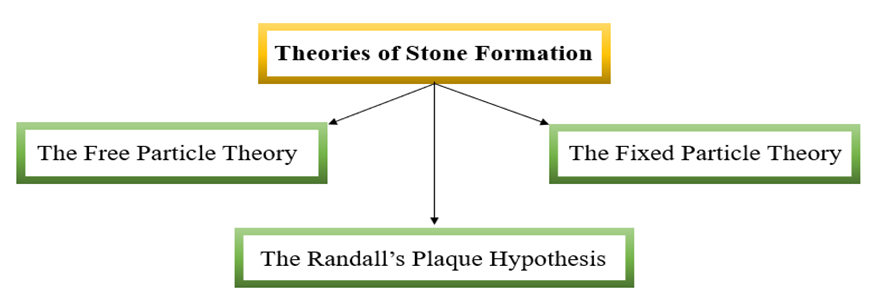
Figure 2. Theories of Stone Formation
The Free Particle Theory
According to the free particle theory, in highly supersaturated urine, stone-forming constituents can spontaneously crystallize within the nephron’s lumen. As these crystals grow or clump together while urine passes through the kidney, they can become large enough to get trapped in a narrow part of the nephron, serving as a starting point for stone formation. [34,35]
The Fixed Particle Theory
The fixed particle theory highlights that newly formed luminal crystals attach to the epithelial surface. When these attached crystals are continuously exposed to supersaturated urine, they eventually grow and serve as focal points for stone formation. [35]
The Randall’s Plaque Hypothesis
Randall plaques are renal papillary lesions in the form of apatite (calcium phosphate) deposits. According to Randall’s plaque hypothesis, these deposits are a primary factor in stone formation. [36] The hypothesis suggests that stone formation begins in the basement membrane of the thin limbs of the loop of Henle, where apatite deposits first appear. These deposits then spread to the interstitium and erode through the urothelium, becoming constantly exposed to urine. This exposure allows them to act as a nidus for organic substances in the urine. [35,37] Various urinary proteins, such as osteopontin and tamm-Horsfall protein, along with lipids and glycosaminoglycans, accumulate on the exposed plaques, forming a multilayered matrix and triggering the formation of calcium oxalate (CaOx) stones. [38]
Types Of Renal Stones
The chemical composition of kidney stones is influenced by abnormalities in the urine’s chemical composition. These stones vary in size, shape, and mineralogical composition. [39] Stones in the urinary tract can be found in the kidneys, ureters, and urinary bladder. Kidney stones are classified as either staghorn, which fill multiple major and minor calyces, or non-staghorn. Non-staghorn stones are further categorized based on their location as calyceal or pelvic, while ureteral stones are identified as proximal, middle, or distal. [35].
Kidney stones are typically classified into five types based on differences in mineral composition and pathogenesis, as follows: [39]
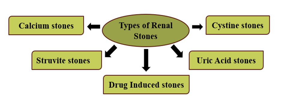
Figure 3. Types of Renal Stones
Calcium Stones:
Calcium-containing kidney stones (approximately 50% of cases) are made of calcium oxalate (CaOX). About 5% of stones are composed of calcium phosphate (CaP), with the rest 45% consisting of a mixture of calcium phosphate and calcium oxalate.. Calcium stones are the most common type of renal stones, making up approximately 80% of all urinary calculi. [39] These stones typically appear in white, black, or grey colours. [40] All calcium stones are radiopaque [41,42] and are visible in X-ray imaging and non-contrast computed tomography (CT) scanning. [41] Urinary pH of 5.0 to 6.5 promotes the formation of calcium oxalate stones, whereas calcium phosphate stones form when the pH is greater than 7.5. [39] An alkaline urine pH encourages the development of calcium phosphate-containing stones. Predisposing factors for calcium oxalate (CaOx) and calcium phosphate (CaP) stone formation include low urine volume, hyperuricosuria, hypocitraturia, hypercalciuria, and hyperoxaluria. [43] A high sodium diet raises the risk of kidney stones by increasing calcium phosphate saturation in the urine and decreasing the inhibitory activity against calcium oxalate crystallization by lowering the excretion of citrate in the urine. The recurrence of calcium stones is greater than other types of kidney stones. [44]
Struvite Stones (Magnesium Ammonium Phosphate Stones):
Struvite stones, which account for 10–15% of cases, are also known as infection stones or triple phosphate stones. [39] They are composed of three phosphates: ammonium phosphate, magnesium phosphate, and calcium phosphate. [45] Struvite stones occur in patients with chronic urinary tract infections that produce urease. The most common pathogen is Proteus mirabilis, while less common pathogens include Klebsiella pneumoniae, Pseudomonas aeruginosa, and Enterobacter. [46,47,48] An alkaline urine pH Favors the formation of struvite stones, as alkalinity promotes the conversion of urea to ammonia by urease. Struvite stones typically form as staghorn calculi. [41,42] And stones are usually light brown or whitish. [49] Although struvite stones are radiopaque, their appearance is faint in radiographic images. [41,42] Women are more likely to develop this type of stone than men. Escherichia coli is not capable of splitting urea and is not associated with struvite stones. [50]
Uric Acid Stones or Urate:
Uric acid stones have an occurrence of about 8-10% of all stone types. [42] Diets high in purines, particularly those rich in animal proteins like meat and fish, result in hyperuricosuria (excess uric acid in the urine). This, combined with low urine volume and a low urinary pH (below 5.05), exacerbates the formation of uric acid stones. [47,51] Uric acid stones are typically oval and smooth in shape, and they often appear yellowish (Scheinman, 2003). Uric acid stones are radiolucent and therefore not clearly visible in X-ray imaging, but are lucid in non-contrast CT. [41] People with gouty arthritis may develop kidney stones. The most common cause of uric acid nephrolithiasis is idiopathic [50], and uric acid stones are more prevalent in men than in women. [39]
Cystine Stones:
These stones comprise less than 2% of all stone types. [39] Cystine stones form due to poor reabsorption of cystine in the renal tubules, which is linked to a hereditary autosomal recessive disorder that ultimately results in cystinuria. [39,42] Low urine pH and urine volume accelerate cystine stone formation. Cystine stones are faintly radiopaque, [41,42] often large and staghorn. They appear smooth and are pale yellow. [52]
Drug-Induced Stones:
This accounts for about 1% of all stone types. [39] Drugs such as guaifenesin, triamterene, atazanavir, and sulfa drugs can induce kidney stones. For instance, people who take the protease inhibitor indinavir sulfate, a drug used to treat HIV infection, are at risk of developing kidney stones. [46] Such lithogenic drugs or their metabolites may deposit to form a nidus or attach to existing renal calculi. Additionally, these drugs can induce the formation of calculi through their metabolic actions by interfering with calcium oxalate or purine metabolism. [50] Drug stones are radiolucent and do not appear in CT scans. [53]
Miscellaneous Stones:
Other types of stones that are rarely found include silicate stones, xanthine stones, and 2,8-dihydroxyadenine stones. [54]
Table 1: Classification of Kidney Stones
|
Type of Stones |
Incidence Rate |
Appearance (Colour) |
X-ray findings |
Crystal Shape |
Risk Factors |
|
Calcium Oxalate, Calcium Phosphate |
80% |
White, Black, or Grey |
Radiopaque, Spherical (Staghorns are rare) |
Envelope, Amorphous |
low urine volume, hyperuricosuria, hypocitraturia, hypercalciuria, and hyperoxaluria |
|
Uric Acid |
8-10% |
Yellowish |
Radiolucent, Staghorns possible |
Diamond, rhomboid |
Hyperuricosuria, low urine volume and a low urinary pH, Gout |
|
Struvite |
10-15% |
Brown or Whitish |
Radiopaque, Staghorns common |
Coffin-lid |
Increased Urine pH and Chronic urinary tract infections |
|
Cystine |
2% |
Pale Yellow |
Faintly radiopaque, Staghorns common |
Hexagonal |
Low urine pH, urine volume, and Inherited disorder |
|
Drug-Induced |
1% |
Yellow-Brown, or Reddish |
Radiolucent |
Needle-shaped and may vary in shape |
- |
Kidney Stone Compositions
Urinary stones consist of both crystalline and non-crystalline phases, or organic material (the matrix), in their chemical composition. Urinary stones' organic matrix is made up of macromolecules such proteins, lipids, carbohydrates, and glycosaminoglycans (GAGs). These chemicals have a major impact on whether kidney stones occur or not. Proteins (64%), non-amino carbohydrates (9.6%), hexosamine as glucosamine (5%), water (10%), and inorganic ash (10.4%) make up the majority of the stone matrix. Kidney stones are built using the matrix as a template.All stone matrices contain phospholipids (8.6% of the total lipid), which represent about 10.3% of the stone matrix. Phospholipids found in cell membranes, which are a component of the organic matrix, encourage the development of calcium phosphate and calcium oxalate stones. [55] The main ingredient in the matrix of all kinds of stones is albumin. [56] A quarter of patients with calcium phosphate (CaP) develop brushite-containing stones, which are a hard phosphate mineral with an increasing incidence rate. CaP can exist in the urinary tract as brushite, carbonate apatite, or hydroxyapatite. Shock waves and ultrasonic lithotripsy treatments do not affect brushite. [57]
Symptoms Of Kidney Stones
Management And Treatment Of Urolithiasis
Medical Therapy
Non-steroidal anti-inflammatory drugs (NSAIDs) such as diclofenac and ketorolac are used as first-line treatment to relieve symptoms in renal colic, which is primarily caused by an obstructing calculus. [3,27] Opioids, such as morphine, codeine, pethidine, and tramadol, are used as alternatives to NSAIDs. [3] Treatment for hypercalciuria involves the use of sodium cellulose phosphate and thiazide diuretics. [12,58] For the treatment of hyperoxaluria, Oxalobacter formigenes, an oxalate-degrading bacterium that colonizes in the stomach when taken orally, is being explored extensively [59] and has shown encouraging results in a few trials. [60] In general, potassium citrate is used to treat hypocitraturia and calcium oxalate (CaOx) stones. [61] Hyperuricosuria is treated with allopurinol. Acetazolamide, sodium bicarbonate, potassium citrate, and sodium citrate are alkalizing agents that are used to dissolve uric acid, xanthine, and cystine stones. D-Penicillamine, α-mercaptopropionyl glycine [62], and tiopronin are used to treat cystine stones [63], while acetohydroxamic acid is used to treat struvite stones. [12] To induce spontaneous expulsion of calculi, medical expulsive therapy (MET) uses calcium channel blockers (nifedipine) or α-adrenergic blockers [64], such as tamsulosin, alfuzosin [59], terazosin, and doxazosin. [65] In addition to tamsulosin, corticosteroids such as deflazacort are used for MET. [66] Urinary calculi can be dissolved by chemolytic dissolution therapy, which uses chemicals such as hemiacidrin, N-acetylcysteine, and THAM-E (tris[hydroxymethyl]aminomethane). [62]
Surgical Intervention
Over three decades, there has been a dramatic change in the surgical instruments used to remove urinary tract stones. [67,68,69] For stone removal, laparoscopic surgery and open surgery have mostly been replaced by highly advanced, non-invasive or minimally invasive modalities such as extracorporeal shock wave lithotripsy (ESWL), percutaneous nephrolithotomy (PCNL), and flexible ureteroscopy. [3,70,71] The use of holmium yttrium–aluminium–garnet (Ho: YAG) lasers for intracorporeal lithotripsy [72], micro-PCNL [73], robotic flexible ureteroscopy [74], robotic nephrolithotomy, and robotic pyelolithotomy [75] are examples of recent advancements in these surgical instruments. The most widely used non-invasive method for treating proximal urinary tract calculi at the moment is ESWL. [76,77]
Table 2: Surgical Intervention for Kidney Stones [67,77]
|
Surgical methods |
Indications |
Method |
|
Extracorporeal shock-wave lithotripsy (ESWL) |
Stone size < 2cm, Upper urinary tract |
Most widely used non-invasive |
|
Percutaneous nephrolithotomy (PCNL) |
Stone size > 2cm, Mid and distal ureteral stones |
Invasive but effective for large or hard stones |
|
Ureteroscopy |
Stone size < 2cm, Lower urinary tract |
Minimally invasive |
Diagnostic Investigations
Examining the patient's dietary, family, and medical histories also helps in diagnosis. A patient's medical history can reveal any illnesses they may have, as well as any medications or treatments they may have recently taken or received, which may be risk factors for urolithiasis. The patient's eating habits and any family history of renal stone disease may also aid in identifying the condition and its possible cause. [78] Although they are common in renal stone disease, unbearable flank pain and haematuria are not always indicative of urinary tract stones. It is basically essential to do the diagnostic assessments listed below in order to rule out false alarms and confirm the presence of renal stones.
Urine analysis:
It is the initial diagnostic procedure used to determine whether urinary tract stones are present. Urine volume, pH, calcium, creatinine, sodium, phosphate, oxalate, citrate, uric acid, and cystine levels are all measured, and the presence of blood is visualized [41,79]. Evaluation by appearance, dipstick, chemical testing, and microscopic inspection is all part of urine analysis. Reddish pee is a sign of haematuria, while cloudy urine typically indicates the presence of bacteria and pus. Typically, dipstick analysis is used to measure the pH and specific gravity of urine as well as the presence of blood, leukocyte esterase, nitrite, and protein (albumin). [79] The pH of urine can occasionally reveal the type of stone that is present. For example, it is widely known that an acidic pH is favourable for the creation of cystine and uric acid stones, whereas an alkaline pH encourages the formation of struvite and calcium phosphate stones. [79,80] Urine with pus typically implies an illness. It is evident that struvite stones are present if pus is discovered in alkaline urine; however, if pus is found in acidic urine, it can be inferred that the infection is secondary and that the stone may be organic, such as a uric acid stone, cystine stone, or xanthine stone. [80] To establish the existence of a UTI, urine culture is typically conducted. [79] White blood cells (WBCs), red blood cells (RBCs), and crystals that form stones are all examined under a microscope. While an elevated RBC count suggests haematuria, an elevated WBC count once more suggests a UTI. [80] Under a microscope, stones can take many different shapes, such as dumbbell-shaped calcium oxalate monohydrate crystals (COM), tetrahedral or bipyramidal-shaped calcium oxalate dihydrate crystals (COD), narrow and elongated calcium phosphate stones, struvite stones that resemble rectangular prisms like coffin lids, uric acid stones that resemble yellow or reddish-brown diamond-shaped crystals (rhomboidal) or needles, and cystine stones that have a hexagonal shape. [80,81,82] On the other hand, 2,8-Dihydroxyadenine stones are round, brown crystals [30]. Determining the underlying metabolic disorders and risk factors is aided by testing urine for the presence of substances such as calcium, creatinine, salt, phosphate, oxalate, citrate, magnesium, uric acid, and cystine [83].
Stone analysis:
An essential component of research into recurrent stone formers is stone analysis, which gives an overview of the mineral components of stones and, consequently, of the variables and metabolic conditions that may be connected to stone formation. This aids in appropriate medical management. In order to identify the components of a stone and their locations within it, stone analysis entails analyzing the entire crust and core of the stone. The results are then summarized in order to strategically determine the underlying reason and, consequently, guide the diagnosis and therapy. The two most often used methods for analyzing stones are X-ray crystallography and infrared spectroscopy. [84,85]
Serum analysis:
As markers of renal function and underlying metabolic reasons, serum analysis includes measuring the levels of 166 of urea, uric acid, creatinine, sodium, potassium, bicarbonate, albumin, calcium, magnesium, and phosphate [79,80]. The glomerular filtration rate and renal tissue integrity, which are negatively impacted in renal stone disease, are depicted by serum urea, uric acid, and creatinine levels. [86] Serum parathyroid hormone levels must be measured in order to check for hyperparathyroidism if increased calcium levels are found in the blood. [78] Since leucocytosis is evident in infected patients, haematological work is done in addition to serum analysis to determine the leukocyte count. [79]
Imaging:
The most crucial diagnostic technique that confirms the diagnosis based on laboratory testing, physical examination, and family history is imaging. It offers a wealth of information on stones and is essential in deciding what kind of treatment is best for a patient. [79,80]
Table 3: Imaging Test for the Evaluation of Kidney Stones [79,80]
|
Procedure |
Advantages |
Disadvantages |
|
Kidney-uterine bladder (KUB) |
Widely available, cost-effective, Minimal Radiation, useful for follow-up of radiopaque stones |
Poor sensitivity |
|
Ultrasound |
Widely available, no radiation exposure, useful for follow-up of radiopaque stones |
Poor sensitivity for small calculi, moderately expensive |
|
Intravenous Pyelography (IVP) |
Helpful in visualizing renal function and anatomy, the diagnosis of obstructing stones |
Intravenous contrast required, may miss non-obstructing stones, Moderate radiation exposure |
|
computed tomography (CT) |
New gold standard, highly sensitive and specific, detects radiolucent stones, no intravenous contrast needed |
Expensive, Moderate radiation exposure |
One of the initial imaging tests is kidney-uterine bladder (KUB) radiography, which is essentially a simple abdominal X-ray. It facilitates finding the stones and imagining their size, shape, and quantity. Compared to less radio-dense uric acid, struvite, and cystine stones, it is more effective at detecting calcium-rich, radiopaque stones. Bowel gas, stool, and extra-urinary calcifications severely restrict its effectiveness and, thus, its applicability, despite the fact that it is very cost-effective. Additionally, there is a significant danger of radiation exposure. [83, 87] Nonetheless, it is still widely utilized for initial detection, fluoroscopically guided shock wave lithotripsy, and subsequent follow-ups [79]. High-frequency sound waves are used in ultrasound, an imaging technique, to create an image of solid objects like stones by bouncing or echoing off of them. Since real-time ultrasound does not carry the dangers of radiation exposure, such as teratogenicity, mutagenicity, or carcinogenicity to the fetus, it is currently being employed as a first-line imaging tool for urolithiasis during pregnancy. [79,88] Additionally, it is the preferred imaging modality for identifying and finding kidney stones in children. [79] Except for its inability to identify ureteral calculi, the method is highly affordable and capable of detecting all kinds of stones. [89] Iodinated contrast media, which travels in blood and is eventually filtered by the kidneys and removed from the ureters and bladder during micturition, is given to the patient intravenously as part of the Intravenous Pyelography (IVP) procedure. The structure and operation of the urinary system, as well as any obstruction or stone therein, are clearly depicted by a series of X-rays of the kidneys, ureters, and bladder obtained at predetermined intervals using contrast medium [3]. It is adept at identifying the stone and offering details on its location, size, and shape as well as the kidneys' structure and function, as well as the stone's surroundings and degree of obstruction. Despite having higher specificity and sensitivity than KUB radiography and ultrasonography, its acceptability is continuously declining because of the contrast media's possible side effects, which can include anything from nephrotoxicity and anaphylactic reactions to bradycardia, flushing, and nausea. [87] Patients on metformin medication, pregnant women, those with impaired renal function, and those allergic to contrast media should not use it. [89,90] The X-ray beam used in computed tomography (CT) is spun around the patient's body to create a series of pictures, which are then reconstructed in three dimensions. Due to its speed, accuracy, and effectiveness in identifying all kinds of stones at any location—all without the requirement for contrast media—noncontrast helical CT has grown in popularity. It has mostly supplanted IVP and is about to surpass all other imaging modalities. It is known to have an accuracy of 96 98% and sensitivity and specificity of 96-100%. [79,87,91] It has the benefit of offering details on the type of stone, the degree of blockage, renal anatomy and physiology, and any additional or non-urological causes of flank discomfort, such as pancreatitis, appendicitis, or gynecological abnormalities. [79,83] The technique's sole disadvantage is the high level of ionizing radiation needed for imaging, which primarily restricts its usage in youngsters and pregnant women. Dual energy CT (DECT) and low-dose unenhanced CT have recently been offered as solutions to this problem. DECT uses variations in the X-ray attenuation characteristics of the components of stones to ascertain the mineral composition of stones. It has two X-ray sources and two detector units. According to DECT, stones come in a variety of colours based on their type. [79,91,92,93] A relatively new imaging method is digital tomosynthesis. Compared to the commonly used non-contrast CT, it has been demonstrated to carry a significantly lower risk of radiation exposure and may offer additional advantages and broader adoption. [79] A radiopharmaceutical substance labelled with technetium-99 is administered intravenously as part of isotope renography, also known as nuclear imaging. A gamma camera detects the radiation emitted as the radioactive substance passes down the urinary tract, producing images of the same. [3,79] Urinary stones can be imaged using magnetic resonance imaging, which uses radio waves, magnets, and the body's inherent magnetic properties. In certain cases, paramagnetic contrast media must be administered for the imaging. [3,79] Due to its superior soft tissue contrast and lack of ionizing radiation risk, it was found to be useful in visualizing pathological changes caused by stones in the urinary tract of pregnant and paediatric patients [79,88]. However, high doses of paramagnetic contrast were later found to be teratogenic [79]. However, it is a safer substitute because contrast media administration is not required [88].
CONCLUSION
Urolithiasis is a common and frequently recurring urinary condition that continues to grow as a global health concern due to its increasing prevalence and the complications it causes. A thorough comprehension of its underlying mechanisms—including urinary solute supersaturation, crystal formation, and retention—is crucial to understanding stone development. The free-particle theory offers further insight into how these stones originate and progress. Timely and accurate diagnosis using imaging techniques and biochemical testing is essential for effective treatment. Current management options range from non-invasive drug therapies and advanced surgical procedures, with a strong emphasis on preventing recurrence. An in-depth grasp of urolithiasis—from its underlying causes to clinical management—is vital for improving individual outcomes and shaping effective public health strategies.
REFERENCES


Nikita Bankoti, Shashi Bisht, Pushpendra Yadav, Chandrakanta*, Urolithiasis: An Update on Epidemiology, Diagnosis, And Treatment Options, Int. J. of Pharm. Sci., 2025, Vol 3, Issue 7, 978-994. https://doi.org/10.5281/zenodo.15834142
 10.5281/zenodo.15834142