View Article

  • Artificial Intelligence in Wound Care: Revolutionizing Therapeutics, Management, and Precision Healing Predictions

  • Dr. M.C. Saxena College of Pharmacy, 171, Barawankala, Mall Road, IIM Rd, Dubagga, Lucknow, Uttar Pradesh 226101, India.

Abstract

With the population aging and chronic diseases becoming more widespread, intricate interventions for high-risk patients with multiple health issues and wounds that are difficult to heal have become increasingly common. Healthcare professionals are challenged to provide safe and effective care for all patients at the same time. When skin wounds are not managed properly, the possibility of infection and complications increases. This can hinder healing and negatively impact patients’ quality of life. Technology based on artificial intelligence (AI) has shown considerable benefits in the processing and analysis of image data. Recently, methods and algorithms based on AI—such as automated learning, deep-learning learning, and neural networks—have been thoroughly investigated in the field of wound care and research. They have proven effective for supporting clinical decisions regarding wound diagnosis, medical care, anticipation, and rehabilitation. Nonetheless, difficulties persist in realizing a closed-loop care system for the thorough integration of AI in wound management, which includes wound diagnosis, observation, and medical care. This review thoroughly encapsulates recent progress in the use of AI for wound repair. In particular, it addresses the function of AI in categorizing types of injuries, assessing wounds, classifying wound tissue types, overseeing and forecasting wound conditions, and tailoring treatment to individual patients. Moreover, the review discusses the difficulties and constraints that AI encounters in managing wounds. In addition, the review considers current limitations and examines potential future directions.

Keywords

Wound Healing, Artificial Intelligence, Chronic wound prediction, Deep learning, Burn degree assessment

Introduction

The largest organ in the body, the skin plays a vital role in immunological responses, maintaining temperatures, defending tissues and organs within the body from environmental harm, and fending off alien microbes.  Physical, chemical, and disease-related injuries can cause small-scale harm to adjacent blood vessels and cells in addition to impairing the skin's barrier function and aesthetics.  Over eleven million Americans get acute wounds each year, and over 6 million have persistent wounds, which drives up yearly wound care costs to over $30 billion.  In addition to placing a heavy burden on healthcare systems and generating a great deal of psychological and financial strain on society, severe skin injuries can result in infection, amputation, systemic problems, and even death. In order to lower disability and mortality, it is clinically important to encourage wound repair and improve the quality of healing, particularly by minimising the creation of scars. [1,2]. The current approaches to wound care, such as dressing changes, grafting of skin, artificial dermis, dressings for wounds, adverse pressure medication, and the use of growth hormones and inflammatory substances, are constrained by their poor overall effectiveness, single functionality, and less than ideal results.  These strategies also have a lot of problems, like inadequate chronic wound healing, a high risk of infection, a lack of precise and real-time assessment, a lack of individualized care, a lack of healthcare resources, and heavy financial constraints.  Several interrelated phases, such as hemostasis, inflammatory processes, angiogenesis, superficial tissue regenerating and remodeling, are involved in wound repair.  It is still difficult to predict the exact healing trajectory and the most effective treatment therapies since wound healing processes are complicated and dynamic. [3-6]. Monitoring and assessing wound states, as well as guiding medical choices are facilitated by precise wound assessment, which includes lesion degree and type.  Building on this basis, wound healing and regeneration of skin can be successfully encouraged by individualized treatment plans that utilize the body's natural healing mechanisms. [7] A state-of-the-art technical science, artificial intelligence (AI) is used to mimic, expand, and improve human intelligence.  Machine learning, support vector machines, electronic health records, neural networks and deep learning are some of the AI technologies that have been developed to far.  Global healthcare systems are undergoing a fundamental transformation as a result of AI's exceptional ability to analyze, display, comprehend, and interpret complicated medical data. [8-12] In order to improve diagnostic speed and precision while optimising healthcare assets, artificial intelligence (AI) can, for instance, integrate medical imagery such as CT scans, magnetic resonance imaging, and three-dimensional ultrasound reconstructions for automated diagnosis.  AI approaches may also effectively process vast volumes of unorganized data to retrieve important information, train iteratively to reliably diagnose complicated illnesses, and offer suggestions for individualized treatment. [13,14]. Conventional wound evaluation and therapy can be laborious and susceptible to diagnostic errors, and it demands competent and skilled clinical practitioners.  AI can now effectively help doctors diagnose wounds by utilizing vast datasets, including a variety of textual and picture data. It then helps in creating treatment plans and improving wound care, which improves therapy results and patient outcomes 20. In addition to assisting clinical physicians, AI also trains younger nursing teams with a lack of expertise, facilitating remote healthcare and improving overall service quality.  Even though AI-based approaches can help with wound care decision-making, the majority of current research focuses on diagnosis and evaluation methodologies, paying little consideration to outcomes of therapy and tactics. [15-17]. There are still obstacles in the way of developing a closed-loop care system for the all-encompassing use of AI in wound management, which should include wound diagnosis, monitoring, and therapy. [18] There are currently no thorough reviews in this field.  In light of this, this paper offers a thorough summary of current developments in AI applications in wound repair therapeutics.  It discusses the present applications of AI in the following areas: injury type classification, area and depth of wound, wound tissue type, wound observation and prediction, and the creation of individualized treatment plans.  The constraints and potential future of artificial intelligence in wound care are also covered in the study.  This paper addresses the difficulties and possible solutions while also proving the usefulness and effectiveness of AI in improving wound diagnosis and therapy through in-depth study and also in order to provide the groundwork for future advancements, this article attempts to present a thorough summary of the existing uses of AI in dermal wound evaluation and healing time prediction.

  1. AI assessment and categorization of wound types

Numerous factors, such as the injury's etiology, site, extent, dimension, exposure to the outside world, severity, healing period, and possible risk of infection, can be used to categorize skin injuries. Diabetes foot ulcers (DFU), artery ulcers (AU), lower limb ulcers of the veins (VU), ulcers due to pressure (PU), and wounds from surgery are among the primary forms of wounds seen in clinical settings. [19] The creation of high-precision models for classification and precise wound classification is essential for evaluation, planning of therapy, and prognosis since various injury types call for distinct treatment modalities.  To meet this demand, researchers have put up a number of creative AI models.  Convolutional layers, layers of pooling, and fully interconnected layers make up CNN in DL. These layers are capable of automatically extracting local features from input, with a primary focus on recovering fundamental characteristics such picture edges and textures.  For example, DFUNet, a unique CNN (convolutional neural network) architecture, is frequently used to categorize diabetic foot ulcers. To better capture feature variations in pictures and distinguish between diabetic foot ulcers and healthy skin, the framework integrates the classic convolution layer with the parallel convolution layer. These characteristics include edge, intensity, shifts in color, and more.  With a cross-validation coefficient of ten and an area under the curve (AUC) of 0.961, DFUNet can reliably distinguish between DFU and normal skin, even on tiny datasets. [20]. As a sub-field of machine learning, deep learning (DL) has the ability to autonomously extract characteristics from raw data, handle high-dimensional data efficiently, and have exceptional sophisticated perception and task understanding.  By adding more layers and neurons or altering the network topology, it may adjust to various tasks and data-set sizes.  CNN has been effectively employed in several research to categorize DFU and VU. [21] According to researcher, created a high-precision classification algorithm that matches current injury severity rating systems by utilizing machine learning methods.  Even without medical staff, this concept can use wearable technology to rapidly and precisely triage patients in large-scale catastrophe sites.  With the VGG16 network serving as an identification model. [22] In another study, it was effectively divided persistent wounds into four categories using comprehensible AI tools X-AI-cwc, machine learning, and data enrichment approaches.  With prediction accuracy of 95.36% for DFU, 100% for capillary injuries, 100% for injuries caused by pressure, and 99.2% for surgical wounds, the average F1 score was 0.76. [23]. Additionally, DFUNet can shorten processing times by processing incoming data more precisely and effectively.  Additionally, DFUNet can shorten processing times by processing incoming data more precisely and effectively.  Large volumes of data may be processed quickly and effectively by machine learning algorithms, which can also automatically identify useful aspects from the data and make precise predictions and classifications.  The objective of dynamically maximizing the error between expected and actual outcomes is accomplished by improving the error between observed and predicted results, which aids computers in learning all the nonlinear relationships between variables.  Strong generalization ability, adaptability for data with high dimensions, and apathy to data loss are some of SVM's benefits as a machine learning technique. In light of this, researcher created an SVM system that can evaluate smartphone photos and identify locations of chronic wounds based on picture color channels.  This technique, which has a typical accuracy of 73.3% and precision of 94.6%, employs an SVM to identify wound borders and distinguish between portions of the wound and healthy tissue by considering color and wavelet properties. [24] Another study used 2,957 images from the Singapore Advanced Institutes Image Registration Center to create an interpretable AI model that classified vascular images in Asian populations and identified PU, venous leg ulcers (VLU), surgical site infections, and neuroischemic ulcers (NIU) with an average classification accuracy of 95.9%. [25]

2.1 Evaluation of burn degree

For wound care and therapy to be effective, an accurate evaluation of the severity of burns is essential [26].  An incorrect assessment may cause wound care to be postponed, which might negatively impact the results of subsequent treatments [27].  By assessing the total body surface area injured, the depth of burns, and the degree of scarring, machine learning (AI) is used in modern medical procedures to evaluate the severity of burns [28].  Furthermore, the method known as Spatial Frequency-Domain Imaging (SFDI), which makes use of the connection between histologic data and changes in tissue properties, has shown itself to be a very useful tool.  By examining photos taken at different wavelengths and frequencies, this technique can forecast the extent of burns during a 24-hour period [29-31].  The accuracy of these predictions is further increased by the application of SVM, or Support Vector Machine, classifiers. [32]. The efficiency of AI in assessing burn depth at the same time [33].  Their AI model effectively recognized four different levels of burn severity [34] using semantic segmentation of images from polarized high-performance light cameras: superficial (I), superficial to intermediate (II), medium to deep (III), and deep to full thickness (IV), with impressive accuracy rates of up to 92%.  Building such a model needs a large training dataset in addition to intensive learning and training.  However, there is a chance that the system might be further improved by acquiring additional photos, which is both feasible and encouraging for future developments. [33,35]

  1. AI-assisted evaluation of wounds

Important details on a wound's cause, severity, length, status changes, and expected healing are included in its geometric shape and look. [36] In order to diagnose, plan treatments, and anticipate prognoses, wound measurements—such as size, area, or volume—are critical. They also play a significant role in determining how quickly wounds heal.  Important markers of wound healing and the efficacy of therapy include tracking the decrease in wound area or volume and the development of granulation/epithelial tissue. [37] Nonetheless, many doctors continue to struggle to guarantee measurement accuracy and consistency.  Conventional wound assessment uses precise measuring techniques with digital cameras, paper rulers, and depth probes to determine wound area and volume, as well as visual components (such as inflammation, tissue granulation, and evaluation of wound exudation). [38] AI's powers in edge recognition, contrast augmentation, and denoising have led to its widespread use in medical picture segmentation.  You Only Look Once (YOLO), single shot multibox detector, and regions of interest (ROI)-CNN are often used detection models; YOLO, GoogLeNet, AlexNet, ResNet, and VGG are frequently used classification models.  According to studies, using segment and outlier removal approaches can increase DL's classification accuracy when it comes to differentiating burn regions from the surrounding healthy skin. [39]. Determining the area and depth of the wound is essential for assessing the severity of skin injuries and for formulating clinical treatment plans.  Although automated support for burn evaluation is already provided by quick and portable computer-aided diagnostic (CAD) equipment and laser Doppler imaging, their high maintenance costs prevent widespread usage in hospitals. [40] On the other hand, DL imaging systems that rely on spectroscopy and photography provide easier access to solutions.  When used in conjunction with DL algorithms (such CNN and variational autoencoders), such systems can help physicians diagnose wounds and guide therapeutic care. [41,42] In conclusion, doctors may use more precise and reliable evaluation instruments thanks to AI-enhanced wound measuring techniques.  This enhances patient outcomes by optimizing treatment options, improving monitoring of the healing process, and improving the precision of diagnostic and therapy.

3.1 Skin wound assessment

According to a study,  support vector machines (SVM) to precisely identify wound borders in order to optimize the region chosen for automated analysis [24].  By using the conditional random field approach, these limits were further honed.  Optical theory, cross-channel picture comparison, and a fuzzy spectral clustering segmentation technique are used to estimate the extent of the wound [43].  However, these techniques are limited to two-dimensional images.  Edward et al. created a 3D point cloud of the wound's borders and structure using a vision-laser scanner in order to record the three-dimensional architecture of wounds. [44]. By lowering infection rates, speeding up healing, and minimizing human error, combining this type of scanner with vertical robots increases accuracy and patient outcomes.  Additionally, to reduce scanning time, a specially created auxiliary laser LED was used to offer an additional measurement point, utilizing a computerized neural network. [45] Accurate three-dimensional imaging of chronic wounds was made possible in partnership with RSI by connecting digital photography with optical raster using a computerized scanner and picture processing software (DigiSkin). [46] Additionally, a recently created convolutional encoder-decoder network—a variation of ConvNet—is used in an integrated system that integrates visual data with deep learning techniques for lesion delineation and area estimate.  In addition to being dependable and computationally efficient, this system has the ability to identify wound infections and forecast the course of healing. [47]

Table 1. Comparison of AI methods regarding wound measurement

S. No

Wound Measurement Method

Principle

Application

Merits

Limitations

1.

Automatic image analysis pipeline [23]

Computer vision, object detection algorithms (YOLO)

Automated measurement of wound size and automatic assessment of average wound closure percentage. High fidelity results on unseen data with minimal human intervention

Automated and enables high fidelity results

  1. Not good at dealing with some of the challenges like occlusion and blur.


  1. Quantitative measurements are not exactly aligned

2.

Fuzzy spectral clustering [25]

Gray scale based fuzzy similarity measure, spectral clustering segmentation algorithm

Accurate depiction of the wound area and automatic calculation of the contrast between wound and non-wound areas

Effective depiction of wound areas in non-uniformly illuminated images

  1. The wounds that are near to heal or the images having very low (i.e. nearly zero) contrast between healed wound area and healthy skin are not accurately segmented.


  1. The method is not completely automatic.

3.

Vision laser scanner [20]

Use laser ranging scanning to generate 3D point cloud, artificial neural network estimation method

Accurate 3D reconstruction of wound margins and topology

Simultaneous generation of 3D point clouds of wound skin and its edges

  1. The scanner can only deal with small size wound (~3-inch length)

4.

Integrated system [18]

Convolutional encoder-decoder networks (a variant of ConvNet), Hough transformation, computer vision tasks

Wound segmentation in an end-to-end different manner and estimation of wound surface area by transformation of pixel length to actual length

High computational efficiency, validity and reliability as a multifunctional, integrated and unified framework system


The application of AI technology differs depending on the kind of wound.  Burn wounds, ulcers from diabetes, ulcers caused by pressure, lower-limb venous ulcers, and chronically infected wounds are among the clinical skin wounds that are often treated.  Regular evaluation of ulcer wounds is improved by the non-contact measures made possible through the combination of AI with machine learning and image technology [48].  Regular hospital visits are less necessary as a result of patients being able to share pictures of their lesions from the comfort of their own homes.  Additionally, utilizing information from electronic health records in conjunction with machine learning (ML) algorithms has demonstrated remarkable efficacy in forecasting the onset of pressure ulcers. [49]. Based on individual patient information from medical records, AI algorithms are able to personalize forecasts.  AI-driven methods for determining burn depth have shown considerable clinical use in circumstances of burn wounds [50].  Furthermore,by using a network of neurons to simulate skin contraction for durations exceeding a year in order to anticipate the healing trajectories of burn wounds [51].  ML systems trained with extensive health data, including specific wound state descriptors, have successfully predicted the risks of surgical site infections (SSIs). [52] The stability and accuracy of wound assessments have been increased with the combination of many optical wound assessment instruments and multi-modal imaging technologies.  In addition to offering precise measures of wound surface and volume, these technological developments also provide information about the tissue composition of the wound bed [53].  The convenience and organization of managing digital imaging and wound-related data has significantly increased with the ongoing advancement of intelligent information assessment systems.  Advanced intelligent surveillance systems, which are essential for increasing healing rates and hastening patient recovery, have been made possible by these advancements. [54]

3.2 AI-based wound area measurement

For clinical therapy, a precise estimate of the wound area is essential, especially in burn cases where a poor assessment of the area might result in inappropriate fluid resuscitation, raising the risk of shock, fluid overload, kidney damage, and compartment syndrome. [55] The foundation of measuring wounds is high-quality imagery. [38] In difficult situations with dark skin tones, blurry wound margins, uneven forms, diseased tissue, and NT, digital wound measuring systems (DWMS) show excellent accuracy. [56]  Clinical criteria for three-dimensional volume as well as depth analysis have not yet been reached by three-dimensional measurement systems (3D-DWMS), despite their proven dependability in two-dimensional area analysis. [57,58]. A study found that combining three-dimensional reconstruction data with wound photos increased the accuracy of burn area measurement, outperforming conventional geometric area computation techniques.  Deep convolutional neural networks (DCNN) are a kind of deep learning (DL) that automatically learns object features in photos. They use local perceptual characteristics and translation invariance to increase the efficacy of local features, which is crucial for complicated burns.  In light of this, scientists have created a DCNN framework for autonomous tissue and wound segmentation that, when paired with a variety of datasets, can successfully get around the drawbacks of conventional burn area assessment techniques.  A DL-based approach for burn wound recognition, segmentation, and TBSA% computation was presented in a research. [59,60]. Researchers used annotated photos of burn wounds and healthy images to train U-Net and MASK R-CNN models.  With a Dice coefficient of 0.9496, the MASK R-CNN model in conjunction with ResNet101 outperformed the U-Net model in conjunction with ResNet101 for burn wound picture segmentation.  The efficacy of CNN based on datasets for DFU and VLU segmentation was examined in another study.  CNN based on the egNet, LinkNet, U-Net, and Unet-VGG16 algorithms was trained on a sacral PU dataset under supervised learning.  The findings demonstrated that the CNN based on U-Net successfully and quickly segregated the wounds. [61,62]. Treatment procedures in the healthcare industry have grown more effective due to the rapid advancement of AI technology and the growing popularity of mobile devices like smartphones and smartwatches. [38] The advancement of distant wound area measuring and assessment has been greatly aided by the combination of accurate pattern recognition technologies with cameras found in smartphones and tablets.  By merging DL MobileNetsV2 with labeled datasets, a completely automated wound segmentation system based on mobile devices was able to separate wounds from natural photos.  This model may operate on mobile devices with a DC because of its straightforward foundation and minimal processing demands. [63] In addition, researchers created the AutoTrace framework a DCNN model that can be used on mobile devices and allows for accurate and impartial tissue and wound prediction. [64]  Resarcher assessed the precision of three different DL frameworks (U-Net, V-Net, and Seg Net models) in splitting DFU regions on mobile clients in order to accomplish high-precision data analysis with minimal data.  With the highest accuracy possible, the U-Net model did well in this assignment.  According to these investigations, mobile terminals provide a wide range of potential applications and are the best medium for acquiring wound images and transmitting data. [65,66]. In order to improve measurement accuracy, a multi-step process involving photo capturing, shades of gray converting, muddle interpreting, threshold categorization, injury recognition, and growth and deterioration of wound areas has also been proposed for a handheld wound area measurement method.  Using DL models that are supported by the OpenCV framework, this approach performs exceptionally well on tasks like deep burn segmentation, palm segmentation, and complete wound segmentation. [67] The Swift mobile application now offers the benefit of a non-contact, intuitive wound assessment tool thanks to research that integrated FLIRTM infrared cameras, which provide skin temperature readings comparable to clinical reference thermometers.  This makes it possible for clinical physicians to photograph, measure, and monitor the temperature and size of wounds from a single visit to the next, making it appropriate for patients and their family members to monitor at home. [68]  In order to assess the accuracy of the C4W mobile application's length, breadth, and area measurements, researchers used it to track the DFU healing process for chronic wounds and compared it with more conventional measurement techniques.  When measuring the length, breadth, and size of DFU wounds in eight patients, the method demonstrated dependability. [69]. A full evaluation still requires the creation of labeled datasets for scar depth and specialized evaluation structures for more in-depth wound analysis and treatment, even if many modern technologies mainly concentrate on lesion segmentation to facilitate diagnosis.  Improving the models' adaptability to a greater variety of wound forms and clinical situations is one of the main objectives for future research, since it will allow for more thorough and accurate wound evaluation.  To sum up, the use of DL and other modern technologies in wound delineation and analysis gives medical professionals automated, quick, and precise ways to evaluate wounds.  The advancement and improvement of these advancements will boost medical effectiveness, aid in patient recovery, and offer more robust support for wound care. Future applications in self-monitoring and remote healthcare have even more potential due to continuous technology improvements.

3.3 AI-based wound depth measuring

Nowadays, determining the depth of the wound serves as the foundation for developing clinical treatment strategies in addition to being an essential step in determining the severity of injuries.  Inaccurate burn evaluations can result in poor wound care, which frequently causes unneeded procedures or delayed recovery. [70] The main foundation for determining the depth of a burn is variations in the color and texture of the wound, which represent different levels of dermal capillary damage.  Even though clinical evaluation is still a commonly used technique, a delayed or inaccurate early diagnosis might result in improper therapy and have an impact on prognosis.  Thus, there is substantial theoretical and practical utility in applying image analysis technologies to help diagnose burn depth. [71] Outliers can reduce accuracy, particularly when training models, even though several AI algorithms are being created and proven to be effective for analyzing wound images.  Based on algorithms for supervised learning, the SVM and k-nearest neighbor (KNN) methods of classification may isolate data points from various categories as much as feasible in order to eliminate outliers. In order to increase the categorization precision for deep wounds, wound beds, and partial wounds by eliminating outliers, a multi-stage strategy based on Z-test and univariate analysis was devised. This was based on the development of multiple burn classification models utilizing SVM and k-nearest neighbor (KNN) categorization methods. The accuracy was 76% when 10-fold cross-validation was used. [72] By incorporating learned transfer into the convolutional neural network-based ResNet50 model, researchers in another study addressed the lack of unique scene photos and were able to achieve a diagnosis accuracy of 80% for three categories of burns (superficial, intermediate, and deep). [73]

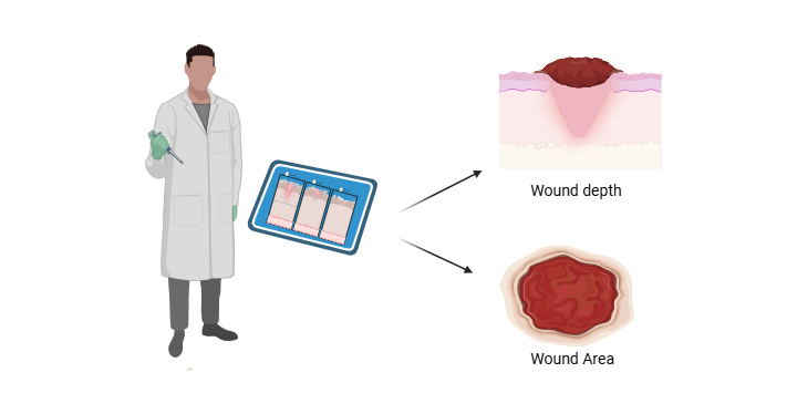
Fig.1 - AI helped in wound depth and area measuring

Additionally, multimodal AI burns diagnosis systems have been built using RGB pictures and ultrasound; the texture information from the ultrasound increase the depth classifiers' accuracy to 80%. [74] The U-Net CNN can precisely partition target areas by combining high-level semantic characteristics with low-level detail data.  To determine the precise depth of a wound, researchers used this model in conjunction with a high-performance split optical camera.  According to the research's findings, the diagnostic accuracy for pediatric burns is around 97%. [75] Multi-parameter and high-dimensional techniques are frequently employed in conventional wound image analysis and processing to get high accuracy, which makes processing complicated pictures challenging.  With fewer parameters, the VGG-16 network built on DCNN can efficiently extract local features from pictures.  Additionally, data dimensionality, processing, and parameter count are decreased by employing pooling layers to preserve the image's primary attributes.  In light of this, Despo et al. created an end-to-end DL model to assess certain burn wound characteristics and employed an enhanced completely convolution network (FCN) DL technique centered on the VGG-16 model for burn classification.  Four different kinds of wound photos were used to train the model, and it successfully classified the images. [76]. Burn depth may be automatically predicted with AI technology, and diagnosis accuracy can be greatly increased by combining picture capture with DL algorithms.  To further improve accuracy, it is necessary to choose suitable imaging modalities, increase datasets, and optimize algorithms, as the existing approaches only get an accuracy of about 90%.  In conclusion, determining the precise depth of burns is still a crucial and difficult part of evaluating injuries.  Even with advancements in burn depth identification, chronic wounds' irregular geometries and notable color fluctuations, as well as the comparable look of wounds with varying depths and tissue compositions, imply that current technologies might not be enough for an accurate assessment. Although computerized & objective diagnostic techniques are the best and most extensively used method, doctors' clinical knowledge and experience are still necessary.

4. Segmentation of wound tissue types and AI assessment

The types of tissue present in a lésion serve as vital indicators of the healing process.  By estimating the tissue components accurately, clinicians can choose suitable dressings, recognize wounds that may not heal, quickly refer patients to specialists, customize treatment based on the patient's condition, and enhance wound care and healing results.  Especially in resource-limited settings, clinicians can be assisted or augmented by high-performance automatic classifiers in the classification of wound tissue types. [77] Utilizing AI for image recognition provides a powerful means of automatically categorizing wound tissues.  Nonetheless, factors such as surrounding noise, variability, and inaccuracies in image capture can impact automatic wound classification. [78] A method utilizing CNN for cells evaluation of stress perforation images was proposed by the researcher.  They retrieved the ROI from the raw perforated images, eliminated noise, and obtained 5X5 pixel sections from every ROI for accurate classification.  This approach yielded a categorizing precision of 92.01% for various tissue types, including GT, drain, and NT.  In subsequent research, persisting wound images were divided into nXn patches and input into a deep neural network (DNN) for feature extraction and SVM classifier training.  This method reached an accuracy rate of 86.4% when distinguishing seven cells types (NT, ablation, well-being GT, non-healthy GT, excessive granulation, contaminated, and epidermalized) [79,80] Conversely, recent research has suggested an automated approach to wound assessment that combines manual color rectification with deep learning models, including versions of EfficientNet and MobileNetV2 embedded with the U-Net architecture.  This approach compares images under varying illumination, length, and lens conditions with effectiveness, demonstrating excellence in segmenting wounded areas and GT. However, it still necessitates enhancements for ET and NT segmentation. [81]. Methods based on AI, employing a multi-view approach and superpixel FCN techniques, have greatly improved the effectiveness of perforated tissue classification. [82] At present, strategies for enhancing images have been applied to boost the precision of tissue categorizing in persistent wounds. [83] And by implementing a superpixel segmentation method that utilizes linear iterative clustering (LIC) along with five-dimensional color and visual plane space.  With the use of LIC, which has robust pixel division and precise limits fitting capabilities, visual division can be performed quickly and effectively. [84] An app for classifying wound tissue can aid patients in real-time monitoring of wound dynamics, which can ease dread during the course of home treatment.  In the field of telemedicine, a mobile injury capture system has been created by researchers to gather images of wounds using the cameras on smart devices.  The “Complex Wound DB” is a new image database developed to categorize complex wounds into five groups: non-wound area, GT, fibrocyte tissue, dry inflammation, and hemorrhage.  This dataset, which consists of specifically 27 images identified by four medical specialists, is publiquement available. [85] At present, researcher have created a mobile app called Deep-wound, which is built on a multi-label CNN system and can categorize wound images to assist in daily wound care. [86] Additionally, automated remote classification of wound tissue will offer doctors valuable guidance, assisting them in conducting thorough wound assessments and developing accurate treatment plans. The classification of wound tissue at present depends on the analysis of characteristics of individual pixels or clusters of pixels (superpixels) within the image, followed by their categorization into various tissue types (e.g., GT, scar, necrosis).  This approach can resolve serious diagnostic issues.  Nonetheless, due to the great similarity of wound tissues, especially in between GT and contaminated tissues, it is essential to optimize multi-view techniques and ultrapixel FCN methods.  Based on this, the accuracy of identification can be improved further by merging images of surface wounds with pathological sections.

5. AI supervision and anticipation of wound healing

It is especially difficult to predict the trajectories of wound healing, but this is essential for accomplishing wound resolution. [87] By predicting wound healing, physicians can choose the most suitable treatment plans, leading to improved efficiency and effectiveness in wound care. AI facilitates accurate wound measurements from targeted data and utilizes diagnostic information to forecast lesion healing progressions by instinctively learning from and overseeing extensive amounts of clinical records.  In addition, forecasting the duration of wound healing assists healthcare professionals in devising treatment plans, establishing practical patient expectations, and possibly enhancing outcomes while lowering expenses. [88,89]

5.1 Prediction of chronic wounds

Chronic wounds represent a considerable worldwide challenge, characterized by localized injuries to skin and tissue that have a disrupted physiological healing response. [90] According to the Wound Healing Association, chronic wounds are characterized by a failure to return damaged tissue to its habituelle configuration and function within a regular timeframe and without delay. [91]  The recovery phase for chronic wounds usually takes more than four weeks, which greatly diminishes the standard of life and well-being of those affected and contributes to higher mortality rates. [92] Thus, accurate evaluation, forecasting, and handling of persistent wounds are vital for alleviating the burden on the healthcare system and improving the pace and standard of patient recovery. [93]. In medical practice, routine lower ankle wounds like vascular, hypertensive, tension, and venous ulcers present a significant danger to aged, who are more vulnerable due to numerous age-related changes.  Among these changes are a heightened occurrence of chronic ailments such as heart-related disease and diabetes, as well as issues like reduced mobility, incontinence, low body weight, inadequate nutritional condition, and cognitive impairment. [94-96]. Intrinsic changes associated with aging that affect skin wound healing—like altered inflammatory responses, lower amounts of supporting embedded structure and growth factors, retarded keratinization, and reduced angiogenic activity—play a role in the slower rates of wound closure observed in older adults. [97]  The occurrence of such wounds has risen significantly with the aging society, thereby increasing the reliance on restricted healthcare resources. [98] To tackle this issue, scientists have created a wearable sensor driven by AI that is connected to sophisticated wound dressing bandages. For the purpose of monitoring chronic wounds and determining their healing stages, this system employs a deep-rooted artificially-designed neural network (ANN) algorithm. [99]. This near-field detecting système offers essential information for making treatment decisions and evaluating how effective wound care medications are.  Chakraborty et al. have proposed a telemedicine model that employs Linear Discriminant Analysis for tissue type classification, attaining a tissues prediction precision. [100] With this approach, persistent wound healing statuses can be diagnosed remotely, providing clinicians with statistical tissue compositional data to support their decision-making.  Furthermore, the evolution of statistical computing has accelerated the creation of a number of promising machine learning methods. [101]. Chronic wounds pose a major challenge to global health, making precise diagnosis and successful treatment essential for promoting healing and preventing additional complications. [102] AI is being used more and more in healthcare for predictive analysis of medical data, adjusting fluently to new details. [103] Even although digital medical documents are widely employed for wound documentation, managing and tracking all facets of patient care for individuals with chronic wounds remains challenging. [104] Nevertheless, utilizing machine learning and big data analytics holds considerable potential for lowering treatment costs, shortening the duration of simulations, and improving the comprehensive excellence of care. [105]

5.2 Previsioning the duration of wound healing

Being able to estimate the duration of wound healing is of great clinical importance, as it allows doctors to quickly adapt treatment strategies to the specific requirements of each patient.[106] Doctors can use precise forecasts to determine if a patient needs several healing operations or if an early closure is possible, as well as to identify the optimal timeframe for healing traumatological wounds.  With this predictive ability, the time until wound closure is shortened and the likelihood of wound-related complications and failures is reduced. [107]. Evaluating the depth of the skin surface and scabs is essential for comprehending the epidermal wound healing process, as it offers crucial perspectives into the normality of re-epithelialization. [108] Visual Uniformity Tomography (OCT) is a real-time imaging method that does not require surgery and allows for the assessment of tissue micro-structures in cross-section.  By combining OCT with AI algorithms, it becomes possible to automate the measurement of epithelial tissue and scab thickness, which aids in forecasting wound healing durations. [108] Nevertheless, the prediction of healing processes in amputation wounds constitutes a complex challenge. This is attributable to factors like severe cardiac ischemia and the lack of trustworthy assessment tools.  To tackle this issue, Squiers et al. developed a new imaging system for capturing multiple-view photographs of the inferior limbs. [109] The accuracy of predictions concerning amputation wound healing has improved by analyzing these images alongside the patient's clinical risk factors using machine learning algorithms.  Moreover, this strategy may decrease the need for further surgeries and the incidence of delayed healing. In 2020, researchers from China created an artificial model based on CNN for recognizing burn depth, and it was effectively employed to anticipate the restoration time of flammable wounds.  By examining the burn's depth, this model provided an accurate estimation of the damaged healing progression. [110]. The precise anticipation of healing times poses a constant desafio for clinicians due to the complexity and dynamism of the wound healing process.  Nonetheless, as extensive data sets become more accessible and computing capabilities improve, AI-based models are likely to become essential for predicting and evaluating wound healing timelines. [111]

  1. AI-supported creation of individualized wound care products

In contemporary medical technology, the use of AI is fostering advancement in skin prototype creation, simulation of wound healing processes, development of pharmaceuticals, and smart dressings, offering new insights and resources for wound management. At present, studies have advanced hybrid models that merge volume, membranes, and one-dimensional prototypes to create 3d geometrical and mechanical representations of skin/subcutaneous complexes, incorporating intricate internal structures via automation. [112] Models of artificial skin created with AI provide a novel viewpoint for research on wound healing and ease the incorporation of products designed for wound repair or observation into these models. This significantly propels fundamental investigations in this area forward and enhances the efficacy of product development. [113] In the field of drug innovation, AI applications have shown outstanding accomplishments.  Utilizing AI for the screening of new antimicrobial peptides fosters innovation that spans from surface antimicrobials to deep drug development.  Even though the majority of screening studies continue to take place in in-vitro and in in vivo-based, the combination of algorithmic and statistical-based strategies with DL models opens up new avenues for drug development. [114]. Furthermore, studies utilizing sequencing results from the skin of diabetic patients and AI-driven bioinformatics have pinpointed a possible therapeutic agent, Trichostatin A, as well as a posible objective, histone deacetylase 4 (HDAC4), for the repair of diabetic wounds. [115]  An AI-nanomaterial detecting system for the ultra-selective identification of combustible organic compounds has also been developed by researchers.  This system employs integrated altered silicon-based microwire field-effect transistors along with various salt molecules.  The sensor can efficiently identify 11 VOCs even amidst physical/chemical interference after being integrated with a computer-generated neurological network model, providing a promising method for recognizing VOCs in wounds. [116]  While information on AI-assisted micro-stuffs as antibiofilm agents are limited, earlier investigations indicate that the integration of various AI usages with microbial films and wounds may result in the creation of additional small-sized wound management devices. [117]. At present, AI-supported wearable sensors for detecting and managing wounds have been created.  A adaptable AI-guided carryable detecting device utilizing a DANN method for persistent wound surveillance and short-distance interaction was suggested by researcher that, it interacts with uninterrupted bandages that are connected to MXene, integriert with wound dressing, and tuned to radiofrequency. [118] Moreover, a microneedle sensor patch that utilizes DL has been created and trained with fluorescence frequency data utilizing the KNN model in order to perform multiple-variate categorizing of wound contamination subtypes.  Smartphone-captured fluorescence visuals can be combined to perceive pH values, facilitating precise and trustworthy wound supervision.   AI's use in bio 3D printing technology is developing, especially in supportive functions and data-oriented production in 3D printing processes. [Fig. 2][119,120]

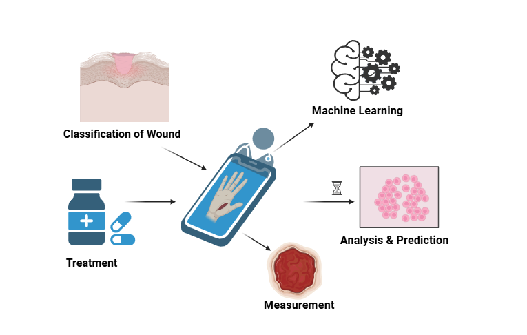
Fig. 2 Creation of personalized wound care with the help of AI

The combination of 3D embossing innovation and AI continuously improves the accuracy, adaptability, and adaptation of numerous materials. [121] Based on Gaussian method regression, scientists have optimized DL models to effectively anticipate the printing capacity scores of bio-inks. [122] Moreover, following the optimization of DL models, a study suggested an AI-assisted high-throughput printing process conditional selection system. This system includes programmed pneumatic extruding bio-printers and AI-assisted image evaluation methodologies that can forecast the printability of bio-inks and optimize the printing process to create higher-quality three-dimensional printed hydrogel-based dressings. [123] The enhancement of the bio-ink 3D printing process has been accomplished through DL methods, offering new perspectives for the creation of hydrogel inks with superior quality in three-dimensional printing. The integration of sophisticated AI and the creation of novel materials are leading to extraordinary innovations and breakthroughs in wound treatment, presenting extensive prospects for development and potential future medical progress.  As AI continues to be applied and developed, it is anticipated that wound diagnosing and treatment procedures will be optimized, leading to the creation of personalized treatment products. This will result in more precise and individualized medical services for patients, enhancing the efficiency and effectiveness of treatment.

  1. Discussion and Visions

7.1 Pros and cons of AI concepts and algorithms

Wound care encompasses activities like image evaluation, cells categorizing, dimensions measuring, and temporal surveillance. These tasks are frequently time-consuming and susceptible to biases from assessors. [124] AI is transforming conventional approaches to wound diagnosis and management, allowing for enhanced precision and smartness in the assessment process. It is possible to greatly save clinicians’ time, lessen the financial burdens on patients, and enhance their standard of life through the use of a comprehensive wound identification, prognosis, and treatment plan system based on AI. At present, AI-supported systems for wound diagnosis and treatment establish an integrated framework that involves automatic identification, analysis, summarization, comprehension, learning, planning, and updating. This allows for a continuous evaluation of the complete wound healing process. [125] Provides a summary of the applications, benefits, restrictions, and scalability of existing AI frameworks and formulas in the context of wound healing.  Shallow-ML models, appropriate for different data types such as organized and unorganized data, can recognize and utilize the most pertinent features to enhance prediction accuracy.  Healing outcomes are predicted by evaluating individual records and treatment strategies data using algorithms like random forests and SVM, as well as optimizing treatment plans according to specific patient characteristics.

7.2 Challenges and techniques of handling

While AI needs extensive data support, the absence of standardized guidelines and protocols for wound data collection has resulted in hospitals creating methods tailored to their specific needs. This has complicated the creation of comprehensive databases for wound care.  The advancement of AI in wound care is impeded by the difficulties researchers encounter in acquiring extensive and all-encompassing datasets related to wound care.  Even when using extensive data collection, non-specific or unobserved factors can still affect wound healing consequences, resulting in DL models that make incomplete or inaccurate predictions. Consequently, it is necessary to create standardized systems for wound data collection and to develop recording devices that are easy to use. This will improve operability and make it easier to document wound care quickly in different environments. [124] Nonetheless, the contribution of AI to digitalizing wound diagnose and recovery anticipation has not been thoroughly investigated.  It is essential to find appropriate wound determination tools and to further regulate the testing and implementation of AI-based electronic wound evaluation tools.  This will assist in identifying the wound types that are most appropriate for AI administration and the specific algorithms that yield the best results. [126]. The application of AI in identifying wounds is primarily restricted to color detection, and it does not possess the capability to predict details regarding wound flexibility and exudate. Because damaged sinuses and perforations cannot be examined, developing devices are unable to determine longitudinally wound details, necessitating manual evaluations for data collection.  This suggests that new strategies are required to gather, analyze, and handle large quantities of data in order to overcome these limitations. The long-term impacts of using AI programmes, such as the efficiency and standard of wound recovery, have not yet been assessed in many studies.  Nonetheless, several initial studies have indicated a high level of satisfaction with AI in medical practice from both clinicians and patients. [127,128]  This implementation allows for the mechanized evaluation of wound subtypes and related diseases.  AI can create tailored care recommendations—like those concerning how often to sanitize a wound, which dressing to use, and what medication dosing to follow—based on past medical data. This can assist with clinical decision-making and enhance patient recovery management.

7.3 Summary and prospect

AI is advancing faster than ever before, especially in the field of medicine, where it greatly improves quick image analysis, diagnosis, risk forecasting, and supplementary treatment. Specific instances of AI utilizations in skin wound managing.  In the past ten years, the rapid development of computer processing expertise has made it easier to integrate AI systems more thoroughly into various medical imaging technologies, including X-ray, ultrasound, digital image processing, and magneto resonance-based imaging. Machine learning (ML) and deep learning (DL) have played a crucial role in the analysis of medical images derived from these technologies, showcasing impressive accuracy and dependability. AI can perform a wide range of functions, such as aiding in diagnosis, therapy selection, risk prediction, disease stratification, medical error reduction, and productivity enhancement.  Aaron Jones and colleagues executed a quasi-experimental design in four different contexts of the Australian Health Service in a significant study.  Data were collected from both the standardized and interventional groups, showing that during the intervention, 101 of the 132 wounds improved, with an average reduction in wound size of 53.99%.  This study highlights how effective and practical AI is in managing wounds. [129]. The prospects for the use of AI in managing skin wounds are encouraging, especially with the emergence of Explainable Artificial Intelligence (XAI) grounded in deep learning (DL) for medical image analysis.  XAI is developing into an essential tool that improves AI's capacity to provide new insights into data, thus augmenting the repository base with elements of new discovery.  With the growing prevalence of DL-based methods, the need for clarification is intensifying—particularly in vital domains like medical imagery evaluation, which is essential for evaluating skin wounds.  In addition to various imaging methods and AI-integrated systems, remote consultation systems driven by AI that utilize mobile devices and tablets for data collection and connectability are becoming increasingly popular. [130,131]. The field of wound care has been dramatically transformed by AI, revolutionizing how wounds are assessed, measured, classified, and predicted.  Currently, the use of AI in skin wounds primarily focuses on two domains: analysis of wound images and data integration. [132] Nonetheless, it is still in progress to develop AI-based technologies to a specification appropriate for clinical use that ensures the provision of high-quality wound care. AI has the potential to greatly improve wound care practice by establishing rigorous criteria for wound data acquisition and developing recording systems that are more efficient and easier to use.  This will ensure that patients have a more thorough and higher-quality healthcare experience.

Future Aspects of AI in Wound Healing

Artificial intelligence (AI) holds significant promise for advancing wound healing and management, primarily through improved assessment, prediction, and personalized treatment strategies. The integration of AI in this domain reflects the broader trend of leveraging technological advancements to enhance healthcare delivery and outcomes. [133,134]

Image-Based Analysis and Prediction

One of the key areas where AI is making strides is in the image-based assessment of wounds. AI systems, particularly those employing machine learning and deep learning algorithms, can analyze wound images to assist clinicians in diagnosing and monitoring wounds and predicting healing trajectories. These AI-based platforms can process large datasets of wound images and provide insights that are crucial for creating efficient care pathways and improving patient outcomes. [133,135]

Improving Wound Management Efficiency

AI helps streamline the wound management process by enabling the automation of wound measurement and classification. The prediction of healing times is another area where AI shows promise, helping healthcare professionals to better allocate resources and prioritize care. This is particularly beneficial in environments dealing with high volumes of chronic wounds, which are often a marker of underlying health issues like diabetes and obesity. [135,136]

Personalized and Remote Care

AI can facilitate personalized wound care by tailoring treatments based on predicted wound healing outcomes. Additionally, remote consultation systems leveraging AI can improve accessibility and continuity of care. These systems make use of devices such as smartphones and tablets to capture health data, thus enabling real-time monitoring and early intervention, reducing the need for frequent hospital visits. [137]

Challenges and Future Directions

Despite its potential, the integration of AI in wound care faces several challenges. Data diversity and the need for comprehensive datasets to train AI models are critical issues. Ethical concerns around data privacy and algorithm transparency, as well as the equitable distribution of AI technologies, must also be addressed to ensure that AI's benefits are maximally realized across different patient demographics and healthcare systems. [134,136]. The future of AI in wound healing is likely to focus on overcoming these challenges through collaboration between AI developers and the healthcare community. Innovations in AI could lead to a closed-loop care system in wound management, encompassing diagnosis, monitoring, and treatment, thereby providing comprehensive and integrated care solutions. [134]

CONCLUSIONS

Artificial Intelligence (AI) is poised to revolutionize wound care through advancements in therapeutics, management, and precision healing predictions. The integration of AI technologies in wound care not only holds the promise of enhancing therapeutic outcomes but also offers significant improvements in patient management and healing predictions. AI has demonstrated efficacy in transforming wound assessment processes. It automates the measurement, classification, and analysis of wounds, thereby reducing the time burden on healthcare professionals while increasing accuracy. Image-based AI systems are particularly noteworthy, as they improve the ability to diagnose and assess therapy effectiveness by analyzing comprehensive datasets collected over time. These systems facilitate more precise predictions of healing trajectories, assisting clinicians in making more informed decisions about treatment plans. AI is also reshaping therapeutic strategies by enabling personalized treatment plans based on predictive analytics. By processing vast amounts of data, AI can suggest targeted interventions that align closely with the patient's specific wound characteristics and healing progress. Furthermore, AI's role in wound management extends to real-time monitoring and data-driven care, thus ensuring timely interventions and proactive management of potential complications. While these advancements are promising, there are challenges to be addressed in achieving a comprehensive, closed-loop AI application system for wound care. Issues such as data privacy, model interpretability, and regulatory compliance require careful consideration to facilitate seamless integration into clinical workflows. Ethical considerations, including equitable access and potential biases in AI models, must also be addressed to ensure responsible implementation. In conclusion, AI's potential to revolutionize wound care is vast, offering innovative solutions for therapeutic, management, and predictive challenges. By overcoming existing barriers and focusing on research and collaboration, AI-driven advancements can significantly enhance the healing process and improve patient outcomes. While a comprehensive essay could delve deeper into each aspect, this overview highlights the transformative potential of AI in wound care.

REFERENCES

  1. Blanpain C, Fuchs E. Epidermal homeostasis: a balancing act of stem cells in the skin. Nat Rev Mol Cell Biol. 2009 Mar;10(3):207-17. doi: 10.1038/nrm2636. Epub 2009 Feb 11. PMID: 19209183; PMCID: PMC2760218.
  2. Eurosurveillance editorial team. WHO launches the World Health Statistics 2012. Euro Surveill. 2012 May 17;17(20):20175. PMID: 22642947.
  3. Li J, Zhou C, Luo C, Qian B, Liu S, Zeng Y. et al. N-acetyl cysteine-loaded graphene oxide-collagen hybrid membrane for scarless wound healing. Theranostics. 2019;9:5839–53. doi: 10.7150/thno.34480.
  4. Wang PH, Huang BS, Horng HC, Yeh CC, Chen YJ. Wound healing. J Chin Med Assoc. 2018;81:94–101. doi: 10.1016/j.jcma.2017.11.002.
  5. Daniel H, Suoqin J, Peng S, Rachel C, Morgan D, Quy N. et al. Defining epidermal basal cell states during skin homeostasis and wound healing using single-cell transcriptomics. Cell reports. 2021;30:3932–47.
  6. Kolimi P, Narala S, Nyavanandi D, Youssef AAA, Dudhipala N. Innovative Treatment Strategies to Accelerate Wound Healing: Trajectory and Recent Advancements. Cells. 2022 Aug 6;11(15):2439. doi: 10.3390/cells11152439. PMID: 35954282; PMCID: PMC9367945.
  7. Nagle SM, Stevens KA, Wilbraham SC. Wound Assessment. 2023 Jun 26. In: StatPearls [Internet]. Treasure Island (FL): StatPearls Publishing; 2025 Jan–. PMID: 29489199.
  8. Dallmann AC, Sheridan M, Mattke S, Ennis W. Prediction of Healing Trajectory of Chronic Wounds Using a Machine Learning Approach. Adv Wound Care (New Rochelle). 2024 Nov 6. doi: 10.1089/wound.2024.0095. Epub ahead of print. PMID: 39508072.
  9. Fourati Kallel I, Kammoun JK, Lajnef H, Ben Ali S. Intelligent progress monitoring of healing wound tissues based on classification models. Biomed Phys Eng Express. 2025 Mar 26;11(3). doi: 10.1088/2057-1976/adc137. PMID: 40096753.
  10. Fife CE, Eckert KA. Harnessing electronic healthcare data for wound care research: Standards for reporting observational registry data obtained directly from electronic health records. Wound Repair Regen. 2017 Apr;25(2):192-209. doi: 10.1111/wrr.12523. Epub 2017 Apr 27. PMID: 28370796.
  11. Ohura N, Mitsuno R, Sakisaka M, Terabe Y, Morishige Y, Uchiyama A, Okoshi T, Shinji I, Takushima A. Convolutional neural networks for wound detection: the role of artificial intelligence in wound care. J Wound Care. 2019 Oct 1;28(Sup10):S13-S24. doi: 10.12968/jowc.2019.28.Sup10.S13. PMID: 31600101.
  12. Mehta AS, Teymoori S, Recendez C, Fregoso D, Gallegos A, Yang HY, Aslankoohi E, Rolandi M, Isseroff RR, Zhao M, Gomez M. Quantifying innervation facilitated by deep learning in wound healing. Sci Rep. 2023 Oct 6;13(1):16885. doi: 10.1038/s41598-023-42743-5. PMID: 37803028; PMCID: PMC10558471.
  13. Li S, Mohamedi AH, Senkowsky J, Nair A, Tang L. Imaging in Chronic Wound Diagnostics. Adv Wound Care (New Rochelle). 2020 May 1;9(5):245-263. doi: 10.1089/wound.2019.0967. Epub 2020 Mar 19. PMID: 32226649; PMCID: PMC7099416.
  14. Zhao K, Jiang B, Zhang S, Zhang L, Zhang L, Feng Y. et al. Measurement accuracy and repeatability of RECIST-defined pulmonary lesions and lymph nodes in Ultra-Low-Dose CT based on deep learning image reconstruction. Cancers (Basel) 2022;14:5016.
  15. Chairat S, Chaichulee S, Dissaneewate T, Wangkulangkul P, Kongpanichakul L. AI-Assisted Assessment of Wound Tissue with Automatic Color and Measurement Calibration on Images Taken with a Smartphone. Healthcare (Basel). 2023 Jan 16;11(2):273. doi: 10.3390/healthcare11020273. PMID: 36673641; PMCID: PMC9858639.
  16. Huang S, Dang J, Sheckter CC, Yenikomshian HA, Gillenwater J. A systematic review of machine learning and automation in burn wound evaluation: A promising but developing frontier. Burns. 2021 Dec;47(8):1691-1704. doi: 10.1016/j.burns.2021.07.007. Epub 2021 Jul 15. PMID: 34419331.
  17. Encarnação R, Manuel T, Palheira H, Neves-Amado J, Alves P. Artificial Intelligence in Wound Care Education: Protocol for a Scoping Review. Nurs Rep. 2024 Mar 14;14(1):627-640. doi: 10.3390/nursrep14010048. PMID: 38535720; PMCID: PMC10975757.
  18. L
  19. Zhang L, Wang S, Tan M, Zhou H, Tang Y, Zou Y. Efficacy of oxidized regenerated cellulose/collagen dressing for management of skin wounds: a systematic review and meta-analysis. Evid Based Complement Alternat Med. 2021;2021:1058671.
  20. Goyal M, Reeves ND, Davison AK, Rajbhandari S, Spragg J, Yap MH. DFUNet: convolutional neural networks for diabetic foot ulcer classification. IEEE Transactions on Emerging Topics in Computational Intelligence. 2018;4:728–39.
  21. Malihi L, Hüsers J, Richter ML, Moelleken M, Przysucha M, Busch D. et al. Automatic wound type classification with convolutional neural networks. Stud Health Technol Inform. 2022;295:281–4.
  22. Kim D, You S, So S, Lee J, Yook S, Jang DP. et al. A data-driven artificial intelligence model for remote triage in the prehospital environment. PLoS One. 2018;13:e0206006.
  23. Sarp S, Kuzlu M, Wilson E, Cali U, Guler O. A highly transparent and explainable artificial intelligence tool for chronic wound classification: XAI-CWC. 2021. Avaliable from: https://www.preprints.org/manuscript/202101.0346.
  24. Wang L, Pedersen P, Agu E, Strong D, Tulu B. Area determination of diabetic foot ulcer images using a cascaded two-stage svm based classification. IEEE Trans Biomed Eng. 2017;64:2098–109. doi: 10.1109/TBME.2016.2632522.
  25. Lo ZJ, Mak MHW, Liang S, Chan YM, Goh CC, Lai T. et al. Development of an explainable artificial intelligence model for Asian vascular wound images. Int Wound J. 2024;21:e14565.
  26. Dai F, Zhang D, Su K, Xin N. Burn images segmentation based on burn-GAN. J Burn Care Res. 2021;42:755–762.
  27. Yoshino Y, Ohtsuka M, Kawaguchi M, Sakai K, Hashimoto A, Hayashi M, Madokoro N, Asano Y, Abe M, Ishii T, Isei T, Ito T, Inoue Y, Imafuku S, Irisawa R, Ohtsuka M, Ogawa F, Kadono T, Kawakami T, Kukino R, Kono T, Kodera M, Takahara M, Tanioka M, Nakanishi T, Nakamura Y, Hasegawa M, Fujimoto M, Fujiwara H, Maekawa T, Matsuo K, Yamasaki O, Le Pavoux A, Tachibana T, Ihn H Wound/Burn Guidelines Committee. The wound/burn guidelines - 6: guidelines for the management of burns. J Dermatol. 2016;43:989–1010.
  28. Huang S, Dang J, Sheckter CC, Yenikomshian HA, Gillenwater J. A systematic review of machine learning and automation in burn wound evaluation: a promising but developing frontier. Burns. 2021;47:1691–1704.
  29. Ponticorvo A, Rowland R, Baldado M, Burmeister DM, Christy RJ, Bernal NP, Durkin AJ. Evaluating clinical observation versus spatial frequency domain imaging (SFDI), laser speckle imaging (LSI) and thermal imaging for the assessment of burn depth. Burns. 2019;45:450–460.
  30. Ponticorvo A, Burmeister DM, Yang B, Choi B, Christy RJ, Durkin AJ. Quantitative assessment of graded burn wounds in a porcine model using spatial frequency domain imaging (SFDI) and laser speckle imaging (LSI) Biomed Opt Express. 2014;5:3467–3481.
  31. Nguyen JQ, Crouzet C, Mai T, Riola K, Uchitel D, Liaw LH, Bernal N, Ponticorvo A, Choi B, Durkin AJ. Spatial frequency domain imaging of burn wounds in a preclinical model of graded burn severity. J Biomed Opt. 2013;18:66010.
  32. Rowland R, Ponticorvo A, Baldado M, Kennedy GT, Burmeister DM, Christy RJ, Bernal NP, Durkin AJ. Burn wound classification model using spatial frequency-domain imaging and machine learning. J Biomed Opt. 2019;24:1–9.
  33. Cirillo MD, Mirdell R, Sjöberg F, Pham TD. Improving burn depth assessment for pediatric scalds by AI based on semantic segmentation of polarized light photography images. Burns. 2021;47:1586–1593.
  34. Hettiaratchy S, Papini R. Initial management of a major burn: II--assessment and resuscitation. BMJ. 2004;329:101–103.
  35. Wilson RH, Rowland R, Kennedy GT, Campbell C, Joe VC, Chin TL, Burmeister DM, Christy RJ, Durkin AJ. Review of machine learning for optical imaging of burn wound severity assessment. J Biomed Opt. 2024;29:020901.
  36. Flanagan M. Wound measurement: can it help us to monitor progression to healing? J Wound Care. 2003;12:189–94.
  37. Rodeheaver GT, Stotts NA. Methods for assessing change in pressure ulcer status. Adv Wound Care. 1995;8:suppl. 34-6.
  38. Ferreira F, Pires IM, Ponciano V, Costa M, Villasana MV, Garcia NM. et al. Experimental study on wound area measurement with mobile devices. Sensors. 2021;21:5762.
  39. Heredia-Juesas J, Grahaml K, Thatcher JE, Fan W, Michael DiMaio J, Martinez-Lorenzo JA. Mahalanobis outier removal for improving the non-viable detection on human injuries. Annu Int Conf IEEE Eng Med Biol Soc. 2018;2018:698–701.
  40. Cirillo M, Mirdell R, Sjberg F, Pham TD. Time-independent prediction of burn depth using deep convolutional neural networks. J Burn Care Res. 2019;40:857–63.
  41. Serrano C, Boloix-Tortosa R, Gómez-Cía T, Acha B. Features identification for automatic burn classification. Burns. 2015;41:1883–90
  42. Xin Deng, Pier Luigi Dragotti. Deep convolutional neural network for multi-modal image restoration and fusion. IEEE Transactions on Pattern Analysis and Machine Intelligence. 2021;43(10):3333–48.
  43. Manohar Dhane D, Maity M, Mungle T, Bar C, Achar A, Kolekar M, Chakraborty C. Fuzzy spectral clustering for automated delineation of chronic wound region using digital images. Comput Biol Med. 2017;89:551–560.
  44. Zhao YM, Currie EH, Kavoussi L, Rabbany SY. Laser scanner for 3D reconstruction of a wound’s edge and topology. Int J Comput Assist Radiol Surg. 2021;16:1761–1773.
  45. Zhao YM, Currie EH, Kavoussi L, Rabbany SY. Laser scanner for 3D reconstruction of a wound’s edge and topology. Int J Comput Assist Radiol Surg. 2021;16:1761–1773.
  46. Körber A, Rietkötter J, Grabbe S, Dissemond J. Three-dimensional documentation of wound healing: first results of a new objective method for measurement. J Dtsch Dermatol Ges. 2006;4:848–854.
  47. Wang C, Yan X, Smith M, Kochhar K, Rubin M, Warren SM, Wrobel J, Lee H. A unified framework for automatic wound segmentation and analysis with deep convolutional neural networks. Annu Int Conf IEEE Eng Med Biol Soc. 2015;2015:2415–2418.
  48. Kairys A, Pauliukiene R, Raudonis V, Ceponis J. Towards home-based diabetic foot ulcer monitoring: a systematic review. Sensors (Basel) 2023;23:3618.
  49. Alderden J, Pepper GA, Wilson A, Whitney JD, Richardson S, Butcher R, Jo Y, Cummins MR. Predicting pressure injury in critical care patients: a machine-learning model. Am J Crit Care. 2018;27:461–468.
  50. Cirillo MD, Mirdell R, Sjöberg F, Pham TD. Time-independent prediction of burn depth using deep convolutional neural networks. J Burn Care Res. 2019;40:857–863.
  51. Egberts G, Schaaphok M, Vermolen F, Zuijlen PV. A Bayesian finite-element trained machine learning approach for predicting post-burn contraction. Neural Comput Appl. 2022;34:8635–8642.
  52. Ke C, Jin Y, Evans H, Lober B, Qian X, Liu J, Huang S. Prognostics of surgical site infections using dynamic health data. J Biomed Inform. 2017;65:22–33.
  53. Paul DW, Ghassemi P, Ramella-Roman JC, Prindeze NJ, Moffatt LT, Alkhalil A, Shupp JW. Noninvasive imaging technologies for cutaneous wound assessment: a review. Wound Repair Regen. 2015;23:149–162.
  54. Lucas Y, Niri R, Treuillet S, Douzi H, Castaneda B. Wound size imaging: ready for smart assessment and monitoring. Adv Wound Care (New Rochelle) 2021;10:641–661.
  55. Mandrycky C, Wang Z, Kim K, Kim DH. 3D bioprinting for engineering complex tissues. Biotechnol Adv. 2016;34:422–34.
  56. Alonso MC, Mohammed HT, Fraser RD, Ramirez Garcia Luna JL, Mannion D. Comparison of wound surface area measurements obtained using clinically validated artificial intelligence-based technology versus manual methods and the effect of measurement method on debridement code reimbursement cost. Wounds. 2023;35:e330–8.
  57. Retrouvey H, Chan J, Shahrokhi S. Comparison of two-dimensional methods versus three-dimensional scanning systems in the assessment of total body surface area estimation in burn patients. Burns. 2018;44:195–200.
  58. Jun D, Choi H, Kim J, Lee M, Kim S, Jo D. et al. Efficacy of the mobile three-dimensional wound measurement system in pressure ulcer assessment. Journal of Wound Management and Research. 2019;15:78–84.
  59. Sheng Jiajun, Haihang Jin, Jian Liu. et al. Application of three - dimensional wound analyzer in the small wound area measurement during the process of wound healing. J Burn Care Res. 2018;39:268–73.
  60. Chang CW, Lai F, Christian M, Chen YC, Hsu C, Chen YS. et al. Deep learning-assisted burn wound diagnosis: Diagnostic model development study. JMIR Med Inform. 2021;9:e22798.
  61. Ohura N, Mitsuno R, Sakisaka M, Terabe Y, Morishige Y, Uchiyama A. et al. Convolutional neural networks for wound detection: the role of artificial intelligence in wound care. J Wound Care. 2019;28:S13–24.
  62. Cui C, Thurnhofer-Hemsi K, Soroushmehr R, Mishra A, Lopez-Rubio E. Diabetic wound segmentation using convolutional neural networks. EMBC. 2019;2019:1002–5.
  63. Wang C, Anisuzzaman DM, Williamson V, Dhar MK, Rostami B, Niezgoda J. et al. Fully automatic wound segmentation with deep convolutional neural networks. Sci Rep. 2020;10:21897.
  64. Ramachandram D, Ramirez-GarciaLuna JL, Fraser RDJ, Martínez-Jiménez MA, Arriaga-Caballero JE, Allport J. Fully automated wound tissue segmentation using deep learning on mobile devices: Cohort study. JMIR Mhealth Uhealth. 2022;10:e36977.
  65. Rania N, Douzi H, Yves L, Sylvie T. Semantic Segmentation of Diabetic Foot Ulcer Images: Dealing with Small Dataset in DL Approaches. Image and Signal Processing: 9th International Conference. 2020. pp. 162–9.
  66. Ge K, Wu M, Liu H, Gong J, Zhang Y, Hu Q. et al. Wound documentation by using 3g mobile as acquisition terminal: An appropriate proposal for community wound care. Int J Low Extrem Wounds. 2015;14:200–3.
  67. Chang CW, Ho CY, Lai F, Christian M, Huang SC, Chang DH, Chen YS. Application of multiple deep learning models for automatic burn wound assessment. Burns. 2023;49:1039–51.
  68. Wang SC, Anderson JA, Evans R, Woo K, Beland B, Sasseville D, Moreau L. Point-of-care wound visioning technology: Reproducibility and accuracy of a wound measurement app. PloS one. 2017;12:e0183139.
  69. Chan KS, Chan YM, Tan AHM, Liang S, Lo ZJ. Clinical validation of an artificial intelligence-enabled wound imaging mobile application in diabetic foot ulcers. Int Wound J. 2021;19:114–24.
  70. .Stan M, Henk H, Jozef V, Ali P, Phillip B. Assessment of burn depth and burn wound healing potential. Burns. 2023;49:1003–16.
  71. Acha B, Serrano C, Fondon I, Gomez-Cia T. Burn depth analysis using multidimensional scaling applied to psychophysical experiment data. IEEE Transactions on Medical Imaging. 2013;32:1111–20.
  72. Li W, Mo W, Zhang X, Squiers JJ, Lu Y, Sellke EW. et al. Outlier detection and removal improves accuracy of machine learning approach to multispectral burn diagnostic imaging. Journal of Biomedical Optics. 2015;20:121305.
  73. Wang Y, Ke Z, He Z, Chen X, Zhang Y, Xie P. et al. Real-time burn depth assessment using artificial networks: a large-scale, multicentre study. Burns. 2020;46:1829–38.
  74. Jacobson MJ, Masry ME, Arrubla DC, Tricas MR, Gnyawali SC, Zhang X. et al. Autonomous multi-modality burn wound characterization using artificial intelligence. Mil Med. 2023;188:674–81.
  75. Cirillo MD, Mirdell R, Sjöberg F, Pham TD. Improving burn depth assessment for pediatric scalds by AI based on semantic segmentation of polarized light photography images. Burns. 2021;47:1586–93.
  76. Despo O, Yeung S, Jopling J, Pridgen B, Sheckter C, Silberstein S, BURNED: towards efficient and accurate burn prognosis using deep learning. 2017. Avaliable from:http://cs231n. stanford. edu/reports/2017/pdfs/507. pdf.
  77. Rostami B, Anisuzzaman D, Wang C, Gopalakrishnan S, Niezgoda J, Yu Z. Multiclass wound image classification using an ensemble deep CNN-based classifier. Comput Biol Med. 2021;134:104536.
  78. Baxter RD, Carter JE, Craig CK, Williams JW, Holmes JH. Improved burn healing classification via artificial intelligence. J Burn Care Res. 2020;41:S60–1.
  79. Zahia S, Sierra-Sosa D, Garcia-Zapirain B, Elmaghraby A. Tissue classification and segmentation of pressure injuries using convolutional neural networks. Computer methods and programs in biomedicine. 2018;159:51–8.
  80. Nejati H, Ghazijahani HA, Abdollahzadeh M, Malekzadeh T, Cheung N-M, Lee K-H, Low L-L. Fine-grained wound tissue analysis using deep neural network. ICASSP: IEEE. 2018. pp. 1010–4.
  81. Chairat S, Chaichulee S, Dissaneewate T, Wangkulangkul P, Kongpanichakul L. AI-assisted assessment of wound tissue with automatic color and measurement calibration on images taken with a smartphone. Healthcare (Basel) 2023;11:273.
  82. Wannous H, Lucas Y, Treuillet S. Enhanced assessment of the wound-healing process by accurate multiview tissue classification. IEEE transactions on Medical Imaging. 2010;30:315–26.
  83. Hüsers J, Hafer G, Heggemann J, Wiemeyer S, Przysucha M, Dissemond J. et al. Automatic classification of diabetic foot ulcer images-A transfer-learning approach to detect wound maceration. Stud Health Technol Inform. 2022;289:301–04.
  84. Niri R, Douzi H, Lucas Y, Treuillet S. A superpixel-wise fully convolutional neural network approach for diabetic foot ulcer tissue classification. ICPR International Workshops and Challenges: Virtual Event. 2021. pp. 308–20.
  85. Pereira TA, Popim RC, Passos LA, Pereira DR, Pereira CR, Papa JP. ComplexWoundDB: A database for automatic complex wound tissue categorization. IWSSIP: IEEE. 2022. pp. 1–4.
  86. Shenoy V, Foster E, Aalami L, Majeed B, Aalami O. Deepwound: Automated postoperative wound assessment and surgical site surveillance through convolutional neural networks. BIBM:IEEE. 2018. pp. 1017–21.
  87. Zhu X, Olsson MM, Bajpai R, Lim VH, Goh LJ. Factors associated with healing outcomes in primary care patients with diabetic foot ulcers: A retrospective study in a multiethnic sample. Adv Skin Wound Care. 2022;35:22–9.
  88. Kim YJ, Kelley BP, Nasser JS, Chung KC. Implementing precision medicine and artificial intelligence in plastic surgery: concepts and future prospects. Plast Reconstr Surg Glob Open. 2019;7:e2113.
  89. Jin X, Liu C, Xu T, Su L, Zhang X. Artificial intelligence biosensors: Challenges and prospects. Biosens Bioelectron. 2020;165:112412.
  90. Lazarus GS, Cooper DM, Knighton DR, Margolis DJ, Pecoraro RE, Rodeheaver G, Robson MC. Definitions and guidelines for assessment of wounds and evaluation of healing. Arch Dermatol. 1994;130:489–493.
  91. Kirsner RS. The Wound Healing Society chronic wound ulcer healing guidelines update of the 2006 guidelines--blending old with new. Wound Repair Regen. 2016;24:110–111.
  92. van Koppen CJ, Hartmann RW. Advances in the treatment of chronic wounds: a patent review. Expert Opin Ther Pat. 2015;25:931–937.
  93. Kolimi P, Narala S, Nyavanandi D, Youssef AAA, Dudhipala N. Innovative treatment strategies to accelerate wound healing: trajectory and recent advancements. Cells. 2022;11:2439.
  94. Bowers S, Franco E. Chronic wounds: evaluation and management. Am Fam Physician. 2020;101:159–166.
  95. Alam W, Hasson J, Reed M. Clinical approach to chronic wound management in older adults. J Am Geriatr Soc. 2021;69:2327–2334.
  96. Gould L, Abadir P, Brem H, Carter M, Conner-Kerr T, Davidson J, DiPietro L, Falanga V, Fife C, Gardner S, Grice E, Harmon J, Hazzard WR, High KP, Houghton P, Jacobson N, Kirsner RS, Kovacs EJ, Margolis D, McFarland Horne F, Reed MJ, Sullivan DH, Thom S, Tomic-Canic M, Walston J, Whitney J, Williams J, Zieman S, Schmader K. Chronic wound repair and healing in older adults: current status and future research. Wound Repair Regen. 2015;23:1–13.
  97. Inouye SK, Studenski S, Tinetti ME, Kuchel GA. Geriatric syndromes: clinical, research, and policy implications of a core geriatric concept. J Am Geriatr Soc. 2007;55:780–791.
  98. Powers JG, Higham C, Broussard K, Phillips TJ. Wound healing and treating wounds: chronic wound care and management. J Am Acad Dermatol. 2016;74:607–25.
  99. Gould L, Abadir P, Brem H, Carter M, Conner-Kerr T, Davidson J, DiPietro L, Falanga V, Fife C, Gardner S, Grice E, Harmon J, Hazzard WR, High KP, Houghton P, Jacobson N, Kirsner RS, Kovacs EJ, Margolis D, McFarland Horne F, Reed MJ, Sullivan DH, Thom S, Tomic-Canic M, Walston J, Whitney J, Williams J, Zieman S, Schmader K. Chronic wound repair and healing in older adults: current status and future research. Wound Repair Regen. 2015;23:1–13.
  100. Chakraborty C, Gupta B, Ghosh SK, Das DK, Chakraborty C. Telemedicine supported chronic wound tissue prediction using classification approaches. J Med Syst. 2016;40:68.
  101. Veli?kovi? VM, Spelman T, Clark M, Probst S, Armstrong DG, Steyerberg E. Individualized risk prediction for improved chronic wound management. Adv Wound Care (New Rochelle) 2023;12:387–398. doi: 10.1089/wound.2022.0017.
  102. Haalboom M. Chronic wounds: innovations in diagnostics and therapeutics. Curr Med Chem. 2018;25:5772–5781. doi: 10.2174/0929867324666170710120556.
  103. Rajesh E, Basheer S, Dhanaraj RK, Yadav S, Kadry S, Khan MA, Kim YJ, Cha JH. Machine learning for online automatic prediction of common disease attributes using never-ending image learner. Diagnostics (Basel) 2022;13:95. doi: 10.3390/diagnostics13010095.
  104. Woods JS, Saxena M, Nagamine T, Howell RS, Criscitelli T, Gorenstein S, M Gillette B. The future of data-driven wound care. AORN J. 2018;107:455–463. doi: 10.1002/aorn.12102.
  105. Khan S, Khan HU, Nazir S. Systematic analysis of healthcare big data analytics for efficient care and disease diagnosing. Sci Rep. 2022;12:22377. doi: 10.1038/s41598-022-26090-5.
  106. Sheehan P, Jones P, Giurini JM, Caselli A, Veves A. Percent change in wound area of diabetic foot ulcers over a 4-week period is a robust predictor of complete healing in a 12-week prospective trial. Plast Reconstr Surg. 2006;117(Suppl):239S–244S. doi: 10.1097/01.prs.0000222891.74489.33.
  107. Lisboa FA, Dente CJ, Schobel SA, Khatri V, Potter BK, Kirk AD, Elster EA. Utilizing precision medicine to estimate timing for surgical closure of traumatic extremity wounds. Ann Surg. 2019;270:535–543. doi: 10.1097/SLA.0000000000003470.
  108. Rousselle P, Braye F, Dayan G. Re-epithelialization of adult skin wounds: cellular mechanisms and therapeutic strategies. Adv Drug Deliv Rev. 2019;146:344–365. doi: 10.1016/j.addr.2018.06.019.
  109. Ji Y, Yang S, Zhou K, Rocliffe HR, Pellicoro A, Cash JL, Wang R, Li C, Huang Z. Deep-learning approach for automated thickness measurement of epithelial tissue and scab using optical coherence tomography. J Biomed Opt. 2022;27:015002. doi: 10.1117/1.JBO.27.1.015002.
  110. Squiers JJ, Thatcher JE, Bastawros DS, Applewhite AJ, Baxter RD, Yi F, Quan P, Yu S, DiMaio JM, Gable DR. Machine learning analysis of multispectral imaging and clinical risk factors to predict amputation wound healing. J Vasc Surg. 2022;75:279–285. doi: 10.1016/j.jvs.2021.06.478.
  111. Wang Y, Ke Z, He Z, Chen X, Zhang Y, Xie P, Li T, Zhou J, Li F, Yang C, Zhang P, Huang C, Kai L. Real-time burn depth assessment using artificial networks: a large-scale, multicentre study. Burns. 2020;46:1829–1838. doi: 10.1016/j.burns.2020.07.010.
  112. Herlin C, Gilles B, Subsol G, Captier G. Generic 3D geometrical and mechanical modeling of the skin/subcutaneous complex by a procedural hybrid method. Biomedical Simulation: 6th International Symposium. 2014. pp. 173–81.
  113. Wang M, Luo Y, Wang T, Wan C, Pan L, Pan S. et al. Artificial skin perception. Adv Mater. 2021;33:2003014. doi: 10.1002/adma.202003014.
  114. Singh S, Kaur N, Gehlot A. Application of artificial intelligence in drug design: A review. Comput Biol Med. 2024;179:108810. doi: 10.1016/j.compbiomed.2024.108810.
  115. Xue Y, Chen C, Tan R, Zhang J, Fang Q, Jin R. et al. Artificial intelligence-assisted bioinformatics, microneedle, and diabetic wound healing: a "new deal" of an old drug. ACS Appl Mater Interfaces. 2022;14:37396–409. doi: 10.1021/acsami.2c08994.
  116. Wang B, Cancilla JC, Torrecilla JS, Haick H. Artificial sensing intelligence with silicon nanowires for ultraselective detection in the gas phase. Nano Lett. 2014;14:933–8. doi: 10.1021/nl404335p.
  117. Mallick S, Nag M, Lahiri D, Pandit S, Sarkar T, Pati S. et al. Engineered nanotechnology: An effective therapeutic platform for the chronic cutaneous wound. Nanomaterials (Basel) 2022;12:778. doi: 10.3390/nano12050778.
  118. Kalasin S, Sangnuang P, Surareungchai W. Intelligent wearable sensors interconnected with advanced wound dressing bandages for contactless chronic skin monitoring: Artificial intelligence for predicting tissue regeneration. Anal Chem. 2022;94:6842–52. doi: 10.1021/acs.analchem.2c00782.
  119. Xiao J, Zhou Z, Zhong G, Xu T, Zhang X. Self-sterilizing microneedle sensing patches for machine learning-enabled wound pH visual monitoring. Adv Funct Mater. 2024;34:2315067.
  120. Wang Z, Liang X, Wang G, Wang X, Chen Y. Emerging bioprinting for wound healing. Adv Mater. 2023: 2304738.
  121. Kim N, Lee H, Han G, Kang M, Park S, Kim DE. et al. 3D-printed functional hydrogel by dna-induced biomineralization for accelerated diabetic wound healing. Adv Sci (Weinh) 2023;10:2300816. doi: 10.1002/advs.202300816.
  122. Chen B, Dong J, Ruelas M, Ye X, He J, Yao R. et al. Artificial intelligence-assisted high-throughput screening of printing conditions of hydrogel architectures for accelerated diabetic wound healing. Adv Funct Mater. 2022;32:2201843.
  123. Kim N, Lee H, Han G, Kang M, Park S, Kim DE. et al. 3D-printed functional hydrogel by dna-induced biomineralization for accelerated diabetic wound healing. Adv Sci (Weinh) 2023;10:2300816. doi: 10.1002/advs.202300816.
  124. Tehsin S, Kausar S, Jameel A. Diabetic wounds and artificial intelligence: A mini-review. World J Clin Cases. 2023;11:84–91. doi: 10.12998/wjcc.v11.i1.84.
  125. Dang Thi Thanh L, Tuan DP. Unveiling the role of artificial intelligence for wound assessment and wound healing prediction. Exploration of medicine. 2023;4:589–611.
  126. Jopling JK, Pridgen BC, Yeung S. Setting assessment standards for artificial intelligence computer vision wound annotations. JAMA Netw Open. 2021;4:e217851. doi: 10.1001/jamanetworkopen.2021.7851.
  127. Yee A, Harmon J, Yi S. Quantitative monitoring wound healing status through three-dimensional imaging on mobile platforms. J Am Coll Clin Wound Spec. 2016;8:21–27. doi: 10.1016/j.jccw.2017.11.001.
  128. Gupta S, Gupta SS, McMath K, Sugandh S. Enhancing complex wound care by leveraging artificial intelligence: an artificial intelligence chatbot software study. Wounds. 2023;35:e265–7
  129.  Barakat-Johnson M, Jones A, Burger M, Leong T, Frotjold A, Randall S, Fethney J, Coyer F. Reshaping wound care: evaluation of an artificial intelligence app to improve wound assessment and management. Stud Health Technol Inform. 2024;310:941–945. doi: 10.3233/SHTI231103.
  130. Santorsola M, Lescai F. The promise of explainable deep learning for omics data analysis: adding new discovery tools to AI. N Biotechnol. 2023;77:1–11. doi: 10.1016/j.nbt.2023.06.002.
  131. Karako K, Song P, Chen Y, Tang W. Realizing 5G- and AI-based doctor-to-doctor remote diagnosis: opportunities, challenges, and prospects. Biosci Trends. 2020;14:314–317. doi: 10.5582/bst.2020.03364.
  132. Xing MY, Li MX, Li J, Li B. Advances in the application of artificial intelligence in the field of chronic wound care. Adv Clin Med. 2022;12:11013–11018.
  133. Anisuzzaman, D. M., Niezgoda, J., Rostami, B., Wang, C., Yu, Z., & Gopalakrishnan, S. (2021). Image-Based Artificial Intelligence in Wound Assessment: A Systematic Review. Advances in Wound Care, 11(12), 687–709.
  134. Le, D. T. P., & Pham, T. D. (2023). Unveiling the role of artificial intelligence for wound assessment and wound healing prediction. Exploration of Medicine, 589–611.
  135. Chen, M.-Y. (2024). Progress in the application of artificial intelligence in skin wound assessment and prediction of healing time. American Journal of Translational Research, 16(7), 2765–2776.
  136. Ganesan, O., Morris, M. X., Orgill, D., & Guo, L. (2024). A review of artificial intelligence in wound care. Artificial Intelligence Surgery, 4(4), 364–75.
  137. Dabas, M., Schwartz, D., Beeckman, D., & Gefen, A. (2022). Application of Artificial Intelligence Methodologies to Chronic Wound Care and Management: A Scoping Review. Advances in Wound Care, 12(4), 205–240.

Reference

  1. Blanpain C, Fuchs E. Epidermal homeostasis: a balancing act of stem cells in the skin. Nat Rev Mol Cell Biol. 2009 Mar;10(3):207-17. doi: 10.1038/nrm2636. Epub 2009 Feb 11. PMID: 19209183; PMCID: PMC2760218.
  2. Eurosurveillance editorial team. WHO launches the World Health Statistics 2012. Euro Surveill. 2012 May 17;17(20):20175. PMID: 22642947.
  3. Li J, Zhou C, Luo C, Qian B, Liu S, Zeng Y. et al. N-acetyl cysteine-loaded graphene oxide-collagen hybrid membrane for scarless wound healing. Theranostics. 2019;9:5839–53. doi: 10.7150/thno.34480.
  4. Wang PH, Huang BS, Horng HC, Yeh CC, Chen YJ. Wound healing. J Chin Med Assoc. 2018;81:94–101. doi: 10.1016/j.jcma.2017.11.002.
  5. Daniel H, Suoqin J, Peng S, Rachel C, Morgan D, Quy N. et al. Defining epidermal basal cell states during skin homeostasis and wound healing using single-cell transcriptomics. Cell reports. 2021;30:3932–47.
  6. Kolimi P, Narala S, Nyavanandi D, Youssef AAA, Dudhipala N. Innovative Treatment Strategies to Accelerate Wound Healing: Trajectory and Recent Advancements. Cells. 2022 Aug 6;11(15):2439. doi: 10.3390/cells11152439. PMID: 35954282; PMCID: PMC9367945.
  7. Nagle SM, Stevens KA, Wilbraham SC. Wound Assessment. 2023 Jun 26. In: StatPearls [Internet]. Treasure Island (FL): StatPearls Publishing; 2025 Jan–. PMID: 29489199.
  8. Dallmann AC, Sheridan M, Mattke S, Ennis W. Prediction of Healing Trajectory of Chronic Wounds Using a Machine Learning Approach. Adv Wound Care (New Rochelle). 2024 Nov 6. doi: 10.1089/wound.2024.0095. Epub ahead of print. PMID: 39508072.
  9. Fourati Kallel I, Kammoun JK, Lajnef H, Ben Ali S. Intelligent progress monitoring of healing wound tissues based on classification models. Biomed Phys Eng Express. 2025 Mar 26;11(3). doi: 10.1088/2057-1976/adc137. PMID: 40096753.
  10. Fife CE, Eckert KA. Harnessing electronic healthcare data for wound care research: Standards for reporting observational registry data obtained directly from electronic health records. Wound Repair Regen. 2017 Apr;25(2):192-209. doi: 10.1111/wrr.12523. Epub 2017 Apr 27. PMID: 28370796.
  11. Ohura N, Mitsuno R, Sakisaka M, Terabe Y, Morishige Y, Uchiyama A, Okoshi T, Shinji I, Takushima A. Convolutional neural networks for wound detection: the role of artificial intelligence in wound care. J Wound Care. 2019 Oct 1;28(Sup10):S13-S24. doi: 10.12968/jowc.2019.28.Sup10.S13. PMID: 31600101.
  12. Mehta AS, Teymoori S, Recendez C, Fregoso D, Gallegos A, Yang HY, Aslankoohi E, Rolandi M, Isseroff RR, Zhao M, Gomez M. Quantifying innervation facilitated by deep learning in wound healing. Sci Rep. 2023 Oct 6;13(1):16885. doi: 10.1038/s41598-023-42743-5. PMID: 37803028; PMCID: PMC10558471.
  13. Li S, Mohamedi AH, Senkowsky J, Nair A, Tang L. Imaging in Chronic Wound Diagnostics. Adv Wound Care (New Rochelle). 2020 May 1;9(5):245-263. doi: 10.1089/wound.2019.0967. Epub 2020 Mar 19. PMID: 32226649; PMCID: PMC7099416.
  14. Zhao K, Jiang B, Zhang S, Zhang L, Zhang L, Feng Y. et al. Measurement accuracy and repeatability of RECIST-defined pulmonary lesions and lymph nodes in Ultra-Low-Dose CT based on deep learning image reconstruction. Cancers (Basel) 2022;14:5016.
  15. Chairat S, Chaichulee S, Dissaneewate T, Wangkulangkul P, Kongpanichakul L. AI-Assisted Assessment of Wound Tissue with Automatic Color and Measurement Calibration on Images Taken with a Smartphone. Healthcare (Basel). 2023 Jan 16;11(2):273. doi: 10.3390/healthcare11020273. PMID: 36673641; PMCID: PMC9858639.
  16. Huang S, Dang J, Sheckter CC, Yenikomshian HA, Gillenwater J. A systematic review of machine learning and automation in burn wound evaluation: A promising but developing frontier. Burns. 2021 Dec;47(8):1691-1704. doi: 10.1016/j.burns.2021.07.007. Epub 2021 Jul 15. PMID: 34419331.
  17. Encarnação R, Manuel T, Palheira H, Neves-Amado J, Alves P. Artificial Intelligence in Wound Care Education: Protocol for a Scoping Review. Nurs Rep. 2024 Mar 14;14(1):627-640. doi: 10.3390/nursrep14010048. PMID: 38535720; PMCID: PMC10975757.
  18. L
  19. Zhang L, Wang S, Tan M, Zhou H, Tang Y, Zou Y. Efficacy of oxidized regenerated cellulose/collagen dressing for management of skin wounds: a systematic review and meta-analysis. Evid Based Complement Alternat Med. 2021;2021:1058671.
  20. Goyal M, Reeves ND, Davison AK, Rajbhandari S, Spragg J, Yap MH. DFUNet: convolutional neural networks for diabetic foot ulcer classification. IEEE Transactions on Emerging Topics in Computational Intelligence. 2018;4:728–39.
  21. Malihi L, Hüsers J, Richter ML, Moelleken M, Przysucha M, Busch D. et al. Automatic wound type classification with convolutional neural networks. Stud Health Technol Inform. 2022;295:281–4.
  22. Kim D, You S, So S, Lee J, Yook S, Jang DP. et al. A data-driven artificial intelligence model for remote triage in the prehospital environment. PLoS One. 2018;13:e0206006.
  23. Sarp S, Kuzlu M, Wilson E, Cali U, Guler O. A highly transparent and explainable artificial intelligence tool for chronic wound classification: XAI-CWC. 2021. Avaliable from: https://www.preprints.org/manuscript/202101.0346.
  24. Wang L, Pedersen P, Agu E, Strong D, Tulu B. Area determination of diabetic foot ulcer images using a cascaded two-stage svm based classification. IEEE Trans Biomed Eng. 2017;64:2098–109. doi: 10.1109/TBME.2016.2632522.
  25. Lo ZJ, Mak MHW, Liang S, Chan YM, Goh CC, Lai T. et al. Development of an explainable artificial intelligence model for Asian vascular wound images. Int Wound J. 2024;21:e14565.
  26. Dai F, Zhang D, Su K, Xin N. Burn images segmentation based on burn-GAN. J Burn Care Res. 2021;42:755–762.
  27. Yoshino Y, Ohtsuka M, Kawaguchi M, Sakai K, Hashimoto A, Hayashi M, Madokoro N, Asano Y, Abe M, Ishii T, Isei T, Ito T, Inoue Y, Imafuku S, Irisawa R, Ohtsuka M, Ogawa F, Kadono T, Kawakami T, Kukino R, Kono T, Kodera M, Takahara M, Tanioka M, Nakanishi T, Nakamura Y, Hasegawa M, Fujimoto M, Fujiwara H, Maekawa T, Matsuo K, Yamasaki O, Le Pavoux A, Tachibana T, Ihn H Wound/Burn Guidelines Committee. The wound/burn guidelines - 6: guidelines for the management of burns. J Dermatol. 2016;43:989–1010.
  28. Huang S, Dang J, Sheckter CC, Yenikomshian HA, Gillenwater J. A systematic review of machine learning and automation in burn wound evaluation: a promising but developing frontier. Burns. 2021;47:1691–1704.
  29. Ponticorvo A, Rowland R, Baldado M, Burmeister DM, Christy RJ, Bernal NP, Durkin AJ. Evaluating clinical observation versus spatial frequency domain imaging (SFDI), laser speckle imaging (LSI) and thermal imaging for the assessment of burn depth. Burns. 2019;45:450–460.
  30. Ponticorvo A, Burmeister DM, Yang B, Choi B, Christy RJ, Durkin AJ. Quantitative assessment of graded burn wounds in a porcine model using spatial frequency domain imaging (SFDI) and laser speckle imaging (LSI) Biomed Opt Express. 2014;5:3467–3481.
  31. Nguyen JQ, Crouzet C, Mai T, Riola K, Uchitel D, Liaw LH, Bernal N, Ponticorvo A, Choi B, Durkin AJ. Spatial frequency domain imaging of burn wounds in a preclinical model of graded burn severity. J Biomed Opt. 2013;18:66010.
  32. Rowland R, Ponticorvo A, Baldado M, Kennedy GT, Burmeister DM, Christy RJ, Bernal NP, Durkin AJ. Burn wound classification model using spatial frequency-domain imaging and machine learning. J Biomed Opt. 2019;24:1–9.
  33. Cirillo MD, Mirdell R, Sjöberg F, Pham TD. Improving burn depth assessment for pediatric scalds by AI based on semantic segmentation of polarized light photography images. Burns. 2021;47:1586–1593.
  34. Hettiaratchy S, Papini R. Initial management of a major burn: II--assessment and resuscitation. BMJ. 2004;329:101–103.
  35. Wilson RH, Rowland R, Kennedy GT, Campbell C, Joe VC, Chin TL, Burmeister DM, Christy RJ, Durkin AJ. Review of machine learning for optical imaging of burn wound severity assessment. J Biomed Opt. 2024;29:020901.
  36. Flanagan M. Wound measurement: can it help us to monitor progression to healing? J Wound Care. 2003;12:189–94.
  37. Rodeheaver GT, Stotts NA. Methods for assessing change in pressure ulcer status. Adv Wound Care. 1995;8:suppl. 34-6.
  38. Ferreira F, Pires IM, Ponciano V, Costa M, Villasana MV, Garcia NM. et al. Experimental study on wound area measurement with mobile devices. Sensors. 2021;21:5762.
  39. Heredia-Juesas J, Grahaml K, Thatcher JE, Fan W, Michael DiMaio J, Martinez-Lorenzo JA. Mahalanobis outier removal for improving the non-viable detection on human injuries. Annu Int Conf IEEE Eng Med Biol Soc. 2018;2018:698–701.
  40. Cirillo M, Mirdell R, Sjberg F, Pham TD. Time-independent prediction of burn depth using deep convolutional neural networks. J Burn Care Res. 2019;40:857–63.
  41. Serrano C, Boloix-Tortosa R, Gómez-Cía T, Acha B. Features identification for automatic burn classification. Burns. 2015;41:1883–90
  42. Xin Deng, Pier Luigi Dragotti. Deep convolutional neural network for multi-modal image restoration and fusion. IEEE Transactions on Pattern Analysis and Machine Intelligence. 2021;43(10):3333–48.
  43. Manohar Dhane D, Maity M, Mungle T, Bar C, Achar A, Kolekar M, Chakraborty C. Fuzzy spectral clustering for automated delineation of chronic wound region using digital images. Comput Biol Med. 2017;89:551–560.
  44. Zhao YM, Currie EH, Kavoussi L, Rabbany SY. Laser scanner for 3D reconstruction of a wound’s edge and topology. Int J Comput Assist Radiol Surg. 2021;16:1761–1773.
  45. Zhao YM, Currie EH, Kavoussi L, Rabbany SY. Laser scanner for 3D reconstruction of a wound’s edge and topology. Int J Comput Assist Radiol Surg. 2021;16:1761–1773.
  46. Körber A, Rietkötter J, Grabbe S, Dissemond J. Three-dimensional documentation of wound healing: first results of a new objective method for measurement. J Dtsch Dermatol Ges. 2006;4:848–854.
  47. Wang C, Yan X, Smith M, Kochhar K, Rubin M, Warren SM, Wrobel J, Lee H. A unified framework for automatic wound segmentation and analysis with deep convolutional neural networks. Annu Int Conf IEEE Eng Med Biol Soc. 2015;2015:2415–2418.
  48. Kairys A, Pauliukiene R, Raudonis V, Ceponis J. Towards home-based diabetic foot ulcer monitoring: a systematic review. Sensors (Basel) 2023;23:3618.
  49. Alderden J, Pepper GA, Wilson A, Whitney JD, Richardson S, Butcher R, Jo Y, Cummins MR. Predicting pressure injury in critical care patients: a machine-learning model. Am J Crit Care. 2018;27:461–468.
  50. Cirillo MD, Mirdell R, Sjöberg F, Pham TD. Time-independent prediction of burn depth using deep convolutional neural networks. J Burn Care Res. 2019;40:857–863.
  51. Egberts G, Schaaphok M, Vermolen F, Zuijlen PV. A Bayesian finite-element trained machine learning approach for predicting post-burn contraction. Neural Comput Appl. 2022;34:8635–8642.
  52. Ke C, Jin Y, Evans H, Lober B, Qian X, Liu J, Huang S. Prognostics of surgical site infections using dynamic health data. J Biomed Inform. 2017;65:22–33.
  53. Paul DW, Ghassemi P, Ramella-Roman JC, Prindeze NJ, Moffatt LT, Alkhalil A, Shupp JW. Noninvasive imaging technologies for cutaneous wound assessment: a review. Wound Repair Regen. 2015;23:149–162.
  54. Lucas Y, Niri R, Treuillet S, Douzi H, Castaneda B. Wound size imaging: ready for smart assessment and monitoring. Adv Wound Care (New Rochelle) 2021;10:641–661.
  55. Mandrycky C, Wang Z, Kim K, Kim DH. 3D bioprinting for engineering complex tissues. Biotechnol Adv. 2016;34:422–34.
  56. Alonso MC, Mohammed HT, Fraser RD, Ramirez Garcia Luna JL, Mannion D. Comparison of wound surface area measurements obtained using clinically validated artificial intelligence-based technology versus manual methods and the effect of measurement method on debridement code reimbursement cost. Wounds. 2023;35:e330–8.
  57. Retrouvey H, Chan J, Shahrokhi S. Comparison of two-dimensional methods versus three-dimensional scanning systems in the assessment of total body surface area estimation in burn patients. Burns. 2018;44:195–200.
  58. Jun D, Choi H, Kim J, Lee M, Kim S, Jo D. et al. Efficacy of the mobile three-dimensional wound measurement system in pressure ulcer assessment. Journal of Wound Management and Research. 2019;15:78–84.
  59. Sheng Jiajun, Haihang Jin, Jian Liu. et al. Application of three - dimensional wound analyzer in the small wound area measurement during the process of wound healing. J Burn Care Res. 2018;39:268–73.
  60. Chang CW, Lai F, Christian M, Chen YC, Hsu C, Chen YS. et al. Deep learning-assisted burn wound diagnosis: Diagnostic model development study. JMIR Med Inform. 2021;9:e22798.
  61. Ohura N, Mitsuno R, Sakisaka M, Terabe Y, Morishige Y, Uchiyama A. et al. Convolutional neural networks for wound detection: the role of artificial intelligence in wound care. J Wound Care. 2019;28:S13–24.
  62. Cui C, Thurnhofer-Hemsi K, Soroushmehr R, Mishra A, Lopez-Rubio E. Diabetic wound segmentation using convolutional neural networks. EMBC. 2019;2019:1002–5.
  63. Wang C, Anisuzzaman DM, Williamson V, Dhar MK, Rostami B, Niezgoda J. et al. Fully automatic wound segmentation with deep convolutional neural networks. Sci Rep. 2020;10:21897.
  64. Ramachandram D, Ramirez-GarciaLuna JL, Fraser RDJ, Martínez-Jiménez MA, Arriaga-Caballero JE, Allport J. Fully automated wound tissue segmentation using deep learning on mobile devices: Cohort study. JMIR Mhealth Uhealth. 2022;10:e36977.
  65. Rania N, Douzi H, Yves L, Sylvie T. Semantic Segmentation of Diabetic Foot Ulcer Images: Dealing with Small Dataset in DL Approaches. Image and Signal Processing: 9th International Conference. 2020. pp. 162–9.
  66. Ge K, Wu M, Liu H, Gong J, Zhang Y, Hu Q. et al. Wound documentation by using 3g mobile as acquisition terminal: An appropriate proposal for community wound care. Int J Low Extrem Wounds. 2015;14:200–3.
  67. Chang CW, Ho CY, Lai F, Christian M, Huang SC, Chang DH, Chen YS. Application of multiple deep learning models for automatic burn wound assessment. Burns. 2023;49:1039–51.
  68. Wang SC, Anderson JA, Evans R, Woo K, Beland B, Sasseville D, Moreau L. Point-of-care wound visioning technology: Reproducibility and accuracy of a wound measurement app. PloS one. 2017;12:e0183139.
  69. Chan KS, Chan YM, Tan AHM, Liang S, Lo ZJ. Clinical validation of an artificial intelligence-enabled wound imaging mobile application in diabetic foot ulcers. Int Wound J. 2021;19:114–24.
  70. .Stan M, Henk H, Jozef V, Ali P, Phillip B. Assessment of burn depth and burn wound healing potential. Burns. 2023;49:1003–16.
  71. Acha B, Serrano C, Fondon I, Gomez-Cia T. Burn depth analysis using multidimensional scaling applied to psychophysical experiment data. IEEE Transactions on Medical Imaging. 2013;32:1111–20.
  72. Li W, Mo W, Zhang X, Squiers JJ, Lu Y, Sellke EW. et al. Outlier detection and removal improves accuracy of machine learning approach to multispectral burn diagnostic imaging. Journal of Biomedical Optics. 2015;20:121305.
  73. Wang Y, Ke Z, He Z, Chen X, Zhang Y, Xie P. et al. Real-time burn depth assessment using artificial networks: a large-scale, multicentre study. Burns. 2020;46:1829–38.
  74. Jacobson MJ, Masry ME, Arrubla DC, Tricas MR, Gnyawali SC, Zhang X. et al. Autonomous multi-modality burn wound characterization using artificial intelligence. Mil Med. 2023;188:674–81.
  75. Cirillo MD, Mirdell R, Sjöberg F, Pham TD. Improving burn depth assessment for pediatric scalds by AI based on semantic segmentation of polarized light photography images. Burns. 2021;47:1586–93.
  76. Despo O, Yeung S, Jopling J, Pridgen B, Sheckter C, Silberstein S, BURNED: towards efficient and accurate burn prognosis using deep learning. 2017. Avaliable from:http://cs231n. stanford. edu/reports/2017/pdfs/507. pdf.
  77. Rostami B, Anisuzzaman D, Wang C, Gopalakrishnan S, Niezgoda J, Yu Z. Multiclass wound image classification using an ensemble deep CNN-based classifier. Comput Biol Med. 2021;134:104536.
  78. Baxter RD, Carter JE, Craig CK, Williams JW, Holmes JH. Improved burn healing classification via artificial intelligence. J Burn Care Res. 2020;41:S60–1.
  79. Zahia S, Sierra-Sosa D, Garcia-Zapirain B, Elmaghraby A. Tissue classification and segmentation of pressure injuries using convolutional neural networks. Computer methods and programs in biomedicine. 2018;159:51–8.
  80. Nejati H, Ghazijahani HA, Abdollahzadeh M, Malekzadeh T, Cheung N-M, Lee K-H, Low L-L. Fine-grained wound tissue analysis using deep neural network. ICASSP: IEEE. 2018. pp. 1010–4.
  81. Chairat S, Chaichulee S, Dissaneewate T, Wangkulangkul P, Kongpanichakul L. AI-assisted assessment of wound tissue with automatic color and measurement calibration on images taken with a smartphone. Healthcare (Basel) 2023;11:273.
  82. Wannous H, Lucas Y, Treuillet S. Enhanced assessment of the wound-healing process by accurate multiview tissue classification. IEEE transactions on Medical Imaging. 2010;30:315–26.
  83. Hüsers J, Hafer G, Heggemann J, Wiemeyer S, Przysucha M, Dissemond J. et al. Automatic classification of diabetic foot ulcer images-A transfer-learning approach to detect wound maceration. Stud Health Technol Inform. 2022;289:301–04.
  84. Niri R, Douzi H, Lucas Y, Treuillet S. A superpixel-wise fully convolutional neural network approach for diabetic foot ulcer tissue classification. ICPR International Workshops and Challenges: Virtual Event. 2021. pp. 308–20.
  85. Pereira TA, Popim RC, Passos LA, Pereira DR, Pereira CR, Papa JP. ComplexWoundDB: A database for automatic complex wound tissue categorization. IWSSIP: IEEE. 2022. pp. 1–4.
  86. Shenoy V, Foster E, Aalami L, Majeed B, Aalami O. Deepwound: Automated postoperative wound assessment and surgical site surveillance through convolutional neural networks. BIBM:IEEE. 2018. pp. 1017–21.
  87. Zhu X, Olsson MM, Bajpai R, Lim VH, Goh LJ. Factors associated with healing outcomes in primary care patients with diabetic foot ulcers: A retrospective study in a multiethnic sample. Adv Skin Wound Care. 2022;35:22–9.
  88. Kim YJ, Kelley BP, Nasser JS, Chung KC. Implementing precision medicine and artificial intelligence in plastic surgery: concepts and future prospects. Plast Reconstr Surg Glob Open. 2019;7:e2113.
  89. Jin X, Liu C, Xu T, Su L, Zhang X. Artificial intelligence biosensors: Challenges and prospects. Biosens Bioelectron. 2020;165:112412.
  90. Lazarus GS, Cooper DM, Knighton DR, Margolis DJ, Pecoraro RE, Rodeheaver G, Robson MC. Definitions and guidelines for assessment of wounds and evaluation of healing. Arch Dermatol. 1994;130:489–493.
  91. Kirsner RS. The Wound Healing Society chronic wound ulcer healing guidelines update of the 2006 guidelines--blending old with new. Wound Repair Regen. 2016;24:110–111.
  92. van Koppen CJ, Hartmann RW. Advances in the treatment of chronic wounds: a patent review. Expert Opin Ther Pat. 2015;25:931–937.
  93. Kolimi P, Narala S, Nyavanandi D, Youssef AAA, Dudhipala N. Innovative treatment strategies to accelerate wound healing: trajectory and recent advancements. Cells. 2022;11:2439.
  94. Bowers S, Franco E. Chronic wounds: evaluation and management. Am Fam Physician. 2020;101:159–166.
  95. Alam W, Hasson J, Reed M. Clinical approach to chronic wound management in older adults. J Am Geriatr Soc. 2021;69:2327–2334.
  96. Gould L, Abadir P, Brem H, Carter M, Conner-Kerr T, Davidson J, DiPietro L, Falanga V, Fife C, Gardner S, Grice E, Harmon J, Hazzard WR, High KP, Houghton P, Jacobson N, Kirsner RS, Kovacs EJ, Margolis D, McFarland Horne F, Reed MJ, Sullivan DH, Thom S, Tomic-Canic M, Walston J, Whitney J, Williams J, Zieman S, Schmader K. Chronic wound repair and healing in older adults: current status and future research. Wound Repair Regen. 2015;23:1–13.
  97. Inouye SK, Studenski S, Tinetti ME, Kuchel GA. Geriatric syndromes: clinical, research, and policy implications of a core geriatric concept. J Am Geriatr Soc. 2007;55:780–791.
  98. Powers JG, Higham C, Broussard K, Phillips TJ. Wound healing and treating wounds: chronic wound care and management. J Am Acad Dermatol. 2016;74:607–25.
  99. Gould L, Abadir P, Brem H, Carter M, Conner-Kerr T, Davidson J, DiPietro L, Falanga V, Fife C, Gardner S, Grice E, Harmon J, Hazzard WR, High KP, Houghton P, Jacobson N, Kirsner RS, Kovacs EJ, Margolis D, McFarland Horne F, Reed MJ, Sullivan DH, Thom S, Tomic-Canic M, Walston J, Whitney J, Williams J, Zieman S, Schmader K. Chronic wound repair and healing in older adults: current status and future research. Wound Repair Regen. 2015;23:1–13.
  100. Chakraborty C, Gupta B, Ghosh SK, Das DK, Chakraborty C. Telemedicine supported chronic wound tissue prediction using classification approaches. J Med Syst. 2016;40:68.
  101. Veli?kovi? VM, Spelman T, Clark M, Probst S, Armstrong DG, Steyerberg E. Individualized risk prediction for improved chronic wound management. Adv Wound Care (New Rochelle) 2023;12:387–398. doi: 10.1089/wound.2022.0017.
  102. Haalboom M. Chronic wounds: innovations in diagnostics and therapeutics. Curr Med Chem. 2018;25:5772–5781. doi: 10.2174/0929867324666170710120556.
  103. Rajesh E, Basheer S, Dhanaraj RK, Yadav S, Kadry S, Khan MA, Kim YJ, Cha JH. Machine learning for online automatic prediction of common disease attributes using never-ending image learner. Diagnostics (Basel) 2022;13:95. doi: 10.3390/diagnostics13010095.
  104. Woods JS, Saxena M, Nagamine T, Howell RS, Criscitelli T, Gorenstein S, M Gillette B. The future of data-driven wound care. AORN J. 2018;107:455–463. doi: 10.1002/aorn.12102.
  105. Khan S, Khan HU, Nazir S. Systematic analysis of healthcare big data analytics for efficient care and disease diagnosing. Sci Rep. 2022;12:22377. doi: 10.1038/s41598-022-26090-5.
  106. Sheehan P, Jones P, Giurini JM, Caselli A, Veves A. Percent change in wound area of diabetic foot ulcers over a 4-week period is a robust predictor of complete healing in a 12-week prospective trial. Plast Reconstr Surg. 2006;117(Suppl):239S–244S. doi: 10.1097/01.prs.0000222891.74489.33.
  107. Lisboa FA, Dente CJ, Schobel SA, Khatri V, Potter BK, Kirk AD, Elster EA. Utilizing precision medicine to estimate timing for surgical closure of traumatic extremity wounds. Ann Surg. 2019;270:535–543. doi: 10.1097/SLA.0000000000003470.
  108. Rousselle P, Braye F, Dayan G. Re-epithelialization of adult skin wounds: cellular mechanisms and therapeutic strategies. Adv Drug Deliv Rev. 2019;146:344–365. doi: 10.1016/j.addr.2018.06.019.
  109. Ji Y, Yang S, Zhou K, Rocliffe HR, Pellicoro A, Cash JL, Wang R, Li C, Huang Z. Deep-learning approach for automated thickness measurement of epithelial tissue and scab using optical coherence tomography. J Biomed Opt. 2022;27:015002. doi: 10.1117/1.JBO.27.1.015002.
  110. Squiers JJ, Thatcher JE, Bastawros DS, Applewhite AJ, Baxter RD, Yi F, Quan P, Yu S, DiMaio JM, Gable DR. Machine learning analysis of multispectral imaging and clinical risk factors to predict amputation wound healing. J Vasc Surg. 2022;75:279–285. doi: 10.1016/j.jvs.2021.06.478.
  111. Wang Y, Ke Z, He Z, Chen X, Zhang Y, Xie P, Li T, Zhou J, Li F, Yang C, Zhang P, Huang C, Kai L. Real-time burn depth assessment using artificial networks: a large-scale, multicentre study. Burns. 2020;46:1829–1838. doi: 10.1016/j.burns.2020.07.010.
  112. Herlin C, Gilles B, Subsol G, Captier G. Generic 3D geometrical and mechanical modeling of the skin/subcutaneous complex by a procedural hybrid method. Biomedical Simulation: 6th International Symposium. 2014. pp. 173–81.
  113. Wang M, Luo Y, Wang T, Wan C, Pan L, Pan S. et al. Artificial skin perception. Adv Mater. 2021;33:2003014. doi: 10.1002/adma.202003014.
  114. Singh S, Kaur N, Gehlot A. Application of artificial intelligence in drug design: A review. Comput Biol Med. 2024;179:108810. doi: 10.1016/j.compbiomed.2024.108810.
  115. Xue Y, Chen C, Tan R, Zhang J, Fang Q, Jin R. et al. Artificial intelligence-assisted bioinformatics, microneedle, and diabetic wound healing: a "new deal" of an old drug. ACS Appl Mater Interfaces. 2022;14:37396–409. doi: 10.1021/acsami.2c08994.
  116. Wang B, Cancilla JC, Torrecilla JS, Haick H. Artificial sensing intelligence with silicon nanowires for ultraselective detection in the gas phase. Nano Lett. 2014;14:933–8. doi: 10.1021/nl404335p.
  117. Mallick S, Nag M, Lahiri D, Pandit S, Sarkar T, Pati S. et al. Engineered nanotechnology: An effective therapeutic platform for the chronic cutaneous wound. Nanomaterials (Basel) 2022;12:778. doi: 10.3390/nano12050778.
  118. Kalasin S, Sangnuang P, Surareungchai W. Intelligent wearable sensors interconnected with advanced wound dressing bandages for contactless chronic skin monitoring: Artificial intelligence for predicting tissue regeneration. Anal Chem. 2022;94:6842–52. doi: 10.1021/acs.analchem.2c00782.
  119. Xiao J, Zhou Z, Zhong G, Xu T, Zhang X. Self-sterilizing microneedle sensing patches for machine learning-enabled wound pH visual monitoring. Adv Funct Mater. 2024;34:2315067.
  120. Wang Z, Liang X, Wang G, Wang X, Chen Y. Emerging bioprinting for wound healing. Adv Mater. 2023: 2304738.
  121. Kim N, Lee H, Han G, Kang M, Park S, Kim DE. et al. 3D-printed functional hydrogel by dna-induced biomineralization for accelerated diabetic wound healing. Adv Sci (Weinh) 2023;10:2300816. doi: 10.1002/advs.202300816.
  122. Chen B, Dong J, Ruelas M, Ye X, He J, Yao R. et al. Artificial intelligence-assisted high-throughput screening of printing conditions of hydrogel architectures for accelerated diabetic wound healing. Adv Funct Mater. 2022;32:2201843.
  123. Kim N, Lee H, Han G, Kang M, Park S, Kim DE. et al. 3D-printed functional hydrogel by dna-induced biomineralization for accelerated diabetic wound healing. Adv Sci (Weinh) 2023;10:2300816. doi: 10.1002/advs.202300816.
  124. Tehsin S, Kausar S, Jameel A. Diabetic wounds and artificial intelligence: A mini-review. World J Clin Cases. 2023;11:84–91. doi: 10.12998/wjcc.v11.i1.84.
  125. Dang Thi Thanh L, Tuan DP. Unveiling the role of artificial intelligence for wound assessment and wound healing prediction. Exploration of medicine. 2023;4:589–611.
  126. Jopling JK, Pridgen BC, Yeung S. Setting assessment standards for artificial intelligence computer vision wound annotations. JAMA Netw Open. 2021;4:e217851. doi: 10.1001/jamanetworkopen.2021.7851.
  127. Yee A, Harmon J, Yi S. Quantitative monitoring wound healing status through three-dimensional imaging on mobile platforms. J Am Coll Clin Wound Spec. 2016;8:21–27. doi: 10.1016/j.jccw.2017.11.001.
  128. Gupta S, Gupta SS, McMath K, Sugandh S. Enhancing complex wound care by leveraging artificial intelligence: an artificial intelligence chatbot software study. Wounds. 2023;35:e265–7
  129.  Barakat-Johnson M, Jones A, Burger M, Leong T, Frotjold A, Randall S, Fethney J, Coyer F. Reshaping wound care: evaluation of an artificial intelligence app to improve wound assessment and management. Stud Health Technol Inform. 2024;310:941–945. doi: 10.3233/SHTI231103.
  130. Santorsola M, Lescai F. The promise of explainable deep learning for omics data analysis: adding new discovery tools to AI. N Biotechnol. 2023;77:1–11. doi: 10.1016/j.nbt.2023.06.002.
  131. Karako K, Song P, Chen Y, Tang W. Realizing 5G- and AI-based doctor-to-doctor remote diagnosis: opportunities, challenges, and prospects. Biosci Trends. 2020;14:314–317. doi: 10.5582/bst.2020.03364.
  132. Xing MY, Li MX, Li J, Li B. Advances in the application of artificial intelligence in the field of chronic wound care. Adv Clin Med. 2022;12:11013–11018.
  133. Anisuzzaman, D. M., Niezgoda, J., Rostami, B., Wang, C., Yu, Z., & Gopalakrishnan, S. (2021). Image-Based Artificial Intelligence in Wound Assessment: A Systematic Review. Advances in Wound Care, 11(12), 687–709.
  134. Le, D. T. P., & Pham, T. D. (2023). Unveiling the role of artificial intelligence for wound assessment and wound healing prediction. Exploration of Medicine, 589–611.
  135. Chen, M.-Y. (2024). Progress in the application of artificial intelligence in skin wound assessment and prediction of healing time. American Journal of Translational Research, 16(7), 2765–2776.
  136. Ganesan, O., Morris, M. X., Orgill, D., & Guo, L. (2024). A review of artificial intelligence in wound care. Artificial Intelligence Surgery, 4(4), 364–75.
  137. Dabas, M., Schwartz, D., Beeckman, D., & Gefen, A. (2022). Application of Artificial Intelligence Methodologies to Chronic Wound Care and Management: A Scoping Review. Advances in Wound Care, 12(4), 205–240.

Photo
Dr. Shobhit Prakash Sirvastava
Corresponding author

Dr. M.C. Saxena College of Pharmacy, 171, Barawankala, Mall Road, IIM Rd, Dubagga, Lucknow, Uttar Pradesh 226101, India.

Photo
Shivani Maurya
Co-author

Dr. M.C. Saxena College of Pharmacy, 171, Barawankala, Mall Road, IIM Rd, Dubagga, Lucknow, Uttar Pradesh 226101, India.

Shivani Maurya*, Dr. Shobhit Prakash Sirvastava*, Artificial Intelligence in Wound Care: Revolutionizing Therapeutics, Management, and Precision Healing Predictions, Int. J. of Pharm. Sci., 2025, Vol 3, Issue 9, 2291-2317 https://doi.org/10.5281/zenodo.17165845

More related articles
In Vitro Anti inflammatory Activity Of Glochidion ...
Eknath V. Unde , Sneha B. Fand , Amol S. Darade , Ganesh R. Gosav...
A Review of Adverse Effects of Banned Drugs in Ind...
Avnish Rajak, Kavita Lovanshi, Rita Mourya, ...
Formulation And Evaluation of Floating Drug Delive...
Nagendra R., Divyashree P., Nanditha V. V., Venkatesh, K. Hanuman...
Nanoemulsion: An Emerging Approach For The Enhancement Of Solubility In Lipophil...
E. P. Punnya, Ranitha R., Megha P. M., Sreethu K. Sreedharan, ...
Development And Evaluation Of Orodispersible Film Of Telmisartan...
Gunesh N. Dhembre, V. R. Kauthekar, U. T. Jadhao , S. T. Thoke , D. A. Rathod, S. A. Wathore, Priya ...
Ancient methods and Novel methods of Insulin delivery...
Pallavi Saxena, Mohd Shuaib, Abhinay Gupta, ...
Related Articles
A Review on: Pharmacogenomics with AI (Clinical Sematic Network )...
Amol V. Supekar, Tagare Chetan B., Girhe Akshay R., Tanpure Siddharth S., Zirpe Pandhori , ...
Exploring Antidiabetic Herbal Syrups for Paediatric Use: A Comprehensive Review ...
Ashlesha C. Jadhav, Runita S. Karate, Prajakta D. Gawade, Manasi V. Shelke, ...
A Development of Varicose Vein: Today & Tomorrow...
Ishita Vekariya, Vandana Ghul, Morvi Raval, Arati Bhetariya, Dr. Chintankumar Tank, Yash Mori, Drash...
Dapagliflozin Analysis In Diabetes Mellitus: A Critical Review Of Analytical App...
Abhijeet Singh Rana, Kajol, Ankit Sharma, Arti Devi, ...
In Vitro Anti inflammatory Activity Of Glochidion Zeylanicum A. Juss. Leaves Ext...
Eknath V. Unde , Sneha B. Fand , Amol S. Darade , Ganesh R. Gosavi, Archana S. Murgunde, ...
More related articles
In Vitro Anti inflammatory Activity Of Glochidion Zeylanicum A. Juss. Leaves Ext...
Eknath V. Unde , Sneha B. Fand , Amol S. Darade , Ganesh R. Gosavi, Archana S. Murgunde, ...
A Review of Adverse Effects of Banned Drugs in India...
Avnish Rajak, Kavita Lovanshi, Rita Mourya, ...
Formulation And Evaluation of Floating Drug Delivery System of Benazepril...
Nagendra R., Divyashree P., Nanditha V. V., Venkatesh, K. Hanumanthachar Joshi, ...