1,2,3 Fabtech College of Pharmacy, Sangola, Maharashtra, India.
4 Sahyadri college of Pharmacy, Methwade, Sangola, Maharashtra, India
Delonix Regia (Fabaceae), commonly known as the flame tree, is a widely distributed ornamental plant traditionally used in herbal medicine. Various parts of the plant have been studied for their pharmacological properties, Including Antimicrobial, Antioxidant, and Anti-inflammatory effects. The present study evaluates the haemolytic activity of Delonix Regia leaf and flower extracts to determine their cytotoxic potential. Extracts were prepared using different solvents, and their haemolytic effects were assessed on human erythrocytes in vitro. The results revealed concentration-dependent haemolytic activity, with higher haemolysis observed at increased extract concentrations, particularly in methanolic extracts. These findings suggest that while Delonix Regia possesses bioactive compounds with therapeutic potential, careful consideration of cytotoxic effects is necessary for safe medicinal use. Further studies are recommended to isolate and characterize the specific compounds responsible for haemolytic activity.[1] The Fabaceae family includes the decorative tree Delonix regia. The genus Delonix contains two species, such as Delonix elata and Delonix regia Rafin. Delonix regia is a plant that blooms. There are five petals total; four of them are all the same color, but one has a white stripe. It has been utilized in many cultures' traditional medical systems to treat conditions like rheumatism, hemiplagia, inflammation, arthritis, constipation, and leucorrhea. The flowers of Delonix regia are used as tablet binder and as traditional herbal treatments for gynecological conditions.
The tropical ornamental plant Delonix regia, also referred to as the flame tree or royal poinciana, is found throughout Asia, Africa, and the Caribbean. This plant's leaves, blossoms, and bark have all been used historically in folk medicine to cure a range of conditions, including infections, diabetes, and inflammation. Studies assessing the antibacterial, antioxidant, and anti-inflammatory actions of Delonix regia have recently attracted scientific attention due to its pharmacological characteristics. One area of increasing interest is the haemolytic activity of plant extracts, which refers to the ability of compounds to lyse red blood cells. Assessing haemolytic activity is crucial in evaluating the cytotoxicity of bioactive compounds, particularly when considering their potential for therapeutic applications.
Delonix regia Known as "GulMohr," bears an abundance of orange-red flower clusters in the early summer. Fresh flowers are consumed because they have a sweet, tart, and astringent flavour. are said to have anthelmintic properties (Anonymous 1952, 1976). Rural residents of Asia, the far East, and Australia also use them in their cooking.
Gulmohar, regarded as one of the most beautiful tropical trees in the world, is well known for its nutritional and medicinal properties. Tropical and subtropical areas of the world are ideal for gulmohar growth. It does not thrive in areas that receive a lot of sunlight or shade. Although the gulmohar tree is a deciduous evergreen, it is almost extinct in Madagascar, where it is native. This plant is widely known for offering comfortable shade with the help of its feathery foliage. In India, particularly in the warmer months, it is planted alongside roadsides and in gardens.
According to reports, the residents of Patan area in North Gujarat, India, employ Delonix regia in their traditional medicine. while peptic ulcers are being treated. The Yanadi, a tribal group in Andhra Pradesh, India, employed Delonix regia flowers. In several African nations, floral water extracts were also utilized to make traditional, healthful drinks. It is a component of traditional bioproducts and local medicine. big tree with leaves that resemble ferns. Other names for Gulmohar include the peacock flower tree, flame tree, and royal painciana.[2]
Definition of Haemolysis
Haemolysis is the process of red blood cell destruction, leading to the release of haemoglobin into the surrounding fluid. It can be induced by various factors such as toxins, drugs, and certain plant extracts. Haemolysis can have adverse effects on health, including anemia and organ damage.
Hemolysis is the breakdown or destruction of red blood cells (RBCs), leading to the release of hemoglobin into the blood plasma. This process can occur naturally at the end of a red blood cell's life span or abnormally due to various factors such as
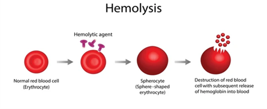
Fig No.1: Hemolysis
Why Does it Occur
Types of Heamolysis
Medications
Aim and Objectives
Investigating Delonix regia Maslin's hemolytic activity is the goal of the study in order to shed light on its possible medical and health ramifications.
Objectives
Review of Literature
Delonix regia, a member of the Fabaceae family, is widely known for its ornamental value and traditional medicinal applications. Various parts of the plant, including the leaves, bark, flowers, and seeds, are used in folk medicine for their anti-inflammatory, antidiabetic, antimicrobial, and antioxidant properties. Recent studies have explored the haemolytic activity of Delonix regia, which is an important parameter in evaluating the cytotoxicity and membrane-disrupting potential of plant extracts. Haemolytic activity refers to the ability of a substance to lyse red blood cells (RBCs), thereby releasing hemoglobin into the surrounding fluid. It is a crucial assay in pharmacological studies, particularly in assessing the biocompatibility and toxicity of plant extracts intended for therapeutic use.
Plan of Work
The present study is designed and carried out by the following methods to evulate the hemolytic activity of Delonix regia
Plant Profile:
Plant Collection and Authentication: The best time for collection of Delonix regia is generally during the flowering and fruiting seasons. Delonix regia plant were collected from Bhivghat ,sangli ,Maharashtra,India.The plant was authenticated by Mr. Tebhurne R.R. M.Sc.B.Ed Botany plant physiology.
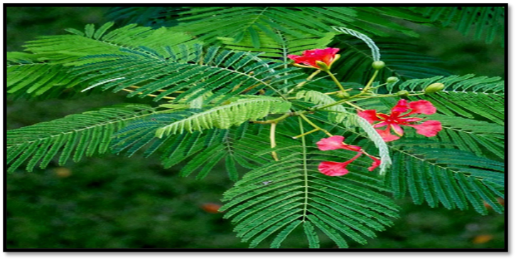
Fig. No.2 : Delonix regia
Description
Uses:
Propagation:
By seeds (which often require scarification or soaking before sowing to improve germination). Can also be propagated vegetatively via cuttings.
Growing Conditions:
Cultivation and Collection:
Cultivation
Climate and Soil
Propagation
Seeds:
Vegetative:
Sowing
Transplantation
Maintenance
Collection
Harvesting of Seeds
Fruit type: Long, woody pod (up to 60 cm).
Time of collection: Pods mature and dry between November to January in most tropical regions.
Collection Method:
Seed Extraction
Storage
MATERIAL AND METHODHOLOGY
1. Preparation of plant Extracts Delonix regia
Collection of Plant Material: Harvest fresh leaves, young shoots, or other plant parts of Delonix regia during the appropriate season as mentioned earlier.
Cleaning and Drying: Clean the collected plant material to remove any dirt or debris. Allow the plant material to air dry in a well-ventilated area away from direct sunlight until it is thoroughly dried. This step helps to prevent the growth of mold and bacteria during storage.
Grinding or Crushing: Once dried, grind or crush the plant material into smaller pieces using a mortar and pestle or a grinder. This increases the surface area of the plant material, facilitating the extraction process.[16]
2. Preparation of Ethanolic Extract
The extraction preparation procedures differed slightly from those detailed in. The leaf sample wasowashed with ordinary water, alleshtly from those detailed in The blender to be ground into powder. Various ratios ored to dry, and then put into for the Soxhlet extraction procedure. 6 to 8 hours after thanol are used as gathered Utilise a muslin cloth to filter it. Centrifuge the far the extract has been gathered 15 minutes at 4,000 rpm and 25 °C. After being gathered the supernatant was retained for drying.[17]
3. Preparation of phosphate Buffer Solution
To prepare a phosphate buffer solution, you will need to mix solutions of monobasic sodium phosphate (NaH2PO4) and dibasic sodium phosphate (Na2HPO4) in appropriate proportions to achieve the desired pH. Here's a general procedure for preparing a phosphate buffer solution[18]
Calculate the Proportions: Determine the desired pH of the buffer solution using a buffer calculator or table. Then, calculate the appropriate proportions of monobasic and dibasic sodium phosphate solutions needed to achieve the desired pH. The Henderson-Hasselbalch equation can be used for this purpose.
Prepare Stock Solutions: Prepare stock solutions of monobasic sodium phosphate (0.2 M) and dibasic sodium phosphate (0.2 M) by dissolving the appropriate amounts of each salt in distilled water. Ensure that the salts are completely dissolved.
Mixing: Slowly add the calculated volumes of the monobasic and dibasic sodium phosphate stock solutions to a clean glass container, while stirring continuously. Continue mixing until the desired pH is reached.
Adjust pH (if Necessary): Measure the pH of the buffer solution using a pH meter or pH indicator paper. If the pH is not within the desired range, adjust it by adding small amounts of either monobasic or dibasic sodium phosphate solution as needed, and then recheck the pH.
Final Dilution (if needed): Once the pH is adjusted to the desired range, adjust the volume of the buffer solution to the final desired volume by adding distilled water if necessary. Mix thoroughly
Filtering (optional): If desired, filter the buffer solution using a sterile filter to remove any particulate matter or impurities.
Sterilization (optional): If the buffer solution is to be used for biological applications, it may be sterilized by autoclaving or filtering through a sterile membrane filter.
Storage: Store the prepared phosphate buffer solution in a clean, sterile container with an airtight lid. It can be stored at room temperature for short-term use or refrigerated for longer-term storage.
4. The process for Making Erythrocyte Cells:
Blood samples were collected from healthy volunteer donors of blood. A sterile saline phosphate buffer solution (PBS Buffer) was used to wash the pellet obtained after centrifuging 5 ml of blood. The cell suspension was added once more to a 0.5% solution of regular saline.
5. Perform the in-vitro hemolytic activity test:
make different concentration of solution and mixed with of the erythrocyte suspension and incubate and centrifuge it and free hemoglobin obtained, measured the absorbance using the UV-Vis spectrophotometer and calculate the % hemolysis.[19]
Phytochemical Investigation:
Table No.1: Phytochemical constituents of Delonix regia exract.
|
Sr. No. |
Name of Test |
Observation |
Inference |
|
1. |
Test For Phenol: Extract Mixed with 2 ml of 2% of Solution of Fecl3 |
Blue/Green Colour |
Phenol present |
|
2. |
Test for Saponin: The Extract was taken in test tube and shaken vigorously with water |
Formation of stable foam |
Saponin present |
|
3. |
Test for Tannins: Extract Mixed with 2% of Fecl3 |
No Black Colour |
Tannin Absent |
|
4. |
Test For Terpenoids: The Extract mixed with choloroform.then 2ml of conc.sulphuric acid was added carefully and shaken gently |
Reddish brown colour observed in the interphase |
Terpenoids Present |
|
5. |
Test for flavonoids: A few drops of sodium hydroxide solution were added to the extract. |
formation of a bright yellow hue. It turns colorless when diluted acid is added. |
Flavonoids present |
|
6. |
Test for glycosides: the extract was mixed with 2ml of glacial acetic acid containing few drops of 2% Fecl3 ; mixture poured into another tube containing 2ml of conc. Sulphuric acid. |
A brown ring at the inerphase |
Carbohydrate present |
|
7. |
Test for protein: The extract treated with few drops of conc. Nitric acid |
Formation of yellow colour. |
Protein Absent |
|
8. |
Test for alkaloids:
To a few ml of filtrate ,1 or 2ml of dragndroff reagent. |
Orange brown coloured ppt. |
Alkaloids present |
|
|
B) Mayers test: To few ml of extract ,2drops of mayers reagents |
Cream coloured ppt. |
Alkaloids present |
|
|
C) Hagers test: To few ml of extact 1or2ml of hagers reagent(saturated solution of picric acid)were added |
Yellow coloured ppt.
|
Alkaloids present
|
|
|
D)wagners test: To few ml of the extract, few drops of wagner reagent (iodine in potassium iodide) |
Reddish brown coloured ppt. |
Alkaloids present |
Experimental Work:
Requirement
Chemical used:
Ethanol,streile (PBS Buffer Solution),Distilled Water,Potassium Dihydrogen Phosphate , Disodium Hydrogen Phosphate.
Apparatus Used:
Test tube, Heparin Tube,Beakers, Stirrer,5ml syringe,Soxhlet Extractor.
Equipment Used :
Centrifugation Machine, Incubator, UV-vis Spectrophotometer [20]
Procedure:
1. Preparation of Plant Extracts Delonix regia
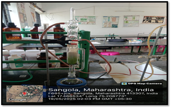
Fig No.3: Preparation of Plant Extract.
The extraction preparation procedures differed slightly from those detailed in. The leaf sample wasowashed with ordinary water, alleshtly from those detailed in The blender to be ground into powder. Various ratios ored to dry, and then put into for the Soxhlet extraction procedure. 6 to 8 hours after ethanol are used as gathered Utilize a muslin cloth to filter it. Centrifuge the far the extract has been gathered 15 minutes at 4,000 rpm and 25 °C. After being gathered the supernatant was retained for drying. [21]
2. Preliminary phytochemical screening of Extraction:
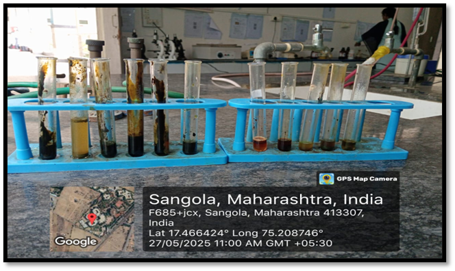
Fig No.4: Phytochemical Screening
3. Preparation of Phosphate Buffer Solution:
To prepare a phosphate buffer solution, you'll need to mix monobasic sodium phosphate (NaH2PO4) and dibasic sodium phosphate (Na2HPO4) in appropriate proportions to achieve the desired pH. Here's a general procedure:
a. Calculate Buffer Ratio: Use the Henderson-Hasselbalch equation to calculate the ratio of monobasic to dibasic sodium phosphate needed to achieve the desired pH. The equation is pH = pKa + log ([A-]/[HA]), where [A-] is the concentration of the conjugate base and [HA] is the concentration of the weak acid.
b. Prepare Stock Solutions: Prepare separate stock solutions of monobasic sodium phosphate (0.1 M) and dibasic sodium phosphate (0.2 M) by dissolving the appropriate amounts of each salt in distilled water. Ensure complete dissolution.
c. Mixing: Mix the stock solutions together in the calculated ratio to achieve the desired pH. For example, to prepare a pH 7.4 buffer, mix 100 ml of 0.1 M monobasic sodium phosphate with 400 ml of 0.2 M dibasic sodium phosphate.
d. Adjust pH (if necessary): Measure the pH of the buffer solution using a pH meter or pH indicator strips. If the pH is not within the desired range, adjust it by adding small amounts of either monobasic or dibasic sodium phosphate solution as needed, and then recheck the pH.
e. Final Dilution (if needed): Once the pH is adjusted to the desired range, adjust the volume of the buffer solution to the final desired volume by adding distilled water if necessary. Mix thoroughly.
f. Filtering (optional): If desired, filter the buffer solution using a sterile filter to remove any particulate matter or impurities.
g. Sterilization (optional): If the buffer solution is to be used for biological applications, it may be sterilized by autoclaving or filtering through a sterile membrane filter.
h. Storage: Store the prepared phosphate buffer solution in a clean, sterile container with an airtight lid. It can be stored at room temperature for short-term use or refrigerated for longer-term storage.[22]
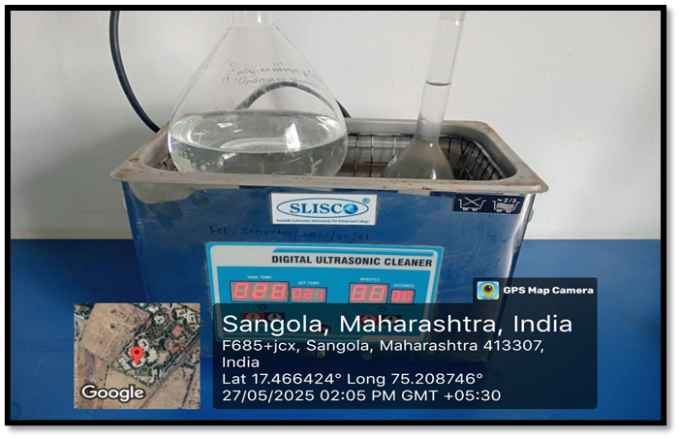
Fig No.5: Preparation of Phosphate Buffer.
4. Preparation of Erythrocyte Cell:
Preparation of erythrocytic cells, also known as red blood cells, involves isolation from whole blood. Here's a general procedure:
a. Blood Collection: Collect whole blood from a suitable animal species or human donor using sterile techniques. Use anticoagulants such as EDTA or heparin to prevent blood clotting during processing.
b. Centrifugation: Transfer the collected blood into centrifuge tubes and centrifuge at low speed (e.g., 200-300 x g) for 10-15 minutes. This separates the blood into layers, with erythrocytic cells settling at the bottom
c. Plasma Removal: Carefully remove the upper layer containing plasma using a pipette or vacuum aspirator. Be careful not to disturb the erythrocytic cell layer
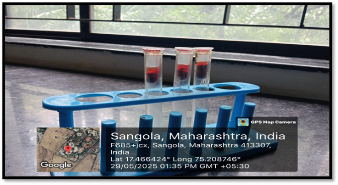
Fig No.6: Separation of Serum and RBCs.
d. Washing: Wash the erythrocytic cell pellet multiple times with an isotonic buffer solution (e.g., phosphate-buffered saline, PBS) to remove any remaining plasma proteins and platelets. Centrifuge the cells after each wash and carefully remove the supernatant
e. Resuspension: After the final wash, resuspend the erythrocytic cells in the desired buffer solution or medium for further experimentation. Adjust the cell concentration as needed using a hemocytometer or automated cell counter
f. Storage: Store the prepared erythrocytic cell suspension in aliquots at appropriate temperatures. Erythrocytes are typically stored at 4°C for short-term use or frozen at -20°C or -80°C for long-term storage. Avoid repeated freeze-thaw cycles to maintain cell integrity.[23]
5. Haemolytic Activity test:
Hemolytic activity testing is a common assay used to assess the ability of substances to cause the lysis or rupture of red blood cells (erythrocytes). Here's a general overview of the hemolytic activity test
a. Preparation of Red Blood Cells (RBCs): Obtain fresh whole blood from a suitable animal species or human donor using sterile techniques. Centrifuge the blood to separate the RBCs from plasma and buffy coat.
b. Washing of RBCs: Wash the RBC pellet multiple times with an isotonic buffer solution (e.g., phosphate-buffered saline, PBS) to remove any residual plasma proteins and platelets. Centrifuge the RBCs after each wash and carefully remove the RBCs.
c. Preparation of Test Samples: Prepare the test samples containing the substance of interest at various concentrations.(25, 50, 50, 75, and 100 g/ml in the saline phosphate buffer) The substance could be a natural product extract, synthetic compound, or pharmaceutical formulation.
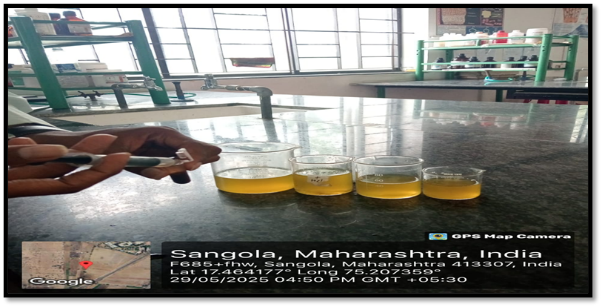
Fig No.7: Preparation of Test Sample
d. Incubation with RBCs: Incubate the RBC suspension with the test samples at physiological conditions (e.g., 37°C, pH 7.4) for a specific period, typically 1-2 hours.
e. Centrifugation: After the incubation period, centrifuge the RBC suspension to separate the intact RBCs (pellet) from any lysed or ruptured RBCs (supernatant).
f. Measurement of Hemolysis: Measure the absorbance of the supernatant at a suitable wavelength (e.g., 540 nm) using a UV-vis spectrophotometer. The absorbance is directly proportional to the amount of hemoglobin released, indicating the degree of hemolysis.
g. Calculation of Hemolytic Activity: Calculate the percentage of hemolysis using the formula: Hemolysis (%) = (Absorbance of test sample - Absorbance of negative control) / (Absorbance of positive control - Absorbance of negative control) × 100 [24,25,26]
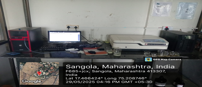
Fig.No.9: Checking Absorbance of Sample.
The Following Formula used to determine the proportion of hemolysis.
% Hemolysis = [(At-An) / (Ac-An)] ×100
Where ,
At: Absorbance of the test sample
An: minimal control absorbance (phosphate buffered saline solution PBS)
Ac: maximum control absorbance ( distilled water)[27]
Observation:
Table No.2 : Absorbance of sample
|
Sr.no |
Concentration |
Absorbance |
Hemolysis % |
Protection |
|
1 |
25ml |
0.017 |
89% |
0.89 |
|
2 |
50ml |
0.061 |
68.15% |
0.68 |
|
3 |
75ml |
0.185 |
14.44% |
0.14 |
|
4 |
100ml |
0.225 |
4.20% |
0.042 |
RESULT:
A helpful method for figuring out the molecular composition of various plant extracts and for discovering bioactive chemicals used in medication development is phytochemical screening. Findings from phytochemical analyses of ethanolic extracts of Delonix regia leaves and stem The Delonix regia screening revealed the absence of reducing sugar but the presence of moisture and elemental components such as carbon, hydrogen, nitrogen, and sulfur. The existence of these chemicals demonstrates the plant's potential for therapeutic use. Tests can be conducted to identify the various phenolic compounds, amino acids, and medicinal value of the plant because neither the stem nor the leaf contain reducing sugar.
Table No.3: Result of Preliminary Phytochemical Screening of Delonix regia
|
Sr. No. |
Constituents |
Observation |
|
|
Ethanolic Extract |
|
|
1 |
Saponins |
+ |
|
2 |
Phenols |
+ |
|
3 |
Tannins |
- |
|
4 |
Terpenoids |
+ |
|
5 |
Flavonoids |
- |
|
6 |
Protiens |
+ |
|
7 |
Carbohydrates |
+ |
|
8. |
Alkaloids |
- |
(-) Indicates the Absence of Compound
(+) Indicates the Presence of Compound
Erythrocytes were used to test the ethanolic extract of Delonix regia's hemolyzing capacity, and the results were reported as a hemolysis percentage. Table No. 4's findings demonstrated that the extracts under study had an impact on hemolysis. Concentrations of 75 g/ml, 4.20%, 50 g/ml, and 68.15% result in the least amount of hemolytic activity, while 25 g/ml, or 89%, results in the highest. The ethanolic extract exhibits the least level of hemolytic activity when it is concentrated at 100 g/ml. The findings also showed that the extract concentration affects the extent of hemolysis. The hemolytic effects of the various test extracts can be grouped into the following categories: 25 g/ml to 100 g/ml, with 50 g/ml to 75 g/ml as the range of values. Medicinal plants include a variety of phytochemical components, including phenol, saponin, and glycosides, which are plentiful and have particular pharmacological effects on humans. Alkaloids, among others. Any chemical that exhibits hemolytic action is often harmful to healthy, normal cells. The four extracts' medium hemolytic response suggests that they have a medium cytotoxicity effect on human erythrocytes. This test is useful for determining whether cytotoxic activity is associated with membrane damage.[28]
Table No.4: Percentage of Hemolysis
|
Sr.no |
Concentration |
Hemolysis% |
|
1 |
25 |
89 % |
|
2 |
50 |
68.15 % |
|
3 |
75 |
14.44 % |
|
4 |
100 |
4.20 % |
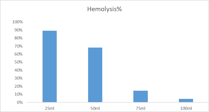
Graph No.1:Graphical Presentation of Haemolysis %
DISCUSSION:
The assessment of in-vitro hemolytic activity of Delonix regia presents an intriguing avenue for understanding the potential biological effects of this plant extract on red blood cells. Hemolysis, the rupture or destruction of red blood cells, can occur due to various factors including chemical agents, toxins, or natural compounds. Delonix regia, also known as the climbing wattle, possesses a diverse array of phytochemicals, making it a subject of interest for biomedical research.
Through in-vitro studies, researchers can elucidate the hemolytic properties of Delonix regia extract by exposing red blood cells to different concentrations of the plant extract and assessing the degree of hemolysis. This process allows for the determination of the extract's cytotoxic effects on erythrocytes, potentially shedding light on its safety profile or therapeutic potential.
The discussion surrounding the research on Delonix regia hemolytic activity necessitates consideration of several key points. Firstly, the concentration-dependent nature of hemolysis should be evaluated to ascertain any dose-response relationships. Additionally, the identification of specific phytochemical constituents responsible for the observed hemolytic effects is crucial for understanding the underlying mechanisms.
Furthermore, the implications of the findings on human health and pharmacological applications should be addressed. While hemolysis may indicate cytotoxicity, it could also signify potential therapeutic benefits such as antimicrobial or anticancer properties. Therefore, a comprehensive discussion should encompass both the risks and benefits associated with Delonix regia extract.[29]
CONCLUSION:
The haemolytic activity assay of Delonix regia leaf extract revealed mild to moderate haemolytic potential in a dose-dependent manner. This suggests the presence of membrane-active phytochemicals, such as saponins or tannins, which may disrupt erythrocyte membranes at higher concentrations. While the extract showed biological activity, its relatively low haemolytic effect at lower concentrations indicates a favorable safety profile for potential pharmacological use. However, further studies including cytotoxicity assays on human cell lines and in vivo toxicity evaluations are necessary to establish its safety and therapeutic potential.
The investigation into the haemolytic activity of Delonix regia leaf extract demonstrated that the plant contains bioactive compounds capable of inducing red blood cell (RBC) membrane lysis in a dose-dependent manner. The observed haemolysis suggests the presence of phytochemicals such as saponins, which are known for their surfactant-like properties that can disrupt cell membranes.
At lower concentrations, the extract exhibited minimal haemolytic activity, indicating relative safety and biocompatibility, which is promising for its potential therapeutic applications. However, as the concentration increased, a notable rise in haemolysis was observed, suggesting a threshold beyond which cytotoxic effects may arise. This dual behavior highlights the importance of dosage considerations in the medicinal use of Delonix regia.
Overall, the study suggests that while Delonix regia exhibits haemolytic properties, these are within an acceptable range at lower concentrations, supporting its traditional medicinal use. Nonetheless, further in-depth studies, including compound isolation, cytotoxicity testing on different human cell lines, and in vivo safety evaluations, are required to fully understand its pharmacological and toxicological profile.[30]
REFERENCES


Akshay Khatal, Amol Pore, Dr. Sanjay Bais, Shantanu Bele, Assessment of In-vitro Haemolytic Activity of Delonix regia, Int. J. of Pharm. Sci., 2025, Vol 3, Issue 9, 81-96. https://doi.org/10.5281/zenodo.17016041
 10.5281/zenodo.17016041
