
We use cookies to ensure our website works properly and to personalise your experience. Cookies policy
Shri Ganpati Institute of Pharmaceutical Sciences and Research, Tembhurni-413211.
Bioresponsive drug delivery systems represent an ideal approach in targeted therapeutic system, offering precise control over drug release in response to specific biological stimuli, such as pH, redox gradients, or enzymatic activity. These systems are especially in the treatment of cancer, cardiovascular diseases, and chronic inflammatory conditions etc where spatial and temporal control of drug release can significantly improve therapeutic outcomes. Sonoporation, a technique which use ultrasound waves to create temporary pores in the cellular membrane, that shows enhancement to the delivery of therapeutic agents. By controlled drug release, sonoporation can improve the efficacy of bioresponsive systems, reducing systemic side effects and increasing the bioavailability of the drugs at the target site. The correlation between bioresponsive drug delivery systems and sonoporation which provides potential for precise medicine, particularly in chemotherapy and gene therapy.
Bioresponsive drug delivery systems are designed to release therapeutic agents in response to specific biological signals, offering a highly targeted work to the treatment. One of the most innovative techniques in this is sonoporation, a process that has ultrasound to temporarily creates small opening to the cell membranes for drug penetration.The concept has a rich history back to the 1950s when the first ultrasound-based therapies were explored. In 1980s, ultrasound was recognized for enhancing the permeability of cell membranes, that phenomenon later known as "sonoporation." However, it wasn’t in demand till the late 1990s.[1-6] During this period, scientists discovered that ultrasound could generate pores in cell membranes without causing permanent damage. This was the key factor for the beginning of using sonoporation as effective therapy, particularly in oncology and gene therapy.In the early 21st century, bioresponsive systems achieved momentum due to advances in nanotechnology and biotechnology. Sonoporation acted as a ‘Sanjivani' method for treating localized diseases like cancer, where high drug concentrations are needed at specific sites without harming healthy tissues. Researchers began coupling sonoporation with microbubbles for further enhancement of drug delivery efficiency, which helps for even more precise control of the drug release process.Today, sonoporation acts as a median between bioresponsive drug delivery and personalized medicine. Its ability to deliver drugs selectively while minimizing systemic side effects is shaping the future of therapies, with ongoing research exploring its potential in various medical fields. (Ben-Gurion University Of The Negev are currently working on the preparation of drug delivery system where nanoparticles having medication and special polymer which helps for distinguishiment of Healthy cells and cancerous cells and in that Sonoporation is a key role player for the development.[7]
CLASSIFICATION
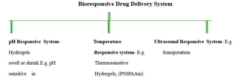
Other-
#Specific Biomolecule Responsive System:- E.g. Antigen/Antibody Responsive System and Nucleic acid Responsive System
#Gas Responsive System:-E.g. CO2 Responsive System(Carriers release drug in response to CO2
#Enzyme Responsive System:- Polymers that are cleaved by specific enzymes.E.g. Proteases & Enzyme sensitive nanocarriers
Each of these types of BRDDS offers unique advantage with specific therapeutic needs making them a versatile tool in new drug delivery System.
Diagram.1. Classification Of Bioresponsive Drug Delivery System[13-14]
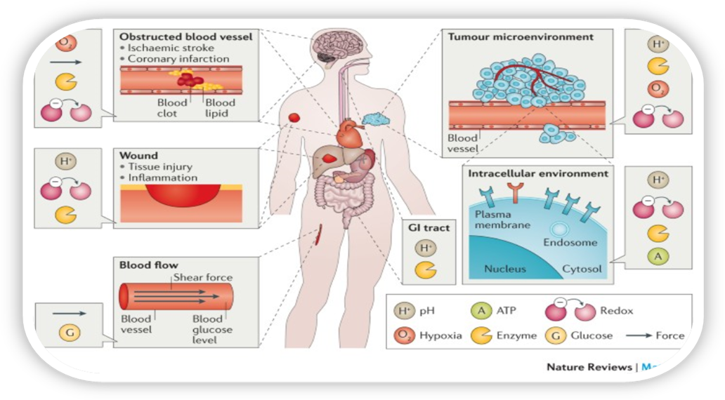
Diagram.2. Bioresponsive Materials[15]
‘Smart’ bioresponsive materials that are sensitive to biological signals or to pathological abnormalities are working on various aspects for the development of next-generations' precision medications. This Review highlights recent advances in the design of smart materials capable of responding to the physiological environment, to biomarkers and to biological particulates. Key design principles, challenges and future directions, including clinical translation, of bioresponsive materials are also discussed.[16]
Overview of Ultrasound
The normal human sound range is from 16 Hz to 15-20,000 Hz (in children and young adults). Beyond this limit, the mechanical vibration is known as ultrasound. The frequencies used in therapy are between 1.0 and 3.0 MHz. Waves are longitudinal wavesof compression and rarefaction. When particles are exposed to a sound wave will oscillate about a fixed point rather than move with the wave itself. As the energy within the sound wave is passed to the material, it will cause oscillation of the particles of that material. Clearly any increase in the molecular vibration in the tissue can result in heat generation, and ultrasound can be used to produce thermal changes in the tissues.[17-20]
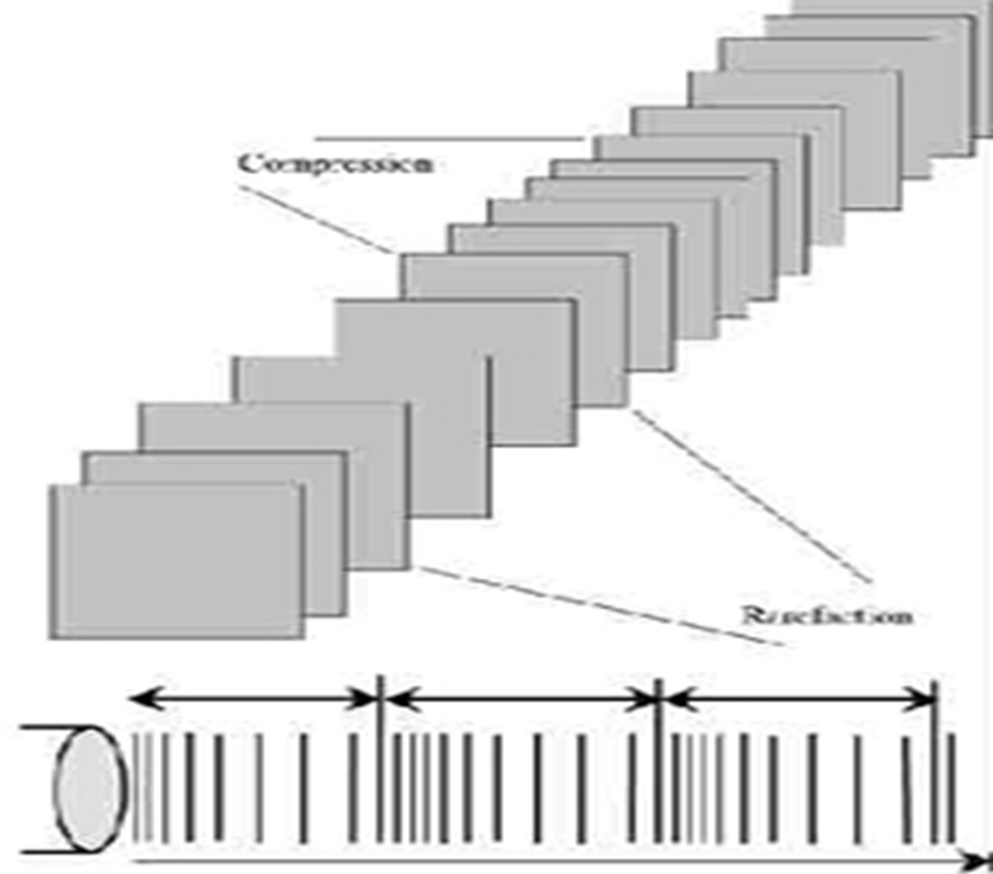
Diagram.3. Ultrasound waveform with compression and rarefaction
Used for Sonoporation Equipments -
#Sonoporation Protocol Software:
Here are the specific steps for the optimization of a sonoporation regimen using a 250-kHz ultrasound transducer, microbubbles, plasmid DNA and human mesenchymal stem cells (hMSCs). The overall goal is to transfer genes to mesenchymal stem cells and assess the resulting protein production. In our previous studies, we achieved targeted bone regeneration via activation of resident stem cells. Sonoporation was applied to deliver bone morphogenetic protein 6 (BMP-6) plasmid to the recruited MSCs within bone fractures to induce bone regeneration and fracture healing. This protocol is focused on in vitro sonoporation validation in support of translational studies of ultrasound-mediated gene delivery. With optimization of a few key parameters in the ultrasound treatment and cell culture conditions, we anticipate that this workflow is suitable for most in vitro experimental models.
In short, this is a protocol uses diagnostic microbubbles and parameters that are applicable for human use.[21]
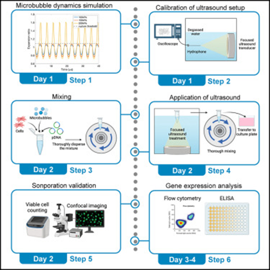
Diagram.4, Step involved in invitro Sonoporation Protocols[21]
#Ultrasound Transducer:
An ultrasound transducer is a key component of ultrasound imaging systems. It converts electrical energy into sound waves and vice versa, enabling the process of capturing internal images of the body. Here's a breakdown of how it works
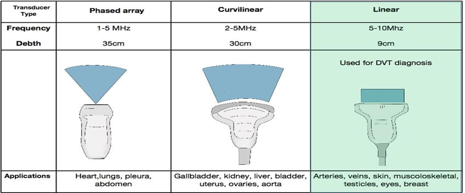
Diagram.5. Types of Transducers
Transmission: The transducer sends high-frequency sound waves (ultrasound) into the body. These sound waves are typically in the range of 2 to 18 MHz, which is above the hearing range for humans.
Reception: When the sound waves hit different tissues, organs, or fluids, they bounce back (echoes) to the transducer. The transducer then converts these echoes into electrical signals.
Image Formation The signals are processed by the ultrasound machine to create real-time images of the body’s internal structures.
Ultrasound is widely used in pregnancy imaging, heart (echocardiography), abdominal organs, and musculoskeletal imaging & In some cases, ultrasound energy is used to treat conditions, like breaking up kidney stones (lithotripsy) or targeting tumors.
#Ultrasound Mediated Gene Transfer
Microbubble Formation: Ultrasound is often used in combination with microbubbles, which are small gas-filled bubbles. These bubbles enhance the efficiency of ultrasound by increasing the mechanical effects on the cell membrane
Cavitation: When exposed to ultrasound waves, the microbubbles oscillate. This causes a phenomenon called cavitation, where the bubbles expand and collapse rapidly. The energy from this cavitation leads to localized mechanical stress.
Membrane Permeable process: The mechanical stress from the collapsing bubbles generates transient pores in the cell membrane, making it temporarily more permeable.
Gene Entry: Genetic material in the surrounding medium (such as plasmid DNA, mRNA, or siRNA) can pass through these pores and enter the cell cytoplasm.
Cell Recovery and Expression: After the pores close, the cell membrane returns to its normal state. The introduced genetic material can then be transported to the nucleus (in the case of DNA) or translated into proteins in the cytoplasm (in the case of RNA), leading to gene expression ,Overall this Sonoporation method used to introduce genetic material in the cell. This technique leverages the mechanical effect of ultrasound waves to enhance the uptake of nucleic acids, such as DNA or RNA, by target cell. UMGT is considered a promising tool for gene therapy because it is relatively safe, targeted, and can be used with various type of genetic material. This ultrasound mediated gene transfer offers a versatile and relatively safe approach to gene delivery, but ongoing research is needed to make this technique more efficient for clinical application [24-28]
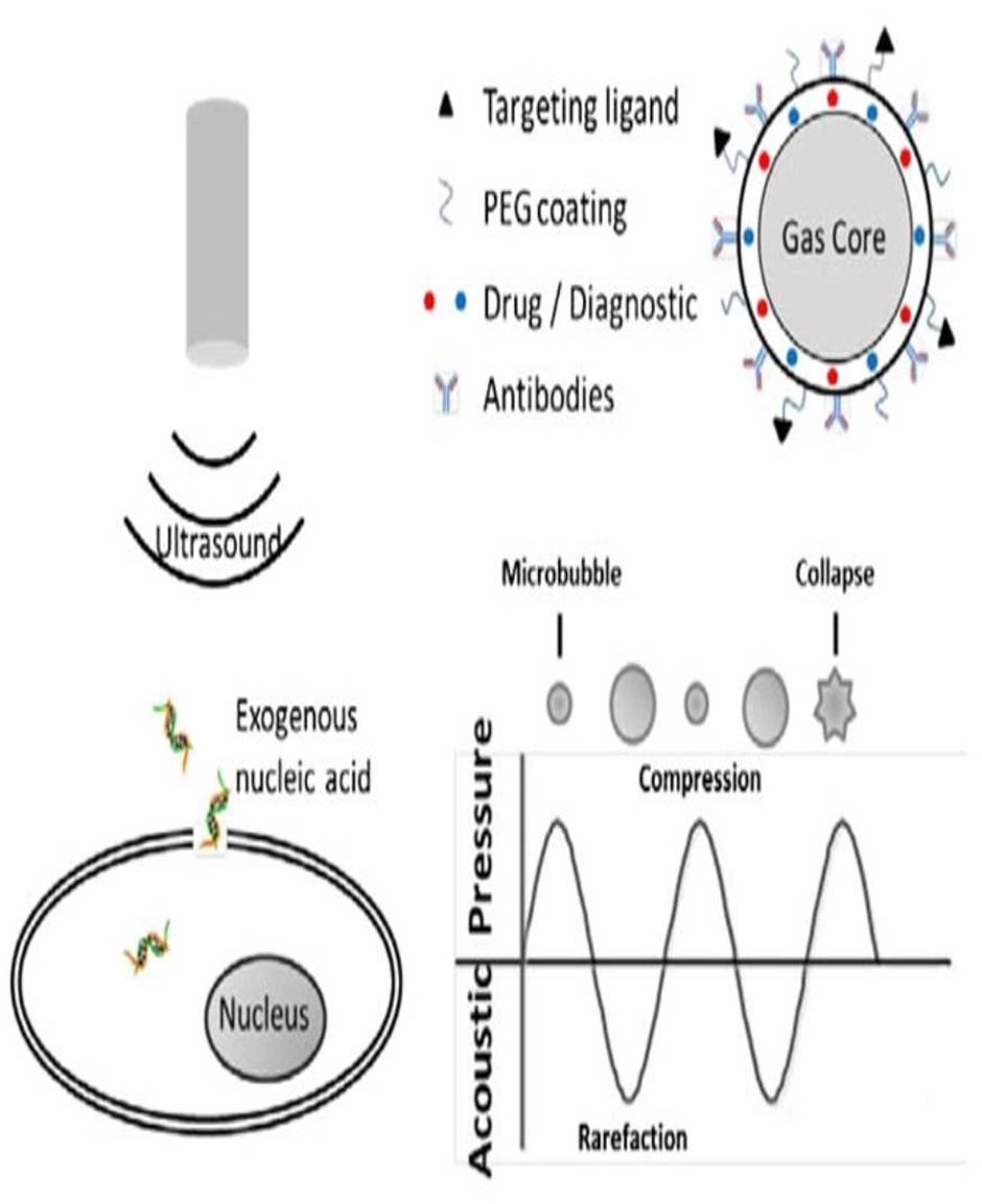
Diagram.6. Genetic info Transfer via Ultrasound
#By using Microbubbles:
This is widely used and key factor for ‘Ultrasound Mediated Gene Transfer Method.’ Ultrasound-responsive lipid microbubbles (ULMBs) are gas-filled microspheres surrounded by a lipid shell that respond to ultrasound stimulation. These microbubbles are widely studied and used in biomedical applications due to their unique properties, particularly in drug delivery and diagnostic imaging. Microbubbles are 1–10 μm diameter gas-filled acoustically-active particles, typically stabilized by a phospholipid monolayer shell.[22-29]
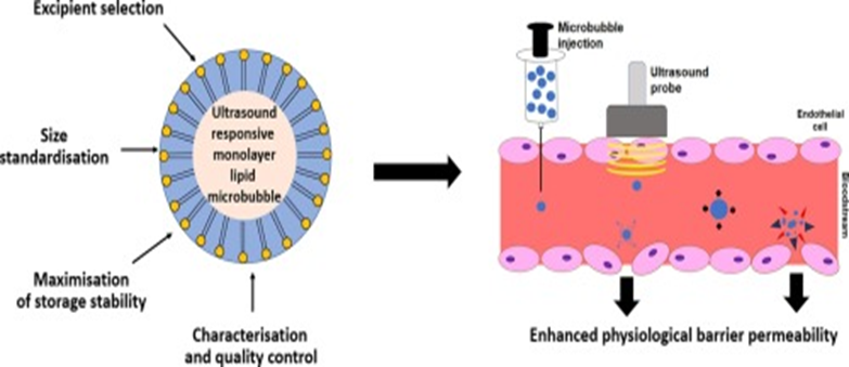
Diagram.7.Ultrasound Responsive Lipid Microbubbles[30]
APPLICATION
Sonoporation involves the use of ultrasound to enhance the permeability of cell membranes, facilitating the delivery of drugs or other molecules into cells. Its applications include:
Gene Therapy: Enhancing the delivery of genes or genetic material into cells to correct genetic disorders.
Drug Delivery: Facilitating the targeted delivery of drugs into specific cells or tissues.
Cancer Treatment: Increasing the effectiveness of chemotherapy and targeted treatments while minimizing collateral damage to healthy tissues.
Vaccine Delivery: Enhancing the uptake of vaccine components to improve immune responses.
Protein Delivery: Facilitating the delivery of proteins into cells for research or therapeutic purposes.
RNA Delivery: Enhancing the delivery of RNA molecules for gene silencing or expression studies.
Stem Cell Engineering: Assisting in the incorporation of stem cells into specific tissues for regenerative medicine.
Intracellular Imaging: Improving the delivery of imaging agents into cells for better visualization under a microscope.
Therapeutic Enzyme Delivery: Facilitating the delivery of therapeutic enzymes to treat enzyme deficiencies or other conditions.
Nucleic Acid Vaccines: Improving the delivery of DNA or RNA vaccines to elicit strong immune responses.
Antisense Oligonucleotide Delivery: Facilitating the delivery of antisense oligonucleotides to modulate gene expression.These diverse applications showcase the versatility of sonoporation in advancing various fields of medicine and research.[31]
RISK ARISES
Sonoporation leads to cell membrane permeability, allowing therapeutic agents, such as drugs or genes, to enter cells. While it has promising potential in targeted drug delivery and gene therapy, several risks are associated with the procedure –
Tissue Damage: High-intensity ultrasound can cause heating and mechanical damage to tissues, potentially leading to burns or injury.
Cellular Damage: While the technique is designed to temporarily open cell membranes, excessive exposure may cause irreversible damage to cells, leading to apoptosis or necrosis.
Uncontrolled Drug Release: Sonoporation may cause unintended release of drugs or genetic material into non-target cells, potentially leading to side effects.
Inflammatory Responses: The process may trigger local inflammatory or immune responses, especially if cavitation (formation of gas bubbles) occurs, which can disrupt surrounding tissues.
Blood Vessel Damage: If sonoporation is used near blood vessels, there is a risk of vessel rupture or leakage, which can cause bleeding or thrombosis.
Long-Term Effects: The long-term impact of sonoporation on tissues and cells is not fully understood, and there may be concerns about potential chronic side effects, especially with repeated treatments.
Difficulty in Precision: Achieving precise control over the ultrasound parameters (such as intensity and frequency) to avoid off-target effects can be challenging.
These risks also involves careful selection of ultrasound parameters and monitoring during the procedure. However, more research is needed to improve its safety profile.[32-35]
COMMERCIALLY AVAILABLE TECHNOLOGIES
Nowadays Sonoporation is mostly in the research and clinical trial phase, with few direct marketed products. However, there are related areas where ultrasound technology has commercialized used in medical practice:
#SonoVue® (Bracco Imaging)Application: Ultrasound contrast agent used for imaging, but its mechanism includes sonoporation-like effects when combined with therapeutic ultrasound. It enhances drug delivery or imaging of targeted areas by making cell membranes more permeable.
Usage: Primarily for enhancing ultrasound imaging in liver and cardiac diagnostics
#Sonocloud® (CarThera)Application: Though not marketed as a standalone drug delivery system, Sonocloud uses focused ultrasound to temporarily open the blood-brain barrier (through sonoporation), allowing drugs like chemotherapy agents to reach brain tumors (e.g., glioblastoma).
Usage: Clinical trials for brain cancer treatment, with promising results in increasing drug penetration into the brain.
#ExoPlex (Ongoing Research and Preclinical Development)Application: Developed by companies researching exosome-based drug delivery, ExoPlex involves ultrasound-activated drug delivery systems. Although not fully marketed, it is an advanced example of integrating sonoporation mechanisms with nanomedicine to target cells for treatment.
#Therapeutic Ultrasound Devices (Physical Therapy)Application: Although simpler, ultrasound devices in physiotherapy claim to enhance healing by improving drug absorption through skin. Some are marketed for treating muscle and joint conditions, indirectly applying sonoporation for localized drug penetration (e.g. topical creams or gels).
Usage: Common in rehabilitation clinics for enhanced healing of soft tissue injuries.
#Liposomal Drug Delivery Systems with Ultrasound Activation Application: Some marketed systems use liposomes that can release their drug payload when activated by ultrasound. This targeted release method is being explored for various cancers and infections. Although still emerging, some companies are developing liposomal products where ultrasound plays a role in targeted delivery.
Examples: Companies like Merrimack Pharmaceuticals have explored this approach in drug delivery.Though sonoporation itself is still in the experimental phase, these examples show the closest commercially available technologies or systems influenced by ultrasound for drug delivery. The field is progressing, and we may see more direct applications in the near future.[36-39]
CONCLUSION
Sonoporation has ultrasound for cell membrane permeability, this holds great promise in advancing targeted drug delivery systems. This method allows convenient way in delivering therapeutic agents, reducing systemic side effects, and improving treatment efficacy, particularly for cancer and genetic disorders. Sonoporation has the potential to revolutionize modern medicine by making treatments more patient-specific and efficient. Continued research and technological advancements are likely to unlock its full potential in clinical applications.
REFERENCES

Farhan Shaikh*, Ashish Sawant, Rahul Mane, Tushar Bansode, Shamir Tamboli Priyanka Khadsare, Bioresponsive Drug Delivery System (Sonoporation), Int. J. of Pharm. Sci., 2025, Vol 3, Issue 5, 5190-5200. https://doi.org/10.5281/zenodo.15562991
 10.5281/zenodo.15562991