
We use cookies to ensure our website works properly and to personalise your experience. Cookies policy
Post Graduate Teaching Department of Biochemistry, Laxminarayan Innovation Technological University, Nagpur.
Around 1.5 million women are diagnosed with breast cancer disease every year, being a major reason women have the highest death rates from cancer. Research in breast cancer is ongoing while the breast cancer cell lines play an important role in molecular diagnostics of the breast cancer, selecting the proper cell line for targeted research is critical. This review introduces classification of breast cancer cell lines, clears some ambiguous information on (MDA-MB- 231, MCF-7, T-47-D, MDA-MB-453, MDA-MB-468 and ZR-75-1) breast cancer cell lines, molecular features, morphological characteristics, functional three-dimensional studies that increase understanding of breast cancer cell lines and help researchers make the proper choice and broaden the selection of cell lines for relevant studies. Lastly, we propose some future directions for studies aimed at breast cancer animal models.
Cancer develops when cells in the body grow abnormally and invade to other parts of body. The process is uncontrolled because the regulatory mechanisms that should control growth and cell division are lost by the cancerous cell (1). Breast cancer is a cancer that arises within breast tissue, generally in the milk-producing ducts (ductal carcinoma) or in lobules (lobular carcinoma). Roughly 1.5 million women are diagnosed every year with breast cancer, and it stands as one of the prominent causes of cancer deaths in women (2). Breast cancer is managed by various primary treatments (3). Research in breast cancer is continuously advancing, and cell lines play key role in molecular diagnostics of breast cancer. With a wide variety of laboratory applications, cell lines are used as an in vitro model in the cancer research (4). The Breast Cancer Cell Lines are used for cancer biology, genetic alterations, and treatment response in vitro. This study aim to understand the complexities of breast cancer and develop new diagnostic and therapeutic options (5). offer several benefits, such as easy to handle and providing an unlimited self replicating source that can be cultivated in infinite quantities, exhibit a relatively high degree of homogeneity and can be easily replaced from frozen reserves if contaminated and the detriment of breast cancer cell line is they are prone to genotypic and phenotypic drift as they are continuously cultured (4).
Classification of Breast cancer cell line
Breast cancer is a very complex and Heterogeneous disease whose classification is based on its molecular features. The key proteins whose expression is vital in the classification of the breast cancer subtypes include human epidermal growth factor receptor 2 (HER2), progesterone receptor (PR) and estrogen receptor, Generally, these subtypes are classified into Luminal A (ER+/PR+, HER2+), Luminal B (ER+/PR+, HER2-), HER2 (HER2 positive)
Triple negative / Basal-like (ER-, PR-, HER2-)
The Luminal A and Luminal B subtypes express estrogen receptors (ER), which constitute a treatment target for hormonal therapies like tamoxifen and letrozole. HER2 subtype therapy is mainly through the targeted agent trastuzumab (Herceptin), which targets HER2 overexpression. Basal-like or triple-negative breast cancer (TNBC) does not have specific targeted therapy because these tumors do not express ER, PR, or HER2. Patients suffering from TNBC have their disease in aggressive forms with poor prognoses and those make the treatment more complicated. Thus, chemotherapy is the primary treatment (6). Triple-negative breast cancer (TNBC) cell lines fall into seven categories: basal-like 1 (BL1), basal-like 2 (BL2), immunomodulatory (IM), mesenchymal (M), mesenchymal stem-like (MSL), luminal androgen receptor (LAR), and unstable (UNS) (7). The identification of molecular targets and precise subtype classification of breast cancers are vital for the development of better treatment and their application for patient-specific therapies (6).
Luminal breast cancer cell lines: Expression of the estrogen receptor (ER) and/or progesterone receptor (PR) is typically a characteristic of luminal breast cancer cell lines. IBEP-1 and IBEP-3 are exceptions in which PR is key for defining their luminal phenotypes. These cell lines show high expression of genes and proteins associated with luminal features, such as ESR1 (Estrogen Receptor Alpha), GATA3 and FOXA1(transcription factors) and luminal keratins (KRT8/18/19). Overexpression of various miRNAs (has-miR-626, has-miR-501-5p, has-miR-760 and has- miR-202) has been showed in miRNA expression profiling on luminal cell lines. Luminal cell lines are generally more differentiated and show lesser migratory capacity because of tight junctions, resembling the features of the tumor. Division of luminal cell lines into luminal A and B subtypes supports tumor subtyping for simple tumor modeling and drug response assays that depend on ER and HER2 status. For example, synergistic effects of tamoxifen and Herceptin for breast cancer treatment were shown on the BT474 (ER+HER2+) cell line, while MCF7 (ER+HER2-) has been commonly used to study tamoxifen-induced cellular responses (8).
Luminal B type of cell lines are usually aggressive and invasive as compared to Luminal A cells because HER2 overexpression is linked to ER down-regulation. The importance of differentiating Luminal B type of cell lines from any other Luminal subtype has been addressed by studies using the ER+HER2+ BT474 cell line, which shown that quiescin-sulfhydryl oxidase 1 expression was associated with a poor prognosis in Luminal B tumours. By drawing the line between these two subtypes, one can better appreciate their distinct biological behavior and treatment responses. (8).
HER2- ER-negative and HER2-positive status are characteristics of HER2 positive cell lines, which also have a similar genetic profile on chromosome 17q12. Genes like HER2, GRB7, PERLD1, STARD3, and C17ORF37 are found in this area, These cell lines exhibit overexpression of specific microRNAs, including has- miR-378, has-miR-29c, has-miR-18a, has-let7b, has-miR-141, has-miR-200c, has-miR-196a and has-miR-640. Cell lines that are HER 2 positive serve as a link between luminal and basal cell lines and display a mix of luminal and basal characteristics. They are more aggressive than luminal cells, especially regarding migration, and their aggressiveness is link to HER2 overexpression, which disturbs cell-cell junctions; they are also more responsive to different drugs and are an excellent model for studying Herceptin. Pearson's correlation test demonstrates a significant correlation between their molecular profile and biological response, which emphasizes their value in research. High ESR1, MAPK1/3, MEK, TYK2, FASN, and GRB7 levels are strongly correlated with a positive drug response, where these proteins are mainly known to promote cell growth. In contrast, high expression of SFN, CAV2, GRB2, RB1, and FLNA predict resistance to therapy, implying that the cells would be less responsive to treatment. Studies performed in cell lines of high HER2 expression have shown that MAPK signaling can predict responses to Herceptin, while the mTOR pathway, Toll-like receptor pathway, N-glycan biosynthesis, and inositol phosphate signalling are involved in Herceptin resistance (8).
Triple-negative cell lines, as indicated by their name, exhibit little to no expression of three key markers: HER2, progesterone receptor (PR) and estrogen receptor (ER). These cell lines are highly diverse and fall into two groups: basal A and basal B cell lines, according to different studies
Triple-negative A subtype, or basal A subtype, is called basal-like because it provides a signature enriched in markers typically found in basal cells. These tumors express basal- specific proteins of various types, such as
The Basal B subtype of triple-negative breast cancer, also known as the mesenchymal cluster or normal-like/claudin-low, is characterized by the overexpression of genes associated with tumor invasion and more aggressive cancer behavior. These comprise genes with functions related to the extracellular matrix and cell migration that include VIM (vimentin), MSN (moesin), different types of collagens (eg. COL3A1, COL6A1/2/3, COL8A1), and a few other important invasion-associated genes, such as MMP2/14 (matrix metalloproteinases), TIMP1 (tissue inhibitor of metalloproteinases), CTSC (cathepsin C), PLAU and PLAUR (urokinase plasminogen activator and receptor), SERPINE1/2 (serine protease inhibitors), and PLAT (tissue-type plasminogen activator). These molecules perform crucial role in remodeling the extracellular matrix, which is important for enabling tumor cells to invade and migrate into surrounding tissues. The tumor cells of this subtype express increased amounts of the signalling factor TGFB1, TGFBR2 (transforming growth factor beta), and AXL, which are major elements for regulating the aggressive characteristics of the tumor. This subtype has been associated with CD44(+) and CD24(-), tumor initiation, metastasis, and therapy resistance cancer stem cell markers. Thus, the above- mentioned molecular features reflect highly invasive and aggressive nature of basal B tumors. Certain proteins (EGFR, CAV1/2, MSN, ETS1) are being utilized to characterize triple- negative breast cancer (TNBC) cells at translational levels. These proteins, when studied along with basal keratins (KRT5/6), CD10, and MET, allow for the identification of a particular subset of TNBC cells called "triple-negative A cells." On the other hand, when these proteins are evaluated together with the stemness marker CD44, they can also identify a different subset designated as "triple-negative B cells." This provides a method to separate various subtypes of TNBC cells based on the molecular and stemness characteristics (8). Different microRNAs are expressed in a distinct manner in the two subtypes of triple-negative breast cancer (TNBC), namely triple-negative A and B cells. MicroRNAs that are overexpressed in A subtypes are hsa-miR-492, hsa-miR-26b, hsa-miR-617, and hsa-miR-155, while others that are expressed more strongly in B subtypes are hsa-miR-125b, hsa-miR-155, hsa-miR-501-5p, hsa-miR-532-3p and hsa-miR-22. Although both subtypes express hsa-miR- 155, it arises from the same precursor in both cases, but it exhibits opposite expression patterns in subtype (A) and subtype (B). Triple-negative A cells represent more differentiated subgroup among triple-negative breast cancer (TNBC) cell lines, these cells might look like luminal or basal cells and on the other hand triple-negative B cells resemble mesenchymal cells and act in an invasive way. Phenotypically, triple-negative A cells resemble the core basal tumor subtype as recognized by basal markers, whereas triple-negative B cells exhibit features associated with epithelial-mesenchymal transition (EMT) and stem-cell markers (8).
Table 1- Immuno-profile of cell lines and List of mRNAs and miRNAs used in identification of each breast cancer cell line subtypes. The literatures where such information is derived are provided right after. (Adapted from (8 and 9)
|
Cell line subtype |
Immuno- profile (9) |
Cell line (9) |
mRNA (8) |
miRNA (8) |
|
|
Luminal A |
ER+, PR+/–, HER2– |
MCF-7, T47D |
HER3, TOB1, TFF3, ER, GATA3, FOXA1, MYB, RET, ZNF278, SPDEF, CRABP2 XBP1, PBX1, MUC1, KRT19/KRT8/KRT18, EGR3, TFF1. |
hsa-miR760 hsa-miR- 626 hsa-miR-501-5p hsamiR-202 |
|
|
Luminal B |
ER+, PR+/–, HER2+ |
ZR-75 |
|||
|
HER2 positive |
ER–, PR–, HER2+ |
MDA- MB-453 |
STARD3, C17ORF37, HER2, GRB7, PERLD1 |
hsa-miR- 29c hsa- let-7b hsa-miR-640 hsa-miR-200c hsa- miR-378 hsa-miR-141 hsa-miR-18 hsa- miR-196a |
|
|
Triple negative |
Basal 1 |
ER–, PR–, HER2– |
MDA- MB-468 |
LAMC2, TRIM29, COL17A1, GABRK, VTCN1, BST2, FABP7, CD10/14/58/59, KRT4/5/6A/6B/13/14/15/16/17, S100A2, SLPI, LYN, ANXA8, BNC1, MET, CD133, ITGA6, ITGB4/6, LAMB3. |
hsa-miR-492 hsa- miR-26b hsa-miR- 617 hsa-miR-155 |
|
Basal 2 |
ER–, PR–, HER2– |
MDA- MB-231 |
COL8A1, MMP2/14, TIMP1, PLAT, CD24(-), CD44, TGFBR2, SERPINE1/2, TGFB1, VIM, SPARC, FN1, FBN1, HAS2, CTSC, PLAU, PLAUR, AXL, PRG1, COL3A1, COL6A1/2/3. |
hsa- miR-501-5p hsa-miR-22 hsa- miR-532-3p hsa-miR-125b hsa-miR- 155 |
|
T-47D
The T-47D cell line was established in November 1974 from pleural effusion fluid collected through thoracocentesis from a 54-year-old patient with inoperable infiltrating ductal carcinoma of the right breast (10). This cell line is similar to the cells found in the mammary gland and is hormone dependent particularly estrogen, which binds to estrogen receptor alpha (ERα) located in the cytoplasm. As a result, this cell line is categorized as ERα-positive and classified as luminal A cell line, known for its sensitivity to estrogen. The T-47D cell line contain features such as desmosomes, tonofilaments, and duct-like vacuoles in the cytoplasm, which suggest it originates from epithelial tissue. Strong evidence for its mammary origin is that, like the original tumor, the cells show fluorescence when tested with goat anti-human casein serum using the direct immunofluorescence method. Additionally, the presence of cytoplasmic steroid receptors, a hallmark of mammary cells, further supports the idea that these cells are of mammary origin. The intensity of this hormone receptor affinity is demonstrated through a radioactive 7-8S component whose binding is greatly diminished with the addition of unmarked hormone. The Scatchard plot, which assesses the relationship between cytosol and increasing concentrations of hormones, shows high affinity binding components in the cytosol for the four steroids (Estrogen, Progesterone, Glucocorticoid, and Androgen Receptor) that were tested. The steroid receptors in T-47D cells have characteristics similar to those in the MCF-7 cell line. Research on the specificity of these receptors, using competition with different hormones, has shown that estradiol and progesterone receptors bind specifically to their corresponding hormones. However, dexamethasone and DHT receptors can also bind to other steroids, though not as effectively as their homologous steroids. The sedimentation of dexamethasone and DHT binding molecules at around 8S on sucrose density gradients indicates that these molecules are authentic receptors. Additionally, Similar cross-competition findings for glucocorticoid and DHT receptors have been reported in various tissues and cell types. For T- 47D cells, estradiol competes for the glucocorticoid receptor, but there is no significant competition observed between dexamethasone and the estradiol receptor (10). T-47D cells show poor migratory and invasive capabilities (11).
ZR-75-1
The ZR-75-1 cell line was derived from a malignant ascitic effusion in a 63-year-old white female in 1978 (12). When cultured, these cells resemble the malignant cells found in the original breast biopsy and those shed into the ascitic fluid. The cells are polygonal, with a large amount of cytoplasm and prominent nuclei, which may contain a single large nucleolus or several chromocenters 2 to 4. They are generally epithelial in nature, with abundant cytoplasm and irregular cell outlines. The cells are occasionally connected by desmosomes and often form rosettes around structures resembling ducts. The cells lining these "ducts" typically have short microvilli extending from their upper surfaces. The cells feature large vacuoles in the cytoplasm that frequently displace the nucleus and are lined with microvilli. These vacuoles contain an amorphous, membranous secretory material. The nuclei are large and lobed, often positioned eccentrically, with one or more distinct nucleoli. The chromatin is typically uniform but can occasionally form small clusters. The cytoplasm is abundant in Golgi complexes, with moderate amounts of smooth endoplasmic reticulum vesicles and rough endoplasmic reticulum strands, which may contain lightly osmiophilic, fluffy material. Free polysomes are plentiful. A moderate quantity of mitochondria of various shapes is present, and tonofibrils are often found in bundles around the nucleus. The cells also contain occasional desmosomes, glycogen rosettes, lipid droplets, and rare multivesicular bodies. The ZR-75-1 cell line has numerous pleomorphic secretory granules with varying densities. These granules are usually located in the apical part of the cell, close to the plasma membrane, or around the membranes of the intracytoplasmic duct-like vacuoles. They can also be found within large intracellular spaces or in duct-like lumens. Each ZR-75-1 cell contains cytoplasmic receptors (17β-Estradiol, 5α- Dihydrotestosterone, Glucocorticoids, and Progesterone) four main types of steroid hormones. These cells are particularly responsive to estrogen and insulin, which makes them a useful model for studying the effects of both hormones. On the other hand, ZR-75-1 cells are specifically suppressed by glucocorticoids and androgens (12). ZR-75-1 cells exhibit low invasiveness (13).
MCF-7
Dr. Soule and his colleagues developed the MCF-7 cell line at the Michigan Cancer Foundation (MCF), named after the foundation in the year 1973. These cells were derived from the pleural effusion of a 69-year-old woman diagnosed with metastatic breast cancer (5). MCF-7 is a commonly used breast cancer cell line that has been maintained and studied by numerous research teams for many years. It is an important model for breast cancer research worldwide, particularly in the development of anticancer treatments. Over the years, MCF-7 has been more informative and useful for clinical applications than any other breast cancer cell line. The cells are positive for estrogen receptors (ER) and progesterone receptors (PR), and they fall under the luminal A molecular subtype. MCF-7 is characterized by its low level of aggressiveness and invasiveness, and it is generally regarded as having low metastatic potential.The human origin of MCF-7 was confirmed through cytogenetic analysis, which showed that most of the chromosomes were metacentric and sub telocentric, with only 21% being acrocentric. Further testing of surface antigens and ribosomal RNA molecular weight in cells from later passages reinforced the human origin of MCF-7 and ruled out contamination by nonhuman cells during subculturing. Additionally, research on isozyme production in MCF-7 demonstrated the synthesis of human glucose-6-phosphatase isozyme B. The Genesis of domes from MCF-7 monolayers, which share structural similarities and hormonal requirements with those formed by mouse mammary epithelial cells, strongly indicates that some functions of differentiated mammary epithelial cells are preserved. This is further supported by the recent discovery of a specific estrogen receptor in MCF-7 cells and early evidence of alpha-lactalbumin production. The biological characteristics of MCF-7 make it an ideal model for efforts to isolate human breast cancer viruses, with such research currently ongoing. Initially, the dominant cells were large, immature, and striated, and did not appear to be of epithelial origin. The exact type of the striated cells was not identified; they may have been mesothelial cells or abnormal fibroblasts. Fibroblasts were present, as indicated by their characteristic morphology and the typical collagen formation in the cultures, which even led to the complete detachment of cells from some tissue culture flasks (14) The original MCF-7 cells typically do not migrate or invade. However, an autocrine loop involving vascular endothelial growth factor (VEGF) promotes migration and invasion of breast cancer cells and express measurable but low amounts of VEGF-A and VEGF-C, and extremely low amounts of VEGF-D (5)
MDA-MB-468
MDA-MB-468 was established in 1977 from the pleural effusion of a 51-year-old woman diagnosed with metastatic breast adenocarcinoma (15) The cell line appears to be of epithelial origin, displaying typical features such as desmosomes, microvilli, and large, irregular nuclei. These characteristics are most prominent in the colonies or plaques seen in some of the cell lines, and least in the MDA-MB-468 line. MDA-MB-468 is a human breast cancer cell line known for having a very high number of epidermal growth factor receptors (EGFR) (16) Epidermal growth factor receptor is a transmembrane protein that demonstrates intrinsic tyrosine kinase activity after binding to one of its cognate ligands. Overexpression and/or amplification of EGFR have been observed in human breast tumors (17). Amplification and overexpression of the EGFRG may occur in some cases. While amplification of the gene is rarely seen in primary breast tumors, some tumors - even those with the highest receptor levels - are suspected of utilizing mechanisms other than gene amplification to produce high levels of EGF receptor RNA and protein (16). Hormone receptor-negative (MDA-MB-468) cell lines exhibit low levels of expression for the TLR2, TLR3, TLR6, and TLR9 genes, while showing high expression of the TLR8 gene. The TLR1, TLR4, TLR5, and TLR7 genes are not expressed. The expression of TLR9 mRNA is notably higher in MDA-MB-468 cells, which also show a significant balance between the upregulation of the BAX gene and the downregulation of the BCL2 gene (18) cell line will serve as an effective model for studying the EGF receptor (16). MDA-MB-468 show higher invasive potential (19) and S100A7 gene regulates the growth and invasion of MDA-MB-468 cells, at least partially by activating the NF-κB pathway and stimulating the expression of its target genes, including MMP-9 and VEGF (20)
MDA-MB-231
MDA-MB-231 is a cell line derived from a single pleural effusion sample collected on October 17, 1973, from a 51-year-old white woman who also had intraductal carcinoma (21).Cells lack the expression of either estrogen receptor (ER) or progesterone receptor (PgR), which implies totally hormone-independent, poorly-differentiated tumors in vivo and highly aggressive, invasive (22).Now known as part of the claudin low molecular subtype, defined by by lower levels of claudin3 and claudin4, a lowered Ki67 expression which is a marker for cell proliferation, increase in EMT (epithelial-mesenchymal transition) related marker and the presence of the characteristics found in normal mammary cancer stem cells (CSCs), For example CD44+CD24-/low profile (23) The MDA-MB-231 cell line was employed to study the genes and pathways that control the metastasis to different sites (bones, brain, or lungs) (22). MDA- MB-231 cells have been widely used to overexpress mPRs for in-depth studies of their functional properties (24). In hypoxic conditions, the MDA-MB-231 cell line exhibited high expression levels of 26 genes, including procollagen-proline 2-oxoglutarate 4-dioxygenase-alpha polypeptides I and II (also known as proline 4-hydroxylase 1 and 2), HIF-1 responsive RTP801, hexokinase 2, Cyclin G2, sin3-associated polypeptide (SAP30), enolase 2 (ENO2), centromere protein F (CENF), transforming growth factor-beta induced gene (TGFBI), pyruvate kinase muscle type (PKM2), human IAP homolog B (BIRC2), glucose transporter protein-III (GLUT3), phosphoglycerate kinase, KIAA0742 protein mRNA, N-myc downstream regulated gene 1 (Ndr1), insulin-induced protein 2, phosphoglucomutase 1 (PGM1), mRNA for highly expressed in cancer (HEC), BCL2/adenovirus E1B 19 kDa interacting protein 3 (BNIP3), BCL2/adenovirus E1B 19 kDa interacting protein 3-like gene (NIX), basic helix–loop–helix domain containing protein-class B 2 mRNA (DEC1), U1-snRNP binding protein homolog (U1SNRNPBP), Bcl2-associated transcription factor (Btf), VEGF, transferrin, and coagulation factor III (TF) (25).
MDA-MB-453
MDA-MB-453 obtained from a pleural effusion with metastatic disease of a 48-year-old Caucasian woman, with a modal chromosome number of 45 (26) in 1978 (27). The MDA-MB- 453 cell line expresses the androgen receptor but is classified as ‘triple-negative’ due to the absence of Her-2/neu protein, progesterone receptor, and estrogen receptor-α protein expression. Unlike estrogen receptorα (ERα) and progesterone receptor (PR), the cell line is negative for both, but positive for androgen receptor (AR); Therefore showing a normal apocrine carcinoma steroid receptor profile. One distinguishing feature of this cell line is its high proliferation in reaction to androgens, a process that could be slowed by anti androgens such as flutamide. Her-2/neu activity is also well established in this cell line, along with a functional interaction between the androgen receptor (AR) and Her-2/neu, mediated through the MAPK/ERK1/2 pathway. These features are commonly found in a significant portion of invasive apocrine breast carcinomas (27). MDA-MB-453 cell line is distinct from other breast cancer cell lines because they exhibit high levels of androgen receptor mRNA while lacking estrogen receptor (ER) and progesterone receptor (PR). Further analysis of protein expression shows the absence of estrogen as well as progesterone receptor, and increased AR mRNA levels are translated into high amounts of functional AR protein. This protein can bind androgens with the expected affinity and trigger the expression of genes known to respond to androgens. Additionally, androgens acting through the AR enhance the binding of PRLR in MDA-MB- 453 breast cancer cells. Further supporting the activity of the androgen receptor (AR) in MDAMB453 cells is the capacity of DHT to drive a transfected gene with an androgen response element in the MMTV LTR linked to CAT. the induction of three natural genes showed these cells to be sensitive to androgens. Typically linked with a more favorably prognosis for patients, a differentiating breast tumor phenotype might be maintained or even caused in part by androgens. The steroid hormone receptor profile of MDA-MB-453 cells shows high AR expression, but no estrogen receptor (ER) or progesterone receptor (PR). It has also been suggested that the androgen receptor is the last of the sex steroid hormone receptors to be lost as tumors transition from a receptor-positive to a receptor-negative phenotype (26).
Table 2 – Breast cancer cell lines (MDA-MB-231, MCF-7, T-47-D, MDA-MB-453, MDA-MB- 468 and ZR-75-1) and their respective medium as per American Type Culture Collection (ATCC) website and data sheet provided by National Center for cell science (NCCS) Courtesy: Ms. Rupal Ramteke.
|
Cell lines |
MCF-7 cell line |
ZR-75-1 cell line |
MDA-MB- 468 cell line |
MDA-MB- 231 cell line |
MDA-MB- 453 cell line |
T47D cell line |
|
Medium |
MEM (E) with NEAA + 10% FBS |
RPMI 1640 + 10% FBS |
Leibovitz- 15 + 10% FBS |
Leibovitz- 15 + 10% FBS |
Leibovitz- 15 + 10% FBS |
RPMI 1640 + 10% FBS |
Cross-contamination
There are many ways in which two cell lines can get mixed up due to poor tissue culture practices and insufficient quality control in many labs, if the same labware is used for different cell lines without proper cleaning, cells from one line can easily contaminate the other. Shared media and reagents between cell lines can also lead to cell transfer. A single cell from an aggressive cell line, like HeLa, can quickly dominate a culture, causing the slower-growing cells to vanish within a few passages. These cross-contaminated cells show minor changes in phenotype due to altered culture conditions. While the cross-contaminated cells might appear similar to the original cells, DNA profiling can help detect contamination by revealing a DNA profile consistent with the donor cell line, providing a reliable way to confirm the authenticity of the culture (28).
Breast cancer cell line in 3 Dimension
Morphology of cell in 3D (three dimensions) differs significantly from that observed in 2D (two dimension). Luminal-type epithelial cells exhibit a typical cobblestone shape and form cell-cell adhesion structures such as E-cadherin in 2D. Basal-type epithelial cells are more spiky and elongated in their morphology and bear markers of EMT, like vimentin (9). On the other hand, three-dimensional cell lines showed four different morphologies: round, mass, grape-like, and stellate (29). Luminal A T47D and MCF-7 cells formed tightly packed structures with strong cell-cell adhesions. In contrast, basal MDA-MB-468, claudin-low MDA-MB-231, and HER2-positive MDA-MB-453 cells formed looser, grape-like or star-shaped structures, reflecting their more invasive characteristics in vitro. Functional three-dimensional studies have significantly improved our understanding of normal breast structure and development. They have helped identify the roles of laminin V and desmogleins in maintaining epithelial cell polarity and normal tissue architecture. These models have provided valuable insights into breast cancer biology, highlighting the role of β1-integrin in cancer progression and showing that the use of blocking antibodies can counteract the malignant behavior of epithelial cells. With growing recognition of the stroma’s influence on breast cancer behavior, and the understanding that basal and luminal breast cancers behave differently when co- cultured with stromal fibroblasts, newer three-dimensional models have incorporated various stromal cells like fibroblasts, macrophages, and endothelial cells (9)
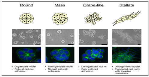
Fig1 - Breast cell line colony morphologies in 3D culture fall into four distinct groups (Adapted from (29)).
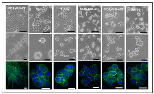
Fig2- Morphologies of (Left to Right: MDA-MB-231, MCF-7, T-47-D, MDA-MB-453, MDA- MB-468 and ZR-75-1) breast cell lines cultured in two- and three-dimensions (Adapted from (29)
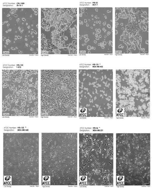
Figure 3- Images of breast cancer cell lines (MDA-MB-231, MCF-7, T-47-D, MDA-MB-453, MDA-MB-468 and ZR-75-1). Courtesy: American Type Culture Collection (ATCC) website.
CONCLUSION
By summarizing and going through all the molecular features of available cell line (on Data sheet of NCCS) for Human Breast cancer, classified them with a subtyping scheme compatible with tumor and resolved some conflicting information of Breast cancer cell lines (MDA-MB- 231, MCF-7, T-47-D, MDA-MB-453, MDA-MB-468 and ZR-75-1) molecular, morphological features, Functional three dimensional studies, which provide greater understanding of breast cancer cell line and it assists us in appropriate selection of and broadens the range of cell lines when performing relevant studies. Many studies are investigating the mechanisms of resistance to chemotherapy in breast cancer cell lines, particularly in triple-negative breast cancer (MDA- MB-231, MDA-MB-453, MDA-MB-468). Understanding how cells become resistant to certain treatments could guide the development of new drugs or combination therapies to overcome resistance, studying the responses of these cell lines to various targeted treatments to predict which therapies may be most effective for individual patients, to better mimic the in vivo conditions of breast cancer, 3D cell culture models and organoids derived from patient tumors. These models more accurately simulate tumor architecture and behavior compared to traditional 2D monolayer cultures, offering insights into tumor progression, drug response, and metastasis, By analyzing gene expression, mutations, and epigenetic changes, new molecular drivers of breast cancer that could serve as biomarkers for diagnosis and prognosis, or as targets for new treatments, investigating the efficacy of immune checkpoint inhibitors, CAR-T cell therapies, and other immune-based treatments, Studies on breast cancer cell lines are also focusing on understanding the processes involved in metastasis, particularly how tumor cells invade surrounding tissues and spread to distant organs. Research in this area aims to identify key molecules and pathways that facilitate metastasis, which could lead to the development of drugs that prevent or halt metastatic spread.
REFERENCES


Rupal Ramteke*, Archana Moon, Tuba Khan, Mohd Vashid Ansari, Comprehensive Review of Breast Cancer Cell Lines: A Guide to Selecting the Optimal Cell Line for Targeted Research, Int. J. 2of Pharm. Sci., 2025, Vol 3, Issue 3, 2580-259 https://doi.org/10.5281/zenodo.15089628
 10.5281/zenodo.15089628