View Article

Abstract

Ketoconazole and Clindamycin are widely used antifungal and antibacterial agents, respectively, and are frequently combined in topical and intravaginal formulations for the treatment of mixed fungal and bacterial infections. Ensuring the quality, safety, and efficacy of such formulations requires reliable and validated analytical methods. The present review critically summarizes the analytical techniques reported for the quantification of Ketoconazole and Clindamycin in bulk drugs, pharmaceutical formulations, and biological matrices. Various spectroscopic methods, including UV and visible spectrophotometry, as well as chromatographic techniques such as RP-HPLC, HPTLC, UPLC, and LC–MS/MS, are discussed with respect to their methodological conditions, sensitivity, and applicability. Special emphasis is placed on stability-indicating chromatographic methods developed in accordance with ICH guidelines, including forced degradation studies. The review also highlights the advantages and limitations of existing methods and identifies the lack of a validated stability-indicating RP-HPLC method for the simultaneous estimation of Ketoconazole and Clindamycin in combined dosage forms. This analytical gap underscores the need for the development of a simple, robust, and regulatory-compliant method suitable for routine quality control and stability studies.

Keywords

Ketoconazole; Clindamycin; Analytical methods; RP-HPLC; UV spectrophotometry; HPTLC; Stability-indicating methods; Pharmaceutical analysis

Introduction

Ketoconazole and Clindamycin are therapeutically important antimicrobial agents that are extensively used in pharmaceutical formulations for the management of infectious diseases. 1Ketoconazole is a broad-spectrum imidazole antifungal agent effective against a wide range of fungal pathogens, particularly Candida species, while Clindamycin is a lincosamide antibiotic with potent activity against Gram-positive and anaerobic bacteria. Due to their complementary mechanisms of action, these drugs are frequently combined in topical and intravaginal formulations for the treatment of mixed fungal and bacterial infections, including vulvovaginal candidiasis and bacterial vaginosis.2-4

The growing clinical use of Ketoconazole–Clindamycin combination products has increased the need for reliable analytical methods to ensure product quality, safety, and therapeutic efficacy.5Accurate quantification of active pharmaceutical ingredients in bulk drugs and finished dosage forms is a critical requirement during formulation development, routine quality control, and regulatory submission processes. Furthermore, stability studies play a vital role in assessing the behavior of pharmaceutical products under various stress conditions and in establishing appropriate storage conditions and shelf life.6-8

Several analytical techniques have been reported in the literature for the estimation of Ketoconazole and Clindamycin individually.9 These include spectroscopic methods such as UV and visible spectrophotometry, chromatographic techniques such as reverse-phase high-performance liquid chromatography (RP-HPLC) and high-performance thin-layer chromatography (HPTLC), as well as advanced hyphenated techniques including ultra-performance liquid chromatography (UPLC) and liquid chromatography–mass spectrometry (LC–MS/MS). Each of these methods offers specific advantages in terms of sensitivity, selectivity, speed, and applicability; however, they also exhibit certain limitations related to cost, complexity, or lack of stability-indicating capability.10

Regulatory guidelines issued by the International Council for Harmonisation (ICH) emphasize the importance of validated, stability-indicating analytical methods for pharmaceutical analysis. Such methods should be capable of accurately quantifying the active drugs in the presence of impurities, excipients, and degradation products formed under stress conditions. Despite the availability of multiple analytical approaches for Ketoconazole and Clindamycin, a comprehensive and consolidated review highlighting their analytical determination, methodological trends, and existing gaps is limited.11

Therefore, the present review aims to critically compile and evaluate the reported analytical methods for the quantification of Ketoconazole and Clindamycin in bulk materials, pharmaceutical formulations, and biological matrices. The review focuses on spectroscopic, chromatographic, and hyphenated techniques, with particular emphasis on stability-indicating methods developed in accordance with ICH guidelines. By summarizing the existing literature and identifying analytical gaps, this review provides a scientific basis for future method development and supports the need for robust and regulatory-compliant analytical methodologies.12

Drug Profile of Ketoconazole13

Table 1:Drug Profile of Ketoconazole

Parameter

Description

Drug Name

Ketoconazole

IUPAC Name

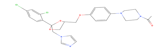
1-[4-(4-{[2-(2,4-dichlorophenyl)-2-[(1H-imidazol-1-yl)methyl]-1,3-dioxolan-4-yl]methoxy}phenyl)piperazin-1-yl]ethan-1-one

Structure

Chemical Class

Imidazole antifungal

Pharmacological Category

Antifungal agent

CAS Number

65277-42-1

Molecular Formula

C??H??Cl?N?O?

Molecular Weight

531.43 g/mol

Physical Appearance

White to off-white crystalline powder

Solubility

Practically insoluble in water; soluble in acidic media

pKa

6.42

Partition Coefficient (Log P)

4.35

Melting Point

~146 °C

Mechanism of Action

Inhibits fungal ergosterol synthesis by blocking cytochrome P450–dependent enzymes

Official Status

BP, USP, Ph. Eur.

Drug Profile of Clindamycin14

Table 2 Drug Profile of Clindamycin

Parameter

Description

Drug Name

Clindamycin

IUPAC Name

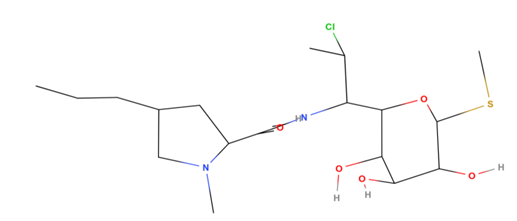
(2S,4R)-N-[(1S,2S)-2-chloro-1-[(2R,3R,4S,5R,6R)-3,4,5-trihydroxy-6-(methylsulfanyl)oxan-2-yl]propyl]-1-methyl-4-propylpyrrolidine-2-carboxamide

Structure

Chemical Class

Lincosamide antibiotic

Pharmacological Category

Antibacterial agent

CAS Number

18323-44-9

Molecular Formula

C??H??ClN?O?S

Molecular Weight

424.98 g/mol

Physical Appearance

White or yellowish crystalline powder

Solubility

Freely soluble in water

pKa

7.6

Partition Coefficient (Log P)

2.16

Melting Point

141–143 °C

Mechanism of Action

Inhibits bacterial protein synthesis by binding to the 50S ribosomal subunit

Official Status

BP, USP, Ph. Eur.

Literature Survey

Reported UV method of Ketoconazole

Table 3. UV Method of Ketoconazole

Sr. No.

Title

Description

Ref

1

UV spectrophotometric method development and validation for estimation of ketoconazole in bulk and pharmaceutical dosage form

Solvent- Methylene Chloride

λmax - 255.2 nm

Linearity- 5-25 μg/ml

15

2

Eco-friendly multivariant green analytical technique for the estimation of ketoconazole by UV spectroscopy in bulk and cream formulation

Solvent- Ethanol-Water (20:80, v/v)

λmax - 226 nm

Linearity- 16-24 μg/ml

16

3

Validation of novel UV Spectrophotometric method for the determination of Ketoconazole in Pharmaceutical Formulation

Solvent- Phosphate buffer with pH 6.8

λmax - 208 nm

Linearity- 10-60 μg/ml

17

4

New visible spectrophotometric method development and validation of ketoconazole in pure and semisolid dosage form

Solvent- Methanol (to dissolve) and DMSO (to makeup)

λmax - 481 nm

Linearity- 5-30 μg/ml

18

5

UV spectrophotometric assay of Ketoconazole oral formulations

Solvent- Purified Water λmax - 240 nm

Linearity- 6.25-100 ppm

19

6

Development and Validation of Stability Indicating UV Spectrophotometric Method for the Determination of Ketoconazole Both in Bulk and Marketed Dosage Formulations

Solvent- Methanol

λmax - 203 nm

Linearity- 2-7 μg/ml

20

7

Spectrophotometric method for the determination of ketoconazole based on amplification reactions

Solvent- Water HPLC Grade λmax - 535 nm

Ketoconazole- 0.2136-1.7088 μg/ml

21

Reported HPLC method of Ketoconazole

Table 4 HPLC Method of Ketoconazole

Sr No.

Title

Description

Ref

1

Environmental benign AQbD based estimation of ketoconazole and beclomethasone by RP-HPLC and multi-analytical UV spectrophotometric method

Mobile Phase: Ethanol: 0.1 M potassium dihydrogen phosphate buffer (pH 2.5) (33:67 v/v);

Stationary phase: ODS reversed-phase column (250 × 4.6 mm, 5 µm);

Flow rate: 1.0 mL/min;

Concentration range: 140–260 µg/mL.

22

2

Stability Indicating RP-HPLC Method For Determination of Ketoconazole in Bulk Drug and in Tablet Dosage Form

Mobile Phase: 0.3% triethylamine in 20 mM potassium dihydrogen phosphate buffer (pH 4.0) : Acetonitrile (68:32 v/v);

Stationary phase: Agilent C8 (150 × 4.6 mm, 5 µm);

Detection wavelength: 232 nm;

Flow rate: 1.0 mL/min; Retention time: 8.97 min;

Concentration range: 10–60 µg/mL.

23

3

Development and validation of reverse-phase HPLC method for estimation of ketoconazole in bulk drug

Mobile Phase: Water: Acetonitrile: Buffer pH 6.8 (51:45:4 v/v);

Stationary phase: Promosil C-18 (250 × 4.6 mm, 5 µm);

Detection wavelength: 238 nm;

Flow rate: 1.0 mL/min;

Retention time: 2.713 min;

Concentration range: 1–50 µg/mL.

24

4

SPE-HPLC method for determination of ketoconazole and clotrimazole residues in cow's milk.

Mobile Phase: Acetonitrile: Sodium acetate buffer (pH 4.6) (85:15 v/v); Stationary phase: Zorbax Eclipse XDB-C18 (250 × 4.6 mm, 5 µm); Detection wavelength: 212 nm; Flow rate: 1.0 mL/min;

Retention time: 5.5 min; Concentration range: 0.1–1.0 µg/mL.

25

5

Application of Validated RP-HPLC Method for Simultaneous Determination of Docetaxel and Ketoconazole in Solid Lipid Nanoparticles

Mobile Phase: Acetonitrile and 0.2% triethylamineamine (pH 6.4) (48:52 v/v);

Stationary phase: Waters Symmetry C18 (250 × 4.5 mm, 5 µm);

Detection wavelength: 230 nm;

Flow rate: 1.0 mL/min; Retention time: 9.5 min;

Concentration range: 0.5–20 µg/mL.

26

6

Chromatographic determination of clotrimazole, ketoconazole and fluconazole in pharmaceutical formulations.

Mobile Phase: Acetonitrile + 25 mM trishydroxymethyl aminomethane buffer (pH 7) (55:45 v/v);

Stationary phase: Bondapak™ C18 (25 cm × 4.6 mm);

Detection wavelength: 260 nm;

Flow rate: 2.0 mL/min;

Retention time: 5.7 min; Concentration range: 80–800 µg/mL.

27

7

An HPLC assay for the determination of ketoconazole in common pharmaceutical preparations

Mobile Phase: 60% acetonitrile in 20 mM disodium hydrogen orthophosphate containing 0.2% v/v diethylamine (pH 4.0);

Stationary phase: Hypersil ODS (200 × 4.6 mm, 3 µm);

Detection wavelength: 232 nm;

Flow rate: 1.5 mL/min;

Retention time: 4.2 min.

28

8

Simple high-performance liquid chromatographic method for determination of ketoconazole in human plasma

Mobile Phase: 0.05 M disodium hydrogen orthophosphate: Acetonitrile (50:50 v/v) (pH 6.0);

Stationary phase: Metaphase KR100-5-C18 (250 × 4.6 mm, 5 µm); Detection wavelength: 260 nm;

Flow rate: 1.5 mL/min;

Retention time: 6.2 min; Concentration range: 62.5–8000 ng/mL.

29

9

Electrochemical detection for high-performance liquid chromatography of ketoconazole in plasma and saliva

Mobile Phase: 0.15 M formic acid and 0.01 M dibutylamine in 50% methanol (pH 3.0);

Stationary phase: Bondapak octadecylsilane (3.9 × 300 mm, 10 µm);

Detection wavelength: 231 nm; Flow rate: 2.0 mL/min;

Retention time: 6.0 min.

30

Reported HPTLC method of Ketoconazole

Table 5 HPTLC method of Ketoconazole

Sr no.

Title

Description

REF

1

High-performance thin-layer chromatographic determination of ketoconazole in pharmaceutical formulations

Mobile Phase: ethanol-acetone-1.0 mol/l H2SO4 (80:10:10) v/v/v

Stationary phase: pre- coated silica gel 60 F254 plates (Merck 10x 20 cm, 0.25-mm thicknes)

λmax :298nm

Rf Value:0.70

Concentration range:3-20µg/ml

31

Reported LC-MS Methods of Ketoconazole

Table 6 LC-MS Method of Ketoconazole

Sr no.

Title

Description

Ref

1

Unique green chromatographic method for the qualitative and quantitative analysis of ketoconazole, its impurities and preservatives from ketoconazole cream formulation: Identification of degradants by Q-ToF LCMS and Robustness by Design of Experiments

Stationary phase:

Waters Acquity UPLC BEH C18 column

Mobile Phase:

A: phosphate buffer

B:phosphate buffer and acetonitrile, 30:70 (v/v)

Flow rate:0.5 mL/min

Detection:225nm

32

Reported UV method of Clindamycin

Table 7 spectrophotometric method of Clindamycin

Sr No.

Title

Description

Ref

1

Development and Validation of a UV-Visible Spectrophotometric Method for Simultaneous Estimation of Curcumin and Clindamycin

Solvent:Water:Methanol

Detection Wavelength:202nm

Linearity:10-60 µg/ml

33

2

UV spectrophotometric method development for estimation of clindamycin phosphate in bulk and dosage form

Solvent:Phosphate buffer saline solution (pH 6.75)

Detection Wavelength:210nm

Linearity:5-30 µg/ml

34

Reported HPLC method of Clindamycin

Table 8 HPLC method of Clindamycin

Sr. No.

Title

Description

Ref

1

Stability-Indicating UPLC Method Development, Validation, and Forced Degradation Studies of Sulfamethoxazole and Clindamycin

Mobile Phase: Methanol: Acetonitrile (80:20 v/v);

Stationary phase: BEH C18 UPLC column (100 mm × 2.1 mm, 1.7 µm); Detection wavelength: 254 nm;

Flow rate: 1.0 mL/min.

35

2

Simultaneous HPLC Determination of Clindamycin Phosphate, Tretinoin, and Preservatives in Gel Dosage Form Using a Novel Stability-Indicating Method

Mobile Phase: (A) 1 mL/L orthophosphoric acid in water, (B) Methanol;

Stationary phase: Agilent C18 (250 × 4.6 mm, 5 µm);

Detection wavelength: 200 nm;

Flow rate: 1.0 mL/min;

Concentration range: 144–336 µg/mL.

36

3

New Methods for Quantification of Amoxicillin and Clindamycin in Human Plasma Using HPLC-UV Detection

Mobile Phase: (A) 5% acetonitrile in phosphate buffer (pH 3), (B) Acetonitrile;

Stationary phase: Poroshell 120 EC-C18 (2.1 × 100 mm, 2.7 µm); Detection wavelength: 204 nm;

Flow rate: 0.5 mL/min;

Concentration range: 1–15 mg/L.

37

4

A Stability-Indicating RP-HPLC Method for the Simultaneous Estimation of Metronidazole, Clindamycin and Clotrimazole in Bulk and Combined Dosage Form

Mobile Phase: Phosphate buffer (pH 4.5) : Methanol: Acetonitrile (30:20:50 v/v);

Stationary phase: Hypersil BDS C18 (250 × 4.6 mm, 5 µm);

Detection wavelength: 210 nm;

Flow rate: 1.0 mL/min;

Retention time: 2.289 min; Concentration range: 25–150 µg/mL.

38

5

A Simple HPLC-UV Method for the Determination of Clindamycin in Human Plasma

Mobile Phase: 0.02 M disodium hydrogen phosphate buffer (pH 2.9) : Acetonitrile (71:29 v/v);

Stationary phase: ACE® C18 (250 × 4.6 mm, 5 µm);

Detection wavelength: 195 nm;

Flow rate: 1.5 mL/min;

Concentration range: 0.5–20 µg/mL.

39

6

RP-HPLC Method Development and Validation for Simultaneous Estimation of Metronidazole, Clindamycin Phosphate and Clotrimazole

Mobile Phase: Potassium dihydrogen phosphate buffer: Acetonitrile (70:30 v/v, pH 2.4);

Stationary phase: Hypersil BDS C8 (250 × 4.6 mm, 5 µm);

Detection wavelength: 210 nm; Flow rate: 2.3 mL/min;

Retention time: 5.712 min; Concentration range: 80–150 µg/mL.

40

7

A New RP-HPLC Method for Estimation of Clindamycin and Adapalene in Gel Formulation

Mobile Phase: Acetonitrile: Phosphate buffer (pH 3.0) (60:40 v/v);

Stationary phase: Luna C18 (250 × 4.6 mm, 5 µm);

Detection wavelength: 210 nm; .

Flow rate: 1.0 mL/min;

Retention time: 3.03 min; Concentration range: 100–500 µg/mL.

41

8

HPLC-UV Assay Method for Clindamycin Palmitate Hydrochloride as Drug Substance and Oral Solution

Mobile Phase: 0.5% triethylamine in water: Methanol (1:9 v/v) adjusted to pH 5.0;

Stationary phase: XTerra RP18 (250 × 4.6 mm, 5 µm);

Detection wavelength: 210 nm;

Flow rate: 1.5 mL/min;

Retention time: 5.77 min.

42

9

Development and Validation of a Gradient HPLC Method for the Determination of Clindamycin and Related Compounds in a Novel Tablet Formulation

Mobile Phase: (A) Carbonate buffer: Acetonitrile (90:10 v/v), (B) Carbonate buffer: Acetonitrile (20:80 v/v); Stationary phase: Waters XTerra RP18 (100 × 4.6 mm, 3.5 µm); Detection wavelength: 214 nm;

Flow rate: 1.0 mL/min.

43

10

A New HPLC-UV Method for the Determination of Clindamycin in Dog Blood Serum

Mobile Phase: Acetonitrile: Phosphate buffer (19:81 v/v) containing 2.5 mM tetrabutylammonium hydrogen sulfate; Stationary phase: Spherisorb ODS-2 C18 (250 × 4 mm, 5 µm);

Detection wavelength: 195 nm;

Flow rate: 1.0 mL/min;

Retention time: 4.0 min; Concentration range: 80–6000 ng/mL.

44

Reported HPTLC method of Clindamycin

Table 9 HPTLC method of Clindamycin

Sr no.

Title

Description

Ref

1

Development and validation of high-performance thin layer chromatographic methods for simultaneous determination of antibacterial pharmaceutical formulations containing clindamycin phosphate

Mobile Phase: Methanol: Acetonitrile (80:20, v/v)

Stationary phase: BEH C18 UPLC column (100 mm × 2.1 mm, 1.7 μm particle size)

λmax :254nm

Flow rate:1.0mL/min

45

Reported LC-MS method of Clindamycin

Table 10 LC-MS method of Clindamycin

Sr no.

Title

Description

REF

1

Quantitative analysis of clindamycin in human plasma by liquid chromatography/electrospray ionisation tandem mass spectrometry using d1-N-ethylclindamycin as internal standard

Stationary phase: 3-mm Supelcosil LC-18 column (33 4.6 mm)

Mobile Phase: Methanol/ water/formic acid (90:10:0.05, v/v/v)

Flow rate: 4mL/min

Detection: Measured in the positive ion mode using multiple reaction monitoring (MRM) with dwell times of 200 ms

Concentration range: 0.05–3.2 µg/ml

46

2.

Determination of clindamycin in human plasma by liquid chromatography–electrospray tandem mass spectrometry: application to the bioequivalence study of clindamycin

Stationary phase: Hypersil column (5 µm, 50 × 4.6 mm);

Mobile phase: Acetonitrile: Water: Trifluoroacetic acid (80:20:0.01 v/v/v); Flow rate: Nebulizing gas (N?) flow set at 74 L/h;

Detection: Positive ion mode using electrospray ionisation with multiple reaction monitoring (MRM);

Dwell time: 200 ms;

Concentration range: 0.0500–20.0 µg/mL.

47

Solvent Usage Pattern in Reported Analytical Methods

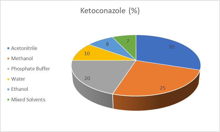
Figure 1: Solvent usage pattern in reported analytical methods for Ketoconazole
Caption: The figure illustrates the relative distribution of solvents employed in UV spectrophotometric, chromatographic, and hyphenated analytical methods reported for Ketoconazole.

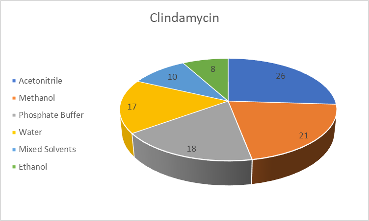
Figure 2: Solvent usage pattern in reported analytical methods for Clindamycin
Caption: The figure represents the proportion of solvents utilized in various analytical techniques, including UV, RP-HPLC, HPTLC, and LC–MS methods, for the analysis of Clindamycin

Current Research on the Ketoconazole–Clindamycin Combination

Recent research indicates growing clinical and pharmaceutical interest in the Ketoconazole–Clindamycin combination, particularly for the treatment of mixed fungal and bacterial infections such as vulvovaginal candidiasis and bacterial vaginosis. Clinical studies and trials have evaluated intravaginal formulations containing both drugs and reported favorable therapeutic outcomes, supporting their complementary antifungal and antibacterial activity. Patent literature further demonstrates active formulation research on this combination in topical and intravaginal dosage forms, confirming pharmaceutical compatibility and commercial relevance.

However, despite clinical use and formulation development, limited attention has been given to analytical method development for this combination. Most reported analytical studies focus on individual estimation of Ketoconazole or Clindamycin, while validated, stability-indicating methods for their simultaneous quantification in combined formulations remain scarce. This highlights a clear need for robust analytical research to support quality control and regulatory requirements for combination products.

CONCLUSION

This review critically summarized the reported analytical methods employed for the quantification of Ketoconazole and Clindamycin using spectroscopic, chromatographic, and hyphenated techniques. UV spectrophotometric methods were found to be simple and cost-effective but limited by poor selectivity and unsuitability for stability assessment. RP-HPLC emerged as the most widely applied technique, offering superior accuracy, precision, and applicability across bulk drugs, pharmaceutical formulations, and biological matrices. HPTLC methods provided rapid and economical alternatives for routine analysis, while advanced techniques such as UPLC and LC–MS/MS demonstrated high sensitivity and specificity, particularly for bioanalytical applications, though their routine use remains restricted due to high cost and operational complexity.

Despite the availability of numerous analytical approaches for individual drugs, a significant research gap exists in the form of a lack of a validated, stability-indicating RP-HPLC method for the simultaneous estimation of Ketoconazole and Clindamycin in combined dosage forms. Many reported methods either do not address forced degradation behavior comprehensively or fail to comply fully with ICH guidelines. Addressing this gap is critical to ensure regulatory compliance, reliable quality control, and accurate stability evaluation of combination formulations. Therefore, the development of a simple, robust, and stability-indicating RP-HPLC method capable of resolving both drugs and their degradation products remains an important and necessary direction for future analytical research.

REFERENCES

  1. Mondal S, Alekya C, Mondal P. A novel rapid approach for the estimation of ketoconazole using reverse-phase ultra-performance liquid chromatography in bulk and tablet dosage form. Pharm Methods. 2020;11(1):1–5.
  2. Annisa V, Sulaiman TNS, Nugroho AK, Nugroho AE. Validation of RP-HPLC UV method for determination of ketoconazole in rabbit plasma and application to pharmacokinetic study. FABAD J Pharm Sci. 2023;48(2):285–294.
  3. Annisa V, Sulaiman TNS, Nugroho AK, Nugroho AE. Validation of RP-HPLC method for quantification of ketoconazole in two-stage model of biorelevant dissolution. Egypt Pharm J. 2023;22:676–682.
  4. Muchakayala SK, Katari NK, Saripella KK, Marisetti VM, Kowtharapu LP. Unique green chromatographic method for qualitative and quantitative analysis of ketoconazole from cream formulation. Sustain Chem Pharm. 2023;36:100865.
  5. Sarfraz S, Hussain S, Raza A, Alrbyawi H, Aljazzar S, Elkaeed E, Somaily H, et al. Simultaneous HPLC determination of clindamycin phosphate, tretinoin, and preservatives in gel dosage form. Inorganics. 2022;10:1–15.
  6. Greibe E, Moser CE, Bruun NE, Hoffmann-Lücke E. New methods for quantification of amoxicillin and clindamycin in human plasma using HPLC with UV detection. J Antimicrob Chemother. 2022;77(9):2437–2440.
  7. Shareef I, Gandla K. Stability-indicating UPLC method development and forced degradation studies of sulfamethoxazole and clindamycin. Saudi J Med Pharm Sci. 2025;11(5):437–443.
  8. Tyagi N, Dave K, Sharma A. Development and validation of high-performance thin-layer chromatographic methods for antibacterial formulations. Int J Pharm Qual Assur. 2024;15(3):747–755.
  9. Kumar V, Sharma A, Jain N. LC-MS/MS method for quantification of clindamycin in human plasma and its pharmacokinetic application. J Chromatogr B. 2019; 1113:253–260.
  10. Costa S, Almeida A, Amaral MH, Sousa Lobo JM. UPLC-MS/MS methods for antifungal drug quantification in biological matrices. Bioanalysis. 2021;13(7):523–536.
  11. Singh J, Kaur H, Bala R. Simultaneous determination of antifungal agents including ketoconazole in biological fluids by LC-MS/MS. Clin Chim Acta. 2017; 467:1–8.
  12. Sharma S, Chhipa RC, Singh R. Ethanol-based novel HPLC method for ketoconazole dissolution analysis. J Pharm Anal. 2015;5(5):288–294.
  13. Wishart DS, Feunang YD, Guo AC, Lo EJ, Marcu A, Grant JR, et al. DrugBank 5.0: a major update to the DrugBank database for 2018. Nucleic Acids Res. 2018;46(D1):D1074–D1082.
  14. Wishart DS, Knox C, Guo AC, Cheng D, Shrivastava S, Tzur D, et al. DrugBank: a comprehensive resource for in silico drug discovery and exploration. Nucleic Acids Res. 2006;34(Database issue):D668–D672.
  15. Ghurghure S., Garad S.S., and Birajdar S., “UV spectrophotometric method development and validation for estimation of ketoconazole in bulk and pharmaceutical dosage form,” Current Trends in Pharmaceutical and Pharmaceutical Chemistry, vol. 5, no. 2, pp. 71–74, 2023.
  16. Kannaiah K.P. and Sugumaran A., “Eco-friendly multivariant green analytical technique for the estimation of drug by UV spectroscopy in bulk and cream formulation,” Química Nova, vol. 45, no. 1, pp. 23–30, 2022.
  17. Shrivastava S., Shrivastava S., and Tiwle R., “Validation of novel UV spectrophotometric method for the determination of ketoconazole in pharmaceutical formulation,” Journal of Pharmaceutical Advanced Research, vol. 3, no. 2, pp. 792–798, 2020.
  18. Madur S., Matole V., Kalshetti M., Patil S., and Sakhare A., “New visible spectrophotometric method development and validation of ketoconazole in pure and semisolid dosage form,” Asian Journal of Research in Chemistry and Pharmaceutical Sciences, vol. 8, no. 1, pp. 7–11, 2020.
  19. Naveed S. and Jaweed L., “UV spectrophotometric assay of ketoconazole oral formulations,” American Journal of Biological and Life Sciences, vol. 2, no. 5, p. 108, 2014.
  20. Kansagra P., Sanghvi G., Purohit P., Vachani A., Sheth N., and Vaishnav D., “Development and validation of stability indicating UV spectrophotometric method for the determination of ketoconazole both in bulk and marketed dosage formulations,” Inventi Rapid: Pharmaceutical Analysis & Quality Assurance, vol. 1, pp. 1–6, 2013.
  21. Shaligram S.R. and Padmaja P., “Spectrophotometric method for the determination of ketoconazole based on amplification reactions,” Journal of Pharmaceutical Analysis, vol. 2, no. 1, pp. 43–47, 2012.
  22. Kannaiah K.P. and Sugumaran A., “Environmental benign AQbD based estimation of ketoconazole and beclomethasone by RP-HPLC and multi-analytical UV spectrophotometric method,” Microchemical Journal, vol. 172, 2022.
  23. Parikh S., Dave J., and Patel J., “Stability indicating RP-HPLC method for determination of ketoconazole in bulk drug and in tablet dosage form,” Journal of Pharmaceutical Science and Medical Research, vol. 1, no. 2, pp. 001–008, 2021.
  24. Sharma S., Chhipa R.C., Singh R., and Alam I., “Development and validation of reverse-phase HPLC method for estimation of ketoconazole in bulk drug,” Pharmacophore, vol. 3, no. 2, pp. 123–129, 2012.
  25. Beigi F.A.K., Imani M., Payehghadr M., and Hosseini H., “SPE-HPLC method for determination of ketoconazole and clotrimazole residues in cow’s milk,” Journal of the Brazilian Chemical Society, vol. 22, no. 9, pp. 1761–1766, 2011.
  26. Venishetty V.K., Parikh N., Sistla R., Ahmed F.J., and Diwan P.V., “Application of validated RP-HPLC method for simultaneous determination of docetaxel and ketoconazole in solid lipid nanoparticles,” Journal of Chromatographic Science, vol. 49, no. 2, pp. 136–141, 2011.
  27. Abdel-Moety E.M., Khattab F.I., Kelani K.M., and AbouAl-Alamein A.M., “Chromatographic determination of clotrimazole, ketoconazole and fluconazole in pharmaceutical formulations,” Il Farmaco, vol. 57, no. 8, pp. 931–938, 2002.
  28. Low A.S. and Wangboonskul J., “An HPLC assay for the determination of ketoconazole in common pharmaceutical preparations,” Analyst, vol. 124, no. 11, pp. 1589–1593, 1999.
  29. Yuen K.H. and Peh K.K., “Simple high-performance liquid chromatographic method for determination of ketoconazole in human plasma,” Journal of Chromatography B: Biomedical Sciences and Applications, vol. 715, no. 2, pp. 436–440, 1998.
  30. Hoffman D.W., Jones-King K.L., Ravaris C.L., and Edkins R.D., “Electrochemical detection for high-performance liquid chromatography of ketoconazole in plasma and saliva,” Analytical Biochemistry, vol. 172, pp. 495–498, 1988.
  31. Saysin S., Liawruangrath B., and Liawruangrath S., “High-performance thin-layer chromatographic determination of ketoconazole in pharmaceutical formulations,” Journal of Cosmetic Science, vol. 61, no. 5, pp. 367–376, 2010.
  32. Muchakayala S.K., Katari N.K., Saripella K.K., Marisetti V.M., and Kowtharapu L.P., “Unique green chromatographic method for the qualitative and quantitative analysis of ketoconazole, its impurities and preservatives from ketoconazole cream formulation: Identification of degradants by Q-ToF LCMS and robustness by design of experiments,” Sustainable Chemistry and Pharmacy, vol. 36, 2023.
  33. “Development and validation of a UV-visible spectrophotometric method for simultaneous estimation of curcumin and clindamycin,” African Journal of Biomedical Research, vol. 27, no. 2S, pp. 632–639, 2024.
  34. Nataraj K.S., Surya Narasimha Raju G.N.V., and Anusha B., “UV spectrophotometric method development for estimation of clindamycin phosphate in bulk and dosage form,” International Journal of Pharmacy and Biological Sciences, vol. 3, no. 4, pp. 164–167, 2013.
  35. Shareef I. and Gandla K., “Stability-indicating UPLC method development, validation, and forced degradation studies of sulfamethoxazole and clindamycin in bulk and formulated dosage forms,” Saudi Journal of Medical and Pharmaceutical Sciences, vol. 11, no. 5, pp. 437–443, 2025.
  36. Sarfraz S., Hussain S., Raza A., Alrbyawi H., Aljazzar S., Elkaeed E., Somaily H., Pashameah R., Alzahrani E., and Farouk A.E., “Simultaneous HPLC determination of clindamycin phosphate, tretinoin, and preservatives in gel dosage form using a novel stability-indicating method,” Inorganics, vol. 10, pp. 1–15, 2022.
  37. Greibe E., Moser C.E., Bruun N.E., and Hoffmann-Lücke E., “New methods for quantification of amoxicillin and clindamycin in human plasma using HPLC with UV detection,” Journal of Antimicrobial Chemotherapy, vol. 77, no. 9, pp. 2437–2440, 2022.
  38. Rajendar L., Potnuri N.R., and Rao R.N., “A stability indicating RP-HPLC method for the simultaneous estimation of metronidazole, clindamycin and clotrimazole in bulk and their combined dosage form,” World Journal of Pharmaceutical Sciences, vol. 3, no. 1, pp. 93–103, 2015.
  39. Mifsud M., Vella J., Ferrito V., Serracino-Inglott A., Azzopardi L.M., and Sammut Bartolo N., “A simple HPLC-UV method for the determination of clindamycin in human plasma,” Journal of Chemical and Pharmaceutical Research, vol. 6, no. 1, pp. 696–704, 2014.
  40. Seethalakshmi N., Chenthilnathan A., and Rama K., “RP-HPLC method development and validation for simultaneous estimation of metronidazole, clindamycin phosphate and clotrimazole in combined pharmaceutical dosage forms,” International Research Journal of Pharmaceutical and Applied Sciences, vol. 4, no. 2, pp. 67–77, 2014.
  41. Khatri R.H., Patel R.B., and Patel M.R., “A new RP-HPLC method for estimation of clindamycin and adapalene in gel formulation: Development and validation consideration,” Thai Journal of Pharmaceutical Sciences, vol. 38, no. 1, pp. 1–56, 2014.
  42. Raju C.B.V.N., Panda G., Nageswara Rao G., and Rockey J., “HPLC-UV assay method for clindamycin palmitate hydrochloride as drug substance and oral solution,” Analytical Letters, vol. 41, no. 11, pp. 2033–2043, 2008.
  43. Platzer D.J. and White B.A., “Development and validation of a gradient HPLC method for the determination of clindamycin and related compounds in a novel tablet formulation,” Journal of Pharmaceutical and Biomedical Analysis, vol. 41, pp. 84–88, 2006.
  44. Batzias G.C., Delis G.A., and Koutsoviti-Papadopoulou M., “A new HPLC/UV method for the determination of clindamycin in dog blood serum,” Journal of Pharmaceutical and Biomedical Analysis, vol. 35, no. 3, pp. 545–554, 2004.
  45. Tyagi N., Dave K., and Sharma A., “Development and validation of high-performance thin layer chromatographic methods for antibacterial formulations,” International Journal of Pharmaceutical Quality Assurance, vol. 15, no. 3, pp. 747–755, 2024.
  46. Rechberger G.N., Fauler G., Windischhofer W., Köfeler H., Erwa W., and Leis H.J., “Quantitative analysis of clindamycin in human plasma by liquid chromatography/electrospray ionisation tandem mass spectrometry using d1-N-ethylclindamycin as internal standard,” Rapid Communications in Mass Spectrometry, vol. 17, no. 2, pp. 135–139, 2003.
  47. Yu L.L., Chao C.K., Liao W.J., Twu T.Y., Liu C.M., Yang T.H., and Lin E.T., “Determination of clindamycin in human plasma by liquid chromatography–electrospray tandem mass spectrometry: Application to the bioequivalence study of clindamycin,” Journal of Chromatography B: Biomedical Sciences and Applications, vol. 724, no. 2, pp. 287–294, 1999.

Reference

  1. Mondal S, Alekya C, Mondal P. A novel rapid approach for the estimation of ketoconazole using reverse-phase ultra-performance liquid chromatography in bulk and tablet dosage form. Pharm Methods. 2020;11(1):1–5.
  2. Annisa V, Sulaiman TNS, Nugroho AK, Nugroho AE. Validation of RP-HPLC UV method for determination of ketoconazole in rabbit plasma and application to pharmacokinetic study. FABAD J Pharm Sci. 2023;48(2):285–294.
  3. Annisa V, Sulaiman TNS, Nugroho AK, Nugroho AE. Validation of RP-HPLC method for quantification of ketoconazole in two-stage model of biorelevant dissolution. Egypt Pharm J. 2023;22:676–682.
  4. Muchakayala SK, Katari NK, Saripella KK, Marisetti VM, Kowtharapu LP. Unique green chromatographic method for qualitative and quantitative analysis of ketoconazole from cream formulation. Sustain Chem Pharm. 2023;36:100865.
  5. Sarfraz S, Hussain S, Raza A, Alrbyawi H, Aljazzar S, Elkaeed E, Somaily H, et al. Simultaneous HPLC determination of clindamycin phosphate, tretinoin, and preservatives in gel dosage form. Inorganics. 2022;10:1–15.
  6. Greibe E, Moser CE, Bruun NE, Hoffmann-Lücke E. New methods for quantification of amoxicillin and clindamycin in human plasma using HPLC with UV detection. J Antimicrob Chemother. 2022;77(9):2437–2440.
  7. Shareef I, Gandla K. Stability-indicating UPLC method development and forced degradation studies of sulfamethoxazole and clindamycin. Saudi J Med Pharm Sci. 2025;11(5):437–443.
  8. Tyagi N, Dave K, Sharma A. Development and validation of high-performance thin-layer chromatographic methods for antibacterial formulations. Int J Pharm Qual Assur. 2024;15(3):747–755.
  9. Kumar V, Sharma A, Jain N. LC-MS/MS method for quantification of clindamycin in human plasma and its pharmacokinetic application. J Chromatogr B. 2019; 1113:253–260.
  10. Costa S, Almeida A, Amaral MH, Sousa Lobo JM. UPLC-MS/MS methods for antifungal drug quantification in biological matrices. Bioanalysis. 2021;13(7):523–536.
  11. Singh J, Kaur H, Bala R. Simultaneous determination of antifungal agents including ketoconazole in biological fluids by LC-MS/MS. Clin Chim Acta. 2017; 467:1–8.
  12. Sharma S, Chhipa RC, Singh R. Ethanol-based novel HPLC method for ketoconazole dissolution analysis. J Pharm Anal. 2015;5(5):288–294.
  13. Wishart DS, Feunang YD, Guo AC, Lo EJ, Marcu A, Grant JR, et al. DrugBank 5.0: a major update to the DrugBank database for 2018. Nucleic Acids Res. 2018;46(D1):D1074–D1082.
  14. Wishart DS, Knox C, Guo AC, Cheng D, Shrivastava S, Tzur D, et al. DrugBank: a comprehensive resource for in silico drug discovery and exploration. Nucleic Acids Res. 2006;34(Database issue):D668–D672.
  15. Ghurghure S., Garad S.S., and Birajdar S., “UV spectrophotometric method development and validation for estimation of ketoconazole in bulk and pharmaceutical dosage form,” Current Trends in Pharmaceutical and Pharmaceutical Chemistry, vol. 5, no. 2, pp. 71–74, 2023.
  16. Kannaiah K.P. and Sugumaran A., “Eco-friendly multivariant green analytical technique for the estimation of drug by UV spectroscopy in bulk and cream formulation,” Química Nova, vol. 45, no. 1, pp. 23–30, 2022.
  17. Shrivastava S., Shrivastava S., and Tiwle R., “Validation of novel UV spectrophotometric method for the determination of ketoconazole in pharmaceutical formulation,” Journal of Pharmaceutical Advanced Research, vol. 3, no. 2, pp. 792–798, 2020.
  18. Madur S., Matole V., Kalshetti M., Patil S., and Sakhare A., “New visible spectrophotometric method development and validation of ketoconazole in pure and semisolid dosage form,” Asian Journal of Research in Chemistry and Pharmaceutical Sciences, vol. 8, no. 1, pp. 7–11, 2020.
  19. Naveed S. and Jaweed L., “UV spectrophotometric assay of ketoconazole oral formulations,” American Journal of Biological and Life Sciences, vol. 2, no. 5, p. 108, 2014.
  20. Kansagra P., Sanghvi G., Purohit P., Vachani A., Sheth N., and Vaishnav D., “Development and validation of stability indicating UV spectrophotometric method for the determination of ketoconazole both in bulk and marketed dosage formulations,” Inventi Rapid: Pharmaceutical Analysis & Quality Assurance, vol. 1, pp. 1–6, 2013.
  21. Shaligram S.R. and Padmaja P., “Spectrophotometric method for the determination of ketoconazole based on amplification reactions,” Journal of Pharmaceutical Analysis, vol. 2, no. 1, pp. 43–47, 2012.
  22. Kannaiah K.P. and Sugumaran A., “Environmental benign AQbD based estimation of ketoconazole and beclomethasone by RP-HPLC and multi-analytical UV spectrophotometric method,” Microchemical Journal, vol. 172, 2022.
  23. Parikh S., Dave J., and Patel J., “Stability indicating RP-HPLC method for determination of ketoconazole in bulk drug and in tablet dosage form,” Journal of Pharmaceutical Science and Medical Research, vol. 1, no. 2, pp. 001–008, 2021.
  24. Sharma S., Chhipa R.C., Singh R., and Alam I., “Development and validation of reverse-phase HPLC method for estimation of ketoconazole in bulk drug,” Pharmacophore, vol. 3, no. 2, pp. 123–129, 2012.
  25. Beigi F.A.K., Imani M., Payehghadr M., and Hosseini H., “SPE-HPLC method for determination of ketoconazole and clotrimazole residues in cow’s milk,” Journal of the Brazilian Chemical Society, vol. 22, no. 9, pp. 1761–1766, 2011.
  26. Venishetty V.K., Parikh N., Sistla R., Ahmed F.J., and Diwan P.V., “Application of validated RP-HPLC method for simultaneous determination of docetaxel and ketoconazole in solid lipid nanoparticles,” Journal of Chromatographic Science, vol. 49, no. 2, pp. 136–141, 2011.
  27. Abdel-Moety E.M., Khattab F.I., Kelani K.M., and AbouAl-Alamein A.M., “Chromatographic determination of clotrimazole, ketoconazole and fluconazole in pharmaceutical formulations,” Il Farmaco, vol. 57, no. 8, pp. 931–938, 2002.
  28. Low A.S. and Wangboonskul J., “An HPLC assay for the determination of ketoconazole in common pharmaceutical preparations,” Analyst, vol. 124, no. 11, pp. 1589–1593, 1999.
  29. Yuen K.H. and Peh K.K., “Simple high-performance liquid chromatographic method for determination of ketoconazole in human plasma,” Journal of Chromatography B: Biomedical Sciences and Applications, vol. 715, no. 2, pp. 436–440, 1998.
  30. Hoffman D.W., Jones-King K.L., Ravaris C.L., and Edkins R.D., “Electrochemical detection for high-performance liquid chromatography of ketoconazole in plasma and saliva,” Analytical Biochemistry, vol. 172, pp. 495–498, 1988.
  31. Saysin S., Liawruangrath B., and Liawruangrath S., “High-performance thin-layer chromatographic determination of ketoconazole in pharmaceutical formulations,” Journal of Cosmetic Science, vol. 61, no. 5, pp. 367–376, 2010.
  32. Muchakayala S.K., Katari N.K., Saripella K.K., Marisetti V.M., and Kowtharapu L.P., “Unique green chromatographic method for the qualitative and quantitative analysis of ketoconazole, its impurities and preservatives from ketoconazole cream formulation: Identification of degradants by Q-ToF LCMS and robustness by design of experiments,” Sustainable Chemistry and Pharmacy, vol. 36, 2023.
  33. “Development and validation of a UV-visible spectrophotometric method for simultaneous estimation of curcumin and clindamycin,” African Journal of Biomedical Research, vol. 27, no. 2S, pp. 632–639, 2024.
  34. Nataraj K.S., Surya Narasimha Raju G.N.V., and Anusha B., “UV spectrophotometric method development for estimation of clindamycin phosphate in bulk and dosage form,” International Journal of Pharmacy and Biological Sciences, vol. 3, no. 4, pp. 164–167, 2013.
  35. Shareef I. and Gandla K., “Stability-indicating UPLC method development, validation, and forced degradation studies of sulfamethoxazole and clindamycin in bulk and formulated dosage forms,” Saudi Journal of Medical and Pharmaceutical Sciences, vol. 11, no. 5, pp. 437–443, 2025.
  36. Sarfraz S., Hussain S., Raza A., Alrbyawi H., Aljazzar S., Elkaeed E., Somaily H., Pashameah R., Alzahrani E., and Farouk A.E., “Simultaneous HPLC determination of clindamycin phosphate, tretinoin, and preservatives in gel dosage form using a novel stability-indicating method,” Inorganics, vol. 10, pp. 1–15, 2022.
  37. Greibe E., Moser C.E., Bruun N.E., and Hoffmann-Lücke E., “New methods for quantification of amoxicillin and clindamycin in human plasma using HPLC with UV detection,” Journal of Antimicrobial Chemotherapy, vol. 77, no. 9, pp. 2437–2440, 2022.
  38. Rajendar L., Potnuri N.R., and Rao R.N., “A stability indicating RP-HPLC method for the simultaneous estimation of metronidazole, clindamycin and clotrimazole in bulk and their combined dosage form,” World Journal of Pharmaceutical Sciences, vol. 3, no. 1, pp. 93–103, 2015.
  39. Mifsud M., Vella J., Ferrito V., Serracino-Inglott A., Azzopardi L.M., and Sammut Bartolo N., “A simple HPLC-UV method for the determination of clindamycin in human plasma,” Journal of Chemical and Pharmaceutical Research, vol. 6, no. 1, pp. 696–704, 2014.
  40. Seethalakshmi N., Chenthilnathan A., and Rama K., “RP-HPLC method development and validation for simultaneous estimation of metronidazole, clindamycin phosphate and clotrimazole in combined pharmaceutical dosage forms,” International Research Journal of Pharmaceutical and Applied Sciences, vol. 4, no. 2, pp. 67–77, 2014.
  41. Khatri R.H., Patel R.B., and Patel M.R., “A new RP-HPLC method for estimation of clindamycin and adapalene in gel formulation: Development and validation consideration,” Thai Journal of Pharmaceutical Sciences, vol. 38, no. 1, pp. 1–56, 2014.
  42. Raju C.B.V.N., Panda G., Nageswara Rao G., and Rockey J., “HPLC-UV assay method for clindamycin palmitate hydrochloride as drug substance and oral solution,” Analytical Letters, vol. 41, no. 11, pp. 2033–2043, 2008.
  43. Platzer D.J. and White B.A., “Development and validation of a gradient HPLC method for the determination of clindamycin and related compounds in a novel tablet formulation,” Journal of Pharmaceutical and Biomedical Analysis, vol. 41, pp. 84–88, 2006.
  44. Batzias G.C., Delis G.A., and Koutsoviti-Papadopoulou M., “A new HPLC/UV method for the determination of clindamycin in dog blood serum,” Journal of Pharmaceutical and Biomedical Analysis, vol. 35, no. 3, pp. 545–554, 2004.
  45. Tyagi N., Dave K., and Sharma A., “Development and validation of high-performance thin layer chromatographic methods for antibacterial formulations,” International Journal of Pharmaceutical Quality Assurance, vol. 15, no. 3, pp. 747–755, 2024.
  46. Rechberger G.N., Fauler G., Windischhofer W., Köfeler H., Erwa W., and Leis H.J., “Quantitative analysis of clindamycin in human plasma by liquid chromatography/electrospray ionisation tandem mass spectrometry using d1-N-ethylclindamycin as internal standard,” Rapid Communications in Mass Spectrometry, vol. 17, no. 2, pp. 135–139, 2003.
  47. Yu L.L., Chao C.K., Liao W.J., Twu T.Y., Liu C.M., Yang T.H., and Lin E.T., “Determination of clindamycin in human plasma by liquid chromatography–electrospray tandem mass spectrometry: Application to the bioequivalence study of clindamycin,” Journal of Chromatography B: Biomedical Sciences and Applications, vol. 724, no. 2, pp. 287–294, 1999.

Photo
Disha Sharma
Corresponding author

Sigma Institute of Pharmacy, Sigma University, Vadodara, Gujarat, India 390019

Photo
Dr. Mitali Dalwadi
Co-author

Sigma Institute of Pharmacy, Sigma University, Vadodara, Gujarat, India 390019

Photo
Dr. Priyanka Patil
Co-author

Sigma Institute of Pharmacy, Sigma University, Vadodara, Gujarat, India 390019

Disha Sharma, Dr. Mitali Dalwadi, Dr. Priyanka Patil, Critical Evaluation of Spectroscopic and Chromatographic Methods for the Quantitative Determination of Ketoconazole and Clindamycin, Int. J. of Pharm. Sci., 2026, Vol 4, Issue 1, 2124-2137. https://doi.org/10.5281/zenodo.18327340

More related articles
Anthelmintic Efficacy of Luffa cylindrica in Relat...
Basavachetan Kharabe, D. Visagaperumal, Dr. Jose Gnana Babu, Vine...
Chiral HPLC Method Development And Validation For ...
SHAIK MASTANAMMA, Chunduri Akhila, M S DHANDAPANI, ...
Formulation and Evaluation of Dexketoprofen Tromet...
Aashish Singh, Dr. Tanu Bhargava, Kamlesh Dashora, Anju Tomar, ...
Herbal and Natural Medicine ...
Vishal Kulkarni , Prashant Naiknavare, Akshay Pawar, Prashant Misal, ...
Formulation And Evaluation Of Topical Microemulsion Of Smilax China...
Ashutosh Lakhera , Shivanand Patil, Meenakshi Kandwal, ...
Related Articles
Structure Biosynthesis And Examination Of Bioactive Small Molecule...
Pravin D. Chand, A. S. sanap, Prachi P. Udapurkar, ...
A Review on Formulation and Evaluation of Turmeric Herbal Cream...
Sharma Nitin, Kaur Sharandeep, Kumar Faneesh, Ujwal, ...
Pathophysiological Mechanisms and Systemic Complications of Polycystic Ovary Syn...
Malarkodi Velraj, Pratyush Mishra, Yasmin Suthar, Varun Arora, Pragi Arora, Tanmay Ghosh, Rishin Bha...
Anthelmintic Efficacy of Luffa cylindrica in Relation to its: Phytoconstituents:...
Basavachetan Kharabe, D. Visagaperumal, Dr. Jose Gnana Babu, Vineeth Chandy, ...
More related articles
Anthelmintic Efficacy of Luffa cylindrica in Relation to its: Phytoconstituents:...
Basavachetan Kharabe, D. Visagaperumal, Dr. Jose Gnana Babu, Vineeth Chandy, ...
Formulation and Evaluation of Dexketoprofen Trometamol Loaded Extended Release C...
Aashish Singh, Dr. Tanu Bhargava, Kamlesh Dashora, Anju Tomar, ...