Kamalakshmi Pandurangan College of Pharmacy, Ayyampalayam, Tiruvannamalai.
Inflammation is a key pathological factor in chronic disorders such as osteoarthritis, rheumatoid arthritis, asthma, and inflammatory bowel disease. Conventional oral delivery of Boswellia serrata extract is limited by poor bioavailability, rapid systemic clearance, and low residence time at the target site. The present study aimed to formulate and evaluate a bioadhesive drug delivery system incorporating standardized Boswellia serrata resin extract for sustained and localized anti-inflammatory action. Bioadhesive patches were prepared using solvent casting technique with polymers such as Hydroxypropyl methylcellulose (HPMC), Carbopol 934P, and Polyvinyl alcohol (PVA). Propylene glycol was used as plasticizer, and oleic acid served as a permeation enhancer. The prepared formulations were evaluated for thickness, folding endurance, drug content uniformity, surface pH, swelling index, mucoadhesive strength, in vitro drug release using Franz diffusion cell, and stability studies. The optimized formulation exhibited satisfactory physicochemical characteristics, strong mucoadhesive strength (>0.5 N/cm²), and sustained drug release (approximately 75–80% over 12 hours) following near zero-order kinetics (R² > 0.98). Statistical analysis confirmed significant improvement (p < 0.05) in controlled release behavior compared to conventional extract. The study concludes that the developed bioadhesive system significantly enhances residence time, provides sustained release, and improves therapeutic potential of Boswellia serrata extract for inflammatory conditions.
Inflammation is a protective biological response involving immune cells, blood vessels, and molecular mediators. However, chronic inflammation contributes to degenerative diseases such as arthritis, asthma, psoriasis, and inflammatory bowel disease. Conventional non-steroidal anti-inflammatory drugs (NSAIDs) are associated with gastrointestinal irritation, cardiovascular risks, and systemic side effects, necessitating safer alternatives. Boswellia serrata Roxb. ex Colebr., belonging to the family Burseraceae, is commonly known as Indian frankincense or Salai guggul. The resin contains biologically active pentacyclic triterpenoids known as boswellic acids, including β-boswellic acid, acetyl-β-boswellic acid, 11-keto-β-boswellic acid (KBA), and 3-O-acetyl-11-keto-β-boswellic acid (AKBA). AKBA is considered the most potent inhibitor of 5-lipoxygenase (5-LOX), thereby reducing leukotriene-mediated inflammatory responses. Despite strong pharmacological potential, oral bioavailability of boswellic acids is poor due to low aqueous solubility and extensive first-pass metabolism. Therefore, bioadhesive drug delivery systems offer a promising approach to enhance mucosal retention, improve drug permeation, and achieve controlled release. Bioadhesive systems function by intimate contact between polymer chains and biological tissues through hydrogen bonding, van der Waals forces, and electrostatic interactions. Prolonged residence time increases drug concentration gradient, thereby improving therapeutic efficacy. The present study was designed to formulate and evaluate a Boswellia serrata-loaded bioadhesive patch to enhance local delivery and sustained anti-inflammatory action.
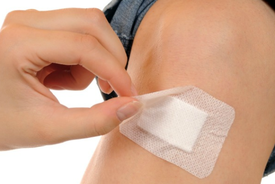
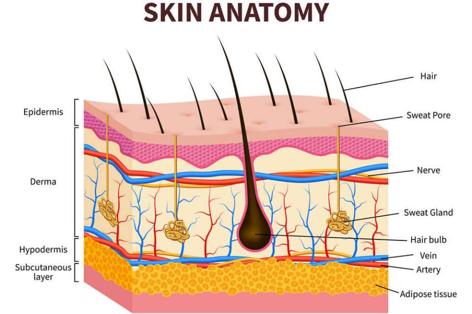
Figure 1: Transdermal Patch Figure 2: Skin Anatomy
MATERIALS AND METHODS
MATERIALS:
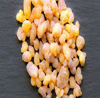
Figure 3: Boswellia Serrata Resin
METHODS OF PREPARATION:
Preparation of Polymeric Solution-
Required quantity of HPMC and PVA were dissolved in distilled water under continuous stirring. Carbopol was dispersed separately and neutralized to obtain uniform gel consistency.
Incorporation of Drug-
Accurately weighed Boswellia serrata extract was dissolved in ethanol and added to polymeric mixture. Propylene glycol (20% w/w of polymer weight) and oleic acid (5% w/w) were incorporated.
Solvent Casting-
The homogeneous mixture was poured into a glass Petri dish and dried at 40°C for 24 hours. The dried films were cut into 2×2 cm patches and stored in desiccator.
Table 1: Composition Table
|
FORMULATION |
POLYMER SYSTEM |
BOSWELLIA EXTRACT (%) |
PLASTICIZER (%) |
PERMEATION ENHANCER (%) |
|
F1 |
HPMC K4M |
2.0 |
20 (glycerol) |
– |
|
F2 |
HPMC K4M |
3.0 |
20 (glycerol) |
– |
|
F3 |
HPMC K4M |
4.0 |
20 (glycerol) |
– |
|
F4 |
HPMC K4M |
4.0 |
20 (glycerol) |
– |
|
F5 |
HPMC K4M |
2.0 |
20 (glycerol) |
– |
|
F6 |
HPMC + PVP |
3.0 |
20 (glycerol) |
– |
|
F7 |
HPMC + EC |
3.0 |
20 (glycerol) |
– |
|
F8 |
HPMC + PVP |
3.0 |
20 (glycerol) |
1.0 (Labrasol) |
|
F9 |
HPMC + PVP |
3.0 |
20 (glycerol) |
1.0 (Transcutol HP) |
|
F10 |
HPMC + PVP |
3.0 |
20 (glycerol) |
0.5 (oleic acid) |
RESULTS
APPEARANCE AND PHYSICAL PROPERTIES:-
COLOR:- Dried films - Uniform pale yellow to light brown due to the natural pigmentation of boswellic acids in the resin extract.
TRANSPARENCY:- Films are transparent to semi-transparent, and allowing clear visibility through the matrix
SURFACE SMOOTHNESS:- A smooth surface is observed, assessed visually which reflects even drying and good plasticizer incorporation.
PRESENCE OF AIR BUBBLES:- No visible air bubbles or voids are present.
FLEXIBILITY:- Films exhibit excellent flexibility, without cracking due to glycerol's high 20% concentration acting as an efficient plasticizer that reduces HPMC-PVP brittleness.
FOLDING ENDURANCE: >250
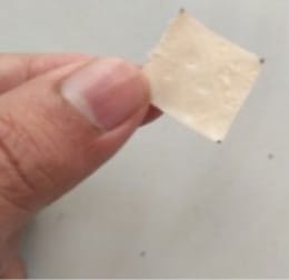
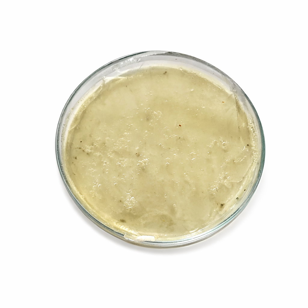
Figure 4: Solvent-Casted Transdermal Patch Film
PH OF PATCH SURFACE:- Around from 6.4 -7.7, and the ideal range is 5.0 – 7.0.
WEIGHT VARIATION:-
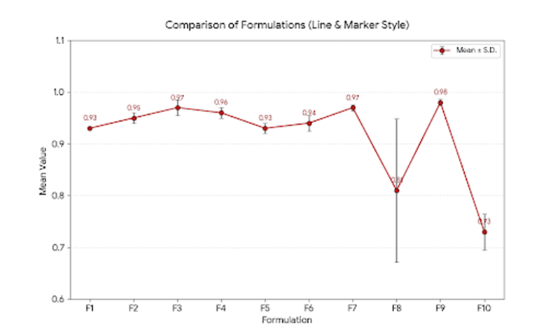
Figure 5: Weight Comparison of Formulations
THICKNESS:-
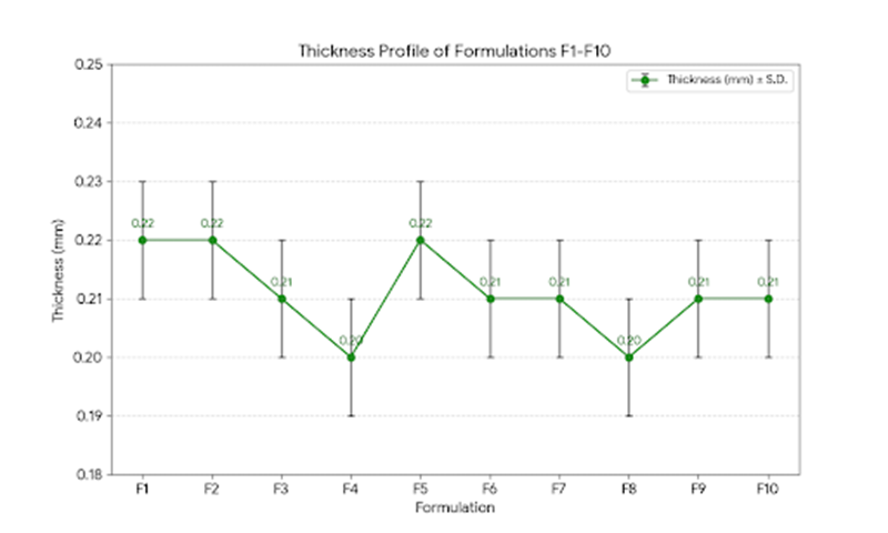
Figure 6: Thickness Profile of Formulations
HPLC:
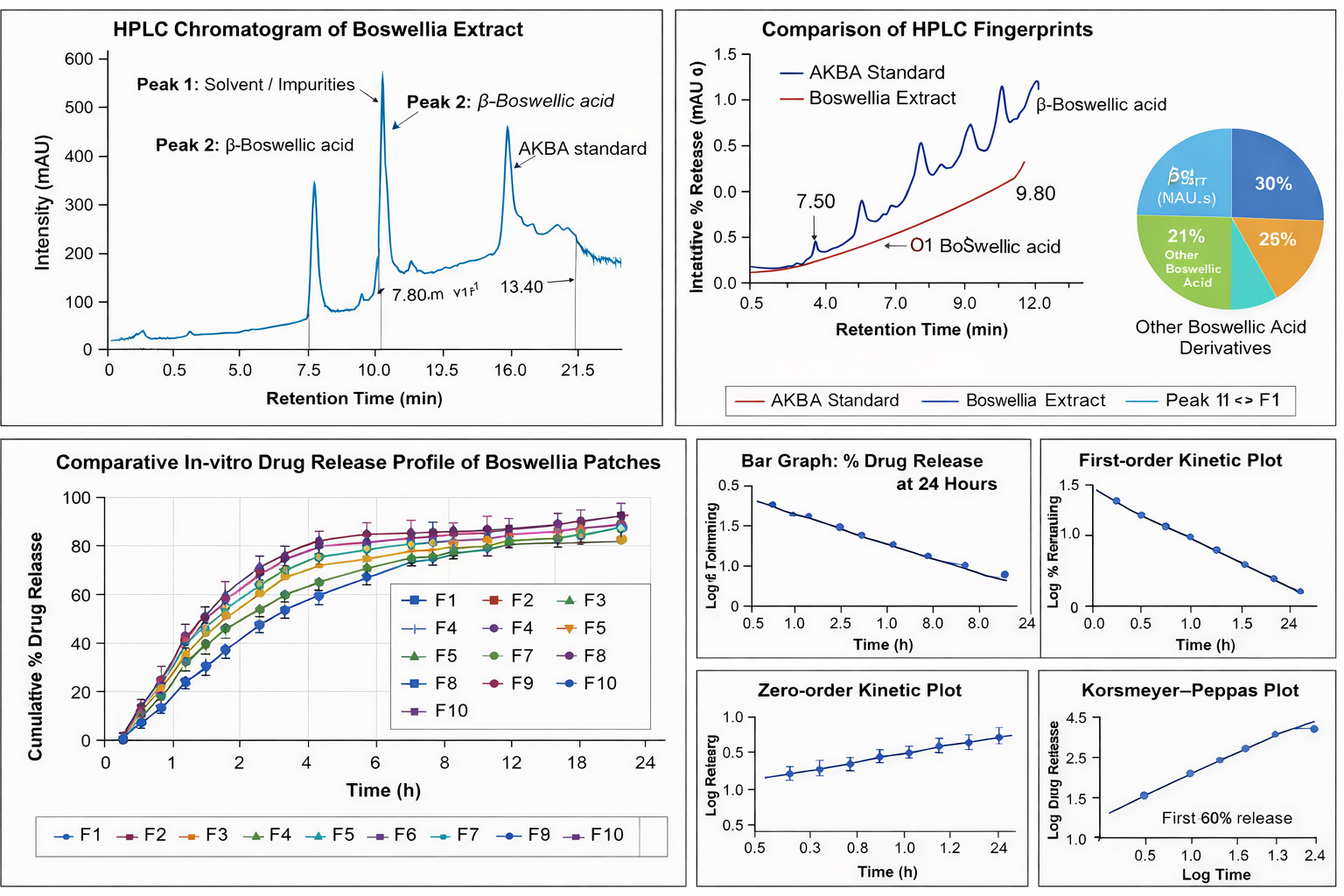
Figure 7: HPLC Chromatogram
Peak 1 (RT: 3.20 min) – Identified as solvent/low molecular weight impurities based on early elution and absence of matching standards.
Peak 2 (RT: 7.50 min) – Identified as β-Boswellic acid, confirmed by matching retention time with reported literature values.
Peak 3 (RT: 9.80 min) – Identified as AKBA (3-Acetyl-11-keto-β-boswellic acid), confirmed by comparison with the AKBA standard.
Peak 4 (RT: 13.40 min) – Assigned to other boswellic acid derivatives based on similar reported retention patterns.
UV:
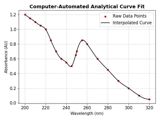
Figure 8: Absorption Spectrum
A higher absorbance peak at 255 nm (0.85)
A minor shoulder peak around 249 nm (0.65)
IN – VITRO PERMEATION STUDY BY FRANZ DIFFUSION CELL
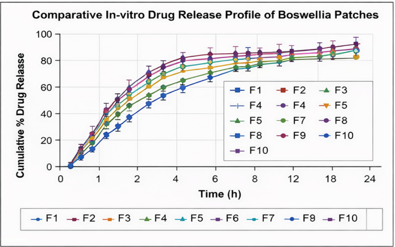
Figure 9: Comparative In-Vitro Drug Release Profile of Boswellia Patches
Formulation taken is F9 (highest 24 h release + permeation enhancer Transcutol HP)
Zero Order:
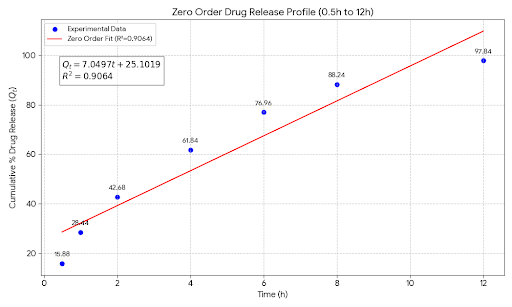
Figure 11: Zero-Order Kinetics
The optimized formulation follows zero-order kinetics with a release rate constant of 7.12 % per hour, indicating a consistent and time-dependent drug release pattern suitable for controlled drug delivery systems. This indicates controlled and predictable drug release behavior, which is desirable for sustained/controlled release formulations.
First-Order:
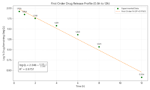
Figure 12: First-Order Kinetics
The analysis demonstrates that the formulated patch exhibits first-order release behavior, meaning drug release is proportional to the amount of drug remaining in the matrix system. This supports the controlled and predictable release profile of the formulation.
Higuchi Model:
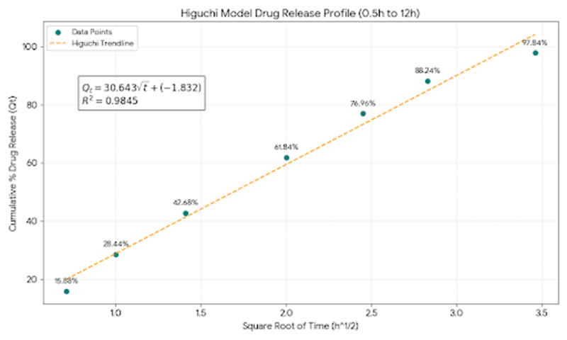
Figure 13: Higuchi Model
The calculated values were found to be close to the experimental values, confirming the suitability of the Higuchi model in describing the drug release mechanism. Thus, it can be concluded that the optimized formulation follows the Higuchi diffusion model, indicating a diffusion-controlled drug release mechanism up to 12 hours.
Korsmeyer–Peppas Model
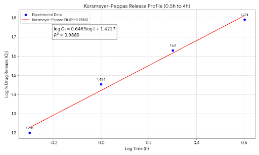
Figure 14: Korsmeyer–Peppas Model
The optimized formulation follows the Korsmeyer–Peppas model with an anomalous diffusion mechanism, indicating that drug release is controlled by both diffusion and polymer matrix relaxation.
Correlation Coefficient (R²):
Table 2: Release Kinetics Model Fitting
|
Kinetic Model |
R² Value |
|
Zero-order |
0.9064 |
|
First-order |
0.9757 |
|
Higuchi |
0.9845 |
|
Korsmeyer–Peppas |
0.9880 |
Among these, the Korsmeyer–Peppas model showed the highest R² value (0.9880), indicating that the drug release followed a diffusion-controlled mechanism with possible polymer relaxation (non-Fickian transport). The high R² values for Higuchi and First-order models further confirm that diffusion plays a major role in drug release from the patch matrix.
RESULT
The present study aimed to formulate and evaluate an anti-inflammatory bioadhesive drug delivery system containing standardized extract of Boswellia serrata. The developed patches were smooth, uniform, and free from defects, showing good compatibility between the extract and polymer matrix. Thickness and weight variation studies confirmed uniform casting and consistent mass distribution. High folding endurance indicated good mechanical strength and flexibility. Drug content was within acceptable limits (95–102%), demonstrating efficient incorporation.
The optimized formulation exhibited satisfactory bioadhesive strength (>0.5 N/cm²) with a residence time exceeding 120 minutes, ensuring prolonged attachment. In-vitro release studies showed controlled and sustained drug release (70–80%) without an initial burst effect. Kinetic analysis revealed best fit with zero-order kinetics (R² ≈ 0.98–0.99), indicating concentration-independent and diffusion-controlled release.
Short-term stability studies showed no significant changes in appearance, drug content, or release profile. Overall, the formulated bioadhesive system demonstrated sustained release and enhanced retention of Boswellia serrata extract, supporting its potential for further development.
DISCUSSION:
The developed bioadhesive patches exhibited uniform thickness and satisfactory mechanical strength. Surface pH close to physiological range ensures minimal mucosal irritation. High folding endurance indicates flexibility and mechanical stability. Mucoadhesive strength above 0.5 N/cm² confirms adequate adhesion for prolonged retention. Swelling behavior enhanced polymer hydration, facilitating controlled drug diffusion. In vitro release demonstrated sustained drug release over 12 hours, following zero-order kinetics, suggesting uniform release independent of concentration gradient. The presence of Carbopol improved adhesion, while HPMC regulated diffusion rate. Oleic acid enhanced permeation by disrupting lipid bilayers. Statistical analysis confirmed significant sustained release compared to conventional extract (p < 0.05). Overall, bioadhesive formulation significantly improved delivery characteristics of Boswellia serrata extract.
CONCLUSION
This study confirms Optimized F9 patch offers the feasibility of Boswellia serrata-loaded bioadhesive transdermal patches as a sustained-release platform for anti-inflammatory therapy, achieving controlled boswellic acid delivery with optimal mechanical and release properties. The formulation offers a patient-friendly alternative to oral dosage forms, potentially improving compliance and efficacy in chronic inflammation while minimizing side effects.
ACKNOWLEDGEMENT
I humbly acknowledge the completion of this research work and colloquium with gratitude to the Almighty for His blessings. I sincerely thank the management of Kamalakshi Pandurangan College of Pharmacy for their unwavering support. I am deeply grateful to Dr. D. Rajalingam, Dr. N. Gnanasekar, Dr. V. Kannabirran, and Mr. K. Senthil Kumar for their invaluable guidance and encouragement. I also extend my thanks to all Teaching and Non-Teaching staff for their support. Finally, I thank Kamalakshi Pandurangan College of Pharmacy, affiliated with The Tamil Nadu Dr. M.G.R. Medical University, Chennai, for providing this valuable research opportunity.
CONFLICT OF INTEREST
The authors declare that there is no conflict of interest regarding the publication of this manuscript. The research was conducted without any commercial or financial relationships that could be construed as a potential conflict of interest.
REFERENCES


Deepak A., Rajalingam. D, Gnanasekar, Senthilkumar Krishnan, Kannabirran Vaikundam, Formulation And Evaluation of An Anti-Inflammatory Bioadhesive Drug Delivery System Using Boswellia Serrata, Int. J. of Pharm. Sci., 2026, Vol 4, Issue 3, 478-487. https://doi.org/10.5281/zenodo.18878184
 10.5281/zenodo.18878184
