Department of Pharmaceutics, Shree Devi College of Pharmacy, Mangaluru – 574142, Karnataka, India
Topical drug delivery systems such as ethosomal gels have gained prominence due to their dual-release control mechanism. The present study aimed to formulate and evaluate an ethosomal gel containing clarithromycin to enhance its transdermal permeation. Clarithromycin-loaded ethosomes were prepared using varying concentrations of phospholipids and ethanol, and characterized for vesicle size, shape, zeta potential, entrapment efficiency, and in vitro drug release using a cellophane membrane. The optimized ethosomes were incorporated into a gel base using Carbopol 974. The resulting ethosomal gels were assessed for pH, spreadability, viscosity, drug content, and drug release behavior. Among the formulations, F6 exhibited optimum physical characteristics with an entrapment efficiency of 81.2%, vesicle size of 4069 nm, zeta potential of 1.36 mV, and a polydispersity index of 0.316. In vitro drug release studies showed a cumulative release of 89.98 µg/cm² over 8 hours, while ex vivo studies using pig skin demonstrated a release of 80.11 µg/cm². The formulation also showed maximum stability under storage at 25?±?2?°C and 60% relative humidity. These findings support the potential of ethosomal gels as effective carriers for the topical delivery of clarithromycin.
For decades, the treatment of acute and chronic illnesses has primarily relied on conventional drug delivery systems, which typically involve the administration of pharmaceutical dosage forms that provide an immediate release of the drug. While such systems are effective in achieving prompt therapeutic concentrations, they often require multiple daily doses to maintain drug levels within the desired therapeutic range. This repeated administration results in significant fluctuations in plasma drug concentration, which may reduce therapeutic efficacy or increase the risk of side effects.¹To overcome these limitations, significant technological advancements have been made in the field of drug delivery. These innovations have led to the development of controlled drug delivery systems, which are specifically designed to regulate the rate and duration of drug release. Controlled release systems offer the advantage of predictable and reproducible drug kinetics, ensuring that drug release follows a consistent and measurable profile from unit to unit. Unlike conventional sustained release systems, controlled release formulations offer superior control over therapeutic drug levels.¹Such systems aim to enhance therapeutic efficacy by maintaining drug concentrations within a narrow therapeutic window for extended periods, reducing the frequency of dosing, improving patient compliance, and maximizing drug utilization. However, controlled drug delivery systems also present challenges, such as the potential toxicity or biocompatibility issues associated with the materials used, complications from implantation or removal, and higher production costs compared to traditional formulations.2The ideal controlled drug delivery system should combine mechanical strength, biocompatibility, ease of use, and safety, while enabling high drug loading and controlled release over time. The development of such systems remains a key focus in pharmaceutical research.² Given this background, the present investigation focuses on exploring novel carrier systems capable of improving therapeutic outcomes through controlled drug delivery.
3. MATERIALS AND METHODS
3.1 Materials: Clarithromycin and soya lecithin were procured from Yarrow Chemicals, Mumbai. Ethanol and Carbopol 974 were obtained from Oxford Lab Fine Chem LLP. Propylene glycol was purchased from Sisco Research Lab Pvt. Ltd., Mumbai. Sodium hydroxide, disodium hydrogen phosphate, and potassium dihydrogen phosphate were supplied by Nice Chemicals (P) Ltd., Kochi, Kerala.
3.2 Preformulation Studies: Preformulation studies represent the initial phase in the rational development of pharmaceutical dosage forms. These studies investigate the physical and chemical characteristics of the drug alone and in combination with excipients. The primary objective is to generate data that aids the formulator in developing stable and bioavailable formulations. The following Preformulation parameters were assessed:
3.2.1Organoleptic Properties:
Organoleptic characteristics such as color and odour were examined visually and compared with pharmacopoeial standards. A small quantity of the drug was placed on butter paper and observed under adequate lighting for color. For odor evaluation, a minimal amount of drug was smelled directly.
3.2.2 Solubility Studies:
Solubility was determined in various solvents including water, ethanol, methanol, isopropanol, benzene, acetone, chloroform, and ether. Excess drug was added to 10 mL of each solvent and agitated in a water bath shaker at 37 ± 0.5?°C for 24 h. After filtration, appropriate dilutions were made with buffer, and drug concentration was determined spectrophotometrically at λ max using the standard calibration curve.3
3.2.3 Determination of Melting Point:
Melting point was assessed using the USP method. A small quantity of drug was placed in a sealed capillary tube, which was inserted into a melting point apparatus. The temperature at which the drug began and completed melting was recorded.
3.2.4 Determination of Drug pH:
The pH of clarithromycin was measured for a freshly prepared 1% solution in ethanol using a calibrated digital pH meter.4
3.2.5 FTIR Study:
Fourier-transform infrared (FTIR) spectroscopy was conducted to identify potential physicochemical interactions between the drug and excipients. Moisture-free samples of clarithromycin, soya lecithin, and Carbopol, as well as their physical mixture, were analyzed using the KBr pellet method. The FTIR spectra were recorded and characteristic peaks were interpreted to identify functional groups and detect possible interactions. FTIR analysis is crucial in evaluating formulation stability and compatibility.5
3.3 Preparation of Ethosomes:
Clarithromycin-loaded ethosomes were formulated using the cold method as described by Touitou (2000).4 The vesicular system comprised phospholipid (2.0–4.0% w/v), ethanol (20–40% v/v), propylene glycol (20% v/v), clarithromycin (0.5% w/v), and distilled water q.s. to 100% v/v.Initially, phospholipid and clarithromycin were dissolved in ethanol under vigorous stirring. Propylene glycol was then added to the solution. The mixture was heated to 40?±?1?°C, and a fine stream of preheated distilled water was slowly added with continuous stirring at 700 rpm using a mechanical stirrer in a closed vessel. The stirring was continued for an additional 5 minutes while maintaining the temperature at 40?±?1?°C. The formulation was allowed to cool to room temperature and subsequently sonicated using a probe sonicator for five cycles of 3 minutes each, with a 1-minute rest between cycles.4
|
Formulation |
Soya lecithin (%w/w) |
Ethanol (% v/v) |
Propylene glycol (%v/v) |
Drug (mg) |
Distilled water (ml) |
|
1 |
2 |
20 |
20 |
500 |
q.s |
|
2 |
4 |
20 |
20 |
500 |
q.s |
|
3 |
3 |
20 |
20 |
500 |
q.s |
|
4 |
4 |
30 |
20 |
500 |
q.s |
|
5 |
3 |
30 |
20 |
500 |
q.s |
|
6 |
2 |
30 |
20 |
500 |
q.s |
|
7 |
3 |
40 |
20 |
500 |
q.s |
|
8 |
4 |
40 |
20 |
500 |
q.s |
|
9 |
2 |
40 |
20 |
500 |
q.s |
Characterization of clarithromycin loaded ethosomes:
3.3.1Vesicle Shape;
The surface morphology and shape of ethosomal vesicles were examined using Scanning Electron Microscopy (SEM). A drop of the ethosomal formulation was mounted on a clear glass stub, air-dried, and coated using a Polaron E 5100 sputter coater. The sample was then visualized under a Scanning Electron Microscope to observe vesicle morphology. 6
3.3.2Vesicle Size;
The particle size analysis of the prepared ethosomal formulation was carried out using a Malvern Zetasizer based on the principle of Dynamic Light Scattering (DLS). This technique was employed to determine the average vesicle size distribution.
3.3.3Zeta Potential;
Zeta potential is indicative of the surface charge and plays a significant role in the stability of the vesicular system. Particles with a zeta potential value greater than ±30 mV are generally considered stable due to sufficient electrostatic repulsion. However, ethosomal formulations often exhibit zeta potential values ranging from −10 to −20 mV. The zeta potential of the developed ethosomal system was determined using a Zetasizer (Malvern, UK) at 25?°C 7.
3.3.4Entrapment Efficiency;
The entrapment efficiency (EE%) of clarithromycin-loaded ethosomes was determined using the ultracentrifugation method. Briefly, 15 mL of the ethosomal formulation was centrifuged at 12,000 rpm for 2 hours. The supernatant was collected, and the concentration of unentrapped drug was quantified using a UV-visible spectrophotometer at 268 nm. Entrapment efficiency was calculated using the following formula:
Entrapment Efficiency ? [?Q t − Q s ? ÷ Q t] × 100
Where,
Qt is the total amount of drug added, and
Qs is the amount of free drug present in the supernatant 8.
3.4 In Vitro Drug Release Studies
In vitro release studies of the ethosomal formulation were conducted using a Franz diffusion cell. The apparatus consists of a donor and a receptor compartment. A cellophane membrane (previously soaked in phosphate buffer pH 7.4) was fixed between the compartments. One millilitre of ethosomal suspension containing clarithromycin was placed in the donor compartment. The receptor compartment contained 50 mL of phosphate buffer (pH 7.4), maintained at 37 ± 0.5 °C, and stirred continuously using a magnetic bead. Samples were withdrawn at predetermined intervals over 8 hours and replaced with fresh buffer to maintain sink conditions. The withdrawn samples were analyzed using UV spectroscopy at 268 nm after appropriate dilutions 9.
Drug Release Kinetics
The release data obtained from in vitro studies were subjected to kinetic modeling to understand the release behavior of clarithromycin from the ethosomal system. The models applied included:
These models were used to elucidate the release mechanism and order of release.
Preparation of Ethosomal gel:
The ethosomal gel was prepared by formulating the 1% carbopol 974 gel. This is obtained by adding 2 ml of 10% (w/v) sodium hydroxide to a solution of the 0.5 g carbopol 974 in 48ml water. The ethosomal gel was prepared by adding the ethosomes suspension to 1% carbopol 974 gels at the ratio of 1:1(w/v) and stirred until homogenous.10
3.5 Evaluation of Ethosomal Gel:
3.5.1 Measurement of pH;
To evaluate the pH, 1 g of clarithromycin-loaded ethosomal gel was dispersed in 100 mL of distilled water using a homogenizer. The pH of the resulting gel dispersion was measured using a digital pH meter by immersing the electrode into the dispersion. The readings were recorded in triplicate, and the average pH value was calculated 4.
3.5.2 Viscosity;
The viscosity of the ethosomal gel was measured using a Brookfield Viscometer (DV-II+Pro, D220). Approximately 50 g of the formulation was transferred into a 50 mL beaker, and spindle number 64 was selected. The measurements were recorded at different rotational speeds (5, 10, 50, and 100 rpm) ensuring that the spindle groove was completely immersed in the sample 11.
3.5.3 Washability;
To assess washability, the gel formulations were applied to the skin, followed by manual washing with water. The ease and extent of removal were observed visually and recorded 12.
3.5.4 Spreadability;
Spreadability is an important parameter to ensure uniform application and therapeutic availability of the gel. It was assessed using a glass slide assembly, in which one slide was fixed to a wooden block and the upper slide was movable. A sample (2–5 g) of the gel was placed between the slides, and weights were added to a pan attached to the upper slide. The time required for the upper slide to travel 10 cm under the load of 80 g was recorded. Lower time indicates better spreadability. Spreadability (S) was calculated using the following formula 13:
s=(M×L)/T
Where,
M = weight tied to the upper slide (g)
L = length of the glass slide (cm)
T = time taken to separate the slides (s)
3.5.5 Drug Content;
To determine the drug content, 100 mg of ethosomal gel was dissolved in 100 mL of phosphate buffer saline (PBS), pH 7.4. A 1 mL aliquot from this solution was further diluted to 100 mL with PBS. The absorbance was measured using a UV-visible spectrophotometer at the λmax of clarithromycin. The drug content was calculated using the following formula 14:
amount of drug=(concentration from the standerd graph×df)/1000

Where,
df = dilution factor.
3.5.6 In Vitro Drug Release Studies;
The in vitro release study was conducted using a Franz diffusion cell with donor and receptor compartments. A cellophane membrane, pre-soaked in PBS (pH 7.4), was fixed between the compartments. 1 g of the ethosomal gel was placed in the donor compartment. The receptor compartment contained 50 mL of PBS, maintained at 37 ± 0.5°C and stirred continuously using a magnetic bead. Samples were withdrawn at regular intervals over 8 hours, and the withdrawn volume was replaced with fresh buffer to maintain sink conditions. The samples were analyzed spectrophotometrically at 268 nm 15.
3.5.7 In Vitro Drug Release Kinetics
The drug release data were analyzed to determine the release mechanism by fitting the results to various kinetic models:
Qt = Q0 + K0t
Where,
Q? = amount of drug released at time t,
Q? = initial amount of drug in solution,
K? = zero-order release rate constant 4.
logc=logc0 - kt2.303

Where,
C? = initial concentration of drug,
k = first-order rate constant,
t = time [50].
Q = KH x t1/2
Where,
Q = cumulative amount of drug released at time t,
K_H = Higuchi release rate constant 4.
M?/M∞ =Ktn
Interpretation of the n value:
3.6 Ex Vivo Skin Permeation Studies:
Ex vivo permeation studies were conducted using pig ear skin, obtained from a local slaughterhouse. The skin was carefully cleaned, and hair was removed. The skin was hydrated in PBS (pH 7.4) before use. The Franz diffusion cell was used, where the dermal side of the skin was exposed to 1 mL of ethosomal gel in the donor compartment. The receptor compartment contained 50 mL of PBS (pH 7.4), stirred at 50 rpm and maintained at 37 ± 0.5°C. 5 mL samples were withdrawn at predetermined intervals up to 8 hours, and the absorbance was measured at 268 nm. Fresh buffer was added after each withdrawal to maintain sink conditions 16.
3.7 Stability Studies:
Stability testing is a critical parameter to ensure shelf life and acceptability of pharmaceutical formulations. It includes monitoring physical and chemical changes such as appearance, drug content, and in vitro drug release. The stability study for the optimized ethosomal gel formulation was conducted as per modified ICH guidelines. Samples were stored under two different conditions in a stability chamber (Labtop Equipment), and analyses were carried out after 4 weeks of storage. The evaluation parameters and results are presented in Table 5.10 17.
4. RESULTS
4.1 Preformulation Studies:
4.1.1 Organoleptic Characteristics;
The organoleptic properties of clarithromycin, including its appearance, color, taste, and odor, were evaluated and compared with literature-reported standards. The findings are presented in Table 4.1.
Table 4.1: Organoleptic Characteristics of Clarithromycin
|
PROPERTIES |
REPORTED |
OBSERVED |
|
Appearance |
Crystalline powder |
Crystalline powder |
|
Color |
White to off white |
White to off white |
|
Taste |
bitter |
bitter |
|
Odor |
Odorless |
Odorless |
4.1.2 Solubility Studies;
Solubility studies were conducted to assess the solubility profile of clarithromycin in various solvents. The drug was found to be soluble in acetone, slightly soluble in methanol, ethanol, and acetonitrile, and practically insoluble in water. The aqueous solubility was recorded as 0.33 mg/L.
4.1.3 Melting Point;
The melting point of clarithromycin was determined using capillary method. The analysis was carried out in triplicate, and the mean melting point was found to be 220?°C.
4.1.4 Determination of pH;
The pH of clarithromycin was evaluated by dispersing the drug in distilled water and measuring using a digital pH meter. The measurement was conducted three times, and the average pH was recorded as 5.6.
4.1.5 FTIR Studies;

FTIR spectra of clarithromycin
FTIR spectra of clarithromycin formulation
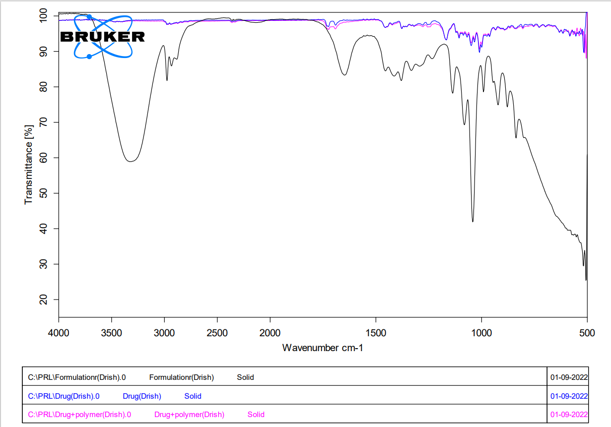
FT-IR spectra of drug with physical mixture
4.2Vesicle size analysis of clarithromycin ethosomes
|
Formulation Code |
Particle size (nm) |
|
F1 |
8123 |
|
F2 |
7642 |
|
F3 |
6554 |
|
F4 |
5326 |
|
F5 |
5268 |
|
F6 |
4069 |
|
F7 |
5187 |
|
F8 |
3241 |
|
F9 |
3826 |
4.3 Drug entrapment efficiency of clarithromycin
|
Serial no |
Formulation code |
Entrapment Efficiency (%) |
|
1 |
F1 |
65.3
|
|
2 |
F2 |
78.9
|
|
3 |
F3 |
63.2
|
|
4 |
F4 |
75.6
|
|
5 |
F5 |
76.3
|
|
6 |
F6 |
82.9
|
|
7 |
F7 |
68.7
|
|
8 |
F8 |
56.5
|
|
9 |
F9 |
55.1
|
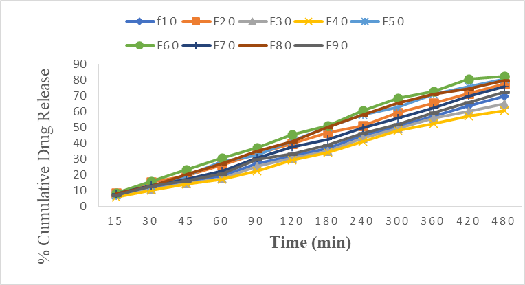
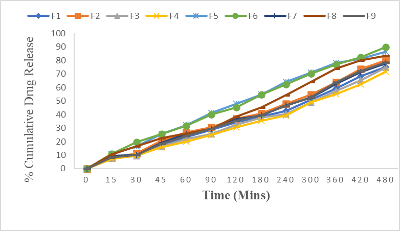
4.4 In vitro drug release profile of clarithromycin ethosomes:
4.5 Evaluation of ethosomal gel incorporated with clarithromycin:
4.5.1 Formulation of ethosomal gel:
Ethosomal gel was prepared by using carbopol as the polymer and prepared gel was smooth and off white in colour. It was characterized by the measurement of pH, viscosity, Spreadability.
4.5.2 Measurement of pH
The pH of the gel was measured as per the procedure and the pH of the ethosomal gel was ranged between 6.8 - 7.0. The pH of the ethosomes was found within the range of pH of skin and would not cause any irritation to skin. Thus prepared ethosomal formulations are suitable for topical application.
4.5.3 Viscosity
The viscosity of the prepared formulations was optimum and was found to be between 5400- 5896 cps
4.5.4 Spreadibilty
The spreading diameter ranges between 12.83- 18.92g/cm2. Thus the spreadability results showed that the ethosomal gel was most effective i.e. it showed best result for spreadability.
4.5.5 Drug content estimation
The drug content of all the formulations was ranged between 65.82-92.90%. Thus it determines that the formulation containing F6 has good drug content when compared to remaining formulations.
4.6 In Vitro Drug Release Studies
Characterization of ethosomal gel containing clarithromycin.
|
Formulation code |
Measurement of pH |
Viscosity (cps) |
Spreadability (g/cm2) |
%Drug content |
|
F1 |
6.9 |
5400 |
12.83 |
65.82 |
|
F2 |
6.9 |
5301 |
14.79 |
79.05 |
|
F3 |
6.8 |
5677 |
16.28 |
81.53 |
|
F4 |
7.0 |
5535 |
15.28 |
72.35 |
|
F5 |
6.8 |
5264 |
13.88 |
90.13 |
|
F6 |
7.0 |
5896 |
18.10 |
92.90 |
|
F7 |
6.8 |
5324 |
18.92 |
68.22 |
|
F8 |
6.7 |
5794 |
15.66 |
91.30 |
|
F9 |
6.7 |
5468 |
13.82 |
83.65 |
In vitro drug release kinetics of clarithromycin ethosomal gel
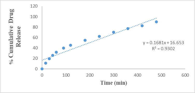
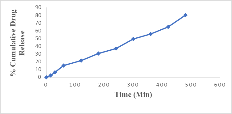
Zero order release kinetics of F6
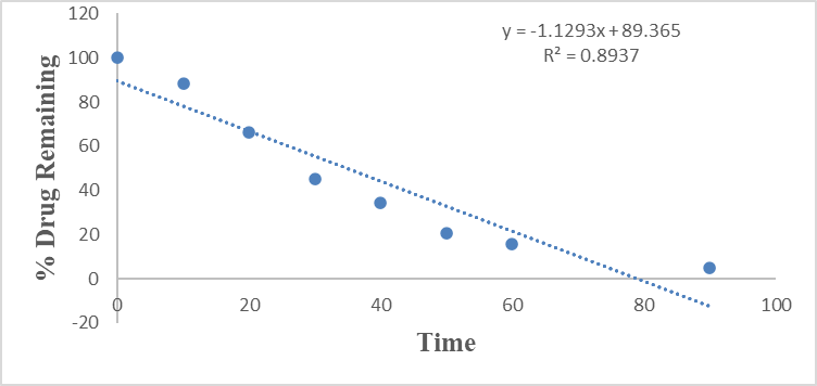
First order release kinetics of F6 formulation
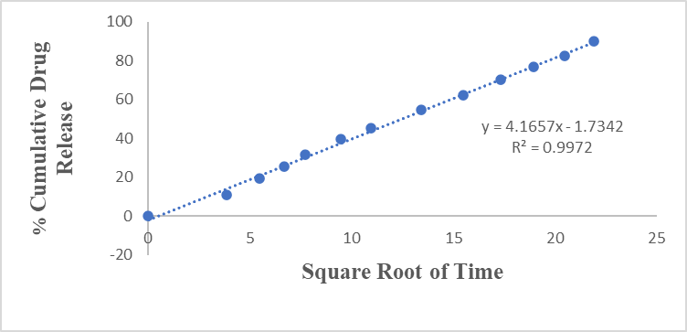
Higuchi model of F6 formulation
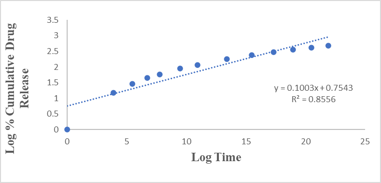
Korsmeyer Peppas model of F6 formulation
Ex vivo cumulative % drug release of ethosomal gel containing clarithromycin
4.7 Stability studies of ethosomal gel containing clarithromycin
|
Formulation code |
Parameters evaluated after the 4 weeks |
Storage temperature |
|
|
250C ± 2 ? C,
60% RH |
400C ± 2 ? C,
60% RH |
||
|
Appearance |
Pale yellow, smooth gel |
Pale yellow, smooth gel |
|
|
pH |
6.9 |
6.8 |
|
|
% Drug content |
90.92 |
88.50 |
|
|
% Drug release |
79.12 |
77.45 |
|
DISCUSSIONS
The present investigation focused on the formulation and evaluation of a clarithromycin-loaded ethosomal gel for transdermal delivery with the aim of enhancing skin permeation and achieving sustained drug release. Preformulation studies confirmed that the organoleptic properties, solubility, pH, and melting point of clarithromycin were consistent with reported literature, indicating its suitability for formulation development. FTIR analysis revealed the presence of characteristic peaks of both the drug and polymer in the formulation, with minor shifts suggesting molecular interactions without chemical incompatibility. Nine ethosomal formulations (F1–F9) containing a fixed drug concentration were prepared and successfully incorporated into a Carbopol 974 gel base. SEM analysis demonstrated that the ethosomes were predominantly spherical with smooth surfaces, indicating stable vesicular structures. Particle size analysis showed that ethanol concentration significantly influenced vesicle size, with higher ethanol concentrations producing smaller vesicles due to membrane fluidization, and vesicle sizes ranged from 3000 to 9000 nm. Drug entrapment efficiency varied among formulations, with the highest entrapment observed in formulation F6 (30% ethanol), while higher ethanol concentrations led to reduced entrapment due to increased membrane permeability and drug leakage. In vitro drug release studies demonstrated controlled and sustained release over 8 h, with formulation F6 showing the highest cumulative drug release. The ethosomal gels exhibited pH values within the physiological skin range, appropriate viscosity, good spreadability, and uniform drug content, indicating suitability for topical application. Release kinetics analysis revealed that most formulations followed the Korsmeyer–Peppas model, while selected formulations including F6 followed the Higuchi model, with release exponent values indicating a non-Fickian diffusion mechanism. Ex vivo permeation studies using pig skin demonstrated enhanced transdermal permeation, with formulation F6 showing high cumulative drug permeation over 8 h and a strong correlation with in vitro release data. Stability studies conducted on the optimized formulation showed no significant changes in physical appearance, pH, drug content, or drug release profile over the study period, confirming the stability of the clarithromycin-loaded ethosomal gel and supporting its potential as an effective transdermal drug delivery system.
CONCLUSION
In the present study, clarithromycin-loaded ethosomes were successfully developed using soya lecithin and ethanol and incorporated into a Carbopol 974 gel base to obtain an ethosomal gel suitable for topical application. Drug–excipient compatibility studies indicated no significant interaction, confirming the appropriateness of the selected formulation components. The preparation method adopted for ethosomes was found to be simple, reproducible, and suitable for consistent formulation development.The formulated ethosomal gels exhibited acceptable physicochemical characteristics, including pH values within the range of 6.8–7.0, which is considered appropriate for topical use. Spreadability studies demonstrated that polymer concentration influenced the spreading behavior of the gels; however, all formulations showed satisfactory spreadability. Viscosity measurements further confirmed that the formulations possessed suitable consistency for topical administration.In vitro drug release studies indicated that formulation F6 containing Carbopol 974 showed comparatively higher drug release over an 8-hour period. Ex vivo permeation studies revealed a slower drug release profile when compared to in vitro results, which may be attributed to the barrier properties of pig skin and the additional diffusion time required for drug permeation. Stability studies of the optimized formulation suggested that the ethosomal gel remained stable under the tested storage conditions.Based on the overall findings, the developed clarithromycin-loaded ethosomal gel demonstrated suitable formulation and release characteristics, indicating its potential as a topical drug delivery system. However, further in vivo and clinical studies would be required to confirm its therapeutic efficacy in the treatment of fungal infections.
REFERANCES


V. Felix Joe, V. Drishya Bharath, Formulation and Characterization of Ethosomes for Transdermal Delivery of Clarithromycin, Int. J. of Pharm. Sci., 2026, Vol 4, Issue 2, 2523-2537. https://doi.org/10.5281/zenodo.18667862
 10.5281/zenodo.18667862
