View Article

Abstract

Genetic disorders, which arise from hereditary mutations, frequently lead to chronic or potentially fatal health issues with limited therapeutic alternatives. Stem cell therapy presents a promising approach by facilitating the regeneration of damaged tissues and the rectification of genetic anomalies. This review examines contemporary strategies employing hematopoietic, mesenchymal, and induced pluripotent stem cells (iPSCs) for the treatment of genetic disorders. The incorporation of gene editing technologies, such as CRISPR-Cas9, has broadened the scope of therapeutic interventions, especially for monogenic disorders, including sickle cell disease, ?-thalassemia, and severe combined immunodeficiency. Additionally, the review addresses emerging applications of these therapies in the context of neurodegenerative and metabolic diseases. Despite persistent challenges concerning safety, delivery mechanisms, and the long-term effectiveness of these treatments, advancements in stem cell-based therapies are progressing swiftly toward clinical implementation. These developments signify a pivotal transition in the realm of genetic medicine, moving from mere symptom management to the prospect of potentially curative interventions.

Keywords

Stem cell , monogenic disorder , genetic disorder , CNS , hPSC

Introduction

Regenerative medicine is recognized as a promising new avenue for treating previously untreatable diseases in contemporary science [1]. This field is inherently multidisciplinary, encompassing areas such as cell biology, genetics, biomechanics, materials science, and computer science [2,3]. The primary objective of regenerative medicine is to restore normal functionality to damaged cells and tissues [4]. Following the discovery of stem cells and a growing awareness of their distinctive properties, these cells have been established as potential therapeutic agents for the repair of organs and tissues, making them excellent candidates for regenerative medicine due to their numerous possible applications [5]. Researchers are now exploring regenerative medicine as an alternative to conventional drug-based therapies, particularly in the context of various diseases, including degenerative conditions [6,7,8,9,10]. The fundamental principle of regenerative medicine involves the regeneration of tissues or organs utilizing cells, with a variety of cell types employed to achieve this goal. However, existing studies have highlighted certain limitations associated with cell therapy. In recent years, several alternatives have been proposed to address these limitations, such as enhancing the application of stem cells for tissue restoration through methods like cell-scaffold combinations, optimized cell cultures with appropriate biochemical characteristics, gene editing, immunomodulation of stem cells, and the use of derivatives from stem cells [11,12,13,14,15]. Nonetheless, the clinical application of these alternatives may be delayed, as further preclinical research is needed due to their status as emerging technologies [16].  Stem cells represent a category of immature cells endowed with the potential to generate and repair various tissues and organs throughout the body due to their unique abilities for proliferation, differentiation, and self-renewal [17]. They have therapeutic effects that enhance physical recovery by regenerating damaged cells to facilitate the healing of organs. Leveraging the inherent properties of stem cells, researchers have harnessed their biological mechanisms for stem-cell-based therapies. The diverse mechanisms through which stem cells can foster tissue regeneration include (1) the inhibition of inflammatory pathways [18,19], (2) the reduction of apoptosis [20,21], (3) the recruitment of other cells [22,23], (4) the stimulation of angiogenesis [24,25], and (5) differentiation [26]. The underlying cause of a disease is a critical factor in determining the appropriate stem cell mechanism to utilize, as well as in guiding the regeneration of tissues or organs. Extensive investigations are necessary to elucidate the key mechanisms involved in the therapeutic application of these cells, and the alignment of stem cell therapeutic mechanisms with disease mechanisms is anticipated to enhance the prospects for developing effective treatments through stem cell utilization. Between 1971 and 2021, a total of 40,183 scholarly articles were published on stem-cell-based therapies, all focused on the discoveries and objectives associated with "Stem Cell Therapy" rooted in the therapeutic effectiveness of stem cells [27]. A genetic disorder can be defined as an impairment of normal physiological and developmental health caused by alterations or mutations in an individual's genomic structure. These disorders can arise from a variety of sources, including chromosomal abnormalities, which are deviations from the typical number or arrangement of chromosomes, and environmental factors. In some instances, the precise origins of these disorders remain enigmatic, leading to gaps in our understanding of their etiology[28]. Environmental influences, including air pollution and exposure to hazardous waste sites, have been identified as significant contributors to an increased risk of structural birth defects among vulnerable populations[29]. Various physical environmental factors also play a crucial role; for instance, maternal drug use, infections acquired during pregnancy, and exposure to harmful chemicals can adversely affect fetal development, further complicating the landscape of genetic disorders. The implications of these genetic anomalies extend far beyond the initial diagnosis. The treatment and management of genetic disorders are often financially burdensome and fraught with challenges. In many cases, complete recovery is not a feasible outcome, leaving individuals and their families to navigate the complexities of ongoing care and potential lifelong implications. Additionally, many genetic disorders are classified as lethal, posing a significant threat to affected individuals. It is essential to note that lethality in genetic disorders often occurs through recessive inheritance patterns; instead of remaining prevalent in the gene pool, lethal dominant disorders tend to be filtered out by natural selection, as carriers of such conditions typically do not survive long enough to transmit these genes to subsequent generations. The prevalence and frequency of various genetic disorders within a population are influenced by a multitude of factors. These include the genetic makeup of that population, which encompasses not only inherited traits but also historical and socio-economic contexts, as well as the types of environmental exposures prevalent in the geographical area. Thus, the complex interplay between genetic predispositions and environmental conditions necessitates a thorough examination to understand the distribution and impact of genetic disorders in different communities, ultimately framing the future of healthcare strategies and interventions aimed at managing these multifaceted conditions[29,30,31].

How might stem cells be useful to cure genetic diseases:

Intravenous administration of whole bone marrow is a therapeutic approach utilized for patients suffering from acute leukemia, bone marrow aplasia, and congenital immune deficiencies (32). The efficacy of these interventions is contingent upon the characteristics of hematopoietic stem cells, which are rare entities that serve as the benchmark for all research pertaining to adult stem cells. Recent advancements have enabled the isolation and characterization of adult stem cells from diverse tissues, paving the way for innovative cell-based therapies aimed at addressing a variety of genetic disorders, extending beyond hematological conditions (33). A primary application of stem cells in the context of genetic diseases involves the cellular repair of tissues impacted by genetic mutations, wherein stem cells devoid of mutations are transplanted to restore normal functionality; these stem cells are typically sourced from a non-mutated donor that is also a tissue match. Alternatively, stem cells may be harvested from the patient and subsequently genetically modified prior to re-implantation (34). These stem cells exhibit the capacity to differentiate into the specific cell types necessary for the restoration of normal tissue function, and they may also serve as cellular models for exploring genetic and biochemical pathways, particularly when derived from affected individuals. In principle, the treatment of monogenic disorders would entail the rectification of the defective gene via genetic engineering. However, this seemingly straightforward approach is complicated by the fact that the implications of the mutated gene can vary significantly across different cells and tissues, depending on the level of gene expression. For instance, the gene mutation associated with cystic fibrosis is expressed in multiple epithelial tissues and results in a range of symptoms (35). Consequently, gene therapy aimed at cystic fibrosis has primarily targeted the lung epithelium, as it is more accessible and because excessive mucus production is a predominant cause of mortality. In contrast, identifying a straightforward target for gene therapy in Huntington’s disease—a neurodegenerative condition affecting numerous regions of the brain—is less clear, unless the intervention is directed at the germ line or early developmental stage. Conversely, hemophilia and sickle cell anemia, which are hematological disorders, are associated with genes that perform more specialized functions, enabling therapeutic manipulation of thestem cells responsible for generating the affected blood cell types (36). It thus appears not unexpected that the only stem cell therapies currently employed in clinical settings—including those involving genetically modified stem cells—are predominantly focused on blood disorders (33), with all existing therapies utilizing adult stem cells, many of which are autologous. Genetically engineered stem cell therapy is anticipated to be most effectively applied in the context of monogenic diseases, as it typically requires the modification or insertion of a single gene. Nevertheless, discernible therapeutic strategies may still pose challenges when the target gene exerts influence across multiple tissues, necessitating intervention at various sites of transplantation. Overall, the most promising therapies for stem cell transplantation will likely emerge in scenarios where a single cell type can be transferred to a specific location. Some stem cell therapies hold the potential for curative outcomes, such as the application of genetically modified hematopoietic stem cells in the treatment of severe combined immune deficiency (37), while others may only aim to ameliorate specific symptoms. A pertinent example of the latter is the transplantation of genetically modified GABAergic neurons, sourced from stem cells, into the brain striatum to address the motor symptoms associated with Huntington’s disease.

Advancing Cell Therapy For Neurodegenrative diseases:

NSCs are responsible for the construction of the CNS, generating various types of neurons and glial cells, and hold great promise for replacement therapies in conditions where different cell types are lost(41). Clinical trials have explored the use of human fetal-derived NSCs for neurological ailments such as Batten disease, Pelizaeus-Merzbacher disease, spinal cord injuries, AMD, and AD(42). The transplantation of NSCs has been shown to be safe and has exhibited signs of positive effectiveness. A human fetal-derived NSC line that was modified to express GDNF enhanced motor neuron survival and extended the lifespan of animals in a SOD1 rat model of ALS(43,44), and showed safety in an early-stage clinical trial (NCT05306457)(45). Administering cells poses challenges in treating any neurodegenerative disease; typically, the initial method considered is direct cell injection. Numerous studies suggest that using NSCs with high a4b1-integrin expression for intravascular delivery can be effective, with proven brain penetration and improved outcomes observed in an ALS mouse model(46). NSCs exhibit significant regional and temporal diversity, which affects their capacity to produce specific progenitor types(47). NSCs in mice, when patterned to the visual cortex, can survive and form appropriate connections if transplanted into the visual cortex but not when placed in the motor cortex of adult mice(48). Therefore, aligning the NSC product with the target region may be crucial for achieving successful multicellular replacement across various CNS areas. In HD, the degeneration of striatal medium spiny neurons leads to severe deterioration of the basal forebrain and cerebral cortex. Transplanting hESC-derived NSCs into a postnatal day 6 mouse model of HD resulted in better histological outcomes and improved motor and cognitive functions(49). Similarly, the transplantation of hiPSC-derived NPCs led to the production of neurons, astrocytes, and oligodendrocytes, enhancing both cognitive and motor functions in an HD mouse model, with outcomes surpassing those achieved with human GCPs(50). These studies illustrate the effectiveness of utilizing multipotent progenitors to replenish lost CNS cell types. Furthermore, the addition of developing vascular cells alongside NPCs enhanced engraftment and vessel structure in a mouse model of stroke, indicating the potential for more complex, mixed cell therapies for extensive CNS repair(51). hPSC-derived cortical organoids that were engrafted into the brains of neonatal rats received thalamocortical and corticocortical inputs, extended their axons, and responded to optogenetic stimulation, which drove reward-seeking behavior(52). In the adult mouse cortex, human cerebral organoids also successfully engrafted, became vascularized, and demonstrated microglial colonization, rhythmic electrical activity, and graft-host connectivity in response to optogenetic stimulation(53). Notably, cortical organoids transplanted into adult mice equipped with electrodes to monitor brain activity integrated well and showed electrophysiological cortical responses to visual stimuli(54). Successful transplantation of organoids into the cerebral cortex of monkeys has also been achieved(55). These findings imply that in the future, hPSC-derived 3D neural grafts could potentially address significant aspects of the multifaceted, regional neural cell loss associated with complex neurodegenerative conditions.

Organoids derived from human pluripotent stem cells for the purposes of drug discovery and assessment:

Human pluripotent stem cells (hPSCs), which encompass both human embryonic stem cells (hESCs) and induced pluripotent stem cells (iPSCs), are characterized by their unique capability to undergo self-renewal while also differentiating into nearly all types of terminally differentiated cells found within the human organism. The initial derivation of hESCs occurred in 1998 from the inner cell mass of pre-implantation embryos(56,57). Subsequently, in 2006, mouse fibroblasts were successfully reprogrammed into pluripotent cells referred to as iPSCs(58). A short time thereafter, human iPSCs (hiPSCs) were also generated(58,59). Since their identification, substantial efforts have been directed towards utilizing hPSCs for applications in disease modeling and drug development. iPSCs have been generated from both patients with specific diseases and healthy individuals to investigate the cellular phenotypes associated with disease progression. Isogenic hPSCs have been employed to examine the effects of individual genes, regulatory regions, or genetic variants (such as single-nucleotide polymorphisms [SNPs]) on cellular differentiation outcomes(60).(Figure1) These models have been extensively utilized as platforms for drug discovery and as predictive tools for drug responses, facilitating the creation of innovative therapeutic candidates and advancing the field of precision medicine. In the initial phases of research utilizing human pluripotent stem cells (hPSCs) for disease modeling, the primary focus was on two-dimensional (2D) cell cultures. While 2D cultures are relatively straightforward to grow and amenable to high-throughput and high-content screening, they exhibit a monotypic nature and lack the complex cell types and appropriate cell-cell interactions characteristic of three-dimensional (3D) structures. Over the past decade, notable advancements have been achieved in the development of 3D organoids, which more accurately replicate the anatomy and physiology of the target organs by closely mimicking gene and protein expression patterns, as well as cellular architecture.

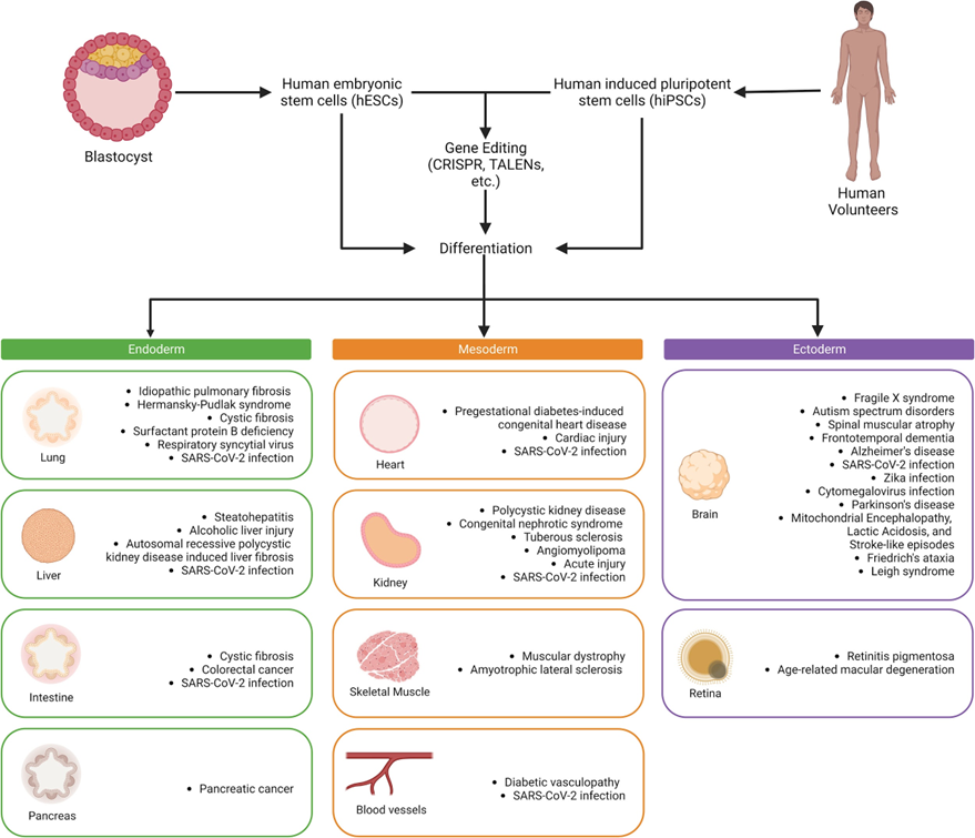
Figure 1. Strategies for using hPSC-derived organoids in disease modeling

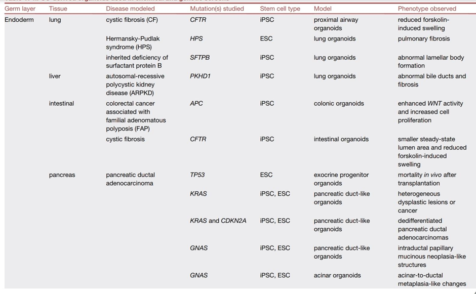
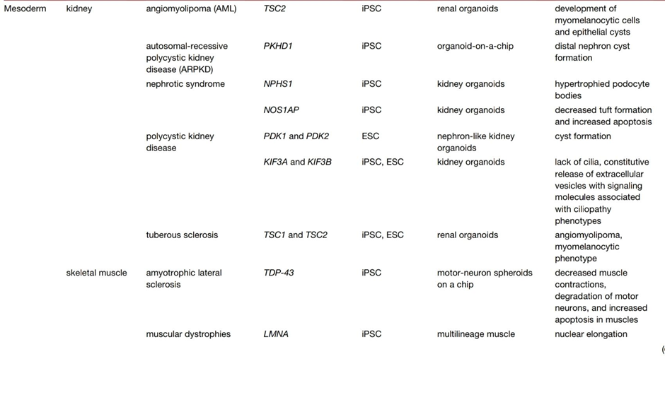
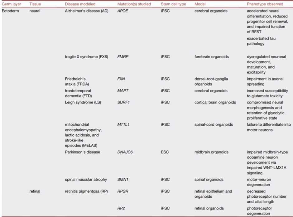
The term "organoids" encompasses a range of definitions; however, they are typically characterized as three-dimensional structures that originate from stem cells, progenitor cells, or differentiated cells. These structures are capable of self-organization, thereby mimicking the architecture of native tissue in vitro(61). Organoids consist of various cell types arranged in an organized manner, offering a more physiologically relevant environment compared to traditional two-dimensional cell cultures. Additionally, their scalable nature renders them suitable for high-throughput drug discovery studies. This aims to encapsulate the advancements made in employing human pluripotent stem cell-derived organoids for drug discovery and genetic screening, as outlined in Table 1.

Table 1: hPSC-derived organoids used in chemical and genetic screens(62-90)

Future prospects of stem cell strategies:

The potential of stem cell-based approaches for addressing neurodegenerative genetic disorders is on the brink of revolutionizing therapeutic methods and the overall drug development process. Organoids derived from human pluripotent stem cells (hPSCs)—notably brain organoids—are becoming instrumental for simulating intricate neurological disorders, including Alzheimer's disease, Parkinson's disease, Huntington's disease, and amyotrophic lateral sclerosis (ALS). These three-dimensional miniaturized organs replicate the cellular structure and function of the human brain, allowing investigators to explore disease mechanisms within a physiologically pertinent context. As advancements in these models continue, with enhancements such as the incorporation of vascular and immune elements, they are anticipated to provide a more precise representation of in vivo conditions. This progression is likely to improve the fidelity of drug screening processes and toxicity evaluations. Furthermore, the integration of stem cell technologies with CRISPR-Cas9 and other precise gene-editing methodologies facilitates the rectification of pathogenic mutations in cells derived from patients, thereby setting the foundation for tailored regenerative therapies. Over time, organoids specific to individual patients could function not solely as platforms for diagnostics and drug testing but also as sources of autologous, genetically corrected cells for transplantation. Moreover, progress in the scalability of organoids, bioprinting techniques, and their incorporation with artificial intelligence is anticipated to enhance high-throughput screening and improve predictive models of treatment responses. From a clinical perspective, the translation of these discoveries into viable treatments will depend on overcoming current limitations such as organoid maturity, vascularization, and functional integration post-transplantation. However, ongoing global collaboration between stem cell biologists, neurologists, and bioengineers is rapidly addressing these challenges. In the years ahead, stem cell-derived organoids will likely become central to personalized medicine, offering hope for curative interventions for devastating neurodegenerative genetic disorders that currently lack effective therapies

CONCLUSION:

Stem cell strategies, ranging from mutation correction to regenerative applications, have emerged as a formidable means for tackling genetic disorders, particularly in the context of neurodegenerative diseases. The advancement of organoids derived from human pluripotent stem cells has significantly transformed our capacity to model these intricate disorders in vitro, thereby providing unparalleled opportunities for advancing the study of disease progression, evaluating drug efficacy, and assessing toxicity within a human-relevant framework. These innovations not only deepen our comprehension of disease mechanisms but also expedite the advancement towards personalized and regenerative therapies. Despite ongoing challenges associated with scalability, clinical translation, and ethical considerations, the continued evolution of stem cell research presents substantial potential for revolutionizing the therapeutic approaches to genetic and neurodegenerative diseases in the foreseeable future.

REFERENCES

  1. Stem Cell Therapy: From Idea to Clinical Practice,by Mohammad Mousaei Ghasroldasht, Jin Seok, Hang-Soo Park, Farzana Begum Liakath Ali and Ayman Al-Hendy,Department of Obstetrics and Gynecology, University of Chicago, Chicago, IL 60637, USA, March 2022 ( cross ref)
  2. Lapteva, L.; Vatsan, R.; Purohit-Sheth, T. Regenerative medicine therapies for rare diseases. Transl. Sci. Rare Dis. 2018, 3, 121-132. [Google Scholar] [CrossRef][PubMed] [Green Version]
  3. Ulia, M.P.; Mantalaris, S. Stem cells bioprocessing: An important milestone to move regenerative medicine research into the clinical arena. Pediatric Res. 2008, 63, 6. [Google Scholar]
  4. Chen, F.-M.; Zhao, Y.-M.; Jin, Y.; Shi, S. Prospects for translational regenerative medicine. Biotechnol. Adv. 2012, 30, 658-672. [Google Scholar] [CrossRef] [PubMed]
  5. Rose, L.F.; Wolf, E.J.; Brindle, T.; Cernich, A.; Dean, W.K.; Dearth, C.L.; Grimm, M.; Kusiak, A.; Nitkin, R.; Potter, K.; et al. The convergence of regenerative medicine and rehabilitation: Federal perspectives. Npj Regen. Med. 2018, 3, 19. [Google Scholar][CrossRef] [Green Version]
  6. Rajabzadeh, N.; Fathi, E.; Farahzadi, R. Stem cell-based regenerative medicine. Stem Cell Investig. 2019, 6, 19. [Google Scholar] [CrossRef]
  7. Rosenthal, N.; Badylak, S. Regenerative medicine: Today's discoveries informing the future of medical practice. Npj Regen. Med. 2016, 1, 16007. [Google Scholar] [CrossRef] [Green Version]
  8. Sivandzade, F.; Cucullo, L. Regenerative stem cell therapy for neurodegenerative diseases: An overview. Int. J. Mol. Sci. 2021, 22, 2153. [Google Scholar] [CrossRef]
  9. Dehkordi, Α.Ν.; Babaheydari, F.M.; Chehelgerdi, M.; Dehkordi, S.R. Skin tissue engineering: Wound healing based on stem-cell-based therapeutic strategies. Stem Cell Res. Ther. 2019, 10, 111. [Google Scholar] [CrossRef] [Green Version]
  10. Granero-Moltó, F.; Weis, J.; Longobardi, L.; Spagnoli, A. Role of mesenchymal stem cells in regenerative medicine: Application to bone and cartilage repair. Expert Opin. Biol. Ther. 2008, 8, 255-268. [Google Scholar] [CrossRef]
  11. Ghasroldasht, M.M.; Matin, M.M.; Mehrjerdi, H.K.; Naderi-Meshkin, H.; Moradi, A.; Rajbaioun, M.; Alipour, F.; Ghasemi, S.; Zare, M.; Mirahmadi, M.; et al. Application of mesenchymal stem cells to enhance non-union bone fracture healing. J. Biomed. Mater. Res. Part A 2018, 107, 301-311. [Google Scholar] [CrossRef]
  12. Howard, D.; Buttery, L.; Shakesheff, K.M.; Roberts, S.J. Tissue engineering: Strategies, stem cells and scaffolds. J. Anat. 2008, 213, 66-72. [Google Scholar] [CrossRef] [PubMed]
  13. Willerth, S.M.; Sakiyama-Elbert, S.E. Combining stem cells and biomaterial scaffolds for constructing tissues and cell delivery. StemJournal 2019, 1, 1-25. [Google Scholar] [CrossRef] [Green Version]
  14.  Boehler, R.M.; Graham, J.; Shea, L.D. Tissue engineering tools for modulation of the immune response. Biotechniques 2011, 51, 239-254. [Google Scholar] [CrossRef] [PubMed] [Green Version]
  15. Andorko, J.I.; Jewell, C.M. Designing biomaterials with immunomodulatory properties for tissue engineering and regenerative medicine. Bioeng. Transl. Med. 2017, 2, 139-155. [Google Scholar] CrossRef] [PubMed] [Green Version]
  16. Goessler, U.R.; Riedel, K.; Hörmann, K.; Riedel, F. Perspectives of gene therapy in stem cell tissue engineering. Cells Tissues Organs 2006, 183, 169-179. [Google Scholar] [CrossRef]
  17. De Pieri, A.; Rochev, Y.; Zeugolis, D.I. Scaffold-free cell-based tissue engineering therapies: Advances, shortfalls and forecast. NPJ Regen. Med. 2021, 6, 18. [Google Scholar] [CrossRef]
  18. Donnelly, H.; Salmeron-Sanchez, M.; Dalby, M.J. Designing stem cell niches for differentiation and self-renewal. J. R. Soc. Interface 2018, 15, 20180388. [Google Scholar] [Cross Ref]
  19.  Planat-Benard, V.; Varin, A.; Casteilla, L. MSCs and Inflammatory cells crosstalk in regenerative medicine: Concerted actions for optimized resolution driven by energy metabolism. Front. Immunol. 2021, 12, 626755. [Google Scholar] [Cross Ref
  20. ]Salari, V.; Mengoni, F.; Del Gallo, F.; Bertini, G.; Fabene, P.F. The anti-inflammatory properties of mesenchymal stem cells in epilepsy: Possible treatments and future perspectives. Int. J. Mol. Sci. 2020, 21, 9683. [Google Scholar] [CrossRef]
  21. He, A.; Jiang, Y.; Gui, C.; Sun, Y.; Li, J.; Wang, J.-A. The antiapoptotic effect of mesenchymal stem cell transplantation on ischemic myocardium is enhanced by anoxic preconditioning. Can. J. Cardiol. 2009, 25, 353-358. [Google Scholar] [CrossRef] [Green Version]
  22.  Li, Z.; Zhang, M.; Tian, Y.; Li, Q.; Huang, X. Mesenchymal stem cells in premature ovarian insufficiency: Mechanisms and prospects. Front. Cell Dev. Biol. 2021, 9, 13. [Google Scholar] [CrossRef] [PubMed]
  23. Schwarz, S.; Huss, R.; Schulz-Siegmund, M.; Vogel, B.; Brandau, S.; Lang, S.; Rotter, N. Bone marrow-derived mesenchymal stem cells migrate to healthy and damaged salivary glands following stem cell infusion. Int. J. Oral Sci. 2014, 6, 154-161. [Google Scholar] [CrossRef] [PubMed]
  24. Rennert, R.C.; Sorkin, M.; Garg, R.K.; Gurtner, G.C. Stem cell recruitment after injury: Lessons for regenerative medicine. Regen. Med. 2012, 7, 833-850. [Google Scholar] [CrossRef] [PubMed] [Green Version]
  25. Jahani, M.; Rezazadeh, D.; Mohammadi, P.; Abdolmaleki, A.; Norooznezhad, A.; Mansouri, K. Regenerative medicine and angiogenesis; challenges and opportunities. Adv. Pharm. Bull. 2020, 10, 490-501. [Google Scholar] [CrossRef] [PubMed]
  26. Tao, H.; Han, Z.; Han, Z.C.; Li, Z. Proangiogenic features of mesenchymal stem cells and their therapeutic applications. Stem Cells Int. 2016, 2016, 1314709. [Google Scholar] [CrossRef] [PubMed] [Green Version]
  27.  Fitzsimmons, R.E.B.; Mazurek, M.S.; Soos, Α.; Simmons, C.A. Mesenchymal stromal/stem cells in regenerative medicine and tissue engineering. Stem Cells Int. 2018, 2018, 8031718. [Google Scholar] [CrossRef]
  28. Fitzsimmons, R.E.B.; Mazurek, M.S.; Soos, Α.; Simmons, C.A. Mesenchymal stromal/stem cells in regenerative medicine and tissue engineering. Stem Cells Int. 2018, 2018, 8031718. [Google Scholar] [CrossRef]
  29. A Comprehensive Review on Various Aspects of Genetic Disorders by,Asad Khan,Dr Suleman,Ihsan Khan and Zahid Khan , 2015 [CrossRef]
  30. Ritz B, Fruen S, Chapa G, Shaw GM, Harris JA (2002). Ambient air pollution and risk of birth defects in southern California. Am. J. epidermal, 155, 17-25.
  31. Risch N, Burchard E, Ziv E, Tang H (2002). Categorization of humans in biomedical research: genes, race and disease. Genome Biol, 3(7), 1-12.
  32. Clarke A, Parsons E, Williams A (1996). Outcomes and process in genetic counselling Clinical Genetics, 50(6), 462-469.
  33. Portela A, Esteller M (2010). Epigenetic modifications and human disease. Nature Biotechnology, 28(10), 1057-1068
  34. Stem cells and genetic disease by A. Mackay-Sim and P. Silburn,National Centre for Adult Stem Cell Research, Eskitis Institute for Cell and Molecular Therapies, Griffith University, Brisbane, QLD, Australia, and Departments of Neurology, Princess Alexandra Hospital and Royal Brisbane Hospital, Brisbane, QLD, Australia , May 2007 [ crossref].
  35. Ballas CB, Zielske SP, Gerson SL (2002) Adult bone marrow stem cells for cell and gene therapies: implications for greater use. J. Cell. Biochem. Suppl. 38, 20–28.
  36. Bordignon C (2006) Stem-cell therapies for blood diseases. Nature 441, 1100–1102.Brinton MA, Nathanson N (1981) Genetic determinants of virus susceptibility: epidemiologic implications of murine models. Epidemiol. Rev. 3, 115–139.
  37. Fischer A, Hacein-Bey-Abina S, Cavazzana-Calvo M (2004) Gene therapy for immunodeficiency diseases. Semin. Hematol.41, 272–278.
  38. Summers KM (1996) Relationship between genotype and phenotype in monogenic diseases: relevance to polygenic diseases. Hum. Mutat. 7, 283–293.
  39. Sadelain M (2006) Recent advances in globin gene transfer for the treatment of beta-thalassemia and sickle cell anemia.Curr. Opin. Hematol. 13, 142–148.
  40. Cavazzana-Calvo M, Hacein-Bey S, de Saint Basile G, Gross F, Yvon E, Nusbaum P, Selz F, Hue C, Certain S,Casanova JL, Bousso P, Deist FL, Fischer A (2000) Gene therapy of human severe combined immunodeficiency(SCID)-X1 disease. Science 288, 669–672.
  41. Zholudeva, L.V., Jin, Y., Qiang, L., Lane, M.A., and Fischer, I. (2021).Preparation of neural stem cells and progenitors: neuronal production and grafting applications. Methods Mol. Biol. 2311, 73–108. https://doi.org/10.1007/978-1-0716-1437-2_7
  42. Tsukamoto, A., Uchida, N., Capela, A., Gorba, T., and Huhn, S. (2013). Clinical translation of human neural stem cells. Stem Cell Res. Ther. 4, 102. https://doi.org/10.1186/scrt313.
  43. Klein, S.M., Behrstock, S., McHugh, J., Hoffmann, K., Wallace, K., Suzuki, M., Aebischer, P., and Svendsen, C.N. (2005). GDNF delivery using human neural progenitor cells in a rat model of ALS. Hum. Gene Ther. 16,509–521. https://doi.org/10.1089/hum.2005.16.509
  44. Thomsen, G.M., Avalos, P., Ma, A.A., Alkaslasi, M., Cho, N., Wyss, L., Vit,J.P., Godoy, M., Suezaki, P., Shelest, O., et al. (2018). Transplantation of neural progenitor cells expressing glial cell line-derived neurotrophic factor into the motor cortex as a strategy to treat amyotrophic lateral scle- rosis. Stem Cells 36, 1122–1131. https://doi.org/10.1002/stem.2825.
  45. Baloh, R.H., Johnson, J.P., Avalos, P., Allred, P., Svendsen, S., Gowing, G., Roxas, K., Wu, A., Donahue, B., Osborne, S., et al. (2022). Transplantation of human neural progenitor cells secreting GDNF into the spinal cord of patients with ALS: a phase 1/2a trial. Nat. Med. 28, 1813–1822.https://doi.org/10.1038/s41591-022-01956-3.
  46. Nizzardo, M., Simone, C., Rizzo, F., Ruggieri, M., Salani, S., Riboldi, G.,Faravelli, I., Zanetta, C., Bresolin, N., Comi, G.P., and Corti, S. (2014).Minimally invasive transplantation of iPSC-derived ALDHhiSSCloVLA4+neural stem cells effectively improves the phenotype of an amyotrophic lateral sclerosis model. Hum. Mol. Genet. 23, 342–354. https://doi.org/10.1093/hmg/ddt425.
  47. Bonnefont, J., and Vanderhaeghen, P. (2021). Neuronal fate acquisition and specification: time for a change. Curr. Opin. Neurobiol. 66,195–204. https://doi.org/10.1016/j.conb.2020.12.006.
  48. Michelsen, K.A., Acosta-Verdugo, S., Benoit-Marand, M., Espuny-Camacho, I., Gaspard, N., Saha, B., Gaillard, A., and Vanderhaeghen, P.(2015). Area-specific reestablishment of damaged circuits in the adult cerebral cortex by cortical neurons derived from mouse embryonic stem cells. Neuron 85, 982–997. https://doi.org/10.1016/j.neuron.2015.02.001.
  49. Reidling, J.C., Relan˜ o-Gine´ s, A., Holley, S.M., Ochaba, J., Moore, C.,Fury, B., Lau, A., Tran, A.H., Yeung, S., Salamati, D., et al. (2018). Human neural stem cell transplantation rescues functional deficits in R6/2 and Q140 Huntington’s disease mice. Stem Cell Rep. 10, 58–72. https://doi.org/10.1016/j.stemcr.2017.11.005.
  50. Park, H.J., Jeon, J., Choi, J., Kim, J.Y., Kim, H.S., Huh, J.Y., Goldman,S.A., and Song, J. (2021). Human iPSC-derived neural precursor cells differentiate into multiple cell types to delay disease progression following transplantation into YAC128 Huntington’s disease mouse model. Cell Prolif. 54, e13082. https://doi.org/10.1111/cpr.13082.
  51. Krzyspiak, J., Yan, J., Ghosh, H.S., Galinski, B., Lituma, P.J., Alvina, K.,Quezada, A., Kee, S., Gronska-Pe - ? ski, M., Tai, Y., et al. (2021). Donor-derived vasculature is required to support neocortical cell grafts after stroke. Stem Cell Res. 59, 102642. https://doi.org/10.1016/j.scr.2021.102642.
  52. Revah, O., Gore, F., Kelley, K.W., Andersen, J., Sakai, N., Chen, X., Li,M.Y., Birey, F., Yang, X., Saw, N.L., et al. (2022). Maturation and circuit integration of transplanted human cortical organoids. Nature 610,319–326. https://doi.org/10.1038/s41586-022-05277-w.
  53. Mansour, A.A., Gonc¸ alves, J.T., Bloyd, C.W., Li, H., Fernandes, S.,Quang, D., Johnston, S., Parylak, S.L., Jin, X., and Gage, F.H. (2018).An in vivo model of functional and vascularized human brain organoids.Nat. Biotechnol. 36, 432–441. https://doi.org/10.1038/nbt.4127.
  54. Wilson, M.N., Thunemann, M., Liu, X., Lu, Y., Puppo, F., Adams, J.W., Kim, J.H., Ramezani, M., Pizzo, D.P., Djurovic, S., et al. (2022). Multi-modal monitoring of human cortical organoids implanted in mice reveal functional connection with visual cortex. Nat. Commun. 13, 7945. https://doi.org/10.1038/s41467-022-35536-3.
  55. Kitahara, T., Sakaguchi, H., Morizane, A., Kikuchi, T., Miyamoto, S., and Takahashi, J. (2020). Axonal extensions along corticospinal tracts from transplanted human cerebral organoids. Stem Cell Rep. 15, 467–481. https://doi.org/10.1016/j.stemcr.2020.06.016.
  56. Human pluripotent-stem-cell-derived organoids for drug discovery and evaluation by ,J. Jeya Vandana, Cassandra Manrique, Lauretta A. Lacko,and Shuibing Chen1, 1)Department of Surgery, Weill Cornell Medicine, 1300 York Avenue, New York, NY 10065, USA.2)Center for Genomic Health, Weill Cornell Medicine, 1300 York Avenue, New York, NY 10065, USA.3)Tri-Institutional PhD Program in Chemical Biology, Weill Cornell Medicine, The Rockefeller University, Memorial Sloan Kettering Cancer, NewYork, NY, USA. 4)Weill Cornell Graduate School of Medical Sciences, Weill Cornell Medical College, New York, NY, USA ,March 2023 (crossref)
  57.  Thomson, J.A., Itskovitz-Eldor, J., Shapiro, S.S., Waknitz, M.A., Swiergiel, J.J., Marshall, V.S., and Jones, J.M. (1998). Embryonic stem cell lines derived from human blastocysts. Science 282, 1145–1147.https://doi.org/10.1126/science.282.5391.1145.
  58. Takahashi, K., and Yamanaka, S. (2006). Induction of pluripotent stem
  59. cells from mouse embryonic and adult fibroblast cultures by defined factors. Cell 126, 663–676. https://doi.org/10.1016/j.cell.2006.07.024.
  60. Yu, J., Vodyanik, M.A., Smuga-Otto, K., Antosiewicz-Bourget, J., Frane,J.L., Tian, S., Nie, J., Jonsdottir, G.A., Ruotti, V., Stewart, R., et al. (2007).Induced pluripotent stem cell lines derived from human somatic cells.Science 318, 1917–1920. https://doi.org/10.1126/science.1151526.
  61.  Hockemeyer, D., and Jaenisch, R. (2016). Induced pluripotent stem cells meet genome editing. Cell Stem Cell 18, 573–586. https://doi.org/10.1016/j.stem.2016.04.013.
  62. Marsee, A., Roos, F.J.M., Verstegen, M.M.A., HPB Organoid Consortium, Gehart, H., de Koning, E., Lemaigre, F., Forbes, S.J., Peng,W.C., Huch, M., et al. (2021). Building consensus on definition and nomenclature of hepatic, pancreatic, and biliary organoids. Cell Stem Cell 28, 816–832. https://doi.org/10.1016/j.stem.2021.04.005.
  63. McCauley, K.B., Hawkins, F., Serra, M., Thomas, D.C., Jacob, A., and Kotton, D.N. (2017). Efficient derivation of functional human airway epithelium from pluripotent stem cells via temporal regulation of Wnt signaling. Cell Stem Cell 20, 844–857.e6. https://doi.org/10.1016/j.stem.2017.03.001.
  64. Strikoudis, A., Cieslak, A., Loffredo, L., Chen, Y.W., Patel, N., Saqi, A.,Lederer, D.J., and Snoeck, H.W. (2019). Modeling of fibrotic lung disease using 3D organoids derived from human pluripotent stem cells. Cell Rep.27, 3709–3723.e5. https://doi.org/10.1016/j.celrep.2019.05.077.
  65. Leibel, S.L., Winquist, A., Tseu, I., Wang, J., Luo, D., Shojaie, S., Nathan,N., Snyder, E., and Post, M. (2019). Reversal of surfactant protein B deficiency in patient specific human induced pluripotent stem cell derived lung organoids by gene therapy. Sci. Rep. 9, 13450. https://doi.org/10.1038/s41598-019-49696-8.
  66.  Guan, Y., Enejder, A., Wang, M., Fang, Z., Cui, L., Chen, S.Y., Wang, J.,Tan, Y., Wu, M., Chen, X., et al. (2021). A human multi-lineage hepatic organoid model for liver fibrosis. Nat. Commun. 12, 6138. https://doi.org/10.1038/s41467-021-26410-9.
  67.  Crespo, M., Vilar, E., Tsai, S.Y., Chang, K., Amin, S., Srinivasan, T.,Zhang, T., Pipalia, N.H., Chen, H.J., Witherspoon, M., et al. (2017).Colonic organoids derived from human induced pluripotent stem cells for modeling colorectal cancer and drug testing. Nat. Med. 23, 878–884. https://doi.org/10.1038/nm.4355.
  68.  Mithal, A., Capilla, A., Heinze, D., Berical, A., Villacorta-Martin, C., Vedaie, M., Jacob, A., Abo, K., Szymaniak, A., Peasley, M., et al. (2020).Generation of mesenchyme free intestinal organoids from human induced pluripotent stem cells. Nat. Commun. 11, 215. https://doi.org/10.1038/s41467-019-13916-6.
  69. 12. Huang, L., Holtzinger, A., Jagan, I., BeGora, M., Lohse, I., Ngai, N., Nostro, C., Wang, R., Muthuswamy, L.B., Crawford, H.C., et al. (2015). Ductal pancreatic cancer modeling and drug screening using human pluripotent stem cell- and patient-derived tumor organoids. Nat. Med. 21, 1364–1371. https://doi.org/10.1038/nm.3973.
  70.  Breunig, M., Merkle, J., Wagner, M., Melzer, M.K., Barth, T.F.E., Engleitner, T., Krumm, J., Wiedenmann, S., Cohrs, C.M., Perkhofer, L., et al.(2021). Modeling plasticity and dysplasia of pancreatic ductal organoids derived from human pluripotent stem cells. Cell Stem Cell 28, 1105–1124.e19. https://doi.org/10.1016/j.stem.2021.03.005.
  71. Huang, L., Desai, R., Conrad, D.N., Leite, N.C., Akshinthala, D., Lim,C.M., Gonzalez, R., Muthuswamy, L.B., Gartner, Z., and Muthuswamy,S.K. (2021). Commitment and oncogene-induced plasticity of human stem cell-derived pancreatic acinar and ductal organoids. Cell Stem Cell 28, 1090–1104.e6. https://doi.org/10.1016/j.stem.2021.03.022.
  72. Hiratsuka, K., Miyoshi, T., Kroll, K.T., Gupta, N.R., Valerius, M.T., Ferrante, T., Yamashita, M., Lewis, J.A., and Morizane, R. (2022).Organoid-on-a-chip model of human ARPKD reveals mechanosensing pathomechanisms for drug discovery. Sci. Adv. 8, eabq0866. https://doi.org/10.1126/sciadv.abq0866.
  73. Hale, L.J., Howden, S.E., Phipson, B., Lonsdale, A., Er, P.X., Ghobrial, I.,Hosawi, S., Wilson, S., Lawlor, K.T., Khan, S., et al. (2018). 3D organoid-derived human glomeruli for personalised podocyte disease modelling and drug screening. Nat. Commun. 9, 5167. https://doi.org/10.1038/s41467-018-07594-z.
  74. Majmundar, A.J., Buerger, F., Forbes, T.A., Klambt, V., Schneider, R., € Deutsch, K., Kitzler, T.M., Howden, S.E., Scurr, M., Tan, K.S., et al.(2021). Recessive NOS1AP variants impair actin remodeling and cause glomerulopathy in humans and mice. Sci. Adv. 7, eabe1386. https://doi.org/10.1126/sciadv.abe1386.
  75. Tran, T., Song, C.J., Nguyen, T., Cheng, S.Y., McMahon, J.A., Yang, R.,Guo, Q., Der, B., Lindstro¨ m, N.O., Lin, D.C.H., and McMahon, A.P.(2022). A scalable organoid model of human autosomal dominant polycystic kidney disease for disease mechanism and drug discovery. Cell Stem Cell 29, 1083–1101.e7. https://doi.org/10.1016/j.stem.2022.06.005.
  76.  Cruz, N.M., Reddy, R., McFaline-Figueroa, J.L., Tran, C., Fu, H., and Freedman, B.S. (2022). Modelling ciliopathy phenotypes in human tissues derived from pluripotent stem cells with genetically ablated cilia.Nat. Biomed. Eng. 6, 463–475. https://doi.org/10.1038/s41551-022-00880-8.
  77.  Pietrobon, A., Yockell-Lelie` vre, J., Flood, T.A., and Stanford, W.L. (2022).Renal organoid modeling of tuberous sclerosis complex reveals lesion features arise from diverse developmental processes. Cell Rep. 40,111048. https://doi.org/10.1016/j.celrep.2022.111048.
  78.  Osaki, T., Uzel, S.G.M., and Kamm, R.D. (2018). Microphysiological 3D model of amyotrophic lateral sclerosis (ALS) from human iPS-derived muscle cells and optogenetic motor neurons. Sci. Adv. 4, eaat5847.https://doi.org/10.1126/sciadv.aat5847.
  79. Maffioletti, S.M., Sarcar, S., Henderson, A.B.H., Mannhardt, I., Pinton, L.,Moyle, L.A., Steele-Stallard, H., Cappellari, O., Wells, K.E., Ferrari, G.,et al. (2018). Three-dimensional human iPSC-derived artificial skeletal muscles model muscular dystrophies and enable multilineage tissue engineering. Cell Rep. 23, 899–908. https://doi.org/10.1016/j.celrep.2018.03.091.
  80.  Meyer, K., Feldman, H.M., Lu, T., Drake, D., Lim, E.T., Ling, K.H., Bishop,N.A., Pan, Y., Seo, J., Lin, Y.T., et al. (2019). REST and neural gene network dysregulation in iPSC models of Alzheimer’s Disease. Cell Rep. 26, 1112–1127.e9. https://doi.org/10.1016/j.celrep.2019.01.023.
  81. Zhao, J., Fu, Y., Yamazaki, Y., Ren, Y., Davis, M.D., Liu, C.C., Lu, W.,Wang, X., Chen, K., Cherukuri, Y., et al. (2020). APOE4 exacerbates synapse loss and neurodegeneration in Alzheimer’s disease patient iPSC-derived cerebral organoids. Nat. Commun. 11, 5540. https://doi.org/10.1038/s41467-020-19264-0.
  82. Kang, Y., Zhou, Y., Li, Y., Han, Y., Xu, J., Niu, W., Li, Z., Liu, S., Feng, H.,Huang, W., et al. (2021). A human forebrain organoid model of fragile X syndrome exhibits altered neurogenesis and highlights new treatment strategies. Nat. Neurosci. 24, 1377–1391. https://doi.org/10.1038/s41593-021-00913-6.
  83. Mazzara, P.G., Muggeo, S., Luoni, M., Massimino, L., Zaghi, M., Valverde, P.T.T., Brusco, S., Marzi, M.J., Palma, C., Colasante, G., et al.(2020). Frataxin gene editing rescues Friedreich’s ataxia pathology in dorsal root ganglia organoid-derived sensory neurons. Nat. Commun.11, 4178. https://doi.org/10.1038/s41467-020-17954-3.
  84. Bowles, K.R., Silva, M.C., Whitney, K., Bertucci, T., Berlind, J.E., Lai,J.D., Garza, J.C., Boles, N.C., Mahali, S., Strang, K.H., et al. (2021).ELAVL4, splicing, and glutamatergic dysfunction precede neuron loss in MAPT mutation cerebral organoids. Cell 184, 4547–4563.e17.https://doi.org/10.1016/j.cell.2021.07.003.
  85.  Inak, G., Rybak-Wolf, A., Lisowski, P., Pentimalli, T.M., Juttner, R., Glazar, P., Uppal, K., Bottani, E., Brunetti, D., Secker, C., et al. (2021). Defective metabolic programming impairs early neuronal morphogenesis in neural cultures and an organoid model of Leigh syndrome. Nat. Commun. 12, 1929. https://doi.org/10.1038/s41467-021-22117-z.
  86.  Khong, Z.J., Soh, B.S., Fan, Y., and Ng, S.Y. (2020). Organoid cultures of MELAS neural cells reveal hyperactive Notch signaling that impacts neurodevelopment. Cell Death Dis. 11, 182. https://doi.org/10.1038/s41419-020-2383-6.
  87.  Wulansari, N., Darsono, W.H.W., Woo, H.J., Chang, M.Y., Kim, J., Bae,E.J., Sun, W., Lee, J.H., Cho, I.J., Shin, H., et al. (2021). Neurodevelopmental defects and neurodegenerative phenotypes in human brain organoids carrying Parkinson’s disease-linked DNAJC6 mutations. Sci. Adv.7, eabb1540. https://doi.org/10.1126/sciadv.abb1540.
  88. Hor, J.H., Soh, E.S.Y., Tan, L.Y., Lim, V.J.W., Santosa, M.M., Ho, B.X.,Fan, Y., Soh, B.S., and Ng, S.Y. (2018). Cell cycle inhibitors protect motor neurons in an organoid model of Spinal Muscular Atrophy. Cell Death Dis.9, 1100. https://doi.org/10.1038/s41419-018-1081-0.
  89. Deng, W.L., Gao, M.L., Lei, X.L., Lv, J.N., Zhao, H., He, K.W., Xia, X.X., Li,L.Y., Chen, Y.C., Li, Y.P., et al. (2018). Gene correction reverses ciliopathy and photoreceptor loss in iPSC-derived retinal organoids from retinitis pigmentosa patients. Stem Cell Rep. 10, 1267–1281. https://doi.org/10.1016/j.stemcr.2018.02.003
  90. .Lane, A., Jovanovic, K., Shortall, C., Ottaviani, D., Panes, A.B., Schwarz,N., Guarascio, R., Hayes, M.J., Palfi, A., Chadderton, N., et al. (2020).Modeling and rescue of RP2 retinitis pigmentosa using iPSC-derivedretinal organoids. Stem Cell Rep. 15, 67–79. https://doi.org/10.1016/j.stemcr.2020.05.007.

Reference

  1. Stem Cell Therapy: From Idea to Clinical Practice,by Mohammad Mousaei Ghasroldasht, Jin Seok, Hang-Soo Park, Farzana Begum Liakath Ali and Ayman Al-Hendy,Department of Obstetrics and Gynecology, University of Chicago, Chicago, IL 60637, USA, March 2022 ( cross ref)
  2. Lapteva, L.; Vatsan, R.; Purohit-Sheth, T. Regenerative medicine therapies for rare diseases. Transl. Sci. Rare Dis. 2018, 3, 121-132. [Google Scholar] [CrossRef][PubMed] [Green Version]
  3. Ulia, M.P.; Mantalaris, S. Stem cells bioprocessing: An important milestone to move regenerative medicine research into the clinical arena. Pediatric Res. 2008, 63, 6. [Google Scholar]
  4. Chen, F.-M.; Zhao, Y.-M.; Jin, Y.; Shi, S. Prospects for translational regenerative medicine. Biotechnol. Adv. 2012, 30, 658-672. [Google Scholar] [CrossRef] [PubMed]
  5. Rose, L.F.; Wolf, E.J.; Brindle, T.; Cernich, A.; Dean, W.K.; Dearth, C.L.; Grimm, M.; Kusiak, A.; Nitkin, R.; Potter, K.; et al. The convergence of regenerative medicine and rehabilitation: Federal perspectives. Npj Regen. Med. 2018, 3, 19. [Google Scholar][CrossRef] [Green Version]
  6. Rajabzadeh, N.; Fathi, E.; Farahzadi, R. Stem cell-based regenerative medicine. Stem Cell Investig. 2019, 6, 19. [Google Scholar] [CrossRef]
  7. Rosenthal, N.; Badylak, S. Regenerative medicine: Today's discoveries informing the future of medical practice. Npj Regen. Med. 2016, 1, 16007. [Google Scholar] [CrossRef] [Green Version]
  8. Sivandzade, F.; Cucullo, L. Regenerative stem cell therapy for neurodegenerative diseases: An overview. Int. J. Mol. Sci. 2021, 22, 2153. [Google Scholar] [CrossRef]
  9. Dehkordi, Α.Ν.; Babaheydari, F.M.; Chehelgerdi, M.; Dehkordi, S.R. Skin tissue engineering: Wound healing based on stem-cell-based therapeutic strategies. Stem Cell Res. Ther. 2019, 10, 111. [Google Scholar] [CrossRef] [Green Version]
  10. Granero-Moltó, F.; Weis, J.; Longobardi, L.; Spagnoli, A. Role of mesenchymal stem cells in regenerative medicine: Application to bone and cartilage repair. Expert Opin. Biol. Ther. 2008, 8, 255-268. [Google Scholar] [CrossRef]
  11. Ghasroldasht, M.M.; Matin, M.M.; Mehrjerdi, H.K.; Naderi-Meshkin, H.; Moradi, A.; Rajbaioun, M.; Alipour, F.; Ghasemi, S.; Zare, M.; Mirahmadi, M.; et al. Application of mesenchymal stem cells to enhance non-union bone fracture healing. J. Biomed. Mater. Res. Part A 2018, 107, 301-311. [Google Scholar] [CrossRef]
  12. Howard, D.; Buttery, L.; Shakesheff, K.M.; Roberts, S.J. Tissue engineering: Strategies, stem cells and scaffolds. J. Anat. 2008, 213, 66-72. [Google Scholar] [CrossRef] [PubMed]
  13. Willerth, S.M.; Sakiyama-Elbert, S.E. Combining stem cells and biomaterial scaffolds for constructing tissues and cell delivery. StemJournal 2019, 1, 1-25. [Google Scholar] [CrossRef] [Green Version]
  14.  Boehler, R.M.; Graham, J.; Shea, L.D. Tissue engineering tools for modulation of the immune response. Biotechniques 2011, 51, 239-254. [Google Scholar] [CrossRef] [PubMed] [Green Version]
  15. Andorko, J.I.; Jewell, C.M. Designing biomaterials with immunomodulatory properties for tissue engineering and regenerative medicine. Bioeng. Transl. Med. 2017, 2, 139-155. [Google Scholar] CrossRef] [PubMed] [Green Version]
  16. Goessler, U.R.; Riedel, K.; Hörmann, K.; Riedel, F. Perspectives of gene therapy in stem cell tissue engineering. Cells Tissues Organs 2006, 183, 169-179. [Google Scholar] [CrossRef]
  17. De Pieri, A.; Rochev, Y.; Zeugolis, D.I. Scaffold-free cell-based tissue engineering therapies: Advances, shortfalls and forecast. NPJ Regen. Med. 2021, 6, 18. [Google Scholar] [CrossRef]
  18. Donnelly, H.; Salmeron-Sanchez, M.; Dalby, M.J. Designing stem cell niches for differentiation and self-renewal. J. R. Soc. Interface 2018, 15, 20180388. [Google Scholar] [Cross Ref]
  19.  Planat-Benard, V.; Varin, A.; Casteilla, L. MSCs and Inflammatory cells crosstalk in regenerative medicine: Concerted actions for optimized resolution driven by energy metabolism. Front. Immunol. 2021, 12, 626755. [Google Scholar] [Cross Ref
  20. ]Salari, V.; Mengoni, F.; Del Gallo, F.; Bertini, G.; Fabene, P.F. The anti-inflammatory properties of mesenchymal stem cells in epilepsy: Possible treatments and future perspectives. Int. J. Mol. Sci. 2020, 21, 9683. [Google Scholar] [CrossRef]
  21. He, A.; Jiang, Y.; Gui, C.; Sun, Y.; Li, J.; Wang, J.-A. The antiapoptotic effect of mesenchymal stem cell transplantation on ischemic myocardium is enhanced by anoxic preconditioning. Can. J. Cardiol. 2009, 25, 353-358. [Google Scholar] [CrossRef] [Green Version]
  22.  Li, Z.; Zhang, M.; Tian, Y.; Li, Q.; Huang, X. Mesenchymal stem cells in premature ovarian insufficiency: Mechanisms and prospects. Front. Cell Dev. Biol. 2021, 9, 13. [Google Scholar] [CrossRef] [PubMed]
  23. Schwarz, S.; Huss, R.; Schulz-Siegmund, M.; Vogel, B.; Brandau, S.; Lang, S.; Rotter, N. Bone marrow-derived mesenchymal stem cells migrate to healthy and damaged salivary glands following stem cell infusion. Int. J. Oral Sci. 2014, 6, 154-161. [Google Scholar] [CrossRef] [PubMed]
  24. Rennert, R.C.; Sorkin, M.; Garg, R.K.; Gurtner, G.C. Stem cell recruitment after injury: Lessons for regenerative medicine. Regen. Med. 2012, 7, 833-850. [Google Scholar] [CrossRef] [PubMed] [Green Version]
  25. Jahani, M.; Rezazadeh, D.; Mohammadi, P.; Abdolmaleki, A.; Norooznezhad, A.; Mansouri, K. Regenerative medicine and angiogenesis; challenges and opportunities. Adv. Pharm. Bull. 2020, 10, 490-501. [Google Scholar] [CrossRef] [PubMed]
  26. Tao, H.; Han, Z.; Han, Z.C.; Li, Z. Proangiogenic features of mesenchymal stem cells and their therapeutic applications. Stem Cells Int. 2016, 2016, 1314709. [Google Scholar] [CrossRef] [PubMed] [Green Version]
  27.  Fitzsimmons, R.E.B.; Mazurek, M.S.; Soos, Α.; Simmons, C.A. Mesenchymal stromal/stem cells in regenerative medicine and tissue engineering. Stem Cells Int. 2018, 2018, 8031718. [Google Scholar] [CrossRef]
  28. Fitzsimmons, R.E.B.; Mazurek, M.S.; Soos, Α.; Simmons, C.A. Mesenchymal stromal/stem cells in regenerative medicine and tissue engineering. Stem Cells Int. 2018, 2018, 8031718. [Google Scholar] [CrossRef]
  29. A Comprehensive Review on Various Aspects of Genetic Disorders by,Asad Khan,Dr Suleman,Ihsan Khan and Zahid Khan , 2015 [CrossRef]
  30. Ritz B, Fruen S, Chapa G, Shaw GM, Harris JA (2002). Ambient air pollution and risk of birth defects in southern California. Am. J. epidermal, 155, 17-25.
  31. Risch N, Burchard E, Ziv E, Tang H (2002). Categorization of humans in biomedical research: genes, race and disease. Genome Biol, 3(7), 1-12.
  32. Clarke A, Parsons E, Williams A (1996). Outcomes and process in genetic counselling Clinical Genetics, 50(6), 462-469.
  33. Portela A, Esteller M (2010). Epigenetic modifications and human disease. Nature Biotechnology, 28(10), 1057-1068
  34. Stem cells and genetic disease by A. Mackay-Sim and P. Silburn,National Centre for Adult Stem Cell Research, Eskitis Institute for Cell and Molecular Therapies, Griffith University, Brisbane, QLD, Australia, and Departments of Neurology, Princess Alexandra Hospital and Royal Brisbane Hospital, Brisbane, QLD, Australia , May 2007 [ crossref].
  35. Ballas CB, Zielske SP, Gerson SL (2002) Adult bone marrow stem cells for cell and gene therapies: implications for greater use. J. Cell. Biochem. Suppl. 38, 20–28.
  36. Bordignon C (2006) Stem-cell therapies for blood diseases. Nature 441, 1100–1102.Brinton MA, Nathanson N (1981) Genetic determinants of virus susceptibility: epidemiologic implications of murine models. Epidemiol. Rev. 3, 115–139.
  37. Fischer A, Hacein-Bey-Abina S, Cavazzana-Calvo M (2004) Gene therapy for immunodeficiency diseases. Semin. Hematol.41, 272–278.
  38. Summers KM (1996) Relationship between genotype and phenotype in monogenic diseases: relevance to polygenic diseases. Hum. Mutat. 7, 283–293.
  39. Sadelain M (2006) Recent advances in globin gene transfer for the treatment of beta-thalassemia and sickle cell anemia.Curr. Opin. Hematol. 13, 142–148.
  40. Cavazzana-Calvo M, Hacein-Bey S, de Saint Basile G, Gross F, Yvon E, Nusbaum P, Selz F, Hue C, Certain S,Casanova JL, Bousso P, Deist FL, Fischer A (2000) Gene therapy of human severe combined immunodeficiency(SCID)-X1 disease. Science 288, 669–672.
  41. Zholudeva, L.V., Jin, Y., Qiang, L., Lane, M.A., and Fischer, I. (2021).Preparation of neural stem cells and progenitors: neuronal production and grafting applications. Methods Mol. Biol. 2311, 73–108. https://doi.org/10.1007/978-1-0716-1437-2_7
  42. Tsukamoto, A., Uchida, N., Capela, A., Gorba, T., and Huhn, S. (2013). Clinical translation of human neural stem cells. Stem Cell Res. Ther. 4, 102. https://doi.org/10.1186/scrt313.
  43. Klein, S.M., Behrstock, S., McHugh, J., Hoffmann, K., Wallace, K., Suzuki, M., Aebischer, P., and Svendsen, C.N. (2005). GDNF delivery using human neural progenitor cells in a rat model of ALS. Hum. Gene Ther. 16,509–521. https://doi.org/10.1089/hum.2005.16.509
  44. Thomsen, G.M., Avalos, P., Ma, A.A., Alkaslasi, M., Cho, N., Wyss, L., Vit,J.P., Godoy, M., Suezaki, P., Shelest, O., et al. (2018). Transplantation of neural progenitor cells expressing glial cell line-derived neurotrophic factor into the motor cortex as a strategy to treat amyotrophic lateral scle- rosis. Stem Cells 36, 1122–1131. https://doi.org/10.1002/stem.2825.
  45. Baloh, R.H., Johnson, J.P., Avalos, P., Allred, P., Svendsen, S., Gowing, G., Roxas, K., Wu, A., Donahue, B., Osborne, S., et al. (2022). Transplantation of human neural progenitor cells secreting GDNF into the spinal cord of patients with ALS: a phase 1/2a trial. Nat. Med. 28, 1813–1822.https://doi.org/10.1038/s41591-022-01956-3.
  46. Nizzardo, M., Simone, C., Rizzo, F., Ruggieri, M., Salani, S., Riboldi, G.,Faravelli, I., Zanetta, C., Bresolin, N., Comi, G.P., and Corti, S. (2014).Minimally invasive transplantation of iPSC-derived ALDHhiSSCloVLA4+neural stem cells effectively improves the phenotype of an amyotrophic lateral sclerosis model. Hum. Mol. Genet. 23, 342–354. https://doi.org/10.1093/hmg/ddt425.
  47. Bonnefont, J., and Vanderhaeghen, P. (2021). Neuronal fate acquisition and specification: time for a change. Curr. Opin. Neurobiol. 66,195–204. https://doi.org/10.1016/j.conb.2020.12.006.
  48. Michelsen, K.A., Acosta-Verdugo, S., Benoit-Marand, M., Espuny-Camacho, I., Gaspard, N., Saha, B., Gaillard, A., and Vanderhaeghen, P.(2015). Area-specific reestablishment of damaged circuits in the adult cerebral cortex by cortical neurons derived from mouse embryonic stem cells. Neuron 85, 982–997. https://doi.org/10.1016/j.neuron.2015.02.001.
  49. Reidling, J.C., Relan˜ o-Gine´ s, A., Holley, S.M., Ochaba, J., Moore, C.,Fury, B., Lau, A., Tran, A.H., Yeung, S., Salamati, D., et al. (2018). Human neural stem cell transplantation rescues functional deficits in R6/2 and Q140 Huntington’s disease mice. Stem Cell Rep. 10, 58–72. https://doi.org/10.1016/j.stemcr.2017.11.005.
  50. Park, H.J., Jeon, J., Choi, J., Kim, J.Y., Kim, H.S., Huh, J.Y., Goldman,S.A., and Song, J. (2021). Human iPSC-derived neural precursor cells differentiate into multiple cell types to delay disease progression following transplantation into YAC128 Huntington’s disease mouse model. Cell Prolif. 54, e13082. https://doi.org/10.1111/cpr.13082.
  51. Krzyspiak, J., Yan, J., Ghosh, H.S., Galinski, B., Lituma, P.J., Alvina, K.,Quezada, A., Kee, S., Gronska-Pe - ? ski, M., Tai, Y., et al. (2021). Donor-derived vasculature is required to support neocortical cell grafts after stroke. Stem Cell Res. 59, 102642. https://doi.org/10.1016/j.scr.2021.102642.
  52. Revah, O., Gore, F., Kelley, K.W., Andersen, J., Sakai, N., Chen, X., Li,M.Y., Birey, F., Yang, X., Saw, N.L., et al. (2022). Maturation and circuit integration of transplanted human cortical organoids. Nature 610,319–326. https://doi.org/10.1038/s41586-022-05277-w.
  53. Mansour, A.A., Gonc¸ alves, J.T., Bloyd, C.W., Li, H., Fernandes, S.,Quang, D., Johnston, S., Parylak, S.L., Jin, X., and Gage, F.H. (2018).An in vivo model of functional and vascularized human brain organoids.Nat. Biotechnol. 36, 432–441. https://doi.org/10.1038/nbt.4127.
  54. Wilson, M.N., Thunemann, M., Liu, X., Lu, Y., Puppo, F., Adams, J.W., Kim, J.H., Ramezani, M., Pizzo, D.P., Djurovic, S., et al. (2022). Multi-modal monitoring of human cortical organoids implanted in mice reveal functional connection with visual cortex. Nat. Commun. 13, 7945. https://doi.org/10.1038/s41467-022-35536-3.
  55. Kitahara, T., Sakaguchi, H., Morizane, A., Kikuchi, T., Miyamoto, S., and Takahashi, J. (2020). Axonal extensions along corticospinal tracts from transplanted human cerebral organoids. Stem Cell Rep. 15, 467–481. https://doi.org/10.1016/j.stemcr.2020.06.016.
  56. Human pluripotent-stem-cell-derived organoids for drug discovery and evaluation by ,J. Jeya Vandana, Cassandra Manrique, Lauretta A. Lacko,and Shuibing Chen1, 1)Department of Surgery, Weill Cornell Medicine, 1300 York Avenue, New York, NY 10065, USA.2)Center for Genomic Health, Weill Cornell Medicine, 1300 York Avenue, New York, NY 10065, USA.3)Tri-Institutional PhD Program in Chemical Biology, Weill Cornell Medicine, The Rockefeller University, Memorial Sloan Kettering Cancer, NewYork, NY, USA. 4)Weill Cornell Graduate School of Medical Sciences, Weill Cornell Medical College, New York, NY, USA ,March 2023 (crossref)
  57.  Thomson, J.A., Itskovitz-Eldor, J., Shapiro, S.S., Waknitz, M.A., Swiergiel, J.J., Marshall, V.S., and Jones, J.M. (1998). Embryonic stem cell lines derived from human blastocysts. Science 282, 1145–1147.https://doi.org/10.1126/science.282.5391.1145.
  58. Takahashi, K., and Yamanaka, S. (2006). Induction of pluripotent stem
  59. cells from mouse embryonic and adult fibroblast cultures by defined factors. Cell 126, 663–676. https://doi.org/10.1016/j.cell.2006.07.024.
  60. Yu, J., Vodyanik, M.A., Smuga-Otto, K., Antosiewicz-Bourget, J., Frane,J.L., Tian, S., Nie, J., Jonsdottir, G.A., Ruotti, V., Stewart, R., et al. (2007).Induced pluripotent stem cell lines derived from human somatic cells.Science 318, 1917–1920. https://doi.org/10.1126/science.1151526.
  61.  Hockemeyer, D., and Jaenisch, R. (2016). Induced pluripotent stem cells meet genome editing. Cell Stem Cell 18, 573–586. https://doi.org/10.1016/j.stem.2016.04.013.
  62. Marsee, A., Roos, F.J.M., Verstegen, M.M.A., HPB Organoid Consortium, Gehart, H., de Koning, E., Lemaigre, F., Forbes, S.J., Peng,W.C., Huch, M., et al. (2021). Building consensus on definition and nomenclature of hepatic, pancreatic, and biliary organoids. Cell Stem Cell 28, 816–832. https://doi.org/10.1016/j.stem.2021.04.005.
  63. McCauley, K.B., Hawkins, F., Serra, M., Thomas, D.C., Jacob, A., and Kotton, D.N. (2017). Efficient derivation of functional human airway epithelium from pluripotent stem cells via temporal regulation of Wnt signaling. Cell Stem Cell 20, 844–857.e6. https://doi.org/10.1016/j.stem.2017.03.001.
  64. Strikoudis, A., Cieslak, A., Loffredo, L., Chen, Y.W., Patel, N., Saqi, A.,Lederer, D.J., and Snoeck, H.W. (2019). Modeling of fibrotic lung disease using 3D organoids derived from human pluripotent stem cells. Cell Rep.27, 3709–3723.e5. https://doi.org/10.1016/j.celrep.2019.05.077.
  65. Leibel, S.L., Winquist, A., Tseu, I., Wang, J., Luo, D., Shojaie, S., Nathan,N., Snyder, E., and Post, M. (2019). Reversal of surfactant protein B deficiency in patient specific human induced pluripotent stem cell derived lung organoids by gene therapy. Sci. Rep. 9, 13450. https://doi.org/10.1038/s41598-019-49696-8.
  66.  Guan, Y., Enejder, A., Wang, M., Fang, Z., Cui, L., Chen, S.Y., Wang, J.,Tan, Y., Wu, M., Chen, X., et al. (2021). A human multi-lineage hepatic organoid model for liver fibrosis. Nat. Commun. 12, 6138. https://doi.org/10.1038/s41467-021-26410-9.
  67.  Crespo, M., Vilar, E., Tsai, S.Y., Chang, K., Amin, S., Srinivasan, T.,Zhang, T., Pipalia, N.H., Chen, H.J., Witherspoon, M., et al. (2017).Colonic organoids derived from human induced pluripotent stem cells for modeling colorectal cancer and drug testing. Nat. Med. 23, 878–884. https://doi.org/10.1038/nm.4355.
  68.  Mithal, A., Capilla, A., Heinze, D., Berical, A., Villacorta-Martin, C., Vedaie, M., Jacob, A., Abo, K., Szymaniak, A., Peasley, M., et al. (2020).Generation of mesenchyme free intestinal organoids from human induced pluripotent stem cells. Nat. Commun. 11, 215. https://doi.org/10.1038/s41467-019-13916-6.
  69. 12. Huang, L., Holtzinger, A., Jagan, I., BeGora, M., Lohse, I., Ngai, N., Nostro, C., Wang, R., Muthuswamy, L.B., Crawford, H.C., et al. (2015). Ductal pancreatic cancer modeling and drug screening using human pluripotent stem cell- and patient-derived tumor organoids. Nat. Med. 21, 1364–1371. https://doi.org/10.1038/nm.3973.
  70.  Breunig, M., Merkle, J., Wagner, M., Melzer, M.K., Barth, T.F.E., Engleitner, T., Krumm, J., Wiedenmann, S., Cohrs, C.M., Perkhofer, L., et al.(2021). Modeling plasticity and dysplasia of pancreatic ductal organoids derived from human pluripotent stem cells. Cell Stem Cell 28, 1105–1124.e19. https://doi.org/10.1016/j.stem.2021.03.005.
  71. Huang, L., Desai, R., Conrad, D.N., Leite, N.C., Akshinthala, D., Lim,C.M., Gonzalez, R., Muthuswamy, L.B., Gartner, Z., and Muthuswamy,S.K. (2021). Commitment and oncogene-induced plasticity of human stem cell-derived pancreatic acinar and ductal organoids. Cell Stem Cell 28, 1090–1104.e6. https://doi.org/10.1016/j.stem.2021.03.022.
  72. Hiratsuka, K., Miyoshi, T., Kroll, K.T., Gupta, N.R., Valerius, M.T., Ferrante, T., Yamashita, M., Lewis, J.A., and Morizane, R. (2022).Organoid-on-a-chip model of human ARPKD reveals mechanosensing pathomechanisms for drug discovery. Sci. Adv. 8, eabq0866. https://doi.org/10.1126/sciadv.abq0866.
  73. Hale, L.J., Howden, S.E., Phipson, B., Lonsdale, A., Er, P.X., Ghobrial, I.,Hosawi, S., Wilson, S., Lawlor, K.T., Khan, S., et al. (2018). 3D organoid-derived human glomeruli for personalised podocyte disease modelling and drug screening. Nat. Commun. 9, 5167. https://doi.org/10.1038/s41467-018-07594-z.
  74. Majmundar, A.J., Buerger, F., Forbes, T.A., Klambt, V., Schneider, R., € Deutsch, K., Kitzler, T.M., Howden, S.E., Scurr, M., Tan, K.S., et al.(2021). Recessive NOS1AP variants impair actin remodeling and cause glomerulopathy in humans and mice. Sci. Adv. 7, eabe1386. https://doi.org/10.1126/sciadv.abe1386.
  75. Tran, T., Song, C.J., Nguyen, T., Cheng, S.Y., McMahon, J.A., Yang, R.,Guo, Q., Der, B., Lindstro¨ m, N.O., Lin, D.C.H., and McMahon, A.P.(2022). A scalable organoid model of human autosomal dominant polycystic kidney disease for disease mechanism and drug discovery. Cell Stem Cell 29, 1083–1101.e7. https://doi.org/10.1016/j.stem.2022.06.005.
  76.  Cruz, N.M., Reddy, R., McFaline-Figueroa, J.L., Tran, C., Fu, H., and Freedman, B.S. (2022). Modelling ciliopathy phenotypes in human tissues derived from pluripotent stem cells with genetically ablated cilia.Nat. Biomed. Eng. 6, 463–475. https://doi.org/10.1038/s41551-022-00880-8.
  77.  Pietrobon, A., Yockell-Lelie` vre, J., Flood, T.A., and Stanford, W.L. (2022).Renal organoid modeling of tuberous sclerosis complex reveals lesion features arise from diverse developmental processes. Cell Rep. 40,111048. https://doi.org/10.1016/j.celrep.2022.111048.
  78.  Osaki, T., Uzel, S.G.M., and Kamm, R.D. (2018). Microphysiological 3D model of amyotrophic lateral sclerosis (ALS) from human iPS-derived muscle cells and optogenetic motor neurons. Sci. Adv. 4, eaat5847.https://doi.org/10.1126/sciadv.aat5847.
  79. Maffioletti, S.M., Sarcar, S., Henderson, A.B.H., Mannhardt, I., Pinton, L.,Moyle, L.A., Steele-Stallard, H., Cappellari, O., Wells, K.E., Ferrari, G.,et al. (2018). Three-dimensional human iPSC-derived artificial skeletal muscles model muscular dystrophies and enable multilineage tissue engineering. Cell Rep. 23, 899–908. https://doi.org/10.1016/j.celrep.2018.03.091.
  80.  Meyer, K., Feldman, H.M., Lu, T., Drake, D., Lim, E.T., Ling, K.H., Bishop,N.A., Pan, Y., Seo, J., Lin, Y.T., et al. (2019). REST and neural gene network dysregulation in iPSC models of Alzheimer’s Disease. Cell Rep. 26, 1112–1127.e9. https://doi.org/10.1016/j.celrep.2019.01.023.
  81. Zhao, J., Fu, Y., Yamazaki, Y., Ren, Y., Davis, M.D., Liu, C.C., Lu, W.,Wang, X., Chen, K., Cherukuri, Y., et al. (2020). APOE4 exacerbates synapse loss and neurodegeneration in Alzheimer’s disease patient iPSC-derived cerebral organoids. Nat. Commun. 11, 5540. https://doi.org/10.1038/s41467-020-19264-0.
  82. Kang, Y., Zhou, Y., Li, Y., Han, Y., Xu, J., Niu, W., Li, Z., Liu, S., Feng, H.,Huang, W., et al. (2021). A human forebrain organoid model of fragile X syndrome exhibits altered neurogenesis and highlights new treatment strategies. Nat. Neurosci. 24, 1377–1391. https://doi.org/10.1038/s41593-021-00913-6.
  83. Mazzara, P.G., Muggeo, S., Luoni, M., Massimino, L., Zaghi, M., Valverde, P.T.T., Brusco, S., Marzi, M.J., Palma, C., Colasante, G., et al.(2020). Frataxin gene editing rescues Friedreich’s ataxia pathology in dorsal root ganglia organoid-derived sensory neurons. Nat. Commun.11, 4178. https://doi.org/10.1038/s41467-020-17954-3.
  84. Bowles, K.R., Silva, M.C., Whitney, K., Bertucci, T., Berlind, J.E., Lai,J.D., Garza, J.C., Boles, N.C., Mahali, S., Strang, K.H., et al. (2021).ELAVL4, splicing, and glutamatergic dysfunction precede neuron loss in MAPT mutation cerebral organoids. Cell 184, 4547–4563.e17.https://doi.org/10.1016/j.cell.2021.07.003.
  85.  Inak, G., Rybak-Wolf, A., Lisowski, P., Pentimalli, T.M., Juttner, R., Glazar, P., Uppal, K., Bottani, E., Brunetti, D., Secker, C., et al. (2021). Defective metabolic programming impairs early neuronal morphogenesis in neural cultures and an organoid model of Leigh syndrome. Nat. Commun. 12, 1929. https://doi.org/10.1038/s41467-021-22117-z.
  86.  Khong, Z.J., Soh, B.S., Fan, Y., and Ng, S.Y. (2020). Organoid cultures of MELAS neural cells reveal hyperactive Notch signaling that impacts neurodevelopment. Cell Death Dis. 11, 182. https://doi.org/10.1038/s41419-020-2383-6.
  87.  Wulansari, N., Darsono, W.H.W., Woo, H.J., Chang, M.Y., Kim, J., Bae,E.J., Sun, W., Lee, J.H., Cho, I.J., Shin, H., et al. (2021). Neurodevelopmental defects and neurodegenerative phenotypes in human brain organoids carrying Parkinson’s disease-linked DNAJC6 mutations. Sci. Adv.7, eabb1540. https://doi.org/10.1126/sciadv.abb1540.
  88. Hor, J.H., Soh, E.S.Y., Tan, L.Y., Lim, V.J.W., Santosa, M.M., Ho, B.X.,Fan, Y., Soh, B.S., and Ng, S.Y. (2018). Cell cycle inhibitors protect motor neurons in an organoid model of Spinal Muscular Atrophy. Cell Death Dis.9, 1100. https://doi.org/10.1038/s41419-018-1081-0.
  89. Deng, W.L., Gao, M.L., Lei, X.L., Lv, J.N., Zhao, H., He, K.W., Xia, X.X., Li,L.Y., Chen, Y.C., Li, Y.P., et al. (2018). Gene correction reverses ciliopathy and photoreceptor loss in iPSC-derived retinal organoids from retinitis pigmentosa patients. Stem Cell Rep. 10, 1267–1281. https://doi.org/10.1016/j.stemcr.2018.02.003
  90. .Lane, A., Jovanovic, K., Shortall, C., Ottaviani, D., Panes, A.B., Schwarz,N., Guarascio, R., Hayes, M.J., Palfi, A., Chadderton, N., et al. (2020).Modeling and rescue of RP2 retinitis pigmentosa using iPSC-derivedretinal organoids. Stem Cell Rep. 15, 67–79. https://doi.org/10.1016/j.stemcr.2020.05.007.

Photo
Saher Fatima Nasir Kazi
Corresponding author

Krantiveer Vasantrao Narayanrao Naik Shikshan Sansthas Institute Of pharmaceutical education and research , nashik

Photo
Megha Shukla
Co-author

Krantiveer Vasantrao Narayanrao Naik Shikshan Sansthas Institute Of pharmaceutical education and research , nashik

Saher Fatema Nasir Kazi, From Mutation to Regeneration: Stem Cell Strategies for Treating Genetic Disorders, Int. J. of Pharm. Sci., 2025, Vol 3, Issue 7, 1576-1591. https://doi.org/10.5281/zenodo.15855504

More related articles
Formulation And Evaluation Of Anti Aging Cream Con...
Aswathi V.V, Navya Ravindran , Risana Mariyam, Fathima Sherin Mus...
To Formulate And Evaluate Polyherbal Tablet For An...
Pratik D. Dhokare, Pratiksha A. Ingale, Babasaheb L. Chopade, Meg...
Mucosal Drug Delivery System...
Dr. C. S. Parameswari, Gonuguntla Sreevani, ...
To Study the Epilepsy Disease...
Sapna Kumari , Preeti Kumari , ...
Microemulsion-Based Topical Therapies: Exploring New Potential ...
Mayuri Mhatre, Gangotri Yadav, Ashish Jain, ...
Targeted Nanoparticle-Based Drug Delivery Systems: Enhancing Therapeutic Efficac...
Sharad Dhotre, Achal Dhotre, Siddhesh Aher, Kartiki Deshmukh, Ishika Dargude, ...
Related Articles
Atomoxetine: A Review On Analytical Method Development And Validation For Quanti...
Jawaharsamuvel R, Mohammed Idrees, Deattu N., Sunitha P. G, ...
A Comprehensive Review About Exploring the Functional Diversity, Structural Comp...
Dr. Mohd Abid, Nadir Khan, Farzeen Shaba, Abu Sehma, Mohd Faizan, Rumana Siddqui, ...
Formulation And Evaluation Of Anti Aging Cream Containing Banana Peel Extract An...
Aswathi V.V, Navya Ravindran , Risana Mariyam, Fathima Sherin Musthafa, Amal Joy, Suha, ...
More related articles
Formulation And Evaluation Of Anti Aging Cream Containing Banana Peel Extract An...
Aswathi V.V, Navya Ravindran , Risana Mariyam, Fathima Sherin Musthafa, Amal Joy, Suha, ...
To Formulate And Evaluate Polyherbal Tablet For Antiurolithatic Activity By Usin...
Pratik D. Dhokare, Pratiksha A. Ingale, Babasaheb L. Chopade, Megha T. Salve, ...
Mucosal Drug Delivery System...
Dr. C. S. Parameswari, Gonuguntla Sreevani, ...