1Miracle College of Science and Management, Alathur, Palakkad, Kerala.
2Department of Microbiology, Sree Narayana Guru College, Coimbatore, Tamil Nadu.
Impetigo is a common skin infection in children which is mainly caused by Staphylococcus sp or Streptococcus pyogenes. The main symptoms of the infection include the formation of honey colored scab over the sores. Managing impetigo can also be difficult due to increasing antibiotic resistance and the need for strict hygiene to prevent reinfection or transmission. In the present study, organism responsible for the Impetigo was isolated, identified and polyherbal extract prepared using the medicinal plants Azadirachta indica, Calotropis gigantea, Wrightia tinctoria and Acalypha indica was prepared. Antimicrobial activities of Individual plant leaves extract and polyherbal preparation was evaluated. A polyherbal gel was prepared as a topical application and its antimicrobial activity was analyzed. Organism causing Impetigo was identified as Staphylococcus aureus. The maximum antimicrobial activity was exhibited by 100 µl of Wrightia tinctoria leaf extract. The polyherbal extract and gel showed the maximum zone of inhibition as 15mm and 13 mm respectively in 100 µl volume samples. Polyherbal gel was analyzed by GC-MS which showed the presence of the compounds having the ant inflammatory, antibacterial and skin smoothening. The prepared polyherbal gel can be used as topical application for the treatment of Impetigo and restoration of skin texture.
Impetigo is a common skin infection characterized by pustule and honey colored crusted erosion. It mainly affects the children. Pyoderma is another name for Impetigo which is a highly contagious skin infection Streptococcus pyogenes was the bacterium responsible for this however Staphylococcus aureus has been reported more in present cases [1, 2]. Common Impetigo is also known as secondary Impetigo, which cause complication to certain systemic diseases including the dermatologic conditions which cause a break in skin [3]. Mainly there are 3 types of impetigo. No bullous impetigo which mainly affects the adult which cause thick, honey colored crusts. Another one is Bullous impetigo which causes large blisters on the skin and finally ecthyma is more serious form of infection than often results from untreated impetigo [4, 5]. Among them bullous impetigo mainly affect neonate. Staphylococcus aureus is isolated from this infectious form. It mainly characterized by fragile, large, flaccid bullae that can rupture and ooze yellow fluid. It will often present for 2-3 weeks without scaring [6]. Another complication is the formation of large and fragile bullae on trunk and extremities and it can also affect the anogenital area and buttocks of infants which is one of the most important cause of ulceration in these regions. It can easily spread to another person when coming in close contact after excoriating the infected area [7]. Topical antibiotics are the main treatment for impetigo. Oral antibiotics like lactamase resistant, narrow spectrum penicillin, amoxicillin, dicloxacillin, doxycycline, broad spectrum penicillin cephalosporin and macrolides were equally effective. Fusidic acid and Mupirocin can be used for the treatment of the infection. In certain cases where topical therapy is impractical or for large bullae impetigo oral antibiotic therapy can be used. Oral antibiotics have certain side effects like nausea as well as it is difficult to apply to the area like eyelids and mouth. Emerging increasing antibiotic resistance and the need for strict hygiene to prevent reinfection or transmission makes Impetigo management a difficult one. There are many natural treatments available for impetigo like aloevera gel, garlic, ginger, grapefruit seed, and eucalyptus neem [8]. Azadirachta indica, commonly known as neem, belongs to the family Meliaceae. It is one of two species in the genus Azadirachta and is native to India and Burma, growing widely in tropical and semi-tropical regions. Neem is known for its antimicrobial, anti-inflammatory, and wound-healing properties, making it valuable in treating skin conditions such as eczema, impetigo, and boils [9]. Calotropis gigantea, known as Erukku in Tamil and commonly referred to as the giant milkweed, belongs to the family Apocynaceae. It is a well-known weed in arid lands and is traditionally used for its antimicrobial and anti-inflammatory effects in managing skin disorders and wounds [10]. Wrightia tinctoria, a small to medium-sized deciduous tree also belonging to the Apocynaceae family, is widely recognized for its therapeutic efficacy against skin diseases like psoriasis and fungal infections due to its antimicrobial activity [11]. Acalypha indica, known as Kuppaimeni in Tamil, belongs to the Euphorbiaceae family. It is a small herb about 60 cm tall with ascending pubescent branches. The plant is traditionally used to treat boils, sores, eczema, and skin infections. Its leaves are rich in phytochemicals such as alkaloids, flavonoids, tannins, phenols, amino acids, carbohydrates, and saponins, contributing to its antibacterial, anti-inflammatory, and wound-healing effects [12]. In the present study, four medicinal plant leaves which shows antimicrobial, anti-inflammatory and skin soothening effects were chosen for the preparation of a polyherbal gel as a topical applicant for Impetigo treatment. Azadirachta indica (Neem) is renowned for treating skin infections, eczema, and acne due to its potent antibacterial, antifungal, and blood-purifying actions. Calotropis gigantea (Arka) is known for its anti-inflammatory and antimicrobial effects, often applied topically in leprosy, wounds, and dermatitis. Wrightia tinctoria traditionally used to manage psoriasis, scabies, and fungal infections due to its anti-skin disease and antimicrobial actions. Acalypha indica is employed for its wound-healing and detoxifying properties, commonly used in skin eruptions, boils, and ulcers. Together, these herbs form a powerful arsenal in Ayurvedic dermatology, supporting skin health naturally and holistically.
MATERIALS AND METHOD
Collection of the sample
The swabs were taken from around the nose and blisters of the suspected patients. Sterile swabs were used to collect samples.
Isolation of the bacteria from clinical sample
The streaking technique was used for the isolation of the bacteria. Streaking was done on nutrient agar, blood agar and mannitol salt agar plate.
Identification and characterization of the Bacterial isolates
The isolated bacteria were identified by using their cultural and morphological characteristics on media. This was followed by microscopic examination of the bacterial isolates examined included shape, elevation, surface edge and consistency. Gram staining and the biochemical test like indole, Methyl Red Test, Voges Proskauer, citrate utilization, TSI, urease, catalase, oxidase tests were employed to confirm the isolated organism [13]
Collection and Preparation of the leaves extract
Azadirachta indica, Calotropis gigantea, Wrightia tinctoria, Acalypha indica were collected from Palakkad district. Collected leaves were washed with tap water and shade dried for one to two weeks. The dried leaves are powdered and stored in airtight containers. 30g of the dried leaves were weighed separately in 4 conical flasks with 200ml of ethanol. Kept at the shaker for 72 hrs. at room temperature [14].
Preparation of the Polyherbal extract
Polyherbal extract was prepared by taking equal amounts of all the leaves and ethanol as solvent. It was allowed to stand for 72 hours at a shaking incubator. Afterwards, each extract was filtered through Whatsman No.1 filter papers. Remaining extracts were dried and stored in Eppendorf tubes at 4°C [15].
Phytochemical Screening of the Prepared Extracts
10 ml each of individual plant extract and polyherbal extracts were collected in screw capped bottles and stored for phytochemical tests.
Qualitative analysis of phytochemicals in the prepared extracts
The phytochemical analysis of the leaves extract was carried out to determine the presence of Alkaloids, Flavonoids, Terpenoids, Phenols, Tannins, Saponins, Steroids and Carbohydrates.
Quantitative analysis of Polyherbal extract - GC-MS Analysis
GC-MS analysis of the polyherbal extract was performed using the equipment Shimadzu Nexis GC -2030 system comprising an AOC-30/20i auto sampler .The column used is SH-I-5Sil MS with the length of 30.0m, inner diameter 0.25mm and film thickness of 0.25µm. The GCMS Software used is GCMS Solutions and the Library used is NIST 20 [16].
Preparation of polyherbal gel
1g of Carbapol 940 was dispersed in 50ml of distilled water with continuous stirring. 5%ethanol extract of the poly herbal formulation were dissolved in 15ml of ethanol with continuous stirring. This ethanol extract solution was added into the polymer solution and mixed well. Methyl paraben is added as a preservative into the mixture and mixed well by magnetic stirrer. After complete dispersion of the extract and preservative the pH was adjusted to neutral by using sodium hydroxide. Glycerin 10ml was added and mixed well in a magnetic stirrer. Distilled water was added and made up-to 100ml [17].
Table 1: Ingredients and quantity used for preparation of polyherbal gel
|
Ingredients |
Quantity |
|
Carbapol 940 |
1g |
|
Polyherbal extract |
5% |
|
Ethanol |
15ml |
|
Glycerin |
10ml |
|
Sodium hydroxide solution |
q. s |
|
Methyl paraben |
0.5g |
|
Distilled water |
q. s up-to 100ml |
Antibacterial Activity of the formulated polyherbal extract and gel
Antibacterial activity of Azadirachta indica, Calotropis gigantea, Wrightia tinctoria, Acalypha indica leaves extracts, poly herbal formulation and poly herbal gel was determined by using agar disc diffusion method on Muller Hinton agar (MHA) medium. The plant extract were dissolved in DMSO (Dimethyl sulfoxide). The test bacterial strain, Staphylococcus sp was cultured in a nutrient broth for 18 hrs. Bacterial broth culture was swabbed on to the sterile Muller Hinton Agar. Sterile discs loaded with 50µl and 100µl concentrations of leaves extracts, polyherbal extract and polyherbal gel were loaded on to the organism seeded plates. All plates were incubated at 37°C for 48 hrs. Zone of clearance around the discs were noted and measured [18].
RESULTS
Growth characteristics of the isolated pathogens from impetigo blisters on various media
Samples were collected from the Impetigo blisters using sterile swabs. The collected swabs were streaked on the sterile Nutrient agar, Mannitol Salt Agar and Blood agar plates. After 24hr of incubation small yellow to golden colored colonies were observed on Nutrient Agar. On blood agar it exhibited a light to golden yellow pigmented colonies with clear zone around the colonies.
Morphological and Biochemical characterization of the of the isolated bacteria
The isolated bacterial colonies were subjected to morphological and biochemical characterization. Purple colored cocci arranged in clusters were observed on Gram staining. Morphological and biochemical test results are tabulated (Table 2).
Table 2: Morphological and biochemical characteristics of the isolated bacteria
|
S. No |
Test |
Result |
|
1 |
Gram stain |
G+ve cocci |
|
2 |
Indole |
-ve |
|
3 |
MR |
+ve |
|
4 |
VP |
+ve |
|
5 |
Citrate |
+ve |
|
6 |
TSI |
A/A |
|
7 |
Urease |
+ve |
|
8 |
Catalase |
+ve |
|
9 |
Oxidase |
-ve |
|
10 |
Coagulase |
+ve |
+ve – Positive, -ve – Negative results
Collection and Preparation of the leaves extracts
The selected medicinal plants Azadirachta indica, Calotropis gigantea, Wrightia tinctoria, Acalypha indica were collected from Palakkad, kerala and extracts were prepared using ethanol. Polyherbal extract was prepared by mixing equal quantity of all the individual leaves powder and extracted using ethanol as the solvent.
Phytochemical screening of the Prepared Extracts
Individual plant extracts and polyherbal extracts were subjected to the preliminary phytochemical screening using standard protocol. The results are given in the table 3.
Table 3: Phytochemical constituents in the leaf extracts and polyherbal formulation
|
S. No. |
Test |
Azadirachta indica |
Calotropis gigantea |
Wrightia tinctoria |
Acalypha indica |
Polyherbal Extracts |
|
1. |
Alkaloids |
Negative |
Positive |
Negative |
Positive |
Positive |
|
2. |
Flavanoids |
Positive |
Positive |
Positive |
Positive |
Positive |
|
3. |
Terpenoids |
Positive |
Positive |
Negative |
Positive |
Positive |
|
4. |
Phenols |
Positive |
Positive |
Positive |
Positive |
Positive |
|
5. |
Tannin |
Positive |
Positive |
Positive |
Positive |
Positive |
|
6. |
Saponins |
Positive |
Positive |
Negative |
Positive |
Positive |
|
7. |
Steroids |
Positive |
Positive |
Positive |
Positive |
Positive |
|
8. |
Carbohydrates |
Positive |
Negative |
Negative |
Positive |
Positive |
GC-MS Analysis
GC-MS analysis is used for the identification and quantitation of volatile and organic compounds present in the polyherbal extract. The GC–MS chromatogram (Fig.1) of polyherbal extract prepared using ethanol recorded 23 peaks corresponding to bioactive compounds, which were recognized by comparing its mass spectra along with their analogs reported in the NIST library. Major compounds present in the polyherbal extract and their biological activities are tabulated (Table 6).
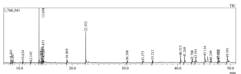
Fig 1: GC– MS Chromatogram of Polyherbal Extract
Table 4: GCMS analysis of the Polyherbal Extract
|
Peak No. |
R. Time |
Area % |
Height % |
A/H |
Name |
Biological Activities |
|
1 |
8.361 |
1.83 |
3.24 |
1.83 |
Azulene |
Anti-inflammatory, antioxidant, ant allergic, |
|
2 |
8.687 |
0.53 |
0.78 |
2.18 |
4-Vinylphenol |
Antioxidant, antimicrobial, aromatic compound |
|
3 |
10.624 |
0.85 |
1.57 |
1.17 |
alpha - Terpinyl acetate |
Antimicrobial, anti-inflammatory, |
|
4 |
12.145 |
0.32 |
0.66 |
1 59 |
2,3,4,5-Tetrahydroxypentanal |
Antioxidant |
|
5 |
13.698 |
17.2 |
33.71 |
1.65 |
Diethyl Phthalate |
Antioxidant |
|
6 |
14.08 |
1.91 |
0.92 |
3.15 |
Methyl hexofuranoside |
Antioxidant, stabilizes membranes, retains moisture in the skin |
|
7 |
14.23 |
3.66 |
6.67 |
3.7 |
Trehalose |
Stress tolerance |
|
8 |
14.451 |
8.89 |
7.6 |
3.76 |
2-O-Methylhexose |
Osmoprotectant |
|
9 |
14.52 |
1.09 |
1.8 |
1.9 |
Glucose |
Antioxidant, antimicrobial, cytoprotective |
|
10 |
18.969 |
2.28 |
2.41 |
3 |
n-Hexadecanoic acid |
Antioxidant, antimicrobial, anticancer, precursor for Vitamin E and K |
|
11 |
22.552 |
19.05 |
19.28 |
3.19 |
Phytol |
Antimicrobial |
|
12 |
30.388 |
1.3 |
1.39 |
3.1 |
Palmitic acid. beta - monoglyceride |
Anti-inflammatory, antioxidant |
|
13 |
33.375 |
0.97 |
0.8 |
3.52 |
Linolenic acid |
Antioxidant, skin-protective, anticancer |
|
14 |
35.212 |
1.45 |
1.63 |
2.87 |
Squalene |
Antioxidant, anti-aging, protects cell membranes, skin health |
|
15 |
40.515 |
5.48 |
4.11 |
4.3 |
dl - alpha - Tocopherol |
Anti-inflammatory, antiviral, antimalarial, |
|
16 |
41.269 |
4.81 |
3.29 |
4.74 |
Nimbin |
Cholesterol-lowering, anti-inflammatory, anticancer, antioxidant |
|
17 |
42.7 |
2.4 |
1.24 |
6.4 |
Campestral |
Antioxidant, hypoglycemic, anti-inflammatory, cholesterol-lowering |
|
18 |
43.391 |
3.9 |
2.09 |
6.04 |
Stigma sterol |
Antioxidant, anti-inflammatory, anticancer, hypolipidemic |
|
19 |
45.116 |
7.6 |
3.74 |
6.58 |
Gamma .- Sitasterol |
Potential anti-inflammatory and anticancer |
|
20 |
46.249 |
1.5 |
0.8 |
6.18 |
24-Noroleana-3,12-diene |
Wound healing, skin regeneration , anti-cancer,anti-inflammatory, antioxidant, |
|
21 |
47.625 |
2.89 |
1.29 |
7.19 |
Cycloartenol |
Wound healing, Anti-inflammatory, Antioxidant, Antimicrobial, Analgesic |
|
22 |
47.732 |
3.4 |
1.73 |
6.4 |
Alpha .- Amyrin |
Wound healing, Anti-inflammatory, analgesic, anticancer, skin barrier repair |
|
23 |
49.391 |
6.5 |
2.73 |
7.7 |
Beta. Amyrin acetate |
Anti-inflammatory, antioxidant, antiallergic, soothing (used in cosmetics) |
Antibacterial assay
Antibacterial activity of the leaves extract against the isolated Staphylococcus sp was performed on Mueller Hinton Agar by disc diffusion method and results are tabulated (Table 5). Leaf extract Wrightia tinctoria showed maximum antibacterial activity against the test bacterium. Disc with 50 µl extract showed zone of clearance of 20 mm and 100µl extract showed zone of clearance 24mm.Antimicrobial activity of the prepared polyherbal extract and polyherbal gel were studied by disc diffusion method and tabulated (Table 6). Antibacterial activity increased with increase in concentration. The polyherbal extract produced larger zone than the polyherbal gel.
Table 5: Antibacterial activities of the leaves extracts on Staphylococcus sp
|
SI. No. |
LEAVES EXTRACT |
ZONE OF INHIBITION (mm) |
|
|
50µl |
100µl |
||
|
1. |
Azadirachta indica |
13mm |
15mm |
|
2. |
Calotropis gigantea |
8mm |
10mm |
|
3. |
Wrightia tinctoria |
20mm |
24mm |
|
4. |
Acalypha indica |
7mm |
10mm |
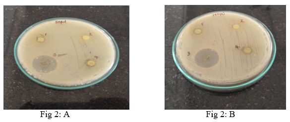
Antibacterial activity of individual leaves extract on MHA (A) 50µl (B) 100µl concentration
Table 6: Antibacterial activity of polyherbal formulation and polyherbal gel
|
S. No. |
Concentration |
Zone of Inhibition |
|
|
Poly Herbal extract |
Poly herbal gel |
||
|
1 |
40µl |
11 |
9 |
|
2 |
60µl |
16 |
13 |
|
3 |
80µl |
22 |
20 |
|
4 |
100µl |
24 |
22 |
DISCUSSION
Impetigo is a prevalent and highly contagious skin infection that primarily impacts children, caused by Staphylococcus aureus and Streptococcus pyogenes. It manifests as red sores or blisters that develop into honey-colored crusts, typically located around the nose and mouth. Although it is usually mild, it can sometimes result in complications like cellulitis and post-streptococcal glomerulonephritis, particularly in environments with poor hygiene [20].
Increasing antibiotic resistance, especially from MRSA and macrolide-resistant S. pyogenes, hampers the effectiveness of treatments such as mupirocin and erythromycin, leading to more prolonged infections and increased healthcare expenses [21, 22]. As a response, herbal treatments are receiving more attention due to their antimicrobial, anti-inflammatory, and wound-healing abilities. Plants like neem, turmeric, and tea tree oil have proven effective against resistant strains, presenting promising alternatives to standard antibiotics and assisting in managing resistance [23].
Traditional medicinal plants like Azadirachta indica, Calotropis gigantea, Wrightia tinctoria, and Acalypha indica are recognized for their abundant phytochemical compositions. In the current study, a polyherbal formulation was prepared using four medicinal plant leaves: Azadirachta indica, Calotropis gigantea, Wrightia tinctoria, and Acalypha indica and validated the existence of significant phytochemicals such as alkaloids, flavonoids, tannins, saponins, sterols, glycosides, and terpenoids in all four plants, aligning with earlier research works [24,25,26]. These constituents contribute to a wide range of pharmacological activities including antibacterial, antifungal, antiviral, anti-inflammatory, antidiabetic, wound healing, hepatoprotective, antioxidant, and anticancer effects, supporting their traditional use and potential for therapeutic applications.
On GC-MS analysis, 23 peaks were detected, and 22 of them showed properties such as antimicrobial, wound healing, skin regeneration, skin barrier repair, analgesic, anti-inflammatory, moisture retention, anti-aging, anticancer, antiviral, anti-malarial, cholesterol-lowering, hypoglycemic, and antioxidant effects. The major components present were β-amyrin acetate, cycloartenol, γ-sitosterol, and campesterol. Rarely detected compounds like 24-Noroleana-3,12-diene (a naturally occurring triterpenoid hydrocarbon) and phytosterol γ-sitosterol were also identified in the polyherbal extract. Out of the 22 biologically active compounds found in the polyherbal extract, 15 were not found in the individual leaves used in the study. These newly formed compounds are believed to be the result of synergistic interactions, and they possess important biological activities desirable in a topical formulation [27].
In antimicrobial activity studies, the results showed that the polyherbal extract could inhibit the growth of the test pathogen, Staphylococcus spp., isolated from impetigo patients. With increasing concentration, the zone of inhibition also increased. At 100?µL concentration, the polyherbal extract produced a 24?mm zone of inhibition. The polyherbal gel produced a 22?mm zone of inhibition, as the gelling agent caused a slow release of compounds into the medium, a desirable property for a topical gel [28].
CONCLUSION
In this study a polyherbal formulation was prepared against the Staphylococcus sp is isolated from impetigo patients. Azadirachta indica, Calotropis gigantea, Wrightia tinctoria and Acalypha indica Plant leaves extract was prepared and preliminary screening was done to determine the phytochemical constituents of the extracts. Equal quantity of the dried leaves was mixed together and extracted with the organic solvent ethanol to obtain polyherbal extract. The polyherbal extract revealed the presence of 23 different bioactive compounds on GCMS analysis. 15 compounds found in the GCMS analysis was not found in the individual plant extract and would have formed due to the interaction of different compounds present in the individual plant extracts. Most of the compounds found in the polyherbal extract exhibited the desirable anti-microbial activities, wound healing properties, skin regeneration, skin barrier repair, anti-inflammatory, analgesic activities. The polyherbal gel formulated using the polyberbal extract showed the slow release of the compounds, one of the desirable property of the topical agent. The novel polyherbal formulation may be a considered for the commercial production of the herbal remedy against Impetigo treatment and restoration of the healthy skin.
REFERENCE

Amala S. Keerthi, Juby T. R.*, GC-MS Characterized Novel Polyherbal Gel for the Treatment of Impetigo: A Study on Its Antimicrobial and Dermatological Benefits, Int. J. of Pharm. Sci., 2025, Vol 3, Issue 9, 3390-3399 https://doi.org/10.5281/zenodo.17226369
 10.5281/zenodo.17226369
