1Professor and head, Department of Conservative Dentistry & Endodontics, Narsinhbhai Patel Dental College and Hospital, Sankalchand Patel University, Visnagar, Gujarat, India.
2Post Graduate Student, Department of Conservative Dentistry & Endodontics, Narsinhbhai Patel Dental College and Hospital, Sankalchand Patel University, Visnagar, Gujarat, India.
3Reader, Department of Conservative Dentistry & Endodontics, Narsinhbhai Patel Dental College and Hospital, Sankalchand Patel University, Visnagar, Gujarat, India.
4Reader, Department of Conservative Dentistry & Endodontics, Narsinhbhai Patel Dental College and Hospital, Sankalchand Patel University, Visnagar, Gujarat, India.
5Post Graduate Student, Department of Conservative Dentistry & Endodontics, Narsinhbhai Patel Dental College and Hospital, Sankalchand Patel University, Visnagar, Gujarat, India.
6Post Graduate Student, Department of Conservative Dentistry & Endodontics, Narsinhbhai Patel Dental College and Hospital, Sankalchand Patel University, Visnagar, Gujarat, India.
In the maxillofacial area, injuries to teeth are commonly observed, particularly coronal fractures affecting front teeth. This case report presents a method for clinically reattaching fractured tooth fragment. After undergoing endodontic treatment, a glass-fibre-reinforced composite root canal post was utilized to enhance retention and distribute stress across the root. The dental restoration was finalized by incorporating the original fragment alongside a dual-cured resin composite. Clinical and radiographic evaluations confirmed the successful placement of the glass fibre-reinforced composite root canal post and restoration, indicating the treatment's effectiveness in preserving the fractured tooth. Thus, it is concluded that reattaching a tooth fragment using a combination of dual-cured resin composite and a glass-fibre-reinforced composite root canal post serves as a viable approach for rehabilitating fractured teeth, providing both satisfactory aesthetics and functionality.
Injuries to both primary and permanent dentition represent a prevalent form of trauma in the maxillofacial region.1 Among these injuries, the maxillary canines, mandibular lateral incisors, and maxillary incisors are most commonly affected.2 Maxillary incisors, in particular, are frequently implicated due to their position in the dental arch, often resulting in crown fractures.3,4 The management of crown fractures is contingent upon various factors, including the extent of the fracture, pulpal condition, biological width, and alveolar bone health.5 When evaluating a fractured tooth, it is essential to consider parameters such as fracture pattern, the presence of associated root fractures, alignment of the fractured tooth fragment, soft tissue condition, aesthetics, occlusion, long-term prognosis, and treatment feasibility.6 Before initiating treatment, thorough clinical assessments should be conducted to evaluate periodontal status, pulpal vitality, coronal integrity, and occlusal alignment.7 Reattaching a tooth fragment, as pioneered by Chosack and Eidelman in 1964, has proven to yield excellent aesthetic and functional outcomes.8 This approach allows for the restoration of contour, natural shape, occlusal harmony, surface texture, and color of the fractured tooth fragment.9 Tooth fragment reattachment presents a conservative, aesthetically pleasing, and cost-effective restorative option, which has been deemed acceptable compared to resin-based composite restorations or full-coverage crowns.10 Advancements in adhesive techniques and restorative materials, particularly dentin hybridization, have facilitated more effective outcomes compared to traditional methods.11 While restoration with cast metal posts may induce coronal wedging stresses, potentially compromising root integrity, fiber-reinforced composite resin posts have demonstrated minimal risk of root fracture.12 Glass fiber posts, introduced in 1901, offer several advantages, including aesthetic appeal, similar modulus of elasticity to dentin, and reinforcement of restored segments through monobloc formation.13 The presented case report illustrates the successful reattachment of a tooth fragment alongside a glass-fibre-reinforced composite post in a patient who achieved restoration of periodontal health, functional rehabilitation, and satisfactory aesthetic results.14,15
CASE REPORT:
A 24 years-old female patient attended the Department of Conservative Dentistry and Endodontics with a chief complaint of a crown fracture of the maxillary right and left central incisors.Patient came to the department after 24 hours of trauma. She had a constant pain in maxillary left central incisor. Extra-oral examination revealed laceration on upper lip and bruise on chin area and intra-oral examination revealed neither lacerations nor evidence of alveolar bone fracture [Figure 1]. Both maxillary central incisors were fractured and teeth showed no mobility. Upon Radiographic examination as well as clinical examination showed a Ellis Class II fracture of the maxillary right central incisor and a Class III fracture of the maxillary left central incisor [Figure 2]. Fractured fragment was presented in maxillary left central incisor and fracture was involved upto the pulp due to this reason patient had a pain, so removal of fracture fragment was done for relieving the pain using local anaesthesia and stored in Normal saline [Figure 3].
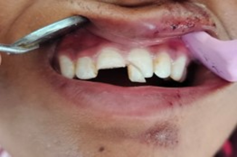
Figure 1: Pre-operative photograph after trauma
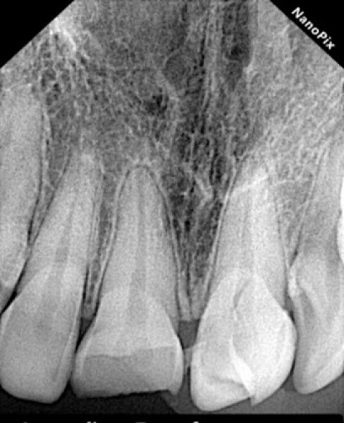
Figure 2: Preoperative radiograph after trauma shows class II Ellis fracture in maxillary right central incisor and class III ellis fracture in maxillary left central
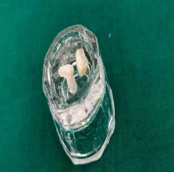
Figure 3: Fractured fragment stored in normal saline
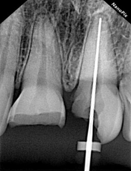
Figure 4: Master apical file till working length
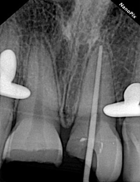
Figure 5: Master apical cone upto the working length
After that root canal treatment was initiated and biomechanical preparation was performed in maxillary left central incisor for pain relief at very same day [Figure 4]. Interim restoration (orafil-G Prevest DenPro) was placed in maxillary left central incisor till further appointment. Patient was recalled after 3 days, She was completely asymptomatic for maxillary left central incisor. Pulp vitality test was performed using pulp vitality tester (Parkell Digitest II). Pulp is vital for maxillary right central incisor and fracture involves till the dentin so decided to direct fracture fragment re-attachment in maxillary right central incisor.For maxillary left central incisor, interim restoration was removed and obturation was initiated. Master apical cone was selected of ISO size 80 at working length [Figure 5] and obturated using endodontic sealer (META BIOMED ADSEAL Root Canal Sealer) and laterally condensed with gutta percha technique.The coronal portion of the root canal obturation was removed with a heat carried instrument for the placement post-system.The post space was prepared with the peeso-reamers no.1- 4(Mani dental peeso-reamers) and checked with radiograph,5mm of gutta percha cone was left at apical 1/3rd for good sealing. Fractured fragment and tooth surface was etched with 37% phosphoric acid gel for 20 secs. then rinsed for 20 secs. and dried with a gentle stream of air.An adhesive(Single bond universal adhesive,3M ESPE),a dual-curing luting system (RelyX U200, 3M ESPE) and a glass fibre reinforced resin post (RelyX Fibre post, 3M) were sequentially applied in maxillary left central incisor.After that it was light cured for 40 secs [Figure 6 and 7].
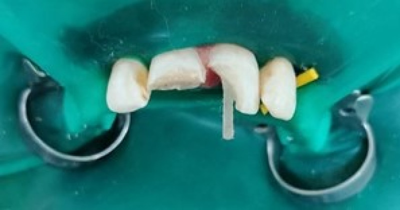
Figure 6: Glass fibre post placement into the maxillary left central incisor
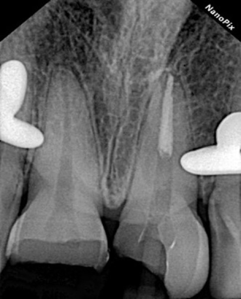
Figure 7: Radiographic evaluation of glass fibre post placement.
Fractured crown fragments were stored in normal saline till the above mentioned treatment was performed. Then, using flowable composite resin (Fusion Flo, Light cured nanohybrid flowable composite resin,Prevest Denpro) fractured crown fragments were adjusted on maxillary right and left central incisors,after ensuring the complete adaptation of fragments it was light cured for 40 secs. The composite resin (Filtek Z250 XT, 3M) was used incrementally above the attached surface of maxillary right and left central incisors then cured for 40secs [Figure 8 and 9].
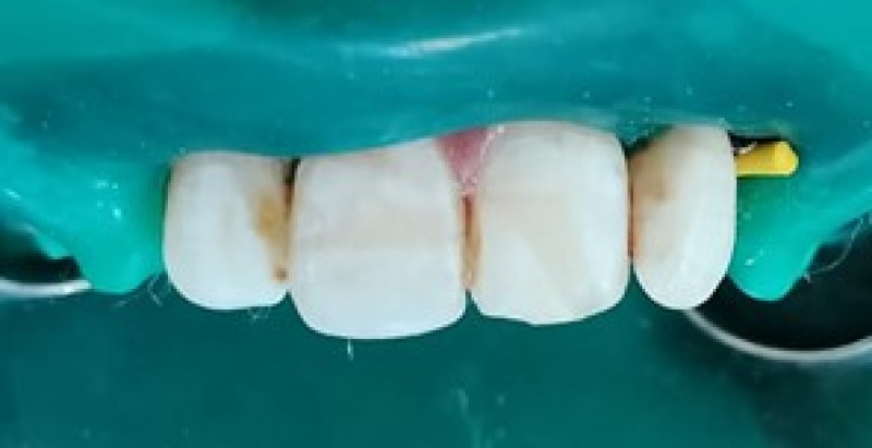
Figure 8: Post-operative photograph after fracture fragments reattachment in maxillary right and left central incisors
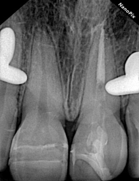
Figure 9: Post-operative radiograph after fracture fragments reattachment in maxillary right and left central incisors.
Residual excess cement was finished using finishing burs.Clinical and radiographic examination of reattachment fragments was done at 3 and 6 months.After follow up at 3 and 6 months there were no mobility,decay,periapical abscess or any surface discolouration observed. [Figure 10 and 11].
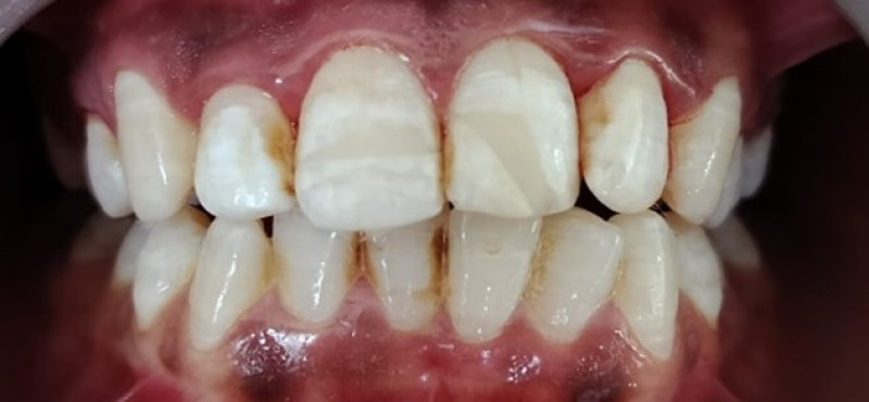
Figure 10: Follow up at 3 months
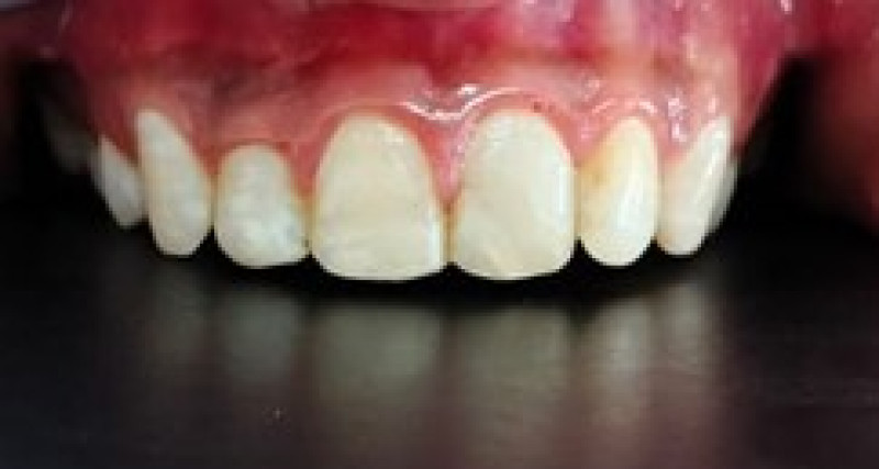
Figure 11: Follow up at 6 months
DISCUSSION
Dental trauma is a common occurrence, with maxillary incisors being frequently involved.1 Several treatment modalities exist for managing such cases, including interventions related to the biological width such as tooth extrusion, crown lengthening followed by fragment reattachment or reconstruction, intentional replantation, and in severe cases, tooth extraction.2It is crucial to promptly restore the function and aesthetics of a fractured tooth. When a fractured crown fragment is available, reattachment is often the preferred initial treatment option.15 Reattachment of a fractured crown fragment can be the most conservative and optimal choice for anterior teeth. The success of reattachment largely depends on swift retrieval of the fragment and its preservation in a physiologic solution or saline to prevent color alteration due to dehydration.16 Our approach involved the use of an adhesive, a dual-curing luting composite system, a glass fibre-reinforced composite root canal post, and the original crown piece. The fractured fragment was stored in normal saline until reattachment.17Glass fibre posts were employed to reinforce the teeth, promoting a monobloc effect with no inherent weak interlayer interface, thus aiding in stress distribution to the remaining radicular dentin. Achieving post retention relies on the resin cement's degree of conversion, which may be compromised in the apical regions of the post space due to limited light exposure.18 Therefore, the use of dual-cure or self-curing composites for post luting is advised. Bonding the post to the tooth structure enhances the prognosis of the repaired tooth by improving post retention and strengthening the tooth structure.19 Self-adhesive dual-cure universal resin cement was utilized for post cementation, facilitating light penetration and enhancing composite resin conversion.20 This approach minimizes the risk of microleakage and promotes strong bond strength to the tooth.Treatment with this approach requires minimal chair time and ensures patient compliance.This comprehensive approach reinforces the repaired segments, enhancing their durability and longevity.21 Follow-up assessments post-treatment demonstrated the success of the combined approach involving glass-fibre-reinforced composite root canal posts and original fragments, with no restorative fractures observed and absence of discoloration.
CONCLUSION
The combined use of a glass fiber reinforced composite post and an original crown fragment is an efficient procedure for the treatment of traumatized anterior teeth that appears to provide excellent aesthetic and functional results.
CONFLICT OF INTEREST
The authors revealed no conflict of interests with regard to this publication.
FINANCIAL SUPPORT AND SPONSORSHIP
Not available
REFERENCES


Kailash Attur, Harvy Shah, Nikunj Patel, Karna Jani, Krunali Jayswal, Shraddha Patel, Harmonizing Crown Fragments The Fusion Of FiberReinforced Posts In The Realm Of Dentistry, Int. J. of Pharm. Sci., 2024, Vol 2, Issue 3, 754-759. https://doi.org/10.5281/zenodo.10850007
 10.5281/zenodo.10850007
