Sankaralingam Bhuvaneswari College of Pharmacy, Anaikuttam, Sivakasi, Virudhunagar. (Affiliated to “The Tamil Nadu Dr M.G.R. Medical University”, Chennai).
Aim: The aim of this review is to highlight the unique properties and versatility of liposomes that make them a preferred nanocarrier platform for precision medicine applications. Objective: To systematically evaluate and summarize the role of liposomes as advanced drug delivery systems in the context of precision medicine. Analyzing the unique properties of liposomes, exploring recent advancements in liposomal formulations, Identifying challenges and future directions in liposome research. Methodology: The review covers the following key aspects of liposomes in precision medicine, 1. Size, structure and composition of liposome 2. Types, Advantages and Disadvantages of liposome 3. Properties and application of liposome 4. Preparation of liposome 5. Evaluation studies for liposomal preparation.
Liposomes were firstly discovered in the 1960's by Bengham. Liposomes are the most explored nanocarriers used in targeted drug delivery systems. The liposomes are spherical particles that encapsulate a fraction of the solvent, in which they freely diffuse (?oat) into their interior. They can have one, several or multiple concentric membranes. Liposomes are constructed of polar lipids which are characterized by having a lipophilic and hydrophilic group on the same molecules (1). Figure:1
1.1 Size And Sstructure Of Liposome
Liposomes are spherical lipid vesicles usually 50–500 nm in diameter. They vary in particle size. Liposomes are circular vesicles with hydrophilic heads and hydrophobic tails, formed by one or more bilayer membranes (2).
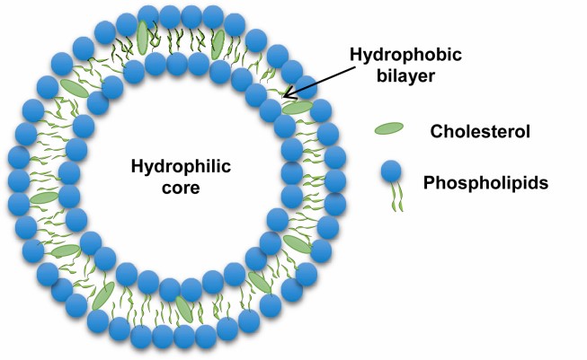
Figure: 1 Structure of Liposome
1.2 Composition of Liposome (3)
The primary building blocks of liposomes are phospholipids, which are amphipathic molecules consisting of a hydrophilic (water-attracting) head and two hydrophobic (water-repelling) tails. The most commonly used phospholipids in liposome formulation include:
2. Phosphatidylethanolamine (PE): Known for its role in membrane fusion and stability.
3. Phosphatidylserine (PS) and Phosphatidylinositol (PI): These phospholipids are less common but can be included for specific functional purposes.
2. Cholesterol: A Stabilizing Agent
Cholesterol is frequently added to liposome formulations to enhance membrane stability and rigidity. By inserting itself between the phospholipid molecules, cholesterol helps to prevent the liposome from becoming too permeable or too rigid, thus improving its structural integrity and longevity.
3. Surface Modifiers
To enhance the functionality and targeting ability of liposomes, various surface modifiers can be incorporated:
1. Polyethylene Glycol (PEG):
PEGylation involves attaching PEG molecules to the liposome surface, which can increase circulation time in the bloodstream by reducing recognition and clearance by the immune system.
2. Ligands: Targeting ligands such as antibodies, peptides, or small molecules can be conjugated to the liposome surface to enable specific binding to target cells or tissues.
4. Internal Contents
The internal contents of liposomes can vary widely depending on their intended use. They can encapsulate:
1. Hydrophilic Drugs: These are trapped in the aqueous core of the liposome.
2. Hydrophobic Drugs: These are incorporated into the lipid bilayer.
5. Additional Components
In some formulations, other components might be added to enhance the properties of the liposome:
1. Charged Lipids: Incorporating cationic or anionic lipids can aid in the delivery of genetic material or enhance interaction with target cells.
2. Steroids and Other Lipids: Certain steroids or specialized lipids can be included to modulate membrane fluidity or other properties. (3)
1.3 Types:(4)
Liposomes are versatile molecules and can be classified in several ways based on their diversity and structural properties, such as composition, shape, size and surface properties.
1. Based on their size, they are classified as:
2. Based on their structural parameters, they are categorized into:
3. Liposomes are synthesized using an extensive range of methods. Based on synthesis, they are classified as:
i. Dehydration and rehydration (DRV): In this process, the small unilamellar vesicles containing the buffer are dried and rehydrated with the aqueous solution, containing the compound needed to be incorporated in the vesicle. This method is generally used to form an oligolamellar vesicle.
ii. Reverse phase evaporation (REV): This technique is based on the formation of inverted micelles. They are formed after sonicating a mixture of a buffered aqueous phase (containing the water-soluble molecules to be encapsulated into the liposomes) and an organic phase in which the amphiphilic molecules are solubilized. The organic solvent is slowly eliminated, converting the inverted micelles into viscous or gel forms. Then the gel state collapses at a critical stage, disrupting some of the inverted micelles. Too many phospholipids in the environment cause a whole bilayer to develop around the remaining micelles, resulting in liposome formation.
iii. Extrusion technique (VET): Here, multilamellar vesicles are pushed through a polycarbonate membrane filter containing pores of the desired size to control the vesicle size distribution.
iv. Freeze and thaw extrusion method (FAT): In this method, liposomes formed using the thin-film method are vortexed with the material needed to be incorporated in the vesicles. This is done until the whole lipid film is suspended. Then, the resulting vesicles are frozen in warm water and vortexed again. This is the most preferable technique when it comes to the formation of multilamellar vesicles or increasing the encapsulation efficiency of the liposomes.
v. Sonication: It’s one of the most extensively used techniques for liposome preparation. Here, multilamellar vesicles formed by other preparation techniques are subjected to sonication, resulting in small unilamellar vesicles.
vi. Solvent dispersion method: It includes ether injection and ethanol injection methods.
vii. Thin-film hydration method: This is the most common and widely used technique. In this method, the lipids are dissolved in an organic solvent, like chloroform or mixtures of chloroform and methanol. Then, the solvent is removed by film deposition under vacuum. The organic solvent is completely evaporated and the lipid residues are again hydrated using an aqueous buffer, resulting in the swelling and hydration of lipids. These lipids lead to the formation of liposomes.
4. Liposomes can also be classified depending on their composition as:
1.4 ADVANTAGE (5)
1.2.5 DISADVANTAGE (6)
1.6 Properties Of Liposomes (7)
1.7 Applicatioin Of Liposomes (8)
Liposomes are the perfect vehicle for the distribution and delivery of drugs due to their biocompatibility, non-toxicity and ability to carry a variety of substances in the required concentrations. Some of their application in a spectrum of areas are mentioned below:
Site-specific drug delivery: Liposomes are suitable vehicles for drug delivery at targeted locations. To form a site-specific drug delivery system, they are combined with opsonins and ligands, such as antibodies, sugar residues, apoproteins, or hormones, which are tagged on the lipid vesicles. This also helps in reducing drug-related toxicity.
Cancer therapeutics: Liposome-based cancer therapeutics are used to treat breast cancer in a site-specific manner. It enhances the pharmacokinetics and pharmacodynamics of the associated drug and delivers it to its intended site, increasing its therapeutic efficiency. For example, Anthracyclines are anticancer drugs. However, they kill the tumor cells and inhibit the other fast-dividing cells of the body, including hair, blood cells and gastrointestinal mucosa. This toxicity to other cells can be prevented by using liposomes. Liposomes encapsulate the drugs and accumulate only at the tumor site, increasing drug efficiency and reducing toxicity.
Transdermal drug delivery: The main challenge in transdermal drug delivery is the penetration of macromolecules and hydrophilic drugs through the stratum corneum (outer layer of the skin). Liposomes have a similar molecular composition as lipid layers and have high permeability. Thus, they are a suitable carrier for the delivery of drugs penetrating the skin.
Treatment of parasitic diseases and infections: Liposomes serve as an ideal carrier to deliver the drugs to treat parasitic diseases, especially those that infect monocytes and macrophages cells, like leishmania. Conventional therapeutic agents, such as amphotericin B, pentamidine, paromomycin and miltefosine, cause several side effects like arrhythmias and gastrointestinal disorders. And in some cases, drug resistance is also observed. In such conditions, drugs encapsulated in liposomes appear suitable for the treatment of leishmaniasis. (8)
1.8 Methods Of Liposomes Preparation (9)
Liposomes can be formulated using different approaches.
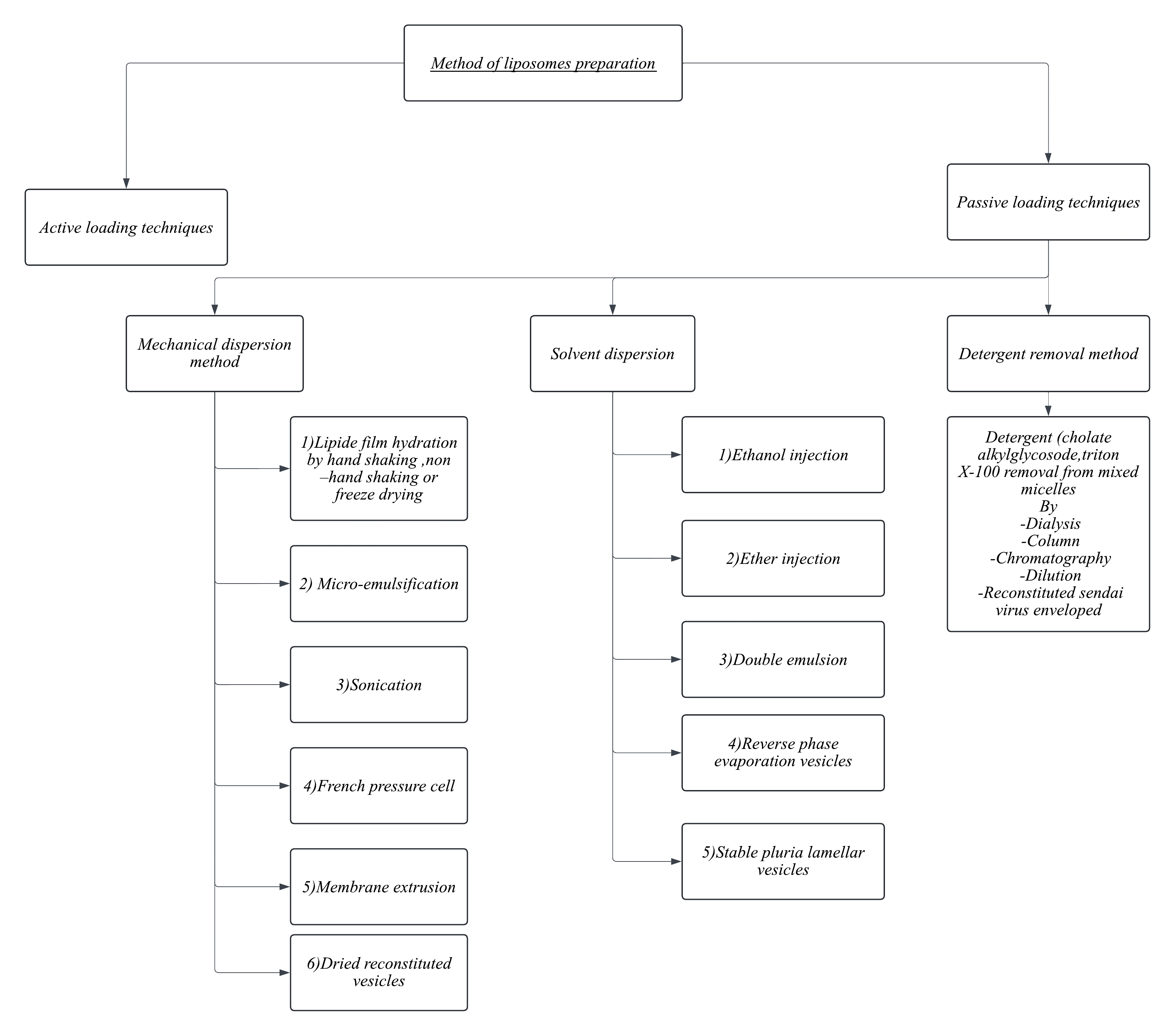
Figure: 4 Flow Chart of Liposome Preparation Methods
Dehydration-Rehydration Method
It is an organic solvent free method to produce LUVs using sonication. This method based on direct dispersing of the lipids at low concentrations into an aqueous solution containing the drug molecules followed by sonication. First, the dehydration step to evaporate water under nitrogen to create multilayered film entrapping the drug molecules. Then, a hydration step to form large vesicles encapsulating the drug molecules. This method is simple but with high heterogeneity of the liposome’s sizes.
pH Jumping Method
Another solvent-free method for liposomes preparation is the pH jumping method. In this method, the aqueous solution of phosphatidic acid and phosphatidylcholine are exposed to almost four-fold increase in pH over a short time to break down MLVs into SUVs. The ratio of phosphatidic acid: phosphatidyl choline determine the percentage of SUVs versus LUVs produced.
Microfluidic Channel Method
The microfluidic channel method has been recently proposed as a novel method for liposomes preparation. In this method, lipids are dissolved in ethanol or isopropanol and the resultant solution is injected upright or in the opposite direction to the aqueous medium within the micro-channels. This method involves continuous axial mixing of the organic and aqueous solutions leads to liposomes formation. Liposomes are stabilized using surfactants to avoid coagulation and separation. Microfluidic channel methods control the mixing process of organic and aqueous phases to achieve reproducible liposomes with proper average size, polydispersity, morphology and lamellarity.
1.9 Evaluation (10)
1. Scanning Electron Microscopy (SEM):
Scanning electron microscopy study was done to determine the surface morphology, size and shape of prepared Frusemide liposomal formulations. The optimized freeze dried liposomal was subjected for Scanning electron microscopy and photographed.
2.Measurement of Zeta Potential (ZP)
The zeta potential means the charges which are present on the surface of liposome. The many time the charge is present on the surface of liposome. This charge is come due to the component or ingredient which was used during the manufacturing. Some charge is must be required on surface of all liposome present in formulation, due to some charge all liposome particle repeal to each other and coagulation of particle are avoided. The zeta potential of liposome was taken in zeta sizer instrument having Malvern software. The analysis of sample was carried out at 25oc with the angle of detection 90o.The ideal zeta potential value must be required in range between +30 to -30mV. These ranges prevent the aggregation of liposomal particle.
3.Determination of Drug Content
Drug content in the preparation was determined by extracting drug from the liposome with 0.1M hydrochloric acid. In this method liposome (50mg) were stirred in 50ml hydrochloric acid until dissolved. It was filtered by Millipore filter paper and drug content was determined, after suitable dilution. At 254nm by UV spectroscopy. The loading efficiency (L) of the liposome was calculated according to following formula, Were, Qn is the amount of drug present in Liposome and Wn is weight of liposome
L (c/o) = (Qn / Wn) × 100
4. Entrapment Efficiency (EE)
It was determined by using the ratio of entrapped drug (mg) to the total drug (mg), which may be expressed by the following formula.
% Drug entrapped PDE=Amount of drug entrapped in liposome Total amount of drug taken initially
x100
5.In-vitro drug release studies
Concentrated liposomal suspension, 0.5 ml was taken in a test tube of opening diameter of 20 mm. The open end was covered with a semi-permeable dialysis membrane (Himedia Laboratories Pvt. Ltd.) and tied with a thread. The test tube was inverted and placed over the surface of 100 ml water present in a 250 ml beaker in such a way that the membrane just touched the water surface. The test tube was secured by a clamp fixed with a stand. The water in the beaker was stirred with a magnetic stirrer so that no vortex could form in the beaker. The temperature was maintained at 37 °C. The drug released from the liposomes permeates across the membrane and enters into the receptor chamber medium. Samples of 2 ml were taken out from the receptor chamber medium, suitably diluted, and the absorbances were taken by UV-spectrophotometer at 263 nm against a blank of fresh medium. At the same time, 2 ml of fresh medium was added to the beaker to keep the volume of the medium constant in the beaker. (10)
CONCLUSION
Liposomes represent a promising platform for drug delivery in precision medicine, combining their ability to enhance drug efficacy with targeted delivery and controlled release mechanisms.
ACKNOWLEDGEMENT
The authors of the paper are grateful to the Correspondent, Principal and all department HOD, as wells as Sankaralingam Bhuvaneswari College Of Pharmacy, Anaikuttam, Sivakasi for providing a first-rate working environment.
REFERENCES

Inigo P.*, Rajesh M., Yashwanth M., Tamilselvi M., Venkatesh Kanna M., Tamilarasi A., Thirumalai Kumar P., Liposomes: The Nanocarrier of Choice in Precision Medicine, Int. J. of Pharm. Sci., 2024, Vol 2, Issue 12, 2401-2408. https://doi.org/10.5281/zenodo.14512226
 10.5281/zenodo.14512226
