Department of Pharmacy, Mewar University. Gangrar, Chittorgarh, Rajasthan-312901
Background: The presence of microbial contamination in injectable pharmaceuticals poses significant risks to patient safety and product efficacy. Ensuring sterility in drug manufacturing is critical for maintaining product integrity and compliance with regulatory standards. Objective: This study aimed to establish a comprehensive microbial surveillance framework for sterile drug manufacturing facilities, focusing on contamination control and sterility assurance. Approach: A systematic approach was employed, which included routine monitoring of microbial presence, identification of contaminants, tracking contamination sources, and conducting risk assessments. The study utilized MALDI-TOF MS for microbial identification, emphasizing the need for facility-specific reference libraries to enhance identification capabilities. Results: The recovery of 60 bacterial isolates from various cleanroom grades and production stages highlighted the widespread presence of microbial contaminants. The limitations of existing MALDI-TOF MS databases, particularly for environmental isolates like Bacillus spp., were identified as a barrier to effective species-level identification. The study demonstrated that MALDI-TOF MS, when supported by adequate database coverage, is a rapid and efficient tool for microbial identification. Conclusion: The findings underscore the critical need for improved contamination control measures in sterile manufacturing processes. Collaboration between MALDI-TOF MS developers and pharmaceutical manufacturers is essential to expand databases and enhance microbial monitoring programs, ultimately ensuring product safety and sterility assurance.
1.1 Introduction to Injectable
Pharmaceuticals that are delivered directly and must be totally devoid of living microorganisms in order to avoid infections and guarantee patient safety are known as sterile Injectables [1].A vital part of contemporary medicine are Injectable drug delivery devices, which provide accurate and quick therapeutic effects by directly delivering medication into the body. Because Injectables avoid the gastrointestinal tract and provide 100% bioavailability and an instantaneous commencement of action, they are especially important in emergency situations and for medications with poor oral absorption [2, 3]. This mode of delivery encompasses various forms including intravenous (IV), intramuscular (IM), subcutaneous (SC), and intradermal (ID) injections, each suited to specific therapeutic needs and pharmacokinetic profiles [3].
The development of Injectable formulations has significantly evolved, incorporating advances in biotechnology, nanotechnology, and materials science to improve drug stability, reduce administration frequency, and enhance patient compliance. Biologics such as monoclonal antibodies, insulin, and vaccines are primarily administered through injections due to their large molecular sizes and susceptibility to enzymatic degradation in the gastrointestinal tract [4]. While injectables offer numerous advantages, they also pose challenges such as pain at the site of administration, risk of infection, and need for trained personnel. Efforts are ongoing to develop less invasive delivery systems and self-administration devices, aiming to improve patient experience and expand access to treatments [5].
1.2 Introduction to Advantages and Disadvantages Injectable
Advantages:
Injectables deliver the drug directly into the bloodstream or target tissues, leading to a quick therapeutic response. This is particularly beneficial in emergency situations [3]
Unlike oral drugs, Injectables avoid the first-pass metabolism in the liver, ensuring complete bioavailability [4]
Large molecules like peptides, proteins, and monoclonal antibodies, which are degraded in the gastrointestinal tract, can be effectively administered via injection [5]
Specific routes like intrathecal or intra-articular injection allow localized drug delivery, reducing systemic side effects [5]
Disadvantages:
Injections can cause pain, discomfort, and anxiety in patients, especially with repeated administration [3].
If not administered under sterile conditions, there is a risk of infection at the injection site [5].
Most Injectable routes require trained healthcare professionals, limiting their use in home settings [6].
Some Injectable drugs, especially biologics, require cold chain storage and are sensitive to temperature and light [6].
Stringent regulatory requirements outline procedures and standards for the manufacturing, testing, and release of these sterile injectables to guarantee their safety and efficacy. Microbial contamination in injectable drugs poses a significant risk, potentially introducing endotoxins, pyrogens, and other harmful contaminants that can lead to severe adverse reactions [6]. To prevent product contamination, proper cleaning techniques and validation are crucial. Implementing efficient preventative measures at every stage of the manufacturing process, from raw materials to final packing and storage, requires an understanding of the origins and routes of contamination [7].
Sterility assurance is achieved by integrating robust design, validation, and operational controls to minimize contamination risks at each production stage. The underlying principle is to build quality into the product by implementing rigorous controls and monitoring systems that consistently reduce the bio burden and prevent microbial ingress [8].
This recognizes that relying solely on end-product testing is insufficient to guarantee sterility due to inherent limitations in sampling and test sensitivity. Effective contamination control strategies must be multifaceted, including thorough cleaning and disinfection, environmental monitoring, personnel training, and validated sterilization processes to ensure the overall sterility and quality of the pharmaceutical product [8].
1.3. Introduction to Microbial Contamination of Pharmaceutical Products
Microbial contamination of pharmaceutical products continues to be a severe challenge to drug manufacturing. Drug products, especially high-risk sterile products such as injection and ophthalmic products, can cause irreversible damage once contamination occurs during the production and storage process, in severe cases, detrimental to the patient’s health and even their lives. It was shown that between 2012 and 2019, more than 50% of all the recalls related to drug products registered by the US Food and Drug Administration (FDA) were associated with microbiological incidents [9,10]. The high frequency of recalls indicates inadequate awareness of contamination risk and poor implementation of relevant control programs. Substantial evidence such as warning letters, alert notifications, and failures suggests a direct relationship between the level of environmental control and the product’s final quality [11]. The company's in-process surveillance approach depends on knowing the kinds and distribution of environmental micro flora throughout drug manufacture [12]. Following the establishment of a pertinent micro flora database, it can be utilized for environmental excursions, product contamination investigations, and other purposes. Trend shifts in the quantity and kind of dominating species could also indicate a flaw in the contamination management strategy (CCS) and prompt certain remedial and preventative measures [13, 14]. Implementing a framework for microbial identification and characterization is essential to a successful approach for controlling microbial contamination. Relatively few research are linked to the microbiological profile in clean pharmaceutical environments, despite the fact that there is consensus and that the most recent rules and recommendations have raised expectations [15]. These studies use a variety of methodologies, from biochemical approaches to genomes, which are costly, complex, or less accurate to market in the business world. It is very desirable to implement a comprehensive monitoring system with a quick, accurate, and economical solution as well as a more quick and effective tool to speed up and increase the quality of the analytical data. Numerous automated and quick methods for microorganism identification and type exist; these are often categorized as either genotypic or phenotypic approaches [16].
1.4. Microbial Contamination Sources and Pathways in Injectable Drugs:
A thorough understanding of how germs can enter the production process and compromise the final product's sterility is necessary for effective contamination control in the manufacturing of Injectable medications. If not procured, evaluated, and handled appropriately, raw materials such as excipients, active pharmaceutical components, and injection water may include microbiological contamination [17]. These raw materials represent a primary source of potential contamination, underscoring the critical importance of stringent quality control measures to ensure their purity[18]. Furthermore, suppliers must adhere to strict quality standards and provide certificates of analysis to verify the microbiological quality of their materials. The manufacturing environment, including the air, personnel, and equipment, can also introduce microorganisms into the production area. Airborne contaminants, such as bacteria, fungi, and mold, can enter the production facility through ventilation systems or human traffic [19]. Personnel working in aseptic environments must undergo extensive training on proper gowning procedures, hygiene practices, and aseptic techniques to minimize the risk of contamination [20]. Compounding pharmacies, which prepare customized medications for individual patients, have also been implicated in contamination-related outbreaks due to inadequate aseptic practices and quality control procedures. The occurrence of substandard and counterfeit antimicrobial drugs can significantly erode public confidence in healthcare systems and governmental oversight [21]. Packaging components such as vials, stoppers, and seals can also introduce microbial contamination if they are not properly sterilized and handled before filling. To guarantee a contamination-free product, good manufacturing practices are directed towards the elimination of microorganisms from the end product [22].
Table No: 1. Microbial Contamination Sources and Pathways in Injectable Drugs.
|
Microorganism |
Source |
Health Risks |
Detection Method |
|
Pseudomonas aeruginosa |
Water systems, raw materials |
Sepsis, endotoxic shock |
PCR, culture methods |
|
Burkholderia cepacia |
Packaging components |
Opportunistic infections |
NGS, MALDI-TOF |
|
Staphylococcus aureus |
Personnel, environment |
Abscesses, bacteremia |
ATP bioluminescence |
|
Candida albicans |
Airborne contamination |
Fungemia, organ failure |
Flow cytometry |
2. Aim:
Microbial Contamination and Sterility Testing in Injectable Pharmaceuticals – Ensuring sterility assurance and contamination control
3. Objectives:
4. MATERIAL AND METHOD
4.1. List of Chemicals and Equipment Used
From June to August 2022, the environmental monitoring program was carried out in a sterile powder for injection drug manufacturing facility in ASPIRO INJECTABLE HYDRABAD. Samples were taken from its two primary production areas: formulation manufacture (MF) and substance manufacture (MS), both areas maintained the 4 grades of clean rooms. Samples were taken from multiple elements of the production process including finished products, raw materials, personnel, production environment, and water. Different pharmacopeial techniques were used: (a) active air sampling using a volumetric air sampler; (b) passive air sampling using settle plates; (c) swabs for irregular surface sampling; (d) contact plates for flat surfaces; (e) 100 mL membrane filtration onto Reasoner’s 2A Agar medium (R2A, bioMérieux, France) for water for injection. Trypticase Soy Agar medium (TSA, bioMérieux, France) was used for the sampling except for the water samples, and a neutralizer to neutralize the effects of disinfectant residues on surfaces was added when necessary. An intensive sampling strategy was implemented, and the sampling frequencies were set according to the potential microbiological contamination risk assessed for different monitoring items and scopes. In general, settle plates were exposed for the duration of operations and changed after 4 h for grades A and B; active air sampling was conducted for each shift. Both settle plates and active air samples were taken for the grade C area for each batch. For hard surfaces like the product contact area, filling machine, and relevant components, sampling was conducted every shift for grades A and B, and each week for grade C. The collected plates were under 30°C–35°C aerobic incubation for 3–5 days for bacterial growth [24,25,26]
Table1 No 2: List of chemicals
|
SL No |
Chemical/Reagent |
Company Name |
|
1 |
Soybean Casein Digest Medium (SCDM/TSB) |
SRL Chemicals |
|
2 |
Fluid Thioglycollate Medium (FTM) |
TM Media |
|
3 |
Sabouraud Dextrose Agar (SDA) |
SRL Chemicals |
|
4 |
Nutrient Agar |
CDH Fine Chemical |
|
5 |
MacConkey Agar |
CDH Fine Chemical |
|
6 |
R2A Agar |
SRL Chemicals |
|
7 |
Polysorbate 80 (Tween 80) |
Matangi Industries |
|
8 |
Lecithin |
Sonic Biochem |
|
9 |
Sodium Thiosulfate |
Ghanshyam Chemicals |
|
10 |
Histidine |
CDH Fine Chemical |
|
11 |
Phosphate Buffered Saline (PBS) |
CDH Fine Chemical |
|
12 |
Ringer’s Solution |
CDH Fine Chemical |
|
13 |
Sterile Water for Injection (SWFI) |
Eurocrit Labs |
|
14 |
Resazurin |
CDH Fine Chemical |
|
15 |
Phenol Red |
CDH Fine Chemical |
|
16 |
Thioglycolic Acid |
CDH Fine Chemical |
|
17 |
Dextrose / Glucose |
CDH Fine Chemical |
|
18 |
Peptone |
CDH Fine Chemical |
|
19 |
Sodium Chloride |
CDH Fine Chemical |
|
20 |
Ethanol (70%) |
Samrat Enterprises |
|
21 |
Isopropyl Alcohol (IPA) |
Samrat Enterprises |
|
22 |
Hydrogen Peroxide |
Indian Peroxide Ltd. |
|
23 |
Peracetic Acid |
CDH Fine Chemical |
|
24 |
Sodium Hypochlorite |
CDH Fine Chemical |
|
25 |
Agar-Agar |
CDH Fine Chemical |
|
26 |
Antifoam A |
CDH Fine Chemical |
|
27 |
Endotoxin-free LAL Reagent |
CDH Fine Chemical |
Table No 3: List of Equipment
|
SL No |
Equipment |
Company |
|
1 |
Laminar Air Flow Cabinet |
Bionics Scientific |
|
2 |
Biosafety Cabinet (Class II) |
Stericox India |
|
3 |
Incubator (30–35°C & 20–25°C) |
HMG India |
|
4 |
Autoclave |
Hamco India |
|
5 |
Hot Air Oven |
Labindia Instruments |
|
6 |
pH Meter |
Microsil India |
|
7 |
Sterility Testing Isolator |
FaBL International |
|
8 |
Sterility Test Pump (Membrane Filtration Unit) |
Aone Scientific |
|
9 |
Vacuum Pump |
Atlas Copco India |
|
10 |
Peristaltic Pump |
Flowtech |
|
11 |
Manifold System |
SOL India |
|
12 |
Sterile Sampling Bottles & Containers |
Plastic Labware India |
|
13 |
Colony Counter |
Electronics India |
|
14 |
Total Organic Carbon (TOC) Analyzer |
Lasany International |
|
15 |
Endotoxin Testing System (LAL Reader) |
Aayur Lifesciences |
|
16 |
Media Dispenser |
Aone Scientific |
|
17 |
Magnetic Stirrer/Hot Plate |
Labindia Instruments |
|
18 |
Analytical Balance |
Microsil India |
|
19 |
Water Bath |
Labindia Instruments |
|
20 |
Refrigerator (2–8°C) |
Labindia Instruments |
|
21 |
Freezer (-20°C) |
Labindia Instruments |
|
22 |
Thermo Hygrometer |
Labindia Instruments |
|
23 |
HEPA Filter Validation Kit |
Bionics Scientific |
4.2. Apparatus
Beakers (100, 250, 500, 1000 ml), Volumetric flask (10, 100 ml), Measuring cylinder (10, 50 ml), Glass rod, Spatula, Conical flask (100, 500 ml), Funnel, Round bottom flask (500 ml), Pipette, Test tube etc.
4. 3. Cultivation and Isolation Studies
The solubility of the drug was carried out in various solvents. A small excess quantity (about 25mg) was taken and put into 10ml of each investigated solvent in a 50ml volumetric flask, and the volume made up to the mark. The solubility study was done at room temperature (25°C). The selected drug of a specific amount was added to each conical flask until undissolved particles were observed even after equilibrium for 6 hours with continuous shaking. The supernatant liquid was analyzed using a UV spectrophotometer for the drug dissolved until two successful readings of analysis were constant. The solubility study of Venlafaxine in various solvents is recorded as well as documented [27,28,29].
4.4. Maldi-T of Ms Identification
Preparations of bacterial isolates for MALDI-TOF MS measurement were done as previously described The on-target extraction preparation method was used to identify all the isolates to establish a standardized rapid routine identification process. Briefly, one fresh colony material was picked and smeared onto a 384 polished steel MSP target (Bruker Daltonik GmbH, Germany) using a toothpick, then one μL of 70% formic acid was added onto the bacterial spot and allowed to air dry afterward add one μL of 10 mg/mL a-cyano-4-hydroxy-cinnamic acid (HCCA, Bruker Daltonik GmbH, Germany) matrix solution (50% acetonitrile; 2.5% trifluoroacetic acid; 47.5% distilled water) and allowed to air dry. The acquisition and analysis of mass spectra were then performed by an Autoflex TOF/TOF mass spectrometer (Bruker Daltonik GmbH, Germany) under the linear positive mode, MALDI Biotyper software package (version Compass) with the reference 7,854 database entries and default parameter settings was used. The Bruker Bacterial Test Standard (BTS) was used for calibration according to the manufacturer’s instructions. Two colony/sample material preparations were analyzed for each isolate, and the higher-scoring identification result was taken.[30]
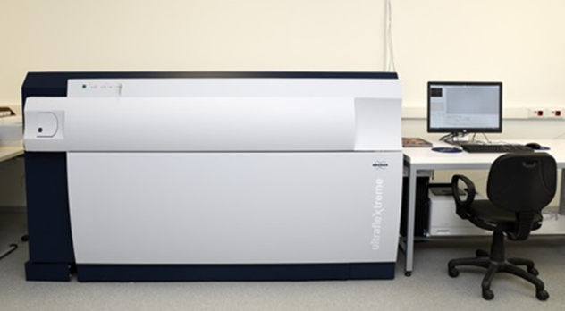
Figure no.1: Maldi-T of Ms Identification
4.5. 16S Rrna Gene Sequencing Analysis
All isolates preserved after purification from the environmental monitoring plan were sub-cultured on TSA medium at 35°C for 24 h during identification. The whole DNA was extracted using a TAKARA DNA extraction kit (Takara Bio Inc., Shiga, Japan) following the manufacturer’s instructions. Primer 27F (5′-AGTTTGATCMTGGCTC AG-3′) and 1492R (5′-GGTTAC CTTGTTACGACTT-3′) were used for PCR amplification. PCR products were resolved and visualized by electrophoresis in gels containing 1.2% agarose and 0.5 lg/mL ethidium bromide. After that, all the amplified DNA fragments were sequenced commercially in both directions (Sangon Co., Ltd., Shanghai), and the sequence reads were analyzed using Lasergene 7.0 (DNAStar, Madison, WI). A query sequence with 98.65% identity to a reference in GenBank was acceptable for species identification [31].
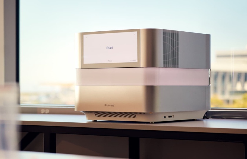
Figure No .2: 16S Rrna Gene Sequencing Analysis
4.6. Whole-Genome Sequencing of Staphylococcus Cohnii
Genomic DNAs of all 33 S. cohnii isolates were extracted from overnight cultures by using a Wizard genomic DNA purification kit (Promega, Beijing, China) and were subjected to whole-genome sequencing using 150 bp pair-end strategies using the Illumina Miseq platform (Illumina Inc., San Diego, CA, United States). All operations followed standard Illumina protocols. Raw reads were trimmed and assembled to contigs using SPAdes version 3.11. Using FDAARGOS_538(GCA_003956025.1) as a reference genome, single-nucleotide polymorphisms (SNPs) were called by the Gingr online software of Harvestsuite.1 The phylogenetic trees were analyzed by parsnp software based on the whole genomes, and the trees were visualized with FigTree software (Version 1.4.3) and Adobe Illustrator software (Version 22.1). Homologous recombination events were detected by Gubbins v3.1.32 and related sites were filtered in the subsequent analysis. After filtering, 172 wgSNPs were obtained [33.34].
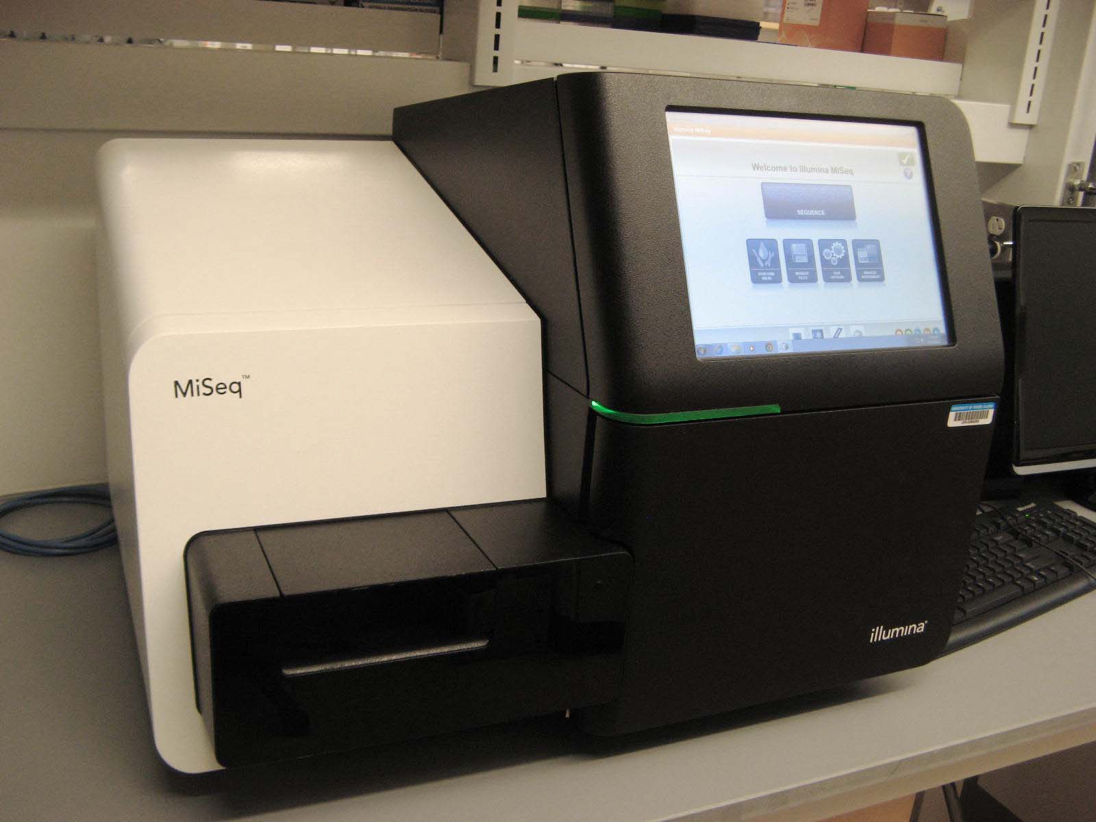
Figure No. 3: Whole-Genome Sequencing
5. RESULT:
5.1: The abundance of microorganisms at different processing locations
A sterile medication manufacturing facility's production process, comprising its supplies, personnel, environment, and production water system, yielded 60 isolates that were collected and processed between June and August 2022. Table No.4 displays the distribution of bacterial contamination. According to the data, the production environment accounted for 48 isolates, or more than 70% of all isolates, while the water system came in second with 12 isolates. Subsequent analysis of the contamination distribution across grade areas revealed that grade C clean areas had the highest rate (52%) with a tendency to decline towards the higher-grade clean room. Numerous causes of contamination were suggested by the increased diversity and quantity of pollutants. Significantly, 12 isolates were found in the grade A production area, indicating that there is serious contamination in this production setting and that immediate action is needed.
Table No.4: The distribution of microorganisms in different locations categorized by number of isolates, genus, and species
|
Environmental monitoring objectives |
Numbers of isolates |
Percentage (%) |
Genus |
Species |
||
|
Raw materials |
24 |
9.34 |
10 |
19 |
||
|
Personnel |
16 |
7.8 |
7 |
8 |
||
|
Production environment |
A |
60 |
48 |
68.36 |
8 |
16 |
|
B |
12 |
13 |
28 |
|||
|
C |
16 |
17 |
31 |
|||
|
Water |
48 |
14.5 |
13 |
24 |
||
|
Total |
88 |
100 |
43 |
94 |
||
5.2. Characterization of Micro flora In the Pharmaceutical Production Environment:
16S rRNA gene sequencing and the multiplex method of MALDI-TOF MS were used to identify the 88 isolates. In addition to the isolation context, growth characteristics, colony shape, and species information, the possible duplicates of the 88 isolates were then eliminated.The multiplex approach of 16S rRNA gene sequencing and MALDI-TOF MS was used to identify the 60 isolates. Table No.5 provides the identification findings of the 41 isolates that were chosen using the MALDI-TOF MS method. According to the manufacturer's threshold interpretation, 189 reliable results were obtained from these samples, including 17 (61.0%) with species-level results (score ≥2.0) and 19 (17.4%) with genus-level results (1.7 ≤ score < 2.0). The remaining 42 isolates (21.6%) received non-reliable identification results (scoring <1.7). Among the 94 unreliable results obtained by MALDI-TOF MS, 79 isolates were identified as species-level by the matching criteria (≥98.65% similarity), and 15 isolates were identified as genus-level. Integrating the results of the whole identification module, it was finally determined that the 241 isolates were from 44 genera and 94 species. With the high-throughput capability, all mass spectrometry identifications can be accomplished within a single batch on the same day.
|
Isolates |
Sample |
Sampling |
Cleanliness Grade |
|
Number |
Code |
Location |
Classification |
|
A1 |
VI-W-2022 WIJ 1 |
Water for Injection |
B |
|
A2 |
VI-W-2022 WIJ 2 |
Water for Injection |
B |
|
A3 |
VI-W-2022 CSR 3 |
Crystallization Room |
B |
|
A4 |
VI-W-2022 CSR4 |
Crystallization Room |
B |
|
A5 |
VI-W-2022 CSR 5 |
Crystallization Room |
B |
|
A6 |
VI-W-2022 CSR 6 |
Crystallization Room |
B |
|
B1 |
VI-F-2022-8 DR |
Distribution Room |
B |
|
B10 |
VI-F-2022-8 AC |
Activated Charcoal |
N/A |
|
B11 |
VI-F-2022-8 AC |
Activated Charcoal |
N/A |
|
B12 |
VI-F-2022-8 AC |
Activated Charcoal |
N/A |
|
B14 |
VI-F-2022-8 AC |
Activated Charcoal |
N/A |
|
B2 |
VI-F-2022-8 CR |
Crystallization Room |
B |
|
B3 |
VI-F-2022-8 WIJ |
Water for Injection |
B |
|
B4 |
VI-F-2022-8 WIJ |
Water for Injection |
B |
|
C4 |
VI-F-2022-8 RS |
RABS gloves |
A |
|
C5 |
VI-F-2022-8 AC |
Activated Charcoal |
N/A |
|
C6 |
VI-F-2022-8 CR |
Crystallization Room |
B |
|
C7 |
VI-F-2022-8 CR |
Crystallization Room |
B |
|
C9 |
VI-F-2022-8 CR |
Crystallization Room |
B |
|
D1 |
?-M-2022-8-1 |
FDP |
N/A |
|
D10 |
?-M-2022-8-2 |
0.22μm Filter |
B |
|
D11 |
?-M-2022-8-3 |
0.22μm Filter |
B |
|
D12 |
?-M-2022-8-4 |
0.22μm Filter |
B |
|
D13 |
?-M-2022-8-5 |
0.22μm Filter |
B |
|
D15 |
?-M-2022-8-6 |
0.22μm Filter |
D |
|
D17 |
?-M-2022-8-7 |
Nitrogen Return |
B |
|
D18 |
?-M-2022-8-8 |
FDP |
N/A |
|
D19 |
?-M-2022-8-9 |
FDP |
N/A |
|
D2 |
?-F-2022-8 CR 1 |
Crystallization Room |
B |
|
D4 |
?-F-2022-8 CR 2 |
Crystallization Room |
B |
|
D5 |
?-F-2022-8 CR 3 |
Crystallization Room |
B |
|
D6 |
?-F-2022-8 HR1 |
Holding Room |
B |
|
D7 |
?-F-2022-8 DB 5 |
Distribution Room |
B |
|
D8 |
?-F-2022-8 CR 6 |
0.22μm Filter |
D |
|
E1 |
?-F-2022-8 CR 7 |
Crystallization Room |
B |
|
E10 |
?-F-2022-8 WIJ |
Water for Injection |
B |
|
E11 |
?-F-2022-8WIJ 9 |
Water for Injection |
B |
|
E12 |
?-F-2022-8 WIJ 10 |
Water for Injection |
B |
|
E13 |
?-F-2022-8 CR 11 |
Crystallization Room |
B |
|
E16 |
?-F-2022-8 DR 12 |
Distribution Room |
B |
|
E2 |
?-F-2022-8 CR 13 |
Trolley |
B |
|
E3 |
?-F-2022-8 CR 14 |
Distribution Room |
B |
|
E4 |
?-F-2022-8 CR 15 |
RABS gloves |
A |
|
F12 |
?-S-20228-3 |
Corridor 1 |
C |
|
F13 |
?-S-20228-4 |
Capping Room1 |
C |
|
F15 |
?-S-20228-5 |
Capping Room1 |
C |
|
F16 |
?-S-20228-6 |
Capping Room1 |
C |
Table No.5: Assessment of MALDI-TOF MS's identification potential for each isolate gathered and the study's most prevalent contaminated genus
|
F17 |
?-S-20228-7 |
Crystallization Room |
B |
|
F18 |
?-S-20228-8 |
Crystallization Room |
B |
|
F19 |
?-S-20228-9 |
Crystallization Room |
B |
|
F20 |
?-S-20228-10 |
Water for Injection |
B |
|
F22 |
?-S-20228-11 |
Water for Injection |
B |
|
F24 |
?-S-20228-12 |
Water for Injection |
B |
|
F25 |
?-S-20228-13 |
Crystallization Room |
B |
|
F26 |
?-S-20228-14 |
Air lock2 |
C |
|
F27 |
?-S-20228-15 |
Air lock2 |
C |
|
F28 |
?-S-20228-16 |
Air lock2 |
C |
|
F29 |
?-S-20228-17 |
Storeroom1 |
C |
|
F3 |
?-S-20228-18 |
Crystallization Room |
B |
|
F30 |
?-S-20228-19 |
Capping Room6 |
C |
The Isolates were numbered according to test batches received by the laboratory The Sample Types codes are defined as a-b-c:
a: Production line, I-Formulation Manufacture; VI-Substance Manufacture;
b: Monitoring objectives and methods, F-viable air sampling using volumetric air; C- air sampling using settle plates; S- swabs or contact plates of the surface; P- personnel, finger plates or gowning sampling; W-water sample and
c: Sampling date;
FDP: Fructose 1,6-bisphosphate;
RABS: Restricted access barrier systems;
Comparing the 16 s rRNA gene sequencing identification results with the MALDI-TOF MS commercial database revealed that of the 60 unreliable MALDI-identifies, 46 isolates from 31 species were not covered by the commercial MALDI database. If we remove those out-of-coverage, the identification capacity of MALDI-TOF MS method could reach 75.8% species-level and 95.4% genus-level. The most significant discrepancy impacted by database coverage was with Bacillus, where the accuracy of species-level identification improved from 30% to 85.7% by removing non-coverage species from the identification set.
Table No.6: Evaluation of the identification capability of MALDI-TOF MS for all isolates collected
|
Genus |
Isolates in this study |
MALDI not covered |
Identification results for all isolates |
Identification results only MALDI covered |
|||
|
Isolates (species) |
Isolates (species) |
Species level |
Genus level |
Species level |
Genus level |
||
|
Staphylococcus |
43 |
87 (10) |
3 (1) |
85.2% (23) |
14.8% (4) |
85.20% |
12.14% |
|
Micrococcus |
5 |
21 (4) |
0 (0) |
81.4% (78) |
13.4% (13) |
83% |
13.80% |
|
Bacillus |
98 |
15 (10) |
13 (6) |
30% (6) |
5% (1) |
85.70% |
14.30% |
|
Actinobacteria |
34 |
11 (4) |
1 (1) |
72.7% (8) |
27.3% (3) |
72.70% |
27.30% |
|
Paenibacillus |
75 |
08 (3) |
0 (0) |
57.1% (8) |
21.4% (3) |
61.50% |
23.10% |
|
In total |
2,96 |
142 (72) |
46 (31) |
60% (147) |
17.4% (42) |
75.40% |
21.50% |
5.3. Contamination Source Tracking of Staphylococcus cohnii Using the WGS Typing Method
The current findings indicate that Staphylococcus spp. is the predominant microbial contaminant in the drug production process, representing 40.25% of the total viable contaminants. Among these, Staphylococcus cohnii was the most significant species, with 33 isolates identified (details provided in Supplementary Tables No:5 and 6). These isolates were recovered from various sources, including grade A (3 isolates), grade B (7), grade C (20) cleanroom environments, personnel (2), and raw materials (1), highlighting its widespread distribution throughout the production process. Notably, environmental monitoring revealed that the majority of S. cohnii isolates (28 out of 33) originated from the formulation manufacturing area (MF), while only five were recovered from the substance manufacturing area (MS).shown in figure No:4
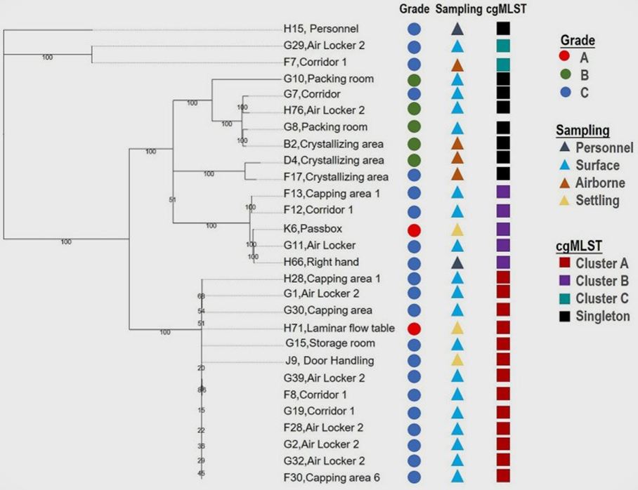
Figure No.4: Result of wgSNP typing of the 28 S. cohnii contaminated
5.4. The micro flora in the pharmaceutical production environment
As demonstrated, the most significant proportion of genus-level contamination was associated with Staphylococcus spp. (40.25%), Micrococcus spp. (11.20%), Bacillus spp. (8.30%), Actinobacteria (5.81%), and Paenibacillus spp. (4.56%), the top 5 genera contributing to 70% of all the isolates. The 19 predominant species accounted for 64% of all 94 species isolated, with a distinct predominance of Staphylococcus spp., which occupied 9 of the 19 species that comprise most of the microflora, of which S. cohnii, S. epidermidis, and S. hominis were detected more than 10 times. Micrococcus luteus, was also shown to be a frequently occurring contaminant. The investigation plan further categorized the monitoring objectives into airborne contamination (using settle plates and volumetric air sampling) and surface contamination (via contact plates and swabs) to enable more targeted analysis. As shown in Figure 5, airborne flora exhibited the greatest diversity in both quantity and species, encompassing 26 different genera. The most frequently recovered organisms were Staphylococcus spp. (69%) and Micrococcus spp. (74%). Among the predominant genera listed in Table 7 (defined as those with ≥ three isolations), four were exclusively detected through airborne sampling. Interestingly, no spore-forming Bacillus species were identified in the air samples—an unexpected finding that contrasts with the common assumption that such spores are primarily transmitted through air. This discrepancy may be attributed to the selection of sampling locations or protocols. Similarly, the high prevalence of Gram-positive cocci detected in the water system could indicate suboptimal sampling practices. These observations highlight key areas for improvement in the design and implementation of future environmental monitoring protocols.In contrast, 58% of the spore-forming bacterial isolates were traced to raw materials, and 32% to environmental surfaces, underscoring the critical role of raw material quality control and proper disinfection procedures during material transfer.
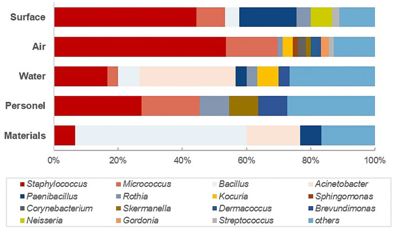
Figure No.5: The distribution of the predominant genus in different monitoring locations.
Table No.7: Most commonly occurring species/genera in the pharmaceutical production environment
|
Species |
Isolation |
Percentage |
|
Staphylococcus cohnii |
30 |
12.69% |
|
Staphylococcus epidermidis |
23 |
10.20% |
|
Micrococcus luteus |
12 |
6.05% |
|
Staphylococcus hominis |
8 |
3.15% |
|
Paenibacillus lautus |
5 |
2.73% |
|
Staphylococcus haemolyticus |
4 |
2.49% |
|
Acinetobacter pittii |
5 |
2.49% |
|
Staphylococcus nepalensis |
4 |
2.07% |
|
Bacillus aryabhattai |
2 |
2.07% |
|
Staphylococcus pasteuri |
5 |
1.66% |
|
Staphylococcus capitis |
3 |
1.66% |
|
Micrococcus cohnii |
2 |
1.66% |
|
Micrococcus antarcticus |
3 |
1.66% |
|
Staphylococcus saccharolyticus |
2 |
1.24% |
|
Staphylococcus argensis |
2 |
1.24% |
|
Rothia amarae |
2 |
1.24% |
|
Bacillus paralicheniformis |
2 |
1.24% |
|
Bacillus megaterium |
4 |
2.07% |
|
Acinetobacter nosocomialis |
2 |
1.24% |
|
Others |
120 |
36.93% |
6. DISCUSSION:
Based on a thorough understanding and evaluation of the processes and facilities of a sterile drug manufacturer, a comprehensive microbial surveillance scheme was established in this study. This scheme encompassed routine monitoring, microbial identification, contamination tracking, and risk assessment. The distribution and diversity of microflora across the entire production chain were systematically examined. Over a period of three months, a total of 292 bacterial isolates were recovered from various sources, covering all areas of the production process and cleanrooms of different classifications. The widespread presence of microbial contaminants underscores a high contamination risk and highlights the urgent need to optimize the surveillance program, including enhanced cleaning and disinfection strategies.In this study, MALDI-TOF MS was employed as the primary method for microbial identification, supplemented by sequencing. This approach enabled rapid and efficient characterization of the environmental flora and facilitated the development of a fast, effective surveillance protocol. MALDI-TOF MS is widely recognized as a simple, high-throughput proteomic tool for the identification of a broad spectrum of bacterial species. Taking database coverage into account, the system achieved identification accuracies of 75.8% at the species level and 95.4% at the genus level, supporting its suitability as a frontline tool for routine environmental monitoring.However, using only the commercial database, genus- and species-level identification rates dropped to 77.4% and 60.0%, respectively. Performance was notably lower for Bacillus spp., with just 35% genus-level and 30% species-level identification accuracy—despite the commercial database containing over 100 Bacillus species. These results fall short of the 90%+ species-level accuracy typically reported in clinical settings (Croxatto et al., 2012; Patel, 2015; Schubert & Kostrzewa, 2017). This discrepancy likely stems from insufficient representation of environmental isolates in the commercial MALDI-TOF MS database. Even when species are included, strain diversity may be limited. Environmental isolates from cleanroom settings are subject to higher stress due to stringent cleaning and disinfection protocols, making them harder to identify than clinical strains. Our findings are consistent with those of Seuylemezian et al. (2018), who reported successful identification of only 8% of bacterial isolates from spacecraft cleanrooms using manufacturer-supplied databases, even under rigorous microbial reduction practices. These results underscore the need to expand MALDI-TOF MS commercial databases with spectra from pharmaceutical cleanroom isolates. Collaboration between MALDI-TOF MS developers and pharmaceutical manufacturers is essential to improve identification performance.Furthermore, considering regional differences in manufacturing environments, product types, and production processes, facilities may harbor unique microbial strains. Developing customized, in-house reference databases using locally isolated strains presents an effective strategy to enhance the accuracy and utility of MALDI-TOF MS systems for environmental monitoring.
7.1. CONCLUSION:
This study established a comprehensive microbial surveillance framework for a sterile drug manufacturing facility, encompassing routine monitoring, microbial identification, contamination tracking, and risk assessment. The recovery of 60 bacterial isolates across various cleanroom grades and production stages highlights the widespread presence of microbial contaminants and underscores the critical need to strengthen contamination control measures.MALDI-TOF MS, supported by sequencing, proved to be a rapid and efficient tool for microbial identification, achieving high accuracy when database coverage was adequate. However, limitations in the commercial MALDI-TOF MS database—particularly for environmental isolates such as Bacillus spp.—significantly impacted species-level identification. These findings align with previous studies and emphasize the need to expand commercial databases with spectra from pharmaceutical cleanroom environments. To improve identification capabilities, it is essential for MALDI-TOF MS developers to collaborate with pharmaceutical manufacturers and to consider the creation of facility-specific reference libraries that reflect local microbial diversity. Such tailored approaches will enhance the effectiveness of microbial monitoring programs and support better contamination control in sterile manufacturing processes.
7.2. Future Recommendation
The findings of this study highlight several critical directions for future improvements in microbial surveillance within sterile pharmaceutical manufacturing environments. One key area is the enhancement of microbial identification accuracy through the expansion and refinement of MALDI-TOF MS databases. Incorporating spectra from environmental and cleanroom-specific isolates, especially stress-adapted strains like Bacillus spp., will significantly improve the identification of non-clinical microorganisms. Collaboration between MALDI-TOF MS developers and pharmaceutical manufacturers is vital to develop more representative and robust databases.Furthermore, creating facility-specific or regional reference libraries using locally isolated strains can address the unique microbial profiles influenced by environmental, geographic, and operational differences. Integrating next-generation sequencing technologies with MALDI-TOF MS could further strengthen the depth and accuracy of microbial identification and tracking.Future studies should also explore the integration of predictive analytics.
Conflicts of Interest
The authors declare that there are no conflicts of interest, whether financial or otherwise.
ACKNOWLEDGEMENTS
The authors wish to thank all researchers for providing an eminent literature source for devising this manuscript.
Consent for Publication
Not Applicable
REFERENCES


Khushbu*, Dr. Peeyush Jain, Dr. Pankaj Chasta, Microbial Contamination and Sterility Testing in Injectable Pharmaceuticals – Ensuring Sterility Assurance and Contamination Control, Int. J. of Pharm. Sci., 2025, Vol 3, Issue 5, 792-809. https://doi.org/10.5281/zenodo.15343825
 10.5281/zenodo.15343825
