View Article

  • Mucoadhesive Drug Delivery Systems for Vaginal Administration

  • 1237Research Scholar at Department of Pharmaceutics, Dr. Shivajirao Kadam College of Pharmacy, Kasabe - Digraj, Maharashtra, India

    4Research Scholar at Department of Pharmaceutics, St.John Institute of Pharmacy and Research, Palghar, Maharashtra, India

    5Graduate Student, Womens college of pharmacy, peth vadgaon, Maharashtra, India

    6Research Scholar at Department of Pharmaceutics, Maratha Vidya Prasarak Samaj College of Pharmacy Nashik, Maharashtra, India                                  

Abstract

The vaginal route of drug administration offers significant advantages for both local and systemic therapy due to its large absorptive surface area, rich vascularization, avoidance of first-pass hepatic metabolism, and reduced gastrointestinal degradation. However, conventional vaginal dosage forms such as creams, gels, tablets, and suppositories often exhibit limited therapeutic effectiveness because of rapid mucus turnover, vaginal self-cleansing, pH variability, formulation leakage, and poor patient compliance. To overcome these limitations, mucoadhesive drug delivery systems (MDDS) have emerged as a promising approach to improve vaginal drug delivery outcomes. By establishing prolonged and intimate contact with the vaginal mucosa through physicochemical interactions between polymers and mucin, mucoadhesive systems enhance formulation residence time. This review provides a comprehensive overview of vaginal anatomy and physiology, challenges in vaginal drug delivery, mechanisms of mucoadhesion, and the role of mucoadhesive polymers. Various mucoadhesive dosage forms, evaluation parameters, therapeutic applications, recent advancements, and future clinical and regulatory challenges are also discussed.

Keywords

Mucoadhesive vaginal drug delivery; Vaginal mucosa; Mucoadhesive polymers; Controlled drug release; Mucus barrier..

Introduction

Because of its unique anatomical, physiological, and biochemical features, the vaginal route of administration is universally acknowledged as a very beneficial method for both local and systemic drug delivery. The vagina offers a comparatively large surface area for medication absorption because it is a muscular, fibromuscular canal bordered with stratified squamous epithelium. Drugs that are ingested can be quickly transported into the systemic circulation thanks to this epithelium's abundance of blood arteries and lymphatic network.[1] Vaginal drug distribution, as opposed to oral administration, avoids exposure to harsh gastrointestinal conditions that might affect drug stability and bioavailability, such as acidic pH, digestive enzymes, and varying gastric emptying durations. Additionally, medications given vaginally can avoid first-pass hepatic processing, which is a significant contributing cause to the decreased systemic availability of many therapeutic medicines. Therefore, vaginal delivery provides a useful substitute for increasing the bioavailability of medications that are poorly absorbed or heavily metabolized when taken orally.[2]Because of these benefits, the vaginal route is especially well-suited for a variety of therapeutic uses, such as the management of infertility, hormone replacement therapy, gynaecological infections, STDs, contraception, and even the systemic delivery of peptides, proteins, and vaccines.[3] Additionally, vaginal drug delivery is linked to easier administration, fewer doses, and better patient convenience—all of which are critical factors in long-term treatments. Additionally, localized drug administration via the vagina minimizes systemic side effects while enabling high drug concentrations at the site of action a significant benefit in the treatment of inflammatory diseases and vaginal infections.[4]The therapeutic efficacy of vaginal medication delivery is nevertheless limited by the disadvantages of traditional dose forms as creams, gels, foams, pills, pessaries, and suppositories, despite the many benefits provided by the vaginal route.[5] These conventional formulations are less suitable for long-term or prolonged therapy because they are often designed for rapid drug release and brief duration of action. The innate self-clearing characteristic of the vaginal canal is a major physiological barrier to successful vaginal medication administration.[6]Constant vaginal fluid renewal, frequent exfoliation of epithelial cells, and ongoing mucus production are characteristics of this process. Because of this, given formulations are quickly eliminated from the vaginal canal, which reduces the amount of time the medication is in touch with the mucosal surface and, as a result, lowers drug absorption and retention.[7]Conventional vaginal dose forms hence frequently show inadequate retention, uneven drug distribution, and less interaction with the vaginal mucosa, resulting in less than ideal therapeutic effects. Furthermore, patients frequently experience discomfort, difficulty, and social embarrassment due to formulation leaks.[8] In order to maintain therapeutic drug levels, these factors often require repeated administration, which may raise the risk of local irritation, disturb the natural vaginal microbiota, and have a detrimental effect on patient compliance. Therefore, despite the vaginal route's inherent benefits, sophisticated drug delivery methods that can get beyond these physiological obstacles and increase treatment efficacy are still desperately needed.[9]Mucoadhesive drug delivery systems (MDDS) have become a viable and successful method for vaginal drug delivery in order to overcome these obstacles. The ability of a substance to stick to a mucosal surface through interactions with the mucus layer or the epithelial tissue is known as mucoadhesion.[10] Mucoadhesive systems are made to make close, long-lasting contact with the vaginal mucosa in order to prevent clearance mechanisms and prolong the formulation's residence time at the administration site. This extended retention lowers the frequency of dosing, improves therapeutic efficacy, and increases drug absorption.[11]The process of mucoadhesion involves several steps, starting with the polymer's initial wetting and hydration, followed by polymer swelling and the subsequent interpenetration of polymer chains into the mucus layer. Alongside this, a number of intermolecular interactions are established between the polymer and mucin glycoproteins, such as hydrophobic interactions, van der Waals forces, hydrogen bonding, and electrostatic attraction.[12] Mucoadhesive drug delivery systems improve formulations' adhesion to the vaginal mucosa through these coupled mechanisms, which increases drug retention and site-specific localization. As a result, these systems can offer controlled and extended drug release, which makes them especially useful for the treatment of long-term illnesses that need constant therapeutic exposure.[13]Numerous mucoadhesive vaginal formulations have been created to address a range of therapeutic requirements. Mucoadhesive gels, pills, films, suppositories, microspheres, and nanoparticles are some of them. Mucoadhesive gels are the most popular of these since they are simple to apply and spread evenly across the vaginal mucosa. While suppositories give prolonged medication release and localized action, mucoadhesive tablets and films enable accurate dosing and enhanced durability. Particulate systems like microspheres and nanoparticles have drawn a lot of attention lately because of their capacity to improve penetration, offer regulated release, and shield medications from deterioration.[14]In order to achieve adhesion and prolonged medication release, conventional mucoadhesive systems mostly rely on synthetic and natural polymers. Because of their superior swelling behavior and potent mucoadhesive qualities, synthetic polymers like polyacrylates are commonly employed. Conversely, natural polymers provide benefits including limited toxicity, biocompatibility, and biodegradability.[15] New Nano carrier-based systems that combine mucoadhesive qualities with controlled release and mucus-penetrating capabilities have been created because to developments in pharmaceutical technology. These cutting-edge solutions are made to go past the diffusion barrier that vaginal mucus presents while still having enough adhesion to guarantee long-term retention.[16]The effectiveness of vaginal MDDS is significantly influenced by the choice of suitable mucoadhesive polymers. Poly(acrylates) like Carbopol, chitosan, cellulose derivatives (such hydroxypropyl methylcellulose and carboxymethyl cellulose), alginates, hyaluronic acid derivatives, and other natural polysaccharides are often used polymers. Each of these polymers has unique physicochemical characteristics that affect their drug release qualities, mechanical strength, swelling capacity, and mucoadhesive behavior.[17]Because polyacrylates contain carboxylic groups that can establish hydrogen bonds with mucin, they are recognized for having a high mucoadhesive strength. Strong electrostatic interactions between negatively charged mucus and chitosan, a cationic polymer, make it very useful for mucoadhesive applications. While alginates and hyaluronic acid derivatives offer biocompatibility and hydration that are advantageous for vaginal applications, cellulose derivatives offer superior film-forming qualities and regulated drug release. To achieve the intended balance between adhesion, medication release, mechanical stability, and patient comfort, the choice of polymer or polymer combination must be carefully optimized.[18]                                                                                                                                                         The dynamic biological milieu of the vagina has a considerable impact on the effectiveness of mucoadhesive medication delivery devices in addition to polymer selection. Changes in pH, enzymatic activity, mucus composition, and hormonal swings related to menstruation, pregnancy, and menopause define the vaginal environment.[19] Polymer swelling, drug stability, and release behavior can all be impacted by vaginal pH, which normally ranges from acidic to neutral depending on physiological circumstances. Vaginal secretions contain enzymes that can break down specific medications or polymers, and mucus turnover can affect how long mucoadhesion lasts.[20]Hormonal fluctuations can also affect the effectiveness of drug delivery by changing vaginal fluid volume, mucus viscosity, and epithelium thickness. Effective MDDS formulations must therefore be thoughtfully created to endure these physiological difficulties while preserving a robust and reliable adherence to the mucosal surface.[21] To increase the robustness and dependability of vaginal MDDS, advanced formulation techniques as pH-responsive systems, enzyme-resistant polymers, and thermosensitive gels are being investigated.[22]All things considered, vaginal mucoadhesive drug delivery systems offer a viable and adaptable platform for improving the management of numerous local and systemic disorders. These systems provide substantial advantages over traditional vaginal formulations by extending residence time, enhancing drug retention, and permitting controlled release. They have shown great promise in the treatment of vaginal infections, hormone administration, contraception, and new uses like microbicide delivery and vaginal immunization.[23]

2. Anatomy and Physiology of the Vaginal Mucosa

The human vagina is a dynamic, intricate organ used for both medicinal and reproductive purposes. Anatomically, it is a fibromuscular tubular canal that runs from the uterine cervix to the external genitalia (vestibule). Its length is around 7–10 cm, and its breadth and thickness vary according to age, hormonal status, and physiological phase. The surrounding fascia and pelvic muscles support the vaginal tube, which is positioned at a 90° angle to the uterus. This allows for flexibility and distension during sexual activity and birthing. The anterior, posterior, and lateral fornices surrounding the cervix are among the anatomical divisions of the vaginal canal, and the walls exhibit transverse folds known as rugae that increase surface area and permit expansion.[24]

2.1 Histological Structure

Three primary layers make up the vaginal wall histologically: the adventitia (outside connective tissue layer), the muscularis (middle smooth muscle layer), and the mucosa (inner layer). The non-keratinized stratified squamous epithelium that makes up the mucosa lacks glands; instead, cervical mucus, vaginal transudate, and exfoliated epithelial cells provide lubrication.[25] The lamina propria, a vascular, collagen-rich layer of connective tissue that supports immune surveillance and cellular turnover, is located underneath the epithelium. While the adventitia comprises collagen and elastin fibres that provide elasticity and resilience, the muscular layer is made up of smooth muscle fibres scattered with elastic tissue to provide structural integrity and contractility.[26]

2.2 Vaginal Mucosa Function and Barrier Properties

Both absorption and defence depend on the vaginal mucosa. Pathogens and mechanical injury are physically prevented by the squamous epithelium and the mucous layer that covers it. Mucus from cervical secretions and transudate produces a viscoelastic covering over the epithelium, even though other mucosal tissues lack goblet cells that make mucus. This mucus, which forms a protective gel and affects medication transport and retention, is made up of water, mucin glycoproteins, enzymes, immunological proteins, and exfoliated cells.[27]The vaginal environment is maintained by a complex interaction between hormonal and microbial variables. Oestrogen promotes cellular glycogen storage and epithelial growth in women of reproductive age. After being exfoliated and broken down, glycogen acts as a nutritional substrate for Lactobacillus species, which ferment the breakdown products of glycogen into lactic acid, keeping the pH between 3.5 and 4.5. Many diseases find this acidic environment to be unfriendly, offering a natural defence. Epithelial thickness, mucus composition, and pH are all dramatically altered by changes in oestrogen levels, which can occur throughout the menstrual cycle, pregnancy, or menopause. These changes have an impact on barrier function and permeability.[28]

2.3 Physiological Variability and Implications for Drug Delivery

The vaginal mucosa's physiological properties differ significantly between biological situations and life stages. For instance, throughout the menstrual cycle, progesterone dominance following ovulation results in distinct mucosal characteristics, whereas oestrogen peaks close to ovulation cause increased epithelial thickness and alterations in mucus consistency. Reduced oestrogen levels during menopause lead to epithelium thinning, decreased mucus production, elevated pH, and an increased risk of infection and dryness. Drug absorption, mucoadhesion, and formulation performance are all directly impacted by these variables.[29] The lamina propria contains a vast network of capillaries that provide good vascularization to the vaginal mucosa. This abundant blood supply promotes both systemic drug absorption and local therapeutic benefits. The vagina is an appealing route for both local and systemic therapy since pharmacokinetic studies have shown that numerous lipophilic and low-molecular-weight drugs can effectively pass through the vaginal epithelium and into the systemic circulation.[30]

2.4 Protective Immune Functions

The vaginal mucosa supports mucosal immunity in addition to its barrier and absorptive functions. It has immune cells that identify and react to microbial threats, including lymphocytes, dendritic cells, and macrophages.[31] Additionally, the mucosal surface secretes antimicrobial peptides and displays pattern recognition receptors that are involved in preserving homeostasis and protecting against infections such as bacterial vaginosis and vulvovaginal candidiasis. By generating hydrogen peroxide and other inhibitory compounds against pathogenic organisms, Lactobacillus species, which are major members of the vaginal microbiota, further strengthen immune protection.[32]

2.5 Relevance to Drug Delivery Systems

The structure and physiology of the vaginal mucosa offer both opportunities and obstacles for medication delivery. While the mucus layer and pH can affect formulation residence duration and drug release profiles, its enormous surface area, permeability, and circulatory network facilitate effective medication absorption. For instance, the viscoelastic gel structure of mucus acts as both a barrier to medication diffusion and a target for mucoadhesive formulations intended to increase absorption and prolong contact duration. To guarantee constant function, formulation design must also take into account dynamic variations in mucus qualities throughout the menstrual cycle.[33]

3. Challenges in Vaginal Drug Delivery

The intricate anatomy and physiology of the vaginal route provide serious obstacles to efficient and reliable treatment results, despite the route's potential benefits for both local and systemic medication delivery. The protective and dynamic vaginal milieu is a significant barrier to vaginal medication administration. The first line of defence against viruses, foreign objects, and irritants is the viscoelastic mucus layer lining the vaginal cavity. Although this layer of mucus aids in the maintenance of vaginal health, it also functions as a chemical and physical barrier that may hinder the diffusion and retention of drugs. Conventional formulations, including gels and suppositories, are quickly cleared by the constant secretion and turnover of mucus, which shortens their residence time and decreases their bioavailability at the target region.[34]Additionally, because Lactobacillus species create lactic acid, the pH of the vaginal environment is normally acidic (between 3.5 and 4.5) in healthy women. Although this acidic environment protects against infections, it can affect the stability and solubility of medications, particularly those that are sensitive to pH. Formulation design is further complicated by pH variations linked to ageing, menstruation, infection, or hormonal changes.[35]Variations in vaginal mucous and epithelial thickness across various physiological conditions present another difficulty. Mucus viscosity, volume, and epithelial cell turnover are all greatly impacted by hormonal changes throughout the menstrual cycle or menopause, which also have an impact on drug absorption and distribution. Additionally, through adhesion and steric interactions, the mucus layer can retain particle delivery systems, causing quick clearance before enough drug release can take place.[36]Complicating matters further are the immune system and vaginal bacteria. Drug disposition and therapeutic efficacy can be altered by microbial composition disruptions, such as those caused by bacterial vaginosis or candidiasis, which can change mucus characteristics, pH, and enzyme activity. All things considered, these physiological obstacles-such as mucus clearance, pH fluctuations, epithelium alterations, and microbiota dynamics-represent significant obstacles to successful and long-lasting vaginal medication administration. To improve retention and absorption, novel formulation techniques such mucoadhesive and mucus-penetrating delivery methods are needed to address these problems.[37]


Table No 1: Challenges in Vaginal Drug Delivery


Challenge


Physiological Basis

Effect on Formulation Performance

Impact on Drug Bioavailability & Efficacy

Formulation Strategies to Overcome


References


Viscoelastic mucus barrier

Vaginal mucus composed of mucins, water, enzymes, and lipids forming a protective gel layer

Limits diffusion of drug molecules and particulate carriers

Reduced penetration and uneven drug distribution

Mucoadhesive polymers, mucus-penetrating nanoparticles


38

Rapid mucus turnover (self-cleansing)

Continuous secretion and renewal of vaginal mucus and fluids

Quick removal of gels, creams, and suppositories

Short residence time; frequent dosing required

Mucoadhesive gels, films, tablets, in situ gelling systems


39


Epithelial cell exfoliation

Constant shedding of superficial epithelial cells

Loss of drug–epithelium contact

Decreased absorption and retention

Bioadhesive systems with prolonged adhesion


40


Acidic vaginal pH (3.5–4.5)

Lactic acid production by Lactobacillus species

Drug instability or precipitation of pH-sensitive drugs

Reduced solubility and therapeutic activity

pH-buffered systems, polymer-based carriers


41


pH variability

Changes during menstruation, menopause, infections, pregnancy

Unpredictable drug release behavior

Variable therapeutic response

pH-responsive polymers, adaptable formulations


42


Hormonal fluctuations

Estrogen/progesterone changes alter mucus thickness and viscosity

Inconsistent formulation performance

Variable absorption across patient populations

Personalized or hormone-responsive delivery systems


43

Mucus entrapment of particles

Adhesive and steric interactions between mucus and particles

Nanoparticles cleared before drug release

Low bioavailability of particulate systems

Surface-modified mucus-penetrating nanoparticles


44


Microbiota alterations

Conditions like bacterial vaginosis or candidiasis alter flora

Changes in pH, enzymes, mucus composition

Altered drug metabolism and efficacy

Microbiota-compatible excipients


45


Enzymatic activity

Presence of proteases and esterases in vaginal fluid

Drug degradation

Reduced drug stability

Enzyme-resistant formulations, encapsulation


46

Patient discomfort & leakage

Fluid nature of conventional dosage forms

Poor retention and acceptability

Reduced compliance

Films, tablets, bioadhesive gels


47


4. Mucoadhesion: mechanisms & theories.

The ability of a substance (often a polymer) to stick to a mucosal surface is known as mucoadhesion. This property increases a formulation's residence duration at the site of delivery, boosting medication absorption and therapeutic efficacy. In pharmaceutical sciences, this phenomenon has been thoroughly investigated as a way to get beyond the conventional dose forms' quick clearance and low retention on mucosal tissues such the oral, nasal, and vaginal mucosa.[48]The contact (or soaking) stage and the consolidation stage are the two broad stages of the mucoadhesion process. Mucoadhesive polymers spread out and make first contact with the mucosal surface during the contact stage, which is frequently made possible by the polymer's swelling and hydration. Interfacial contacts between the polymer and the mucus layer intensify during the consolidation stage, creating adhesive connections that maintain attachment in physiological settings.[49]


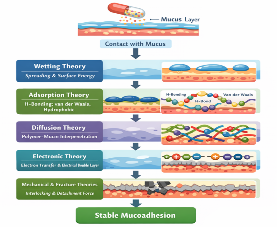
Fig No 1: Theories of Mucoadhesion


4.1 Wetting Theory:This theory highlights the significance of surface energy and spreading in liquid or    low-viscosity mucoadhesive systems. Contact area and adhesion strength are increased when a polymer is more wettable on the mucosal surface.[50]

4.2 Adsorption Theory: Secondary chemical interactions between the mucoadhesive polymer and mucin glycoproteins, including hydrogen bonding, van der Waals forces, and hydrophobic interactions, cause adhesion.[51]

4.3Diffusion idea: According to this idea, adhesion is stabilized by a physical entanglement network formed when polymer and mucin chains interpenetrate. The molecular weight and flexibility of the polymer determine the degree of interpenetration.[52]

4.4 Electronic Theory: An electrical double layer and attractive forces form at the contact as a result of electron transfer between the polymer and mucus, which causes adhesion.[53]

4.5 Mechanical and Fracture Theories: Additional insights into mucoadhesion strength and detachment behavior are provided by mechanical interlocking between polymer and mucosal surface imperfections and the force necessary to break adhesive connections.[54]

5. Mucoadhesive Polymers

Mucoadhesive polymers are essential to mucoadhesive drug delivery systems because they create and sustain close contact between the mucosal surface and the formulation. Through hydrogen bonding, electrostatic attraction, van der Waals forces, and polymer chain interpenetration, these polymers interact with mucus, extending residence time and improving drug absorption. Mucoadhesive polymers can be generically categorised into three types based on their origin and chemical modification: natural, semi-synthetic, and synthetic.[55]

5.1 Natural Polymers

The biocompatibility, biodegradability, and low toxicity of natural polymers make them popular. Pectin, gelatin, xanthan gum, guar gum, chitosan, and alginates are typical examples. Because of its electrostatic interactions with negatively charged mucin, chitosan, a cationic polysaccharide produced from chitin, demonstrates high mucoadhesive characteristics. When hydrated, alginates and pectin create hydrogels that offer prolonged drug release without compromising mucosal compatibility.[56]

5.2 Semi-Synthetic Polymers

Chemically altered natural polymers with enhanced mechanical strength and regulated swelling behaviour are known as semi-synthetic polymers. Vaginal mucoadhesive formulations frequently use cellulose derivatives as hydroxypropyl methylcellulose (HPMC), carboxymethyl cellulose (CMC), and hydroxyethyl cellulose (HEC). These polymers are suitable for tablets, films, and gels because of their good film-forming ability, moderate mucoadhesion, and consistent drug release profiles.[57]

5.3 Synthetic Polymers

Strong and consistent mucoadhesive qualities are provided by synthetic polymers because of their distinct chemical structures. Among the most widely utilized synthetic mucoadhesive polymers are poly(acrylic acid) derivatives as Carbopol and polycarbophil. Extensive hydrogen bonding with mucin is made possible by their high carboxyl group density, which improves adherence and prolongs retention. Controlled-release vaginal medication delivery systems benefit greatly from these polymers.[58]


Table No 2: Mucoadhesive Polymers


Polymer Category

Representative Polymers

Key Properties

Mucoadhesion Mechanism

Advantages in Vaginal Delivery

Limitations


References

Natural Polymers

Chitosan, alginate, pectin, gelatin, xanthan gum, guar gum

Biocompatible, biodegradable, hydrophilic, low toxicity

Hydrogen bonding, electrostatic interaction, gel formation, polymer chain interpenetration

High safety, good patient acceptability, prolonged residence time

Batch variability, lower mechanical strength


59

Chitosan (Natural)

Chitosan and derivatives

Cationic, pH-sensitive, permeation-enhancing

Electrostatic attraction with negatively charged mucin

Strong mucoadhesion, enhanced drug absorption

Solubility dependent on acidic pH


60

Alginate & Pectin (Natural)

Sodium alginate, calcium alginate, pectin

Gel-forming, swellable, moisture responsive

Hydrogen bonding and hydrogel formation

Sustained drug release, good mucosal compatibility

Weak adhesion under high fluid turnover


61

Semi-Synthetic Polymers

HPMC, CMC, HEC

Controlled swelling, film-forming ability, stable viscosity

Hydrogen bonding and polymer chain interpenetration

Reproducible release, good mechanical properties

Moderate mucoadhesive strength


62

Cellulose Derivatives

HPMC, CMC

Non-ionic or anionic, inert, stable

Physical entanglement and hydrogen bonding

Suitable for films, tablets, gels

Limited adhesion alone (often combined with others)


63

Synthetic Polymers

Carbopol, polycarbophil

High molecular weight, pH-sensitive, high viscosity

Extensive hydrogen bonding via carboxyl groups

Strong and prolonged mucoadhesion

Possible irritation at high concentrations


64

Poly(acrylic acid) Derivatives

Carbopol®, polycarbophil

High carboxyl group density, strong swelling

Hydrogen bonding with mucin glycoproteins

Excellent retention, controlled drug release

Requires pH optimization


65

Polymer Combinations

Chitosan+HPMC, Carbopol + HEC

Synergistic mucoadhesion and release control

Combined electrostatic and hydrogen bonding

Improved adhesion and mechanical strength

Formulation complexity


66


6. Types of Mucoadhesive Vaginal Drug Delivery Systems

In order to increase residence duration, improve drug absorption, and improve treatment outcomes, mucoadhesive drug delivery systems (MDDS) for vaginal administration are made to stick to the vaginal mucosa. Conventional dose forms including creams, gels, and tablets frequently show limited retention and require repeated dosing for efficacy due to the vaginal tract's self-cleaning mechanism and constant mucus turnover. By utilizing polymer–mucin interactions to extend contact time and maintain medication release at the target region, mucoadhesive methods overcome these constraints.[67]The two main categories of mucoadhesive vaginal drug delivery methods are advanced particle and innovative technologies, as well as traditional semi-solid and solid formulations. The composition, adhesion method, drug release properties, and therapeutic uses of these dosage forms differ.[68]

6.1 Mucoadhesive Gels

Mucoadhesive gels, which are semi-solid formulations of mucoadhesive polymers like carbopol, hydroxypropyl methylcellulose (HPMC), chitosan, or poloxamer that hydrate upon contact with vaginal fluid and form a viscous gel matrix, are among the most widely used vaginal dosage forms because of their ease of application, flexibility, and capacity to spread evenly across the mucosal surface. The mucoadhesion of these polymers allows for gradual drug release. Mucoadhesive hydrogels that include antivirals or antibiotics to treat infections, contraceptives, or local anti-inflammatory medications are a few examples. Gel strength, swelling, and drug release kinetics are all influenced by the polymer and crosslinking selection.[69]

6.2 Mucoadhesive Tablets

When hydrated, solid dosage forms like mucoadhesive tablets and suppositories are made to progressively swell and stick to the mucosal surface, releasing the medication gradually over time. These formulations frequently include mucoadhesive polymers, such as pectin, chitosan, or polycarbophil, in a matrix that promotes adherence while preserving mechanical integrity.[70] Tablets are small, offer accurate dosage, and are relatively easy to manufacture and store. For ailments such vaginal infections or the preventive administration of microbicides, they can be made for controlled drug release. For instance, chitosan-based mucoadhesive tablets that show prolonged drug release and retention on vaginal mucosa ex vivo have been created for the sustained release of tenofovir, an antiretroviral medication for HIV pre-exposure prophylaxis.[71]

6.3 Mucoadhesive Suppositories

Suppositories are solid or semi-solid inserts that interact with vaginal mucous and release medication when they melt or dissolve at body temperature. They can have a long-lasting effect, particularly if they are made with bio-adhesive polymers that slow down clearance.[72]

6.4 Mucoadhesive Films

Mucoadhesive films are thin, flexible polymeric sheets that are intended to be placed in the vagina. They can stick to the mucosa and provide regulated drug delivery. Films combine the advantages of solid and semi-solid solutions with close mucosal contact, precision dosage, and convenience of insertion.[73] Typically, films are made by solvent casting polymers such polyvinyl alcohol, hyaluronic acid, or HPMC, frequently with plasticisers to guarantee flexibility. They are mechanically sound and can release medications over a few days. In order to treat bacterial vaginosis, a study created cross-linked hyaluronic acid-based mucoadhesive films. The films' ability to stay in place and release therapeutic doses of medication throughout long treatment durations was demonstrated.[74]                    


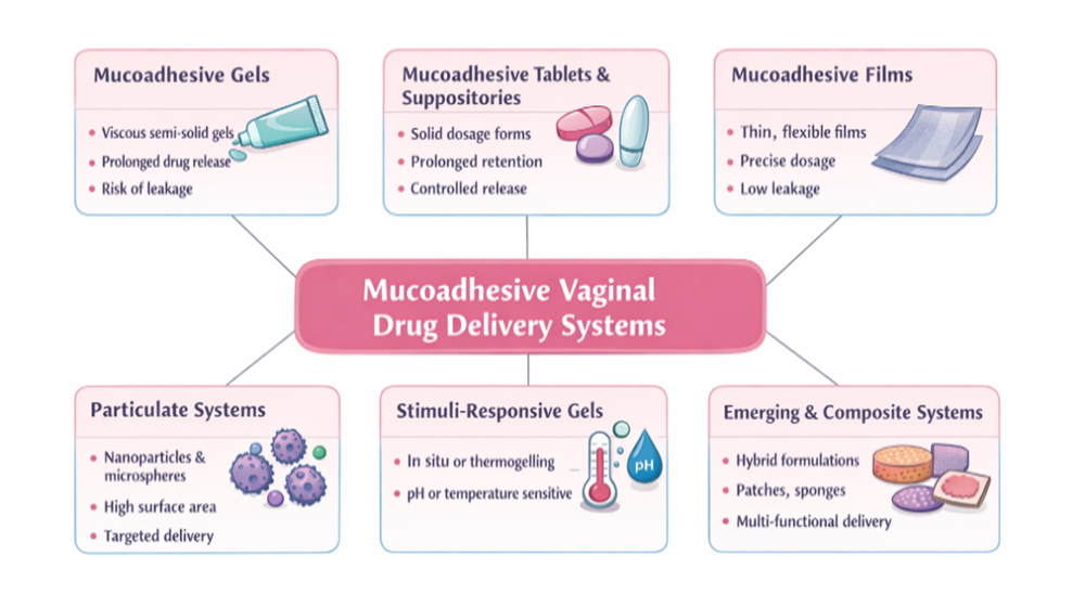
Fig No.2: Types of Mucoadhesive Vaginal Drug Delivery Systems


6.5 Particulate Systems: Microspheres and Nanoparticles

Particulate systems like mucoadhesive microspheres and nanoparticles have been thoroughly studied to further improve drug stability, mucosal contact, and controlled release. These solutions are superior to bulk gels or tablets in terms of surface area augmentation, targeted delivery, and diffusion control.[75] The purpose of microspheres, which typically range in size from 1 to 1000 µm, is to combine medication within a polymer matrix that interacts with mucin. They might be designed to swell or break down and offer sustained release.[76]Nanoparticles, which are usually smaller than 1 µm, provide benefits for surface functionalization and mucosal penetration. To enhance adherence and regulate release, mucoadhesive nanoparticles employ polymers such chitosan, PLGA, PEGylated surfaces, or thiolated derivatives. Reviews show that polymeric nanoparticles, which are designed for both local and systemic activity, are promising vaginal drug delivery vehicles for antimicrobials, antivirals, and anticancer medicines.[77]

6.6 Stimuli-Responsive and In Situ Gelling Systems

Stimuli-responsive or in situ gelling formulations, which change from liquid to gel when exposed to physiological circumstances (temperature, pH, or ions), are a subgroup of mucoadhesive delivery methods.[78] These solutions combine ease of application with improved retention because they often have low viscosity when administered and gel when they come into touch with vaginal secretions.[79]For example, in situ gels based on chitosan and poloxamer blends are examples of thermogelling copolymers that have been investigated for vaginal usage. These formulations create gels that stick to mucosa and prolong medication release by taking advantage of body temperature. Benefits include improved patient comfort and little leaking during implantation.[80]

6.7 Emerging and Composite Delivery Systems

To further improve medication retention and controlled distribution, ongoing research has investigated composite devices that combine various dosage forms, such as films embedded with nanoparticles or tablets with expandable mucoadhesive layers. Although they are less prevalent, advanced techniques include sponges, mucoadhesive patches, and cervix-targeted carriers.[81]

7. Evaluation Parameters for Vaginal Mucoadhesive Systems

In order to guarantee that the system will offer extended residence time, sufficient drug release, and therapeutic efficacy in the intricate vaginal environment, the study of mucoadhesive vaginal drug delivery systems is an essential stage in formulation development. In contrast to traditional dosage forms, mucoadhesive systems need to be evaluated for their adhesion behavior, mechanical integrity, and interaction with mucosal tissue in addition to drug release. To fully characterize these characteristics, several in vitro, ex vivo, and in vitro/permeation models have been documented in the literature.[82]

7.1 Mucoadhesion Testing

Effective adhesion to the vaginal mucosa strongly affects residence time and drug absorption, making it one of the most crucial evaluation metrics. Typical methods include of:
Tensile strength and the force needed to separate the formulation from mucosal tissue are measured using texture analyzers. Typical outputs include detachment force (Fdt) and work of adhesion (Wad).[83] Polymer–mucin contact is evaluated by rheological and indentation studies, which provide information about possible mucoadhesive behavior. Residence time tests (wash-off or immersion models) measure the length of adhesion by subjecting formulations to simulated vaginal fluid in order to replicate the vaginal environment.[84]

7.2 Swelling and Hydration Studies

Mucoadhesive performance is influenced by swelling behavior in two ways: sufficient swelling promotes polymer chain entanglement with mucin, whereas excessive swelling might reduce adherence by diluting functional groups. These investigations measure the amount of water absorbed over time and are frequently associated with mucoadhesive strength.[85]

7.3 In Vitro Drug Release

For mucoadhesive systems to be therapeutically effective, controlled and prolonged drug release is necessary. Release investigations are carried out utilizing Franz diffusion cells or dissolving apparatus under simulated vaginal circumstances (pH ~4.2–4.5) to provide release kinetics, which are then frequently fitted to mathematical models to comprehend release mechanisms.[86]


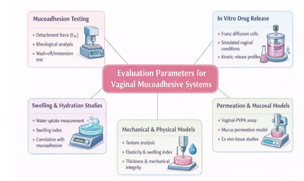
Fig No. 3: Evaluation Parameters for Vaginal Mucoadhesive Systems


7.4 Permeation and Mucosal Models

Drug penetration in the presence of mucus and simulated vaginal fluid is tested using sophisticated in vitro permeation models such as the vaginal-PVPA (Phospholipid Vesicle-based Permeation Assay), which replicates the mucosal barrier. These models offer vital information about how medicine penetrates the mucosa and how formulations interact with mucus.[87]

7.5 Mechanical and Physical Characterization

To make sure the formulation can tolerate handling and self-cleaning forces in vivo, parameters such textural strength, elasticity, swelling index, thickness, and mechanical integrity are assessed.[88]

8. Therapeutic Applications of Vaginal Mucoadhesive Drug Delivery Systems

Because they can increase drug localization, extend residence duration at the site of administration, and improve therapeutic outcomes for both local and systemic applications, vaginal mucoadhesive drug delivery systems are being investigated more and more. By permitting sustained and controlled medication release and overcoming fast clearance caused by vaginal self-cleaning processes, these methods provide notable advantages over traditional formulations.[89]Vaginal illnesses, such as bacterial vaginosis, vulvovaginal candidiasis, and sexually transmitted infections (STIs), are among the main therapeutic uses of mucoadhesive vaginal delivery systems. Improved mucoadhesion improves localized medication action and reduces systemic adverse effects by increasing the retention of antibacterial drugs at the mucosal surface.[90] Mucoadhesive systems based on hydrogel or nanoparticles have been studied for the delivery of antivirals, antifungals, and antibiotics to successfully prevent the growth of pathogens and the formation of biofilms. Additionally, these systems improve drug penetration to the infection site by overcoming diffusion restrictions within mucus.[91]Contraceptive delivery also uses mucoadhesive vaginal systems. Spermicidal chemicals or hormone formulations can be added to polymeric gels, films, or rings for extended release and localized action, which can increase the effectiveness of contraception while lowering dosage frequency and irritation. These systems can serve two purposes at once, such as combining STI prevention with contraception.[92] Vaginal mucoadhesive systems have potential for systemic medication administration of peptides, hormones, and vaccinations in addition to local therapy. Certain medications can avoid first-pass hepatic metabolism due to the vaginal mucosa's strong vascularization, providing an alternate pathway for substances with low oral bioavailability.[93] Mucoadhesive systems have been tested to improve absorption and lower dosage frequency for hormonal therapy (such as oestrogen administration for menopausal symptoms).[94]All things considered, these applications show that mucoadhesive vaginal drug delivery methods not only increase local therapeutic efficacy but also broaden the scope of innovative systemic medicines while improving patient comfort and compliance. It is anticipated that when polymers and formulation techniques continue to be improved, their clinical impact will increase.[95]

9. Recent Advances and Future Perspectives in Vaginal Mucoadhesive Drug Delivery System

Recent studies on vaginal mucoadhesive drug delivery systems (MDDS) have concentrated on using new materials, sophisticated formulations, and multifunctional carriers to improve therapeutic performance and overcome physiological obstacles of the vaginal environment. Significant advancements have been made in customizing mucoadhesive systems for more efficient local and systemic drug delivery, despite the persistent difficulties presented by fast mucus turnover, pH fluctuations, and enzymatic activity.[96]The incorporation of stimuli-responsive and in situ gelling polymers, which alter chemically or physically when they come into touch with vaginal secretions, is one important area of development.[97] In order to improve residence time and drug release management, these systems are made as low-viscosity liquids at the moment of administration but change into gels in response to temperature, pH, or ionic concentrations. This method is a potential option for long-term treatments and continuous drug delivery since it not only improves mucoadhesion but also lowers leakage and improves patient comfort.[98]The application of nano and micro based carriers in mucoadhesive matrices is another significant area of development. Polymeric micelles, liposomes, solid lipid nanoparticles, and nanoparticles (NPs) have all been studied to enhance medication stability, mucus barrier penetration, and targeted action.[99] These carriers' interaction with mucin fibres is increased by surface modification with mucoadhesive polymers or mucus-penetrating ligands, which prolongs retention and improves therapeutic results. These nanosystems have shown promise in delivering hormonal, antiviral, antifungal, and antibacterial drugs more efficiently than traditional versions.[100]The design space for vaginal mucoadhesive systems has also increased due to developments in functionalized polymers and excipients. To optimize medication release and mucosal contact, new biocompatible and biodegradable excipients with multifunctional qualities—such as pH responsiveness, increased swelling, and improved bioadhesive strength are being used.[101] A greater focus on precision formulation engineering, which allows for customized treatment for certain vaginal conditions, is shown in this trend.[102]


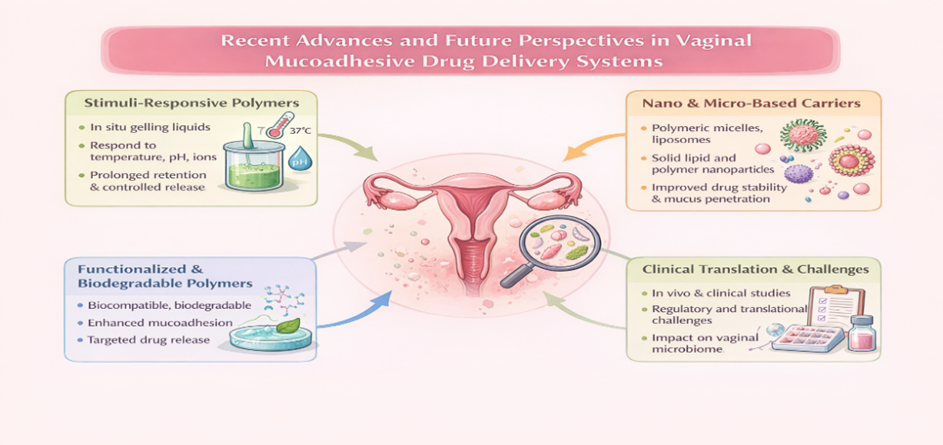
Fig No.4:  Recent Advances and Future Perspectives in Vaginal Mucoadhesive Drug Delivery Systems


Clinical translation and regulatory adoption of these cutting-edge systems are the main areas of focus for the future. Extensive in vivo and clinical evaluations are necessary to establish safety, efficacy, and patient compliance, even though in vitro and ex vivo investigations provide encouraging results.[103] For practical use, more investigation into mucus-penetrating versus mucoadhesive balance, long-term tolerability, and the effect of formulation on the native vaginal flora is essential.[104]The future of vaginal MDDS is being shaped by current developments in stimuli-responsive gels, nano carriers, functional polymers, and formulation excipients.[105] These technologies show great promise for improved local therapy and non-invasive systemic delivery while resolving present vaginal drug delivery constraints with further interdisciplinary research and thorough clinical validation.[106]­­­­­

CONCLUSION

A very promising and adaptable method for enhancing both local and systemic drug therapy via the vaginal channel is the use of vaginal mucoadhesive drug delivery systems (MDDS).[107] The vaginal mucosa is a desirable site for drug delivery due to its distinct anatomical and physiological characteristics, which include extensive vascularization, a wide surface area, and avoidance of first-pass hepatic metabolism. However, quick clearance, poor retention, and unpredictable therapeutic results are common limitations of traditional vaginal dose formulations.[108] By extending residence time, improving mucosal contact, and facilitating sustained and regulated medication release, mucoadhesive systems have been created to address these issues.[109]The design and functionality of vaginal mucoadhesive drug delivery devices have significantly improved because to recent advancements in polymer science.[110] A variety of delivery platforms, such as gels, tablets, films, microspheres, nanoparticles, and in situ gelling systems, have been made possible by the availability of natural, semi-synthetic, and synthetic mucoadhesive polymers.[111] These formulations have shown increased effectiveness in treating hormonal therapy, vaginal infections, contraception, and new uses like peptide and vaccine administration. Additionally, new developments in stimuli-responsive polymers and nanotechnology-based carriers have improved patient acceptance, medication stability, and bioavailability.[112]The successful clinical translation of vaginal mucoadhesive systems necessitates careful evaluation of safety, biocompatibility, impact on vaginal microbiota, and long-term tolerability, despite significant progress. Robust in vivo and clinical trials, mucus–polymer interaction optimization, and regulatory standardization should be the main topics of future study.[113]

REFERENCES

  1. Acarturk F. Mucoadhesive vaginal drug delivery systems. Recent patents on drug delivery & formulation. 2009 Nov 1;3(3):193-205.
  2. Sanchez Armengol E, Veider F, Millotti G, Kali G, Bernkop-Schnürch A, Laffleur F. Exploring the potential of vaginal drug delivery: innovations, efficacy, and therapeutic prospects. Journal of Pharmacy and Pharmacology. 2025 Jun 26;77:1149–1165.
  3. Caramella CM, Rossi S, Ferrari F, Bonferoni MC, Sandri G. Mucoadhesive and thermogelling systems for vaginal drug delivery. Advanced drug delivery reviews. 2015 Sep 15;92:39-52.
  4. Srikrishna S, Cardozo L. The vagina as a route for drug delivery: a review. International urogynecology journal. 2013 Apr;24(4):537-43.
  5. Osma?ek T, Froelich A, Jadach B, Tatarek A, Gadzi?ski P, Falana A, Grali?ska K, Ekert M, Puri V, Wroty?ska-Barczy?ska J, Michniak-Kohn B. Recent advances in polymer-based vaginal drug delivery systems. Pharmaceutics. 2021 Jun 15;13(6):1-49.
  6. Zierden HC, Josyula A, Shapiro RL, Hsueh HT, Hanes J, Ensign LM. Avoiding a sticky situation: bypassing the mucus barrier for improved local drug delivery. Trends in molecular medicine. 2021 May 1;27(5):1-26.
  7. Li WZ, Zhao N, Zhou YQ, Yang LB, Xiao-Ning W, Bao-Hua H, Peng K, Chun-Feng Z. Post-expansile hydrogel foam aerosol of PG-liposomes: a novel delivery system for vaginal drug delivery applications. European journal of pharmaceutical sciences. 2012 Aug 30;47(1):1-8.
  8. Johal HS, Garg T, Rath G, Goyal AK. Advanced topical drug delivery system for the management of vaginal candidiasis. Drug delivery. 2016 Feb 12;23(2):1-14.
  9. Cook MT, Brown MB. Polymeric gels for intravaginal drug delivery. Journal of Controlled Release. 2018 Jan 28;270:1-33.
  10. Dos Santos AM, Carvalho SG, Araujo VH, Carvalho GC, Gremião MP, Chorilli M. Recent advances in hydrogels as strategy for drug delivery intended to vaginal infections. International Journal of Pharmaceutics. 2020 Nov 30;590:1-12.
  11. Gerton ML, Mann BK. Mucoadhesive hyaluronic acid?based films for vaginal delivery of metronidazole. Journal of Biomedical Materials Research Part B: Applied Biomaterials. 2021 Nov;109(11):1-7.
  12. Voltan AR, Quindos G, Alarcón KP, Fusco-Almeida AM, Mendes-Giannini MJ, Chorilli M. Fungal diseases: could nanostructured drug delivery systems be a novel paradigm for therapy?. International Journal of Nanomedicine. 2016 Aug 8:3715-30.
  13. Andersen T, Bleher S, Flaten GE, Tho I, Mattsson S, Škalko-Basnet N. Chitosan in mucoadhesive drug delivery: Focus on local vaginal therapy. Marine drugs. 2015 Jan 7;13(1):1-15.
  14. Hassan AS, Soliman GM, Ali MF, El-Mahdy MM, El-Gindy GE. Mucoadhesive tablets for the vaginal delivery of progesterone: in vitro evaluation and pharmacokinetics/pharmacodynamics in female rabbits. Drug development and industrial pharmacy. 2018 Feb 1;44(2):1-33.
  15. Mirza MA, Panda AK, Asif S, Verma D, Talegaonkar S, Manzoor N, Khan A, Ahmed FJ, Dudeja M, Iqbal Z. A vaginal drug delivery model. Drug delivery. 2016 Oct 12;23(8):1-13.
  16. Araujo VH, de Souza MP, Carvalho GC, Duarte JL, Chorilli M. Chitosan-based systems aimed at local application for vaginal infections. Carbohydrate Polymers. 2021 Jun 1;261:1-11.
  17. Karimunnisa S, Atmaram P. Mucoadhesive nanoliposomal formulation for vaginal delivery of an antifungal. Drug development and industrial pharmacy. 2013 Sep 1;39(9):1-10.
  18. Shatabayeva EO, Kaldybekov DB, Kenessova ZA, Tuleyeva RN, Kudaibergenov SE, Khutoryanskiy VV. Development of mucoadhesive vaginal films for metronidazole delivery Using methacryloylated, crotonoylated, and itaconoylated gelatin blends with poly (vinyl alcohol). AAPS PharmSciTech. 2025 Feb 12;26(2):1-11.
  19. K Maurya S, Pathak K, Bali V. Therapeutic potential of mucoadhesive drug delivery systems-An updated patent review. Recent Patents on Drug Delivery & Formulation. 2010 Nov 1;4(3):1-10.
  20. Furst T, Piette M, Lechanteur A, Evrard B, Piel G. Mucoadhesive cellulosic derivative sponges as drug delivery system for vaginal application. European Journal of Pharmaceutics and Biopharmaceutics. 2015 Sep 1;95:1-8.
  21. Asane GS, Nirmal SA, Rasal KB, Naik AA, Mahadik MS, Rao YM. Polymers for mucoadhesive drug delivery system: a current status. Drug development and industrial pharmacy. 2008 Jan 1;34(11):1-21.
  22. de Carvalho AC, Paiva NF, Demonari IK, Duarte MP, do Couto RO, de Freitas O, Vicentini FT. The potential of films as transmucosal drug delivery systems. Pharmaceutics. 2023 Nov 4;15(11):1-28.
  23. Vigani B, Rossi S, Sandri G, Bonferoni MC, Caramella CM, Ferrari F. Recent advances in the development of in situ gelling drug delivery systems for non-parenteral administration routes. Pharmaceutics. 2020 Sep 10;12(9):1-29.
  24. Li C, Huang Z, Liu Z, Ci L, Liu Z, Liu Y, Yan X, Lu W. Sulfonate-modified phenylboronic acid-rich nanoparticles as a novel mucoadhesive drug delivery system for vaginal administration of protein therapeutics: improved stability, mucin-dependent release and effective intravaginal placement. International Journal of Nanomedicine. 2016 Nov 10:5917-30.
  25. Cazorla-Luna R, Martín-Illana A, Notario-Pérez F, Ruiz-Caro R, Veiga MD. Naturally occurring polyelectrolytes and their use for the development of complex-based mucoadhesive drug delivery systems: An overview. Polymers. 2021 Jul 8;13(14):1-27.
  26. Woodley J. Bioadhesion: new possibilities for drug administration?. Clinical pharmacokinetics. 2001 Feb;40(2):1-8.
  27. Wong TW, Dhanawat M, Rathbone MJ. Vaginal drug delivery: strategies and concerns in polymeric nanoparticle development. Expert Opinion on Drug Delivery. 2014 Sep 1;11(9):1-16.
  28. Ceschel GC, Maffei P, Borgia SL, Ronchi C, Rossi S. Development of a mucoadhesive dosage form for vaginal administration. Drug development and industrial pharmacy. 2001 Jan 1;27(6):1-8.
  29. Martín-Villena MJ, Fernández-Campos F, Calpena-Campmany AC, Bozal-de Febrer N, Ruiz-Martínez MA, Clares-Naveros B. Novel microparticulate systems for the vaginal delivery of nystatin: development and characterization. Carbohydrate polymers. 2013 Apr 15;94(1):1-11.
  30. Nowak J, Laffleur F, Bernkop-Schnürch A. Preactivated hyaluronic acid: A potential mucoadhesive polymer for vaginal delivery. International journal of pharmaceutics. 2015 Jan 15;478(1):1-7.
  31. Tentor F, Siccardi G, Sacco P, Demarchi D, Marsich E, Almdal K, Bose Goswami S, Boisen A. Long lasting mucoadhesive membrane based on alginate and chitosan for intravaginal drug delivery. Journal of Materials Science: Materials in Medicine. 2020 Mar;31(3):1-12.
  32. Ijaz M, Griessinger JA, Mahmood A, Laffleur F, Bernkop-Schnürch A. Thiolated cyclodextrin: development of a mucoadhesive vaginal delivery system for acyclovir. Journal of Pharmaceutical Sciences. 2016 May 1;105(5):1-7.
  33. Gavini E, Sanna V, Juliano C, Bonferoni MC, Giunchedi P. Mucoadhesive vaginal tablets as veterinary delivery system for the controlled release of an antimicrobial drug, acriflavine. AAPS PharmSciTech. 2002 Sep;3(3):1-7.
  34. Chopra S, Motwani SK, Iqbal Z, Talegaonkar S, Ahmad FJ, Khar RK. Optimisation of polyherbal gels for vaginal drug delivery by Box-Behnken statistical design. European journal of pharmaceutics and biopharmaceutics. 2007 Aug 1;67(1):1-12.
  35. Pacheco-Quito EM, Ruiz-Caro R, Rubio J, Tamayo A, Veiga MD. Carrageenan-based acyclovir mucoadhesive vaginal tablets for prevention of genital herpes. Marine drugs. 2020 May 11;18(5):1-19.
  36. Perioli L, Ambrogi V, Pagano C, Massetti E, Rossi C. New solid mucoadhesive systems for benzydamine vaginal administration. Colloids and Surfaces B: Biointerfaces. 2011 Jun 1;84(2):1-8.
  37. Albertini B, Passerini N, Di Sabatino M, Vitali B, Brigidi P, Rodriguez L. Polymer–lipid based mucoadhesive microspheres prepared by spray-congealing for the vaginal delivery of econazole nitrate. European journal of pharmaceutical sciences. 2009 Mar 2;36(4-5):1-11.
  38. Taurin S, Almomen AA, Pollak T, Kim SJ, Maxwell J, Peterson CM, Owen SC, Janat-Amsbury MM. Thermosensitive hydrogels a versatile concept adapted to vaginal drug delivery. Journal of drug targeting. 2018 Aug 9;26(7):1-51.
  39. Berginc K, Suljakovi? S, Škalko-Basnet N, Kristl A. Mucoadhesive liposomes as new formulation for vaginal delivery of curcumin. European journal of pharmaceutics and biopharmaceutics. 2014 May 1;87(1):1-7.
  40. Shapiro RL, DeLong K, Zulfiqar F, Carter D, Better M, Ensign LM. In vitro and ex vivo models for evaluating vaginal drug delivery systems. Advanced drug delivery reviews. 2022 Dec 1;191:1-39.
  41. Owen KB, Smith J, Lubans DR, Ng JY, Lonsdale C. Self-determined motivation and physical activity in children and adolescents: A systematic review and meta-analysis. Preventive medicine. 2014 Oct 1;67:1-10.
  42. Perioli L, Ambrogi V, Pagano C, Scuota S, Rossi C. FG90 chitosan as a new polymer for metronidazole mucoadhesive tablets for vaginal administration. International Journal of Pharmaceutics. 2009 Jul 30;377(1-2):1-8.
  43. Abidin IZ, Murphy EJ, Fehrenbach GW, Rezoagli E, Gately N, Major I. A systematic review of mucoadhesive vaginal tablet testing. Drug Target Insights. 2023 Jan 16;17:1-26.
  44. Ensign LM, Cone R, Hanes J. Oral drug delivery with polymeric nanoparticles: the gastrointestinal mucus barriers. Advanced drug delivery reviews. 2012 May 1;64(6):1-33.
  45. Kenechukwu FC, Attama AA, Ibezim EC, Nnamani PO, Umeyor CE, Uronnachi EM, Gugu TH, Momoh MA, Ofokansi KC, Akpa PA. Surface-modified mucoadhesive microgels as a controlled release system for miconazole nitrate to improve localized treatment of vulvovaginal candidiasis. European journal of pharmaceutical sciences. 2018 Jan 1;111:1-76.
  46. Bonfim AP, Sakita KM, Faria DR, Arita GS, Vendramini FA, Capoci IR, Braga AG, Dos Santos RS, Bruschi ML, Becker TC, de Oliveira Junior AG. Preclinical approaches in vulvovaginal candidiasis treatment with mucoadhesive thermoresponsive systems containing propolis. PLoS One. 2020 Dec 11;15(12):1-20.
  47. Jalalvandi E, Shavandi A. In situ-forming and pH-responsive hydrogel based on chitosan for vaginal delivery of therapeutic agents. Journal of Materials Science: Materials in Medicine. 2018 Oct;29(10):1-11.
  48. Notario-Pérez F, Cazorla-Luna R, Martín-Illana A, Galante J, Ruiz-Caro R, das Neves J, Veiga MD. Design, fabrication and characterisation of drug-loaded vaginal films: State-of-the-art. Journal of Controlled Release. 2020 Nov 10;327:1-23.
  49. Tu?cu-Demiröz F. Vaginal delivery of benzydamine hydrochloride through liposomes dispersed in mucoadhesive gels. Chemical and Pharmaceutical Bulletin. 2017 Jul 1;65(7):1-8.
  50. Martín-Bartolomé L, Ruiz-Caro R, Veiga MD, Notario-Pérez F. Evaluation of polymer combinations in vaginal mucoadhesive tablets for the extended release of acyclovir. European Journal of Pharmaceutical Sciences. 2024 Dec 1;203:1-10.
  51. das Neves J, Amiji M, Sarmento B. Mucoadhesive nanosystems for vaginal microbicide development: friend or foe?. Wiley Interdisciplinary Reviews: Nanomedicine and Nanobiotechnology. 2011 Jul;3(4):1-11.
  52. Li J, Wang K, Lu Z, Huo Y, Juang V, Rodriguez A, Schwendeman A. Characterization of Crinone®: progesterone vaginal gel. International Journal of Pharmaceutics. 2025 Sep 15:1-9.
  53. Karavasili C, Eleftheriadis GK, Gioumouxouzis C, Andriotis EG, Fatouros DG. Mucosal drug delivery and 3D printing technologies: A focus on special patient populations. Advanced Drug Delivery Reviews. 2021 Sep 1;176:1-18.
  54. Sofi HS, Abdal-Hay A, Ivanovski S, Zhang YS, Sheikh FA. Electrospun nanofibers for the delivery of active drugs through nasal, oral and vaginal mucosa: Current status and future perspectives. Materials Science and Engineering: C. 2020 Jun 1;111:1-20.
  55. Mennini N, Casella G, Cirri M, Maestrelli F, Mura P. Development of cyclodextrin hydrogels for vaginal delivery of dehydroepiandrosterone. Journal of Pharmacy and Pharmacology. 2016 Jun;68(6):1-10.
  56. Vani? Ž, Škalko-Basnet N. Nanopharmaceuticals for improved topical vaginal therapy: can they deliver?. European Journal of Pharmaceutical Sciences. 2013 Sep 27;50(1):1-13.
  57. Andrews GP, Donnelly L, Jones DS, Curran RM, Morrow RJ, Woolfson AD, Malcolm RK. Characterization of the rheological, mucoadhesive, and drug release properties of highly structured gel platforms for intravaginal drug delivery. Biomacromolecules. 2009 Sep 14;10(9):1-9.
  58. Sanz R, Clares B, Mallandrich M, Suner-Carbo J, Montes MJ, Calpena AC. Development of a mucoadhesive delivery system for control release of doxepin with application in vaginal pain relief associated with gynecological surgery. International Journal of Pharmaceutics. 2018 Jan 15;535(1-2):1-39.
  59. Giordani B, Abruzzo A, Musazzi UM, Cilurzo F, Nicoletta FP, Dalena F, Parolin C, Vitali B, Cerchiara T, Luppi B, Bigucci F. Freeze-dried matrices based on polyanion polymers for chlorhexidine local release in the buccal and vaginal cavities. Journal of Pharmaceutical Sciences. 2019 Jul 1;108(7):1-11.
  60. Araujo VH, de Souza MP, Carvalho GC, Duarte JL, Chorilli M. Chitosan-based systems aimed at local application for vaginal infections. Carbohydrate Polymers. 2021 Jun 1;261:1-11.
  61. Friedl HE, Dünnhaupt S, Waldner C, Bernkop-Schnürch A. Preactivated thiomers for vaginal drug delivery vehicles. Biomaterials. 2013 Oct 1;34(32):1-8.
  62. Frank LA, Sandri G, D’Autilia F, Contri RV, Bonferoni MC, Caramella C, Frank AG, Pohlmann AR, Guterres SS. Chitosan gel containing polymeric nanocapsules: a new formulation for vaginal drug delivery. International Journal of Nanomedicine. 2014 Jun 28:1-11.
  63. K Garg N, Mangal S, Khambete H, K Sharma P, K Tyagi R. Mucosal delivery of vaccines: role of mucoadhesive/biodegradable polymers. Recent Patents on Drug Delivery & Formulation. 2010 Jun 1;4(2):1-15.
  64. Furst T, Dakwar GR, Zagato E, Lechanteur A, Remaut K, Evrard B, Braeckmans K, Piel G. Freeze-dried mucoadhesive polymeric system containing pegylated lipoplexes: Towards a vaginal sustained released system for siRNA. Journal of Controlled Release. 2016 Aug 28;236:1-11.
  65. Yang TT, Cheng YZ, Qin M, Wang YH, Yu HL, Wang AL, Zhang WF. Thermosensitive chitosan hydrogels containing polymeric microspheres for vaginal drug delivery. BioMed Research International. 2017;2017(1):1-12.
  66. Cazorla-Luna R, Notario-Pérez F, Martín-Illana A, Ruiz-Caro R, Rubio J, Tamayo A, Veiga MD. Bigels based on polyelectrolyte complexes as vaginal drug delivery systems. International Journal of Pharmaceutics. 2025 Jan 25;669:1-15.
  67. Abruzzo A, Giordani B, Parolin C, Vitali B, Protti M, Mercolini L, Cappelletti M, Fedi S, Bigucci F, Cerchiara T, Luppi B. Novel mixed vesicles containing lactobacilli biosurfactant for vaginal delivery of an anti-Candida agent. European Journal of Pharmaceutical Sciences. 2018 Jan 15;112:1-28.
  68. Rossi S, Marciello M, Ferrari F, Puccio A, Bonferoni C, Sandri G, Caramella C. Development of sponge-like dressings for mucosal/transmucosal drug delivery into vaginal cavity. Pharmaceutical Development and Technology. 2012 Apr 1;17(2):1-8.
  69. Moreno MA, Gómez-Mascaraque LG, Arias M, Zampini IC, Sayago JE, Ramos LL, Schmeda-Hirschmann G, López-Rubio A, Isla MI. Electrosprayed chitosan microcapsules as delivery vehicles for vaginal phytoformulations. Carbohydrate Polymers. 2018 Dec 1;201:1-37.
  70. Woolfson AD, Umrethia ML, Kett VL, Malcolm RK. Freeze-dried, mucoadhesive system for vaginal delivery of the HIV microbicide, dapivirine: optimisation by an artificial neural network. International journal of pharmaceutics. 2010 Mar 30;388(1-2):1-8.
  71. Mura P, Maestrelli F, Cirri M, Mennini N. Multiple roles of chitosan in mucosal drug delivery: an updated review. Marine Drugs. 2022 May 20;20(5):1-42.
  72. Thapa R, Pandey P, Parat MO, Gurung S, Parekh HS. Phase transforming in situ gels for sustained and controlled transmucosal drug delivery via the intravaginal route. International Journal of Pharmaceutics. 2024 Apr 25;655:1-15.
  73. Hombach J, Palmberger TF, Bernkop?Schnürch A. Development and in vitro evaluation of a mucoadhesive vaginal delivery system for nystatin. Journal of pharmaceutical sciences. 2009 Feb 1;98(2):1-10.
  74. Campaña-Seoane M, Pérez-Gago A, Vázquez G, Conde N, González P, Martinez A, Martínez X, Varela LG, Herranz M, Aguiar P, Fernández-Ferreiro A. Vaginal residence and pharmacokinetic preclinical study of topical vaginal mucoadhesive W/S emulsions containing ciprofloxacin. International Journal of Pharmaceutics. 2019 Jan 10;554:1-24.
  75. Aguilar-Rosas I, Alcalá-Alcalá S, Llera-Rojas V, Ganem-Rondero A. Preparation and characterization of mucoadhesive nanoparticles of poly (methyl vinyl ether-co-maleic anhydride) containing glycyrrhizic acid intended for vaginal administration. Drug Development and Industrial Pharmacy. 2015 Oct 3;41(10):1-8.
  76. Kumar A, Naik PK, Pradhan D, Ghosh G, Rath G. Mucoadhesive formulations: Innovations, merits, drawbacks, and future outlook. Pharmaceutical Development and Technology. 2020 Aug 8;25(7):1-44.
  77. Han IK, Kim YB, Kang HS, Sul D, Jung WW, Cho HJ, Oh YK. Thermosensitive and mucoadhesive delivery systems of mucosal vaccines. Methods. 2006 Feb 1;38(2):1-6.
  78. Ensign LM, Hoen TE, Maisel K, Cone RA, Hanes JS. Enhanced vaginal drug delivery through the use of hypotonic formulations that induce fluid uptake. Biomaterials. 2013 Sep 1;34(28):1-18.
  79. Endo N, Rahayu LP, Yamamura T, Tanaka H, Tanaka T. Intravaginal administration of progesterone using a new technique for sustained drug release in goats. Journal of Reproduction and Development. 2020;66(5):1-4.
  80. Hernández-González ME, Rodríguez-González CA, Valencia-Gómez LE, Hernández-Paz JF, Jiménez-Vega F, Salcedo M, Olivas-Armendáriz I. Characterization of HPMC and PEG 400 Mucoadhesive Film Loaded with Retinyl Palmitate and Ketorolac for Intravaginal Administration. International Journal of Molecular Sciences. 2024 Nov 26;25(23):1-16.
  81. Tu?cu-Demiröz F, Acartürk F, Özkul A. Preparation and characterization of bioadhesive controlled-release gels of cidofovir for vaginal delivery. Journal of Biomaterials Science, Polymer Edition. 2015 Nov 22;26(17):1-39.
  82. Maestrelli F, Jug M, Cirri M, Kosalec I, Mura P. Characterization and microbiological evaluation of chitosan-alginate microspheres for cefixime vaginal administration. Carbohydrate polymers. 2018 Jul 15;192:1-30.
  83. Giordani B, Melgoza LM, Parolin C, Foschi C, Marangoni A, Abruzzo A, Dalena F, Cerchiara T, Bigucci F, Luppi B, Vitali B. Vaginal Bifidobacterium breve for preventing urogenital infections: Development of delayed release mucoadhesive oral tablets. International Journal of Pharmaceutics. 2018 Oct 25;550(1-2):1-31.
  84. Ré AC, Martins JF, Cunha?Filho M, Gelfuso GM, Aires CP, Gratieri T. New perspectives on the topical management of recurrent candidiasis. Drug Delivery and Translational Research. 2021 Aug;11(4):1-18.
  85. Krogstad EA, Ramanathan R, Nhan C, Kraft JC, Blakney AK, Cao S, Ho RJ, Woodrow KA. Nanoparticle-releasing nanofiber composites for enhanced in vivo vaginal retention. Biomaterials. 2017 Nov 1;144:1-34.
  86. Salmazi R, Calixto G, Bernegossi J, Ramos MA, Bauab TM, Chorilli M. A curcumin-loaded liquid crystal precursor mucoadhesive system for the treatment of vaginal candidiasis. International Journal of Nanomedicine. 2015 Jul 30:1-10.
  87. Fida S, Jalil A, Habib R, Akhlaq M, Mahmood A, Minhas MU, Khan KU, Nawaz A. Development of mucus-penetrating iodine loaded self-emulsifying system for local vaginal delivery. PloS one. 2022 Mar 31;17(3):1-17.
  88. de Araújo PR, Calixto GM, da Silva IC, de Paula Zago LH, Oshiro Junior JA, Pavan FR, Ribeiro AO, Fontana CR, Chorilli M. Mucoadhesive in situ gelling liquid crystalline precursor system to improve the vaginal administration of drugs. AAPS PharmSciTech. 2019 Jun 18;20(6):1-12.
  89. Brako F, Raimi-Abraham BT, Mahalingam S, Craig DQ, Edirisinghe M. The development of progesterone-loaded nanofibers using pressurized gyration: A novel approach to vaginal delivery for the prevention of pre-term birth. International journal of pharmaceutics. 2018 Apr 5;540(1-2):1-25.
  90. Osmari BF, Giuliani LM, Reolon JB, Rigo GV, Tasca T, Cruz L. Gellan gum-based hydrogel containing nanocapsules for vaginal indole-3-carbinol delivery in trichomoniasis treatment. European Journal of Pharmaceutical Sciences. 2020 Aug 1;151:1-40.
  91. Notario-Pérez F, Martín-Illana A, Cazorla-Luna R, Ruiz-Caro R, Peña J, Veiga MD. Improvement of Tenofovir vaginal release from hydrophilic matrices through drug granulation with hydrophobic polymers. European Journal of Pharmaceutical Sciences. 2018 May 30;117:1-12.
  92. Pereira RR, Godoy JS, Svidzinski TI, Bruschi ML. Preparation and characterization of mucoadhesive thermoresponsive systems containing propolis for the treatment of vulvovaginal candidiasis. Journal of Pharmaceutical Sciences. 2013 Apr 1;102(4):1-13.
  93. Baloglu E, Seny?g?t ZA, Karavana SY, Vetter A, Met?n DY, Polat SH, Guneri T, Bernkop-Schnurch A. In vitro evaluation of mucoadhesive vaginal tablets of antifungal drugs prepared with thiolated polymer and development of a new dissolution technique for vaginal formulations. Chemical and Pharmaceutical Bulletin. 2011 Aug 1;59(8):1-7.
  94. Yoo JW, Acharya G, Lee CH. In vivo evaluation of vaginal films for mucosal delivery of nitric oxide. Biomaterials. 2009 Aug 1;30(23-24):1-8.
  95. Zidan AS, Habib MJ. Maximized mucoadhesion and skin permeation of anti-AIDS-loaded niosomal gels. Journal of Pharmaceutical Sciences. 2014 Mar 1;103(3):1-13.
  96. Pereira MN, Reis TA, Matos BN, Cunha-Filho M, Gratieri T, Gelfuso GM. Novel ex vivo protocol using porcine vagina to assess drug permeation from mucoadhesive and colloidal pharmaceutical systems. Colloids and Surfaces B: Biointerfaces. 2017 Oct 1;158:1-27.
  97. Madhavlal PG, Manordas PM. Design and in vitro evaluation of a novel vaginal drug delivery system based on gelucire. Current Drug Delivery. 2009 Apr 1;6(2):1-7.
  98. Martínez-Pérez B, Quintanar-Guerrero D, Tapia-Tapia M, Cisneros-Tamayo R, Zambrano-Zaragoza ML, Alcalá-Alcalá S, Mendoza-Muñoz N, Piñón-Segundo E. Controlled-release biodegradable nanoparticles: From preparation to vaginal applications. European Journal of Pharmaceutical Sciences. 2018 Mar 30;115:1-48.
  99. Almomen A, Cho S, Yang CH, Li Z, Jarboe EA, Peterson CM, Huh KM, Janát-Amsbury MM. Thermosensitive progesterone hydrogel: A safe and effective new formulation for vaginal application. Pharmaceutical research. 2015 Jan 22;32(7):1-14.
  100. Gupta NV, Natasha S, Getyala A, Bhat RS. Bioadhesive vaginal tablets containing spray dried microspheres loaded with clotrimazole drug for treatment of vaginal Candidiasiss. Acta pharmaceutica. 2013 Sep 30;63(3):1-14.
  101. Notario-Pérez F, Cazorla-Luna R, Martín-Illana A, Ruiz-Caro R, Tamayo A, Rubio J, Veiga MD. Optimization of tenofovir release from mucoadhesive vaginal tablets by polymer combination to prevent sexual transmission of HIV. Carbohydrate polymers. 2018 Jan 1;179:1-41.
  102. Khutoryanskiy VV. Advances in mucoadhesion and mucoadhesive polymers. Macromolecular bioscience. 2011 Jun 14;11(6):1-17.
  103. Tarsitano M, Mancuso A, Cristiano MC, Paolino D, Fresta M. In situ swelling formulation of glycerol-monooleate-derived lyotropic liquid crystals proposed for local vaginal application. Molecules. 2022 Sep 23;27(19):1-17.
  104. Nematpour N, Moradipour P, Zangeneh MM, Arkan E, Abdoli M, Behbood L. The application of nanomaterial science in the formulation a novel antibiotic: Assessment of the antifungal properties of mucoadhesive clotrimazole loaded nanofiber versus vaginal films. Materials Science and Engineering: C. 2020 May 1;110:1-12.
  105. Timur SS, ?ahin A, Aytekin E, Öztürk N, Polat KH, Tezel N, Gürsoy RN, Çal?? S. Design and in vitro evaluation of tenofovir-loaded vaginal gels for the prevention of HIV infections. Pharmaceutical development and technology. 2018 Mar 16;23(3):1-23.
  106. Brannigan RP, Khutoryanskiy VV. Progress and current trends in the synthesis of novel polymers with enhanced mucoadhesive properties. Macromolecular Bioscience. 2019 Oct;19(10):1-11.
  107. Rossi S, Ferrari F, Bonferoni MC, Sandri G, Faccendini A, Puccio A, Caramella C. Comparison of poloxamer-and chitosan-based thermally sensitive gels for the treatment of vaginal mucositis. Drug Development and Industrial Pharmacy. 2014 Mar 1;40(3):1-9.
  108. Szyma?ska E, Krzy?owska M, Cal K, Mikolaszek B, Tomaszewski J, Wo?czy?ski S, Winnicka K. Potential of mucoadhesive chitosan glutamate microparticles as microbicide carriers–antiherpes activity and penetration behavior across the human vaginal epithelium. Drug Delivery. 2021 Jan 1;28(1):1-11.
  109. ALI IBRAHIM ES, Ismail S, Fetih G, Shaaban O, Hassanein K, Abdellah NH. Development and characterization of thermosensitive pluronic-based metronidazole in situ gelling formulations for vaginal application. Acta pharmaceutica. 2012 Mar 31;62(1):1-12.
  110. dos Santos Ramos MA, Calixto G, de Toledo LG, Bonifácio BV, dos Santos LC, de Almeida MT, Chorilli M, Bauab TM. Liquid crystal precursor mucoadhesive system as a strategy to improve the prophylactic action of Syngonanthus nitens (Bong.) Ruhland against infection by Candida krusei. International Journal of Nanomedicine. 2015 Dec 16:1-12.
  111. Meng J, Sturgis TF, Youan BB. Engineering tenofovir loaded chitosan nanoparticles to maximize microbicide mucoadhesion. European Journal of Pharmaceutical Sciences. 2011 Sep 18;44(1-2):1-12.
  112. Meng J, Zhang T, Agrahari V, Ezoulin MJ, Youan BB. Comparative biophysical properties of tenofovir-loaded, thiolated and nonthiolated chitosan nanoparticles intended for HIV prevention. Nanomedicine. 2014 Aug 1;9(11):1-18.
  113. Pradines B, Bories C, Vauthier C, Ponchel G, Loiseau PM, Bouchemal K. Drug-free chitosan coated poly (isobutylcyanoacrylate) nanoparticles are active against trichomonas vaginalis and non-toxic towards pig vaginal mucosa. Pharmaceutical research. 2015 Apr 1;32(4):1-8.

Reference

  1. Acarturk F. Mucoadhesive vaginal drug delivery systems. Recent patents on drug delivery & formulation. 2009 Nov 1;3(3):193-205.
  2. Sanchez Armengol E, Veider F, Millotti G, Kali G, Bernkop-Schnürch A, Laffleur F. Exploring the potential of vaginal drug delivery: innovations, efficacy, and therapeutic prospects. Journal of Pharmacy and Pharmacology. 2025 Jun 26;77:1149–1165.
  3. Caramella CM, Rossi S, Ferrari F, Bonferoni MC, Sandri G. Mucoadhesive and thermogelling systems for vaginal drug delivery. Advanced drug delivery reviews. 2015 Sep 15;92:39-52.
  4. Srikrishna S, Cardozo L. The vagina as a route for drug delivery: a review. International urogynecology journal. 2013 Apr;24(4):537-43.
  5. Osma?ek T, Froelich A, Jadach B, Tatarek A, Gadzi?ski P, Falana A, Grali?ska K, Ekert M, Puri V, Wroty?ska-Barczy?ska J, Michniak-Kohn B. Recent advances in polymer-based vaginal drug delivery systems. Pharmaceutics. 2021 Jun 15;13(6):1-49.
  6. Zierden HC, Josyula A, Shapiro RL, Hsueh HT, Hanes J, Ensign LM. Avoiding a sticky situation: bypassing the mucus barrier for improved local drug delivery. Trends in molecular medicine. 2021 May 1;27(5):1-26.
  7. Li WZ, Zhao N, Zhou YQ, Yang LB, Xiao-Ning W, Bao-Hua H, Peng K, Chun-Feng Z. Post-expansile hydrogel foam aerosol of PG-liposomes: a novel delivery system for vaginal drug delivery applications. European journal of pharmaceutical sciences. 2012 Aug 30;47(1):1-8.
  8. Johal HS, Garg T, Rath G, Goyal AK. Advanced topical drug delivery system for the management of vaginal candidiasis. Drug delivery. 2016 Feb 12;23(2):1-14.
  9. Cook MT, Brown MB. Polymeric gels for intravaginal drug delivery. Journal of Controlled Release. 2018 Jan 28;270:1-33.
  10. Dos Santos AM, Carvalho SG, Araujo VH, Carvalho GC, Gremião MP, Chorilli M. Recent advances in hydrogels as strategy for drug delivery intended to vaginal infections. International Journal of Pharmaceutics. 2020 Nov 30;590:1-12.
  11. Gerton ML, Mann BK. Mucoadhesive hyaluronic acid?based films for vaginal delivery of metronidazole. Journal of Biomedical Materials Research Part B: Applied Biomaterials. 2021 Nov;109(11):1-7.
  12. Voltan AR, Quindos G, Alarcón KP, Fusco-Almeida AM, Mendes-Giannini MJ, Chorilli M. Fungal diseases: could nanostructured drug delivery systems be a novel paradigm for therapy?. International Journal of Nanomedicine. 2016 Aug 8:3715-30.
  13. Andersen T, Bleher S, Flaten GE, Tho I, Mattsson S, Škalko-Basnet N. Chitosan in mucoadhesive drug delivery: Focus on local vaginal therapy. Marine drugs. 2015 Jan 7;13(1):1-15.
  14. Hassan AS, Soliman GM, Ali MF, El-Mahdy MM, El-Gindy GE. Mucoadhesive tablets for the vaginal delivery of progesterone: in vitro evaluation and pharmacokinetics/pharmacodynamics in female rabbits. Drug development and industrial pharmacy. 2018 Feb 1;44(2):1-33.
  15. Mirza MA, Panda AK, Asif S, Verma D, Talegaonkar S, Manzoor N, Khan A, Ahmed FJ, Dudeja M, Iqbal Z. A vaginal drug delivery model. Drug delivery. 2016 Oct 12;23(8):1-13.
  16. Araujo VH, de Souza MP, Carvalho GC, Duarte JL, Chorilli M. Chitosan-based systems aimed at local application for vaginal infections. Carbohydrate Polymers. 2021 Jun 1;261:1-11.
  17. Karimunnisa S, Atmaram P. Mucoadhesive nanoliposomal formulation for vaginal delivery of an antifungal. Drug development and industrial pharmacy. 2013 Sep 1;39(9):1-10.
  18. Shatabayeva EO, Kaldybekov DB, Kenessova ZA, Tuleyeva RN, Kudaibergenov SE, Khutoryanskiy VV. Development of mucoadhesive vaginal films for metronidazole delivery Using methacryloylated, crotonoylated, and itaconoylated gelatin blends with poly (vinyl alcohol). AAPS PharmSciTech. 2025 Feb 12;26(2):1-11.
  19. K Maurya S, Pathak K, Bali V. Therapeutic potential of mucoadhesive drug delivery systems-An updated patent review. Recent Patents on Drug Delivery & Formulation. 2010 Nov 1;4(3):1-10.
  20. Furst T, Piette M, Lechanteur A, Evrard B, Piel G. Mucoadhesive cellulosic derivative sponges as drug delivery system for vaginal application. European Journal of Pharmaceutics and Biopharmaceutics. 2015 Sep 1;95:1-8.
  21. Asane GS, Nirmal SA, Rasal KB, Naik AA, Mahadik MS, Rao YM. Polymers for mucoadhesive drug delivery system: a current status. Drug development and industrial pharmacy. 2008 Jan 1;34(11):1-21.
  22. de Carvalho AC, Paiva NF, Demonari IK, Duarte MP, do Couto RO, de Freitas O, Vicentini FT. The potential of films as transmucosal drug delivery systems. Pharmaceutics. 2023 Nov 4;15(11):1-28.
  23. Vigani B, Rossi S, Sandri G, Bonferoni MC, Caramella CM, Ferrari F. Recent advances in the development of in situ gelling drug delivery systems for non-parenteral administration routes. Pharmaceutics. 2020 Sep 10;12(9):1-29.
  24. Li C, Huang Z, Liu Z, Ci L, Liu Z, Liu Y, Yan X, Lu W. Sulfonate-modified phenylboronic acid-rich nanoparticles as a novel mucoadhesive drug delivery system for vaginal administration of protein therapeutics: improved stability, mucin-dependent release and effective intravaginal placement. International Journal of Nanomedicine. 2016 Nov 10:5917-30.
  25. Cazorla-Luna R, Martín-Illana A, Notario-Pérez F, Ruiz-Caro R, Veiga MD. Naturally occurring polyelectrolytes and their use for the development of complex-based mucoadhesive drug delivery systems: An overview. Polymers. 2021 Jul 8;13(14):1-27.
  26. Woodley J. Bioadhesion: new possibilities for drug administration?. Clinical pharmacokinetics. 2001 Feb;40(2):1-8.
  27. Wong TW, Dhanawat M, Rathbone MJ. Vaginal drug delivery: strategies and concerns in polymeric nanoparticle development. Expert Opinion on Drug Delivery. 2014 Sep 1;11(9):1-16.
  28. Ceschel GC, Maffei P, Borgia SL, Ronchi C, Rossi S. Development of a mucoadhesive dosage form for vaginal administration. Drug development and industrial pharmacy. 2001 Jan 1;27(6):1-8.
  29. Martín-Villena MJ, Fernández-Campos F, Calpena-Campmany AC, Bozal-de Febrer N, Ruiz-Martínez MA, Clares-Naveros B. Novel microparticulate systems for the vaginal delivery of nystatin: development and characterization. Carbohydrate polymers. 2013 Apr 15;94(1):1-11.
  30. Nowak J, Laffleur F, Bernkop-Schnürch A. Preactivated hyaluronic acid: A potential mucoadhesive polymer for vaginal delivery. International journal of pharmaceutics. 2015 Jan 15;478(1):1-7.
  31. Tentor F, Siccardi G, Sacco P, Demarchi D, Marsich E, Almdal K, Bose Goswami S, Boisen A. Long lasting mucoadhesive membrane based on alginate and chitosan for intravaginal drug delivery. Journal of Materials Science: Materials in Medicine. 2020 Mar;31(3):1-12.
  32. Ijaz M, Griessinger JA, Mahmood A, Laffleur F, Bernkop-Schnürch A. Thiolated cyclodextrin: development of a mucoadhesive vaginal delivery system for acyclovir. Journal of Pharmaceutical Sciences. 2016 May 1;105(5):1-7.
  33. Gavini E, Sanna V, Juliano C, Bonferoni MC, Giunchedi P. Mucoadhesive vaginal tablets as veterinary delivery system for the controlled release of an antimicrobial drug, acriflavine. AAPS PharmSciTech. 2002 Sep;3(3):1-7.
  34. Chopra S, Motwani SK, Iqbal Z, Talegaonkar S, Ahmad FJ, Khar RK. Optimisation of polyherbal gels for vaginal drug delivery by Box-Behnken statistical design. European journal of pharmaceutics and biopharmaceutics. 2007 Aug 1;67(1):1-12.
  35. Pacheco-Quito EM, Ruiz-Caro R, Rubio J, Tamayo A, Veiga MD. Carrageenan-based acyclovir mucoadhesive vaginal tablets for prevention of genital herpes. Marine drugs. 2020 May 11;18(5):1-19.
  36. Perioli L, Ambrogi V, Pagano C, Massetti E, Rossi C. New solid mucoadhesive systems for benzydamine vaginal administration. Colloids and Surfaces B: Biointerfaces. 2011 Jun 1;84(2):1-8.
  37. Albertini B, Passerini N, Di Sabatino M, Vitali B, Brigidi P, Rodriguez L. Polymer–lipid based mucoadhesive microspheres prepared by spray-congealing for the vaginal delivery of econazole nitrate. European journal of pharmaceutical sciences. 2009 Mar 2;36(4-5):1-11.
  38. Taurin S, Almomen AA, Pollak T, Kim SJ, Maxwell J, Peterson CM, Owen SC, Janat-Amsbury MM. Thermosensitive hydrogels a versatile concept adapted to vaginal drug delivery. Journal of drug targeting. 2018 Aug 9;26(7):1-51.
  39. Berginc K, Suljakovi? S, Škalko-Basnet N, Kristl A. Mucoadhesive liposomes as new formulation for vaginal delivery of curcumin. European journal of pharmaceutics and biopharmaceutics. 2014 May 1;87(1):1-7.
  40. Shapiro RL, DeLong K, Zulfiqar F, Carter D, Better M, Ensign LM. In vitro and ex vivo models for evaluating vaginal drug delivery systems. Advanced drug delivery reviews. 2022 Dec 1;191:1-39.
  41. Owen KB, Smith J, Lubans DR, Ng JY, Lonsdale C. Self-determined motivation and physical activity in children and adolescents: A systematic review and meta-analysis. Preventive medicine. 2014 Oct 1;67:1-10.
  42. Perioli L, Ambrogi V, Pagano C, Scuota S, Rossi C. FG90 chitosan as a new polymer for metronidazole mucoadhesive tablets for vaginal administration. International Journal of Pharmaceutics. 2009 Jul 30;377(1-2):1-8.
  43. Abidin IZ, Murphy EJ, Fehrenbach GW, Rezoagli E, Gately N, Major I. A systematic review of mucoadhesive vaginal tablet testing. Drug Target Insights. 2023 Jan 16;17:1-26.
  44. Ensign LM, Cone R, Hanes J. Oral drug delivery with polymeric nanoparticles: the gastrointestinal mucus barriers. Advanced drug delivery reviews. 2012 May 1;64(6):1-33.
  45. Kenechukwu FC, Attama AA, Ibezim EC, Nnamani PO, Umeyor CE, Uronnachi EM, Gugu TH, Momoh MA, Ofokansi KC, Akpa PA. Surface-modified mucoadhesive microgels as a controlled release system for miconazole nitrate to improve localized treatment of vulvovaginal candidiasis. European journal of pharmaceutical sciences. 2018 Jan 1;111:1-76.
  46. Bonfim AP, Sakita KM, Faria DR, Arita GS, Vendramini FA, Capoci IR, Braga AG, Dos Santos RS, Bruschi ML, Becker TC, de Oliveira Junior AG. Preclinical approaches in vulvovaginal candidiasis treatment with mucoadhesive thermoresponsive systems containing propolis. PLoS One. 2020 Dec 11;15(12):1-20.
  47. Jalalvandi E, Shavandi A. In situ-forming and pH-responsive hydrogel based on chitosan for vaginal delivery of therapeutic agents. Journal of Materials Science: Materials in Medicine. 2018 Oct;29(10):1-11.
  48. Notario-Pérez F, Cazorla-Luna R, Martín-Illana A, Galante J, Ruiz-Caro R, das Neves J, Veiga MD. Design, fabrication and characterisation of drug-loaded vaginal films: State-of-the-art. Journal of Controlled Release. 2020 Nov 10;327:1-23.
  49. Tu?cu-Demiröz F. Vaginal delivery of benzydamine hydrochloride through liposomes dispersed in mucoadhesive gels. Chemical and Pharmaceutical Bulletin. 2017 Jul 1;65(7):1-8.
  50. Martín-Bartolomé L, Ruiz-Caro R, Veiga MD, Notario-Pérez F. Evaluation of polymer combinations in vaginal mucoadhesive tablets for the extended release of acyclovir. European Journal of Pharmaceutical Sciences. 2024 Dec 1;203:1-10.
  51. das Neves J, Amiji M, Sarmento B. Mucoadhesive nanosystems for vaginal microbicide development: friend or foe?. Wiley Interdisciplinary Reviews: Nanomedicine and Nanobiotechnology. 2011 Jul;3(4):1-11.
  52. Li J, Wang K, Lu Z, Huo Y, Juang V, Rodriguez A, Schwendeman A. Characterization of Crinone®: progesterone vaginal gel. International Journal of Pharmaceutics. 2025 Sep 15:1-9.
  53. Karavasili C, Eleftheriadis GK, Gioumouxouzis C, Andriotis EG, Fatouros DG. Mucosal drug delivery and 3D printing technologies: A focus on special patient populations. Advanced Drug Delivery Reviews. 2021 Sep 1;176:1-18.
  54. Sofi HS, Abdal-Hay A, Ivanovski S, Zhang YS, Sheikh FA. Electrospun nanofibers for the delivery of active drugs through nasal, oral and vaginal mucosa: Current status and future perspectives. Materials Science and Engineering: C. 2020 Jun 1;111:1-20.
  55. Mennini N, Casella G, Cirri M, Maestrelli F, Mura P. Development of cyclodextrin hydrogels for vaginal delivery of dehydroepiandrosterone. Journal of Pharmacy and Pharmacology. 2016 Jun;68(6):1-10.
  56. Vani? Ž, Škalko-Basnet N. Nanopharmaceuticals for improved topical vaginal therapy: can they deliver?. European Journal of Pharmaceutical Sciences. 2013 Sep 27;50(1):1-13.
  57. Andrews GP, Donnelly L, Jones DS, Curran RM, Morrow RJ, Woolfson AD, Malcolm RK. Characterization of the rheological, mucoadhesive, and drug release properties of highly structured gel platforms for intravaginal drug delivery. Biomacromolecules. 2009 Sep 14;10(9):1-9.
  58. Sanz R, Clares B, Mallandrich M, Suner-Carbo J, Montes MJ, Calpena AC. Development of a mucoadhesive delivery system for control release of doxepin with application in vaginal pain relief associated with gynecological surgery. International Journal of Pharmaceutics. 2018 Jan 15;535(1-2):1-39.
  59. Giordani B, Abruzzo A, Musazzi UM, Cilurzo F, Nicoletta FP, Dalena F, Parolin C, Vitali B, Cerchiara T, Luppi B, Bigucci F. Freeze-dried matrices based on polyanion polymers for chlorhexidine local release in the buccal and vaginal cavities. Journal of Pharmaceutical Sciences. 2019 Jul 1;108(7):1-11.
  60. Araujo VH, de Souza MP, Carvalho GC, Duarte JL, Chorilli M. Chitosan-based systems aimed at local application for vaginal infections. Carbohydrate Polymers. 2021 Jun 1;261:1-11.
  61. Friedl HE, Dünnhaupt S, Waldner C, Bernkop-Schnürch A. Preactivated thiomers for vaginal drug delivery vehicles. Biomaterials. 2013 Oct 1;34(32):1-8.
  62. Frank LA, Sandri G, D’Autilia F, Contri RV, Bonferoni MC, Caramella C, Frank AG, Pohlmann AR, Guterres SS. Chitosan gel containing polymeric nanocapsules: a new formulation for vaginal drug delivery. International Journal of Nanomedicine. 2014 Jun 28:1-11.
  63. K Garg N, Mangal S, Khambete H, K Sharma P, K Tyagi R. Mucosal delivery of vaccines: role of mucoadhesive/biodegradable polymers. Recent Patents on Drug Delivery & Formulation. 2010 Jun 1;4(2):1-15.
  64. Furst T, Dakwar GR, Zagato E, Lechanteur A, Remaut K, Evrard B, Braeckmans K, Piel G. Freeze-dried mucoadhesive polymeric system containing pegylated lipoplexes: Towards a vaginal sustained released system for siRNA. Journal of Controlled Release. 2016 Aug 28;236:1-11.
  65. Yang TT, Cheng YZ, Qin M, Wang YH, Yu HL, Wang AL, Zhang WF. Thermosensitive chitosan hydrogels containing polymeric microspheres for vaginal drug delivery. BioMed Research International. 2017;2017(1):1-12.
  66. Cazorla-Luna R, Notario-Pérez F, Martín-Illana A, Ruiz-Caro R, Rubio J, Tamayo A, Veiga MD. Bigels based on polyelectrolyte complexes as vaginal drug delivery systems. International Journal of Pharmaceutics. 2025 Jan 25;669:1-15.
  67. Abruzzo A, Giordani B, Parolin C, Vitali B, Protti M, Mercolini L, Cappelletti M, Fedi S, Bigucci F, Cerchiara T, Luppi B. Novel mixed vesicles containing lactobacilli biosurfactant for vaginal delivery of an anti-Candida agent. European Journal of Pharmaceutical Sciences. 2018 Jan 15;112:1-28.
  68. Rossi S, Marciello M, Ferrari F, Puccio A, Bonferoni C, Sandri G, Caramella C. Development of sponge-like dressings for mucosal/transmucosal drug delivery into vaginal cavity. Pharmaceutical Development and Technology. 2012 Apr 1;17(2):1-8.
  69. Moreno MA, Gómez-Mascaraque LG, Arias M, Zampini IC, Sayago JE, Ramos LL, Schmeda-Hirschmann G, López-Rubio A, Isla MI. Electrosprayed chitosan microcapsules as delivery vehicles for vaginal phytoformulations. Carbohydrate Polymers. 2018 Dec 1;201:1-37.
  70. Woolfson AD, Umrethia ML, Kett VL, Malcolm RK. Freeze-dried, mucoadhesive system for vaginal delivery of the HIV microbicide, dapivirine: optimisation by an artificial neural network. International journal of pharmaceutics. 2010 Mar 30;388(1-2):1-8.
  71. Mura P, Maestrelli F, Cirri M, Mennini N. Multiple roles of chitosan in mucosal drug delivery: an updated review. Marine Drugs. 2022 May 20;20(5):1-42.
  72. Thapa R, Pandey P, Parat MO, Gurung S, Parekh HS. Phase transforming in situ gels for sustained and controlled transmucosal drug delivery via the intravaginal route. International Journal of Pharmaceutics. 2024 Apr 25;655:1-15.
  73. Hombach J, Palmberger TF, Bernkop?Schnürch A. Development and in vitro evaluation of a mucoadhesive vaginal delivery system for nystatin. Journal of pharmaceutical sciences. 2009 Feb 1;98(2):1-10.
  74. Campaña-Seoane M, Pérez-Gago A, Vázquez G, Conde N, González P, Martinez A, Martínez X, Varela LG, Herranz M, Aguiar P, Fernández-Ferreiro A. Vaginal residence and pharmacokinetic preclinical study of topical vaginal mucoadhesive W/S emulsions containing ciprofloxacin. International Journal of Pharmaceutics. 2019 Jan 10;554:1-24.
  75. Aguilar-Rosas I, Alcalá-Alcalá S, Llera-Rojas V, Ganem-Rondero A. Preparation and characterization of mucoadhesive nanoparticles of poly (methyl vinyl ether-co-maleic anhydride) containing glycyrrhizic acid intended for vaginal administration. Drug Development and Industrial Pharmacy. 2015 Oct 3;41(10):1-8.
  76. Kumar A, Naik PK, Pradhan D, Ghosh G, Rath G. Mucoadhesive formulations: Innovations, merits, drawbacks, and future outlook. Pharmaceutical Development and Technology. 2020 Aug 8;25(7):1-44.
  77. Han IK, Kim YB, Kang HS, Sul D, Jung WW, Cho HJ, Oh YK. Thermosensitive and mucoadhesive delivery systems of mucosal vaccines. Methods. 2006 Feb 1;38(2):1-6.
  78. Ensign LM, Hoen TE, Maisel K, Cone RA, Hanes JS. Enhanced vaginal drug delivery through the use of hypotonic formulations that induce fluid uptake. Biomaterials. 2013 Sep 1;34(28):1-18.
  79. Endo N, Rahayu LP, Yamamura T, Tanaka H, Tanaka T. Intravaginal administration of progesterone using a new technique for sustained drug release in goats. Journal of Reproduction and Development. 2020;66(5):1-4.
  80. Hernández-González ME, Rodríguez-González CA, Valencia-Gómez LE, Hernández-Paz JF, Jiménez-Vega F, Salcedo M, Olivas-Armendáriz I. Characterization of HPMC and PEG 400 Mucoadhesive Film Loaded with Retinyl Palmitate and Ketorolac for Intravaginal Administration. International Journal of Molecular Sciences. 2024 Nov 26;25(23):1-16.
  81. Tu?cu-Demiröz F, Acartürk F, Özkul A. Preparation and characterization of bioadhesive controlled-release gels of cidofovir for vaginal delivery. Journal of Biomaterials Science, Polymer Edition. 2015 Nov 22;26(17):1-39.
  82. Maestrelli F, Jug M, Cirri M, Kosalec I, Mura P. Characterization and microbiological evaluation of chitosan-alginate microspheres for cefixime vaginal administration. Carbohydrate polymers. 2018 Jul 15;192:1-30.
  83. Giordani B, Melgoza LM, Parolin C, Foschi C, Marangoni A, Abruzzo A, Dalena F, Cerchiara T, Bigucci F, Luppi B, Vitali B. Vaginal Bifidobacterium breve for preventing urogenital infections: Development of delayed release mucoadhesive oral tablets. International Journal of Pharmaceutics. 2018 Oct 25;550(1-2):1-31.
  84. Ré AC, Martins JF, Cunha?Filho M, Gelfuso GM, Aires CP, Gratieri T. New perspectives on the topical management of recurrent candidiasis. Drug Delivery and Translational Research. 2021 Aug;11(4):1-18.
  85. Krogstad EA, Ramanathan R, Nhan C, Kraft JC, Blakney AK, Cao S, Ho RJ, Woodrow KA. Nanoparticle-releasing nanofiber composites for enhanced in vivo vaginal retention. Biomaterials. 2017 Nov 1;144:1-34.
  86. Salmazi R, Calixto G, Bernegossi J, Ramos MA, Bauab TM, Chorilli M. A curcumin-loaded liquid crystal precursor mucoadhesive system for the treatment of vaginal candidiasis. International Journal of Nanomedicine. 2015 Jul 30:1-10.
  87. Fida S, Jalil A, Habib R, Akhlaq M, Mahmood A, Minhas MU, Khan KU, Nawaz A. Development of mucus-penetrating iodine loaded self-emulsifying system for local vaginal delivery. PloS one. 2022 Mar 31;17(3):1-17.
  88. de Araújo PR, Calixto GM, da Silva IC, de Paula Zago LH, Oshiro Junior JA, Pavan FR, Ribeiro AO, Fontana CR, Chorilli M. Mucoadhesive in situ gelling liquid crystalline precursor system to improve the vaginal administration of drugs. AAPS PharmSciTech. 2019 Jun 18;20(6):1-12.
  89. Brako F, Raimi-Abraham BT, Mahalingam S, Craig DQ, Edirisinghe M. The development of progesterone-loaded nanofibers using pressurized gyration: A novel approach to vaginal delivery for the prevention of pre-term birth. International journal of pharmaceutics. 2018 Apr 5;540(1-2):1-25.
  90. Osmari BF, Giuliani LM, Reolon JB, Rigo GV, Tasca T, Cruz L. Gellan gum-based hydrogel containing nanocapsules for vaginal indole-3-carbinol delivery in trichomoniasis treatment. European Journal of Pharmaceutical Sciences. 2020 Aug 1;151:1-40.
  91. Notario-Pérez F, Martín-Illana A, Cazorla-Luna R, Ruiz-Caro R, Peña J, Veiga MD. Improvement of Tenofovir vaginal release from hydrophilic matrices through drug granulation with hydrophobic polymers. European Journal of Pharmaceutical Sciences. 2018 May 30;117:1-12.
  92. Pereira RR, Godoy JS, Svidzinski TI, Bruschi ML. Preparation and characterization of mucoadhesive thermoresponsive systems containing propolis for the treatment of vulvovaginal candidiasis. Journal of Pharmaceutical Sciences. 2013 Apr 1;102(4):1-13.
  93. Baloglu E, Seny?g?t ZA, Karavana SY, Vetter A, Met?n DY, Polat SH, Guneri T, Bernkop-Schnurch A. In vitro evaluation of mucoadhesive vaginal tablets of antifungal drugs prepared with thiolated polymer and development of a new dissolution technique for vaginal formulations. Chemical and Pharmaceutical Bulletin. 2011 Aug 1;59(8):1-7.
  94. Yoo JW, Acharya G, Lee CH. In vivo evaluation of vaginal films for mucosal delivery of nitric oxide. Biomaterials. 2009 Aug 1;30(23-24):1-8.
  95. Zidan AS, Habib MJ. Maximized mucoadhesion and skin permeation of anti-AIDS-loaded niosomal gels. Journal of Pharmaceutical Sciences. 2014 Mar 1;103(3):1-13.
  96. Pereira MN, Reis TA, Matos BN, Cunha-Filho M, Gratieri T, Gelfuso GM. Novel ex vivo protocol using porcine vagina to assess drug permeation from mucoadhesive and colloidal pharmaceutical systems. Colloids and Surfaces B: Biointerfaces. 2017 Oct 1;158:1-27.
  97. Madhavlal PG, Manordas PM. Design and in vitro evaluation of a novel vaginal drug delivery system based on gelucire. Current Drug Delivery. 2009 Apr 1;6(2):1-7.
  98. Martínez-Pérez B, Quintanar-Guerrero D, Tapia-Tapia M, Cisneros-Tamayo R, Zambrano-Zaragoza ML, Alcalá-Alcalá S, Mendoza-Muñoz N, Piñón-Segundo E. Controlled-release biodegradable nanoparticles: From preparation to vaginal applications. European Journal of Pharmaceutical Sciences. 2018 Mar 30;115:1-48.
  99. Almomen A, Cho S, Yang CH, Li Z, Jarboe EA, Peterson CM, Huh KM, Janát-Amsbury MM. Thermosensitive progesterone hydrogel: A safe and effective new formulation for vaginal application. Pharmaceutical research. 2015 Jan 22;32(7):1-14.
  100. Gupta NV, Natasha S, Getyala A, Bhat RS. Bioadhesive vaginal tablets containing spray dried microspheres loaded with clotrimazole drug for treatment of vaginal Candidiasiss. Acta pharmaceutica. 2013 Sep 30;63(3):1-14.
  101. Notario-Pérez F, Cazorla-Luna R, Martín-Illana A, Ruiz-Caro R, Tamayo A, Rubio J, Veiga MD. Optimization of tenofovir release from mucoadhesive vaginal tablets by polymer combination to prevent sexual transmission of HIV. Carbohydrate polymers. 2018 Jan 1;179:1-41.
  102. Khutoryanskiy VV. Advances in mucoadhesion and mucoadhesive polymers. Macromolecular bioscience. 2011 Jun 14;11(6):1-17.
  103. Tarsitano M, Mancuso A, Cristiano MC, Paolino D, Fresta M. In situ swelling formulation of glycerol-monooleate-derived lyotropic liquid crystals proposed for local vaginal application. Molecules. 2022 Sep 23;27(19):1-17.
  104. Nematpour N, Moradipour P, Zangeneh MM, Arkan E, Abdoli M, Behbood L. The application of nanomaterial science in the formulation a novel antibiotic: Assessment of the antifungal properties of mucoadhesive clotrimazole loaded nanofiber versus vaginal films. Materials Science and Engineering: C. 2020 May 1;110:1-12.
  105. Timur SS, ?ahin A, Aytekin E, Öztürk N, Polat KH, Tezel N, Gürsoy RN, Çal?? S. Design and in vitro evaluation of tenofovir-loaded vaginal gels for the prevention of HIV infections. Pharmaceutical development and technology. 2018 Mar 16;23(3):1-23.
  106. Brannigan RP, Khutoryanskiy VV. Progress and current trends in the synthesis of novel polymers with enhanced mucoadhesive properties. Macromolecular Bioscience. 2019 Oct;19(10):1-11.
  107. Rossi S, Ferrari F, Bonferoni MC, Sandri G, Faccendini A, Puccio A, Caramella C. Comparison of poloxamer-and chitosan-based thermally sensitive gels for the treatment of vaginal mucositis. Drug Development and Industrial Pharmacy. 2014 Mar 1;40(3):1-9.
  108. Szyma?ska E, Krzy?owska M, Cal K, Mikolaszek B, Tomaszewski J, Wo?czy?ski S, Winnicka K. Potential of mucoadhesive chitosan glutamate microparticles as microbicide carriers–antiherpes activity and penetration behavior across the human vaginal epithelium. Drug Delivery. 2021 Jan 1;28(1):1-11.
  109. ALI IBRAHIM ES, Ismail S, Fetih G, Shaaban O, Hassanein K, Abdellah NH. Development and characterization of thermosensitive pluronic-based metronidazole in situ gelling formulations for vaginal application. Acta pharmaceutica. 2012 Mar 31;62(1):1-12.
  110. dos Santos Ramos MA, Calixto G, de Toledo LG, Bonifácio BV, dos Santos LC, de Almeida MT, Chorilli M, Bauab TM. Liquid crystal precursor mucoadhesive system as a strategy to improve the prophylactic action of Syngonanthus nitens (Bong.) Ruhland against infection by Candida krusei. International Journal of Nanomedicine. 2015 Dec 16:1-12.
  111. Meng J, Sturgis TF, Youan BB. Engineering tenofovir loaded chitosan nanoparticles to maximize microbicide mucoadhesion. European Journal of Pharmaceutical Sciences. 2011 Sep 18;44(1-2):1-12.
  112. Meng J, Zhang T, Agrahari V, Ezoulin MJ, Youan BB. Comparative biophysical properties of tenofovir-loaded, thiolated and nonthiolated chitosan nanoparticles intended for HIV prevention. Nanomedicine. 2014 Aug 1;9(11):1-18.
  113. Pradines B, Bories C, Vauthier C, Ponchel G, Loiseau PM, Bouchemal K. Drug-free chitosan coated poly (isobutylcyanoacrylate) nanoparticles are active against trichomonas vaginalis and non-toxic towards pig vaginal mucosa. Pharmaceutical research. 2015 Apr 1;32(4):1-8.

Photo
Kadam Swapnil
Corresponding author

Research Scholar at Department of Pharmaceutics, Dr. Shivajirao Kadam College of Pharmacy, Kasabe - Digraj, Maharashtra, India

Photo
Patil Snehit
Co-author

Research Scholar at Department of Pharmaceutics, Dr. Shivajirao Kadam College of Pharmacy, Kasabe - Digraj, Maharashtra, India

Photo
Patil Rajvardhan
Co-author

Research Scholar at Department of Pharmaceutics, Dr. Shivajirao Kadam College of Pharmacy, Kasabe - Digraj, Maharashtra, India

Photo
Shirke Sanket
Co-author

Research Scholar at Department of Pharmaceutics, St.John Institute of Pharmacy and Research, Palghar, Maharashtra, India

Photo
Kadam Vasundhara
Co-author

Graduate Student, Womens college of pharmacy, peth vadgaon, Maharashtra, India

Photo
Sulgudle Shivraj
Co-author

Research Scholar at Department of Pharmaceutics, Maratha Vidya Prasarak Samaj College of Pharmacy Nashik, Maharashtra, India

Photo
Dr. Gejage Santosh
Co-author

Associate Professor Department of Pharmaceutics, Dr. Shivajirao Kadam College of Pharmacy, Kasabe - Digraj, Maharashtra, India

Kadam Swapnil, Patil Snehit, Patil Rajvardhan, Shirke Sanket, Kadam Vasundhara, Sulgudle Shivraj, Dr. Gejage Santosh, Mucoadhesive Drug Delivery Systems for Vaginal Administration, Int. J. of Pharm. Sci., 2026, Vol 4, Issue 2, 1853-1876. https://doi.org/10.5281/zenodo.18620462

Related Articles
Formulation And Evaluation of An Herbal Cream of Psoriasis...
Komal Chavan, Rupali Vijay Kachakure , Madhavi Ashok Kakphale, Shalini Kisan Kawade, Maya Sahebrao J...
Formulation and Evaluation of Polyherbal Transdermal Patch for Gout Therapy...
Sakshi Kachare , Monali Parbhane , Rutuja Jadhav, Ritesh Mali, Madhuja Walunj, Akanksha Rohokale, Sh...
Review on Method Development and Validation of Diclofenac Sodium Using Different...
Lavanya N., Sujatha P. Muchalambe, Likith Kumar K. B., Mamatha T., Mani Sai Santhoshi, Revanth S., K...
Taste Masking of a Certain Bitter Drugs...
Hezam Saleh Mohammed Dhaifallah , ...
More related articles
Taste Masking of a Certain Bitter Drugs...
Hezam Saleh Mohammed Dhaifallah , ...
Formulation And Evaluation of Nanoparticles: A Review...
Muni Raja Lakshmi K, Lavanya E, Naseema Shaik, Vankata Rao Gadde, ...
Taste Masking of a Certain Bitter Drugs...
Hezam Saleh Mohammed Dhaifallah , ...
Formulation And Evaluation of Nanoparticles: A Review...
Muni Raja Lakshmi K, Lavanya E, Naseema Shaik, Vankata Rao Gadde, ...