We use cookies to make sure that our website works properly, as well as some ‘optional’ cookies to personalise content and advertising, provide social media features and analyse how people use our site. Further information can be found in our Cookies policy
Nanotechnology has become an important area of research in cancer treatment because nanoparticles can serve as effective drug delivery systems. Compared to traditional cancer drugs, nanoparticles offer several advantages: they are more stable and biocompatible, they can enter tumors more easily thanks to the enhanced permeability and retention (EPR) effect, and they allow for more precise targeting of cancer cells. A newer approach involves hybrid nanoparticles, which combine the benefits of different types of nanoparticles into one system, making drug delivery even more effective. One of the biggest challenges in cancer therapy is drug resistance, where cancer cells stop responding to treatment. This resistance can occur for several reasons—for example, cancer cells may pump drugs out of themselves (drug efflux), avoid cell death by disabling normal apoptosis pathways, or survive in low-oxygen (hypoxic) environments. Nanoparticles can be engineered to directly target these resistance mechanisms, helping to restore or enhance the effectiveness of cancer treatments. As scientists continue to uncover more ways cancer develops resistance, new nanoparticle systems are being designed to overcome these barriers. Recently, there has also been growing interest in using nanoparticles in cancer immunotherapy, which boosts the body’s own immune system to fight tumors. In this review, we look at how nanoparticles—and especially hybrid nanoparticles—are being used in chemotherapy, targeted therapy, and immunotherapy, and we explain how these systems work both to deliver drugs more effectively and to help reverse drug resistance.
Keywords
nanoparticle, drug delivery, hybrid nanoparticles, targeted cancer therapy, drug resistance
Introduction
Finding better ways to treat cancer is a major global challenge (Siegel et al., 2020). Thanks to new methods and the idea of personalized treatment, outcomes for some cancers have improved.One of the most common cancer treatments is chemotherapy, which works by killing fast-growing cells. The problem is that it does not distinguish between cancer cells and healthy cells. As a result, patients often experience serious side effects such as weakened immunity (from bone marrow suppression), hair loss, and digestive problems (Zitvogel et al., 2008).Because of these issues, researchers have spent decades trying to develop drugs that can target tumor cells more precisely, while sparing healthy cells. Targeted therapy has made important progress in this area, but it still comes with side effects, and cancer cells often develop resistance, making the treatment less effective over time.Cancer is still the second leadingcause of death worldwide, and current treatments are not sufficient for many types of cancer. This is why researchers continue to search for more precise therapies and new ways to overcome drug resistance. Over the past few decades, nanotechnology has become an important tool in medicine, especially for cancer diagnosis and treatment. Nanoparticle (NP)-based drug delivery systems offer many advantages, such as better control over how drugs move through the body, more precise targeting of tumor cells, fewer side effects, and even the ability to help overcome drug resistance (Dadwal et al., 2018; Palazzolo et al., 2018). These nanoparticles are carefully designed in terms of size and properties to match the unique features of tumors. In cancer therapy, nanoparticles work by carrying drugs directly to tumor cells. Once absorbed, they release the drugs to kill cancer cells. The drugs they carry can include both traditional chemotherapy agents and genetic materials, meaning nanoparticles can be used for both chemotherapy and gene therapy (Chen et al., 2015). Nanoparticles also solve a common problem in drug development: some drugs don’t dissolve well in the body. By packaging these drugs inside nanoparticles, they can be safely transported in the bloodstream and delivered where they’re needed (Kipp, 2004; Zhang et al., 2008). Thanks to their small size and surface features, nanoparticles can stay in the body longer, accumulate in tumor tissue, and pass more easily into tumors (Bertrand et al., 2014; Kalyane et al., 2019). At the same time, their targeting ability helps protect normal cells, reducing harmful side effects. For example:
Doxorubicin-loaded PEGylated liposomes caused less heart damage than regular doxorubicin (O’Brien et al., 2004).
Nanoparticle albumin-bound paclitaxel produced fewer side effects and allowed higher tolerated doses than traditional taxanes (Cortes and Saura, 2010).
Beyond chemotherapy and gene therapy, nanoparticles are also being explored for immunotherapy and ablation treatments (Riley and Day, 2017; Yoon et al., 2018). Importantly, nanoparticle-based systems may not only make immunotherapy more effective but also help reprogram the tumor’s immune-suppressing environment, allowing the body’s immune system to better fight cancer (Zang et al., 2017). In recent years, more and more nanotechnology-based cancer drugs have reached the market or advanced to clinical trials. A milestone was achieved in 2010, when the first clinical trial tested a targeted nanoparticle system to deliver small interfering RNA (siRNA) in patients with solid tumors (Davis et al., 2010). Another study showed that a nanoparticle carrying the chemotherapy drug docetaxel (DTXL) was more effective against tumors than the traditional solvent-based version of the drug (Hrkach et al., 2012). A newer development is the use of hybrid nanoparticles, which combine the strengths of different nanoparticles to create more stable and efficient drug delivery systems (Mottaghitalab et al., 2019). Nanoparticles also show promise in tackling multidrug resistance (MDR)—a major obstacle in cancer treatment. They can carry combinations of drugs and block certain resistance mechanisms, such as the efflux transporters that cancer cells use to pump drugs out (Li et al., 2016). Because of this, nanoparticle-based therapies are being investigated for their ability to overcome MDR in cancers like breast cancer (Alimoradi et al., 2018), ovarian cancer (Wang et al., 2018b), and prostate cancer (Zhang J. et al., 2019). Overall, nanotechnology has opened a new era in cancer treatment. Combining nanoscience with medicine offers powerful possibilities, but also raises new challenges that need further research. This review highlights how nanoparticle systems work in cancer therapy, the hurdles still to be overcome, and the promising directions for future studies.
Nanoparticles (NPs) in Cancer Therapy
The effectiveness of nanoparticles in cancer treatment depends a lot on their size, shape, and surface properties (Bahrami et al., 2017). For drug delivery, nanoparticles that are 10 to 100 nanometers (nm) in diameter are usually the most effective. This size range allows them to take advantage of the enhanced permeability and retention (EPR) effect, which helps drugs accumulate inside tumors.
If nanoparticles are too small (less than 10 nm), they can leak into normal tissues and even get filtered out by the kidneys, reducing their effectiveness and potentially causing damage (Venturoli and Rippe, 2005). On the other hand, if they are too large (greater than 100 nm), the immune system’s defense cells, called phagocytes, tend to remove them from circulation (Decuzzi et al., 2009). Surface properties also play a key role. For example, when nanoparticles are coated with hydrophilic materials like polyethylene glycol (PEG), they can avoid being quickly recognized and cleared by the immune system (Yang et al., 2014). This makes them last longer in the bloodstream and gives them more time to reach and penetrate tumors (Perrault et al., 2009; Wong et al., 2011; Yang et al., 2014). In short, the design features of nanoparticles—their size, surface, and structure—directly affect how well they work in treating cancer. Different types of nanoparticles can be used for therapy, each offering unique benefits in targeting and treating tumors.
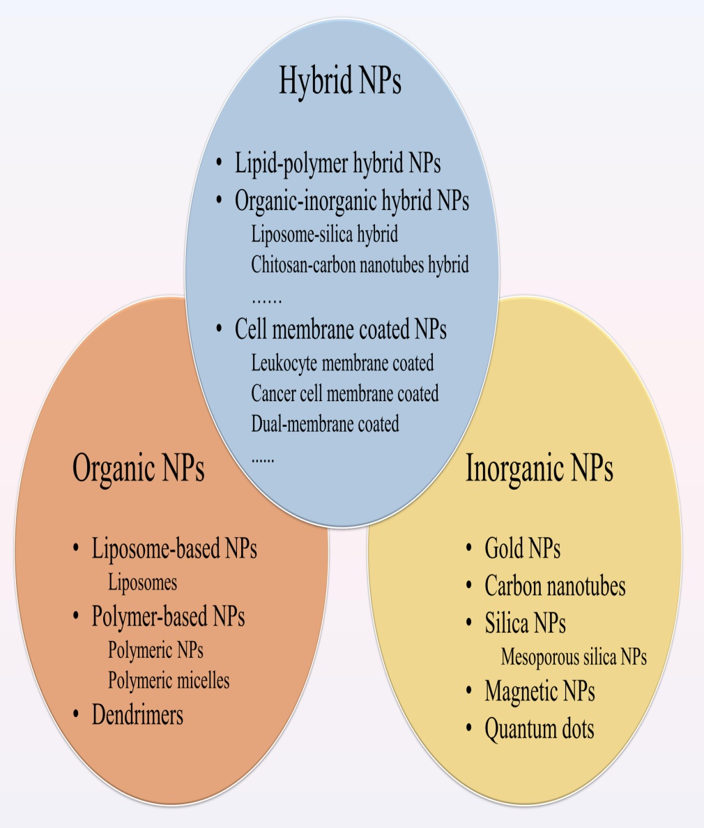
Figure 1. Different types of nanoparticles (NPs) for cancer therapy. NPs applied to drug delivery systems include organic NPs, inorganic NPs and hybrid NPs. The organic NPs contain liposome-based NPs, polymer-based NPs and dendrimers. Among polymer-based NPs, polymeric NPs and polymeric micelles are common. The inorganic NPs consist of gold NPs (Au NPs), carbon nanotubes, silica NPs, magnetic NPs, and quantum dots. Hybrid NPs combine the advantages of different NPs, including lipid-polymer hybrid NPs, organic-inorganic hybrid NPs, and cell membrane-coated NPs.
Organic Nanoparticles (NPs)
Organic nanoparticles have been studied for a long time and come in many forms.
Liposomes
These were the first nanomedicine approved for clinical use.
Think of them as tiny bubbles made of fat (lipids) with a hollow center that can carry both water-loving and fat-loving drugs.
By adjusting their lipid layers, liposomes can mimic cell-like properties, which makes them very effective at delivering drugs.
In cancer therapy, they are widely used to carry drugs like doxorubicin and paclitaxel.
In breast and prostate cancer, liposomes are becoming more common. For example:
Paclitaxel-loaded liposomes work better and last longer in the body compared to the free drug.
Liposomal doxorubicin reduces heart damage while still being effective.
Liposomes can also deliver combinations of drugs, which helps boost treatment results and even overcome drug resistance.
Today, several liposome-based drugs are already being used to treat cancer.
Polymer-based NPs
These are made from repeating chemical building blocks (monomers).
PLGA nanoparticles are the most common. They are safe, biodegradable, and stay longer in the body thanks to the tumor’s leaky blood vessels (the EPR effect).
Dendrimers are tree-like, branched molecules with lots of surface groups. This structure makes them very good at carrying different drugs.
Polymeric micelles form naturally when certain polymers self-assemble in water.
They have a water-hating (hydrophobic) core that can trap insoluble cancer drugs.
Their water-loving (hydrophilic) shell makes them stable in the bloodstream and helps them stay in circulation longer while avoiding quick clearance by the body.
Inorganic Nanoparticles (NPs)
Inorganic nanoparticles are popular in nanomedicine because they have a large surface area and can be easily modified to carry drugs. They’re also relatively easy to make. However, their main drawback is that they often have lower biocompatibility and biodegradability compared to organic nanoparticles.
Some major types include:
Gold nanoparticles (AuNPs):
The most widely studied inorganic NPs.
They are stable, non-toxic, and can be coated with molecules to improve drug targeting.
Useful for cancer therapy because they help drugs build up in tumors and even overcome drug resistance.
They can also be used in gene therapy, photothermal therapy (using heat to kill cancer cells), and immunotherapy.
Carbon nanotubes (CNTs):
Tiny, tube-shaped structures with unique chemical and physical properties.
They can carry drugs like doxorubicin, paclitaxel, and methotrexate to tumors.
When exposed to near-infrared light, CNTs heat up and can destroy cancer cells by thermal ablation.
Silica nanoparticles (SNPs):
“Mesoporous” silica nanoparticles have a sponge-like structure with lots of pores.
This allows them to hold and release large amounts of anticancer drugs.
They are stable, improve drug circulation, and can be engineered to release drugs only at the right time.
Porous silicon versions of SNPs also show potential in immunotherapy by helping the immune system fight cancer.
Magnetic nanoparticles (MNPs):
Made of metals or metal oxides, usually coated with polymers to improve safety.
Can deliver chemotherapy and genes into cancer cells.
Can also be heated with a magnetic field to kill tumors via magnetic hyperthermia.
Hybrid Nanoparticles
Since organic and inorganic NPs each have strengths and weaknesses, scientists are now combining them into hybrid nanoparticles to get the best of both worlds.
Lipid-polymer hybrids:
Have a polymer core and a lipid outer layer.
Combine the stability of polymers with the biocompatibility of lipids.
Can carry both water-loving and fat-loving drugs, making them versatile for cancers like pancreatic, breast, and prostate.
They are easily taken up by cancer cells and avoid quick clearance from the body.
Organic–inorganic hybrids:
Example: Liposome-silica hybrids (LSH) have a silica core surrounded by a lipid shell. These can deliver multiple drugs (like gemcitabine and paclitaxel) and have been shown effective in animal models.
CNTs combined with chitosan polymers have been tested for lung cancer, boosting anticancer activity while lowering toxicity.
Other hybrids mix polymers like PLGA with metals (e.g., gold or manganese), combining drug delivery with hyperthermia to enhance tumor destruction.
This cutting-edge method coats nanoparticles with natural cell membranes (from red blood cells, immune cells, platelets, or even cancer cells).
These coatings allow the NPs to “disguise” themselves, helping them circulate longer and avoid immune clearance.
Example: White blood cell–coated silica NPs avoid immune detection and build up in tumors.
Dual-membrane coatings (like red blood cell + platelet) offer even better stability and targeting.
Multistage delivery systems:
Some smart nanoparticles can change their size or properties inside the tumor.
For instance, large NPs can shrink into smaller ones once inside the tumor microenvironment, helping them penetrate deeper and deliver drugs more effectively.
Mechanisms of Targeting
For cancer treatment, one of the most important features of nanoparticles (NPs) is their ability to target cancer cells specifically. This makes treatment more effective while reducing damage to healthy cells.
Scientists have studied many ways to design nanoparticles for better targeting. To do this, it’s important to understand how tumors grow and how nanoparticles interact with them.
There are two main targeting strategies:
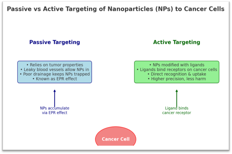
Passive targeting
Relies on the natural properties of tumors.
Tumors often have “leaky” blood vessels and poor drainage, which allows nanoparticles to accumulate in them more easily (this is called the EPR effect – Enhanced Permeability and Retention).
No special modifications are needed for the nanoparticles.
Active targeting
Involves modifying nanoparticles with special molecules (like antibodies, peptides, or sugars) that can bind directly to receptors on cancer cells.
This increases precision, so the drug goes mainly to cancer cells instead of normal ones.
Targeting Cancer Cells
Cancer cells often have special molecules on their surface that normal cells don’t. Nanoparticles (NPs) can be modified to recognize these molecules and deliver drugs more precisely.
Transferrin targeting:
Transferrin is a protein that carries iron into cells. Cancer cells usually have a lot more transferrin receptors than normal cells. By attaching transferrin to nanoparticles, drugs can enter cancer cells more efficiently and even overcome resistance to chemotherapy.
Folic acid targeting:
Folic acid (vitamin B9) is important for DNA building. Many cancers (like ovarian, breast, and lung) have too many folate receptors, while normal cells have very few. Nanoparticles coated with folic acid can specifically target these cancers.
Lectin targeting:
Lectins are proteins that recognize sugars on cell surfaces. Nanoparticles can either carry lectins to attach to cancer cell sugars, or carry sugars to attach to cancer cell lectins. Both ways help nanoparticles stick to cancer cells.
EGFR targeting:
Epidermal growth factor receptor (EGFR) is often overproduced in cancers, helping them grow and spread. Nanoparticles that bind to EGFR (and its variant HER2, common in breast and gastric cancer) can deliver drugs directly to these tumor cells.
Some NPs even combine two targeting molecules at once for extra precision.
Targeting Tumor Blood Vessels (Endothelium)
Instead of targeting cancer cells directly, some nanoparticles attack the blood supply that tumors need to survive.
VEGF/VEGFR: Tumors release VEGF to grow new blood vessels. NPs that block VEGF receptors can starve tumors.
Integrins (αvβ3): These receptors help cancer blood vessels grow and spread. Nanoparticles targeting αvβ3 can block vessel growth and improve anti-VEGF treatments.
VCAM-1: This molecule, overexpressed in tumor blood vessels, can also be a target for NPs.
MMPs (enzymes that remodel tissue): Tumors use MMPs to spread and build new blood vessels. MMP-sensitive nanoparticles can stop tumor growth in cancers like breast, pancreatic, and melanoma.
How Nanoparticles Help Overcome Drug Resistance
One of the biggest problems in cancer treatment is drug resistance. Tumors develop ways to escape chemotherapy, making treatment fail. NPs can help by:
Avoiding drug pumps (efflux transporters): Some cancer cells pump drugs out (like P-glycoprotein). NPs enter cells differently (via endocytosis), delivering drugs deeper inside and bypassing these pumps.
Targeting faulty cell death (apoptosis): Cancer often avoids death by overproducing survival proteins like Bcl-2 and NF-κB. NPs can deliver drugs or siRNA that block these proteins, forcing cancer cells to die.
Example: Co-delivering chemo drugs with ceramide (a molecule that restores tumor suppressor p53) helps trigger cell death.
Fighting hypoxia (low oxygen in tumors): Tumors often lack oxygen, making them resistant to drugs. The protein HIF-1α drives this process. NPs carrying siRNA or inhibitors against HIF-1α make tumors more sensitive to treatment.
Combination therapy in one carrier: NPs can carry multiple drugs (e.g., a chemo + an inhibitor) to attack cancer from different angles, overcoming resistance more effectively.
Role of Nanoparticles in Cancer Immunotherapy
Immunotherapy trains the immune system to fight cancer. NPs are making this approach stronger:
Nanovaccines: NPs carry cancer antigens to immune cells, helping the body recognize and attack tumors.
Artificial APCs (aAPCs): These mimic immune cells that activate T cells, jump-starting an immune attack.
Targeting the tumor microenvironment (TME): NPs can reprogram immune-suppressing cells around tumors (like macrophages and Tregs) so the immune system can attack.
Combination therapy: NPs can carry both chemo and immune drugs, making treatment more powerful with fewer side effects.
CONCLUSION & FUTURE DIRECTIONS
Nanoparticles are revolutionizing cancer treatment. Compared to regular drugs, they:
? Improve targeting and stability ? Reduce side effects ? Overcome drug resistance ? Support combination therapy
Future research is focusing on:
Designing hybrid nanoparticles (mixing organic, inorganic, or biomaterial coatings like cell membranes) for smarter drug delivery.
Understanding how nanoparticles interact with the immune system, since their size, shape, and coating affect response.
Developing NP-based immunotherapy that is safer and more effective, especially by reprogramming the tumor environment.
REFERENCES
Acharya, S., and Sahoo, S. K. (2011). PLGA nanoparticles containing various anticancer agents and tumour delivery by EPR effect. Adv. Drug Deliv. Rev. 63, 170–183. doi: 10.1016/j.addr.2010.10.008
Agarwal, R., and Kaye, S. B. (2003). Ovarian cancer: strategies for overcoming resistance to chemotherapy. Nat. Rev. Cancer 3, 502–516. doi: 10.1038/nrc1123
Alexis, F., Basto, P., Levy-Nissenbaum, E., Radovic-Moreno, A. F., Zhang, L., Pridgen, E., et al. (2008). HER-2-targeted nanoparticle-affibody bioconjugates for cancer therapy. ChemMedChem 3, 1839–1843. doi: 10.1002/cmdc.200800122
Alimoradi, H., Greish, K., Barzegar-Fallah, A., Alshaibani, L., and Pittalà, V. (2018). Nitric oxide-releasing nanoparticles improve doxorubicin anticancer activity. Int. J. Nanomed. 13, 7771–7787. doi: 10.2147/ijn.s187089
Allen, J. D., Brinkhuis, R. F., van Deemter, L., Wijnholds, J., and Schinkel, A. H. (2000). Extensive contribution of the multidrug transporters P-glycoprotein and Mrp1 to basal drug resistance. Cancer Res. 60, 5761–5766.
Almeida, P. V., Shahbazi, M. A., Mäkilä, E., Kaasalainen, M., Salonen, J., Hirvonen, J., et al. (2014). Amine-modified hyaluronic acid-functionalized porous silicon nanoparticles for targeting breast cancer tumors. Nanoscale 6, 10377–10387. doi: 10.1039/c4nr02187h
Amreddy, N., Babu, A., Muralidharan, R., Panneerselvam, J., Srivastava, A., Ahmed, R., et al. (2018). Recent advances in nanoparticle-based cancer drug and gene delivery. Adv. Cancer Res. 137, 115–170. doi: 10.1016/bs.acr.2017.11.003
Amreddy, N., Muralidharan, R., Babu, A., Mehta, M., Johnson, E. V., Zhao, Y. D., et al. (2015). Tumor-targeted and pH-controlled delivery of doxorubicin using gold nanorods for lung cancer therapy. Int. J. Nanomed. 10, 6773–6788. doi: 10.2147/ijn.s93237
Apte, R. S., Chen, D. S., and Ferrara, N. (2019). VEGF in signaling and disease: beyond discovery and development. Cell 176, 1248–1264. doi: 10.1016/j.cell.2019.01.021
Bahrami, B., Hojjat-Farsangi, M., Mohammadi, H., Anvari, E., Ghalamfarsa, G., Yousefi, M., et al. (2017). Nanoparticles and targeted drug delivery in cancer therapy. Immunol. Lett. 190, 64–83. doi: 10.1016/j.imlet.2017.07.015
Bai, F., Wang, C., Lu, Q., Zhao, M., Ban, F. Q., Yu, D. H., et al. (2013). Nanoparticle-mediated drug delivery to tumor neovasculature to combat P-gp expressing multidrug resistant cancer. Biomaterials 34, 6163–6174. doi: 10.1016/j.biomaterials.2013.04.062
Balasubramanian, S., Girija, A. R., Nagaoka, Y., Iwai, S., Suzuki, M., Kizhikkilot, V., et al. (2014). Curcumin and 5-fluorouracil-loaded, folate- and transferrin-decorated polymeric magnetic nanoformulation: a synergistic cancer therapeutic approach, accelerated by magnetic hyperthermia. Int. J. Nanomed. 9, 437–459. doi: 10.2147/ijn.s49882
Basoglu, H., Goncu, B., and Akbas, F. (2018). Magnetic nanoparticle-mediated gene therapy to induce Fas apoptosis pathway in breast cancer. Cancer Gene Ther 25, 141–147. doi: 10.1038/s41417-018-0017-2
Bauleth-Ramos, T., Shahbazi, M. A., Liu, D. F., Fontana, F., Correia, A., Figueiredo, P., et al. (2017). Nutlin-3a and cytokine co-loaded spermine-modified acetalated dextran nanoparticles for cancer chemo-immunotherapy. Adv. Funct. Mater. 27:14. doi: 10.1002/adfm.201703303
Bertrand, N., Wu, J., Xu, X., Kamaly, N., and Farokhzad, O. C. (2014). Cancer nanotechnology: the impact of passive and active targeting in the era of modern cancer biology. Adv. Drug Deliv. Rev. 66, 2–25. doi: 10.1016/j.addr.2013.11.009
Braunová, A., Kostka, L., Sivák, L., Cuchalová, L., Hv?zdová, Z., Laga, R., et al. (2017). Tumor-targeted micelle-forming block copolymers for overcoming of multidrug resistance. J. Control Release 245, 41–51. doi: 10.1016/j.jconrel.2016.11.020
Cagel, M., Tesan, F. C., Bernabeu, E., Salgueiro, M. J., Zubillaga, M. B., Moretton, M. A., et al. (2017). Polymeric mixed micelles as nanomedicines: achievements and perspectives. Eur. J. Pharm. Biopharm. 113, 211–228. doi: 10.1016/j.ejpb.2016.12.019
Carita, A. C., Eloy, J. O., Chorilli, M., Lee, R. J., and Leonardi, G. R. (2018). Recent advances and perspectives in liposomes for cutaneous drug delivery. Curr. Med. Chem. 25, 606–635. doi: 10.2174/0929867324666171009120154
Carmeliet, P., and Jain, R. K. (2000). Angiogenesis in cancer and other diseases. Nature 407, 249–257. doi: 10.1038/35025220
Chen, A. M., Zhang, M., Wei, D., Stueber, D., Taratula, O., Minko, T., et al. (2009). Co-delivery of doxorubicin and Bcl-2 siRNA by mesoporous silica nanoparticles enhances the efficacy of chemotherapy in multidrug-resistant cancer cells. Small 5, 2673–2677. doi: 10.1002/smll.200900621
Chen, X., Zhang, Y., Tang, C., Tian, C., Sun, Q., Su, Z., et al. (2017). Co-delivery of paclitaxel and anti-survivin siRNA via redox-sensitive oligopeptide liposomes for the synergistic treatment of breast cancer and metastasis. Int. J. Pharm. 529, 102–115. doi: 10.1016/j.ijpharm.2017.06.071
Chen, Y., Gao, D. Y., and Huang, L. (2015). In vivo delivery of miRNAs for cancer therapy: challenges and strategies. Adv. Drug Deliv. Rev. 81, 128–141. doi: 10.1016/j.addr.2014.05.009
Chen, Y., Zhu, X., Zhang, X., Liu, B., and Huang, L. (2010). Nanoparticles modified with tumor-targeting scFv deliver siRNA and miRNA for cancer therapy. Mol. Ther. 18, 1650–1656. doi: 10.1038/mt.2010.136
Cheng, C. A., Deng, T., Lin, F. C., Cai, Y., and Zink, J. I. (2019). Supramolecular Nanomachines as Stimuli-Responsive Gatekeepers on Mesoporous Silica Nanoparticles for Antibiotic and Cancer Drug Delivery. Theranostics 9, 3341–3364. doi: 10.7150/thno.34576
heng, H., Wu, Z., Wu, C., Wang, X., Liow, S. S., Li, Z., et al. (2018). Overcoming STC2 mediated drug resistance through drug and gene co-delivery by PHB-PDMAEMA cationic polyester in liver cancer cells. Mater. Sci. Eng. C Mater. Biol. Appl. 83, 210–217. doi: 10.1016/j.msec.2017.08.075
Cheng, J., Gu, Y. J., Cheng, S. H., and Wong, W. T. (2013). Surface functionalized gold nanoparticles for drug delivery. J. Biomed. Nanotechnol. 9, 1362–1369. doi: 10.1166/jbn.2013.1536
Cheow, W. S., and Hadinoto, K. (2011). Factors affecting drug encapsulation and stability of lipid-polymer hybrid nanoparticles. Colloids Surf. B Biointerfaces 85, 214–220. doi: 10.1016/j.colsurfb.2011.02.033
Chintamani, Singh, J. P., Mittal, M. K., Saxena, S., Bansal, A., Bhatia, A., et al. (2005). Role of p-glycoprotein expression in predicting response to neoadjuvant chemotherapy in breast cancer–a prospective clinical study. World J. Surg. Oncol. 3:61. doi: 10.1186/1477-7819-3-61Zylberberg, C., and Matosevic, S. (2016). Pharmaceutical liposomal drug delivery: a review of new delivery systems and a look at the regulatory landscape. Drug Deliv. 23, 3319–3329. doi: 10.1080/10717544.2016.1177136
Zitvogel, L., Apetoh, L., Ghiringhelli, F., and Kroemer, G. (2008). Immunological aspects of cancer chemotherapy. Nat. Rev. Immunol. 8, 59–73. doi: 10.1038/nri2216
Zhou, Q., Zhang, L., Yang, T., and Wu, H. (2018). Stimuli-responsive polymeric micelles for drug delivery and cancer therapy. Int. J. Nanomed. 13, 2921–2942. doi: 10.2147/ijn.s158696
Zhao, Y., Huan, M. L., Liu, M., Cheng, Y., Sun, Y., Cui, H., et al. (2016). Doxorubicin and resveratrol co-delivery nanoparticle to overcome doxorubicin resistance. Sci. Rep. 6:35267. doi: 10.1038/srep35267
Zhao, X., Li, F., Li, Y., Wang, H., Ren, H., Chen, J., et al. (2015). Co-delivery of HIF1α siRNA and gemcitabine via biocompatible lipid-polymer hybrid nanoparticles for effective treatment of pancreatic cancer. Biomaterials 46, 13–25. doi: 10.1016/j.biomaterials.2014.12.028
Zhao, M. D., Li, J. Q., Chen, F. Y., Dong, W., Wen, L. J., Fei, W. D., et al. (2019). Co-Delivery of Curcumin and Paclitaxel by “Core-Shell” targeting amphiphilic copolymer to reverse resistance in the treatment of ovarian cancer. Int. J. Nanomed. 14, 9453–9467. doi: 10.2147/ijn.s224579
Zhang, L., Chan, J. M., Gu, F. X., Rhee, J. W., Wang, A. Z., Radovic-Moreno, A. F., et al. (2008). Self-assembled lipid–polymer hybrid nanoparticles: a robust drug delivery platform. ACS Nano 2, 1696–1702. doi: 10.1021/nn800275r
Zhang, J., Zhang, Q., Lou, Y., Fu, Q., Chen, Q., Wei, T., et al. (2018). Hypoxia-inducible factor-1α/interleukin-1β signaling enhances hepatoma epithelial-mesenchymal transition through macrophages in a hypoxic-inflammatory microenvironment. Hepatology 67, 1872–1889. doi: 10.1002/hep.29681
Zhang, S., Guo, N., Wan, G., Zhang, T., Li, C., Wang, Y., et al. (2019). pH and redox dual-responsive nanoparticles based on disulfide-containing poly(β-amino ester) for combining chemotherapy and COX-2 inhibitor to overcome drug resistance in breast cancer. J. Nanobiotechnol. 17:109. doi: 10.1186/s12951-019-0540-9
Zhang, J., Wang, L., You, X., Xian, T., Wu, J., and Pang, J. (2019). Nanoparticle therapy for prostate cancer: overview and perspectives. Curr. Top. Med. Chem. 19, 57–73. doi: 10.2174/1568026619666190125145836
Zhang, R. X., Ahmed, T., Li, L. Y., Li, J., Abbasi, A. Z., and Wu, X. Y. (2017). Design of nanocarriers for nanoscale drug delivery to enhance cancer treatment using hybrid polymer and lipid building blocks. Nanoscale 9, 1334–1355. doi: 10.1039/c6nr08486a
Zhang, F., Correia, A., Mäkilä, E., Li, W., Salonen, J., Hirvonen, J. J., et al. (2017). Receptor-mediated surface charge inversion platform based on porous silicon nanoparticles for efficient cancer cell recognition and combination therapy. ACS Appl. Mater. Interfaces 9, 10034–10046. doi: 10.1021/acsami.7b02196
Zang, X., Zhao, X., Hu, H., Qiao, M., Deng, Y., and Chen, D. (2017). Nanoparticles for tumor immunotherapy. Eur. J. Pharm. Biopharm. 115, 243–256. doi: 10.1016/j.ejpb.2017.03.013
Yu, B., Song, N., Hu, H., Chen, G., Shen, Y., and Cong, H. (2018). A degradable triple temperature-, pH-, and redox-responsive drug system for cancer chemotherapy. J. Biomed. Mater. Res. A 106, 3203–3210. doi: 10.1002/jbm.a.36515
Yoon, H. Y., Selvan, S. T., Yang, Y., Kim, M. J., Yi, D. K., Kwon, I. C., et al. (2018). Engineering nanoparticle strategies for effective cancer immunotherapy. Biomaterials 178, 597–607. doi: 10.1016/j.biomaterials.2018.03.036
Wong, C., Stylianopoulos, T., Cui, J., Martin, J., Chauhan, V. P., Jiang, W., et al. (2011). Multistage nanoparticle delivery system for deep penetration into tumor tissue. Proc. Natl. Acad. Sci. U.S.A. 108, 2426–2431. doi: 10.1073/pnas.1018382108
Wang, Y., Gao, S., Ye, W. H., Yoon, H. S., and Yang, Y. Y. (2006). Co-delivery of drugs and DNA from cationic core-shell nanoparticles self-assembled from a biodegradable copolymer. Nat. Mater. 5, 791–796. doi: 10.1038/nmat1737.
Reference
Acharya, S., and Sahoo, S. K. (2011). PLGA nanoparticles containing various anticancer agents and tumour delivery by EPR effect. Adv. Drug Deliv. Rev. 63, 170–183. doi: 10.1016/j.addr.2010.10.008
Agarwal, R., and Kaye, S. B. (2003). Ovarian cancer: strategies for overcoming resistance to chemotherapy. Nat. Rev. Cancer 3, 502–516. doi: 10.1038/nrc1123
Alexis, F., Basto, P., Levy-Nissenbaum, E., Radovic-Moreno, A. F., Zhang, L., Pridgen, E., et al. (2008). HER-2-targeted nanoparticle-affibody bioconjugates for cancer therapy. ChemMedChem 3, 1839–1843. doi: 10.1002/cmdc.200800122
Alimoradi, H., Greish, K., Barzegar-Fallah, A., Alshaibani, L., and Pittalà, V. (2018). Nitric oxide-releasing nanoparticles improve doxorubicin anticancer activity. Int. J. Nanomed. 13, 7771–7787. doi: 10.2147/ijn.s187089
Allen, J. D., Brinkhuis, R. F., van Deemter, L., Wijnholds, J., and Schinkel, A. H. (2000). Extensive contribution of the multidrug transporters P-glycoprotein and Mrp1 to basal drug resistance. Cancer Res. 60, 5761–5766.
Almeida, P. V., Shahbazi, M. A., Mäkilä, E., Kaasalainen, M., Salonen, J., Hirvonen, J., et al. (2014). Amine-modified hyaluronic acid-functionalized porous silicon nanoparticles for targeting breast cancer tumors. Nanoscale 6, 10377–10387. doi: 10.1039/c4nr02187h
Amreddy, N., Babu, A., Muralidharan, R., Panneerselvam, J., Srivastava, A., Ahmed, R., et al. (2018). Recent advances in nanoparticle-based cancer drug and gene delivery. Adv. Cancer Res. 137, 115–170. doi: 10.1016/bs.acr.2017.11.003
Amreddy, N., Muralidharan, R., Babu, A., Mehta, M., Johnson, E. V., Zhao, Y. D., et al. (2015). Tumor-targeted and pH-controlled delivery of doxorubicin using gold nanorods for lung cancer therapy. Int. J. Nanomed. 10, 6773–6788. doi: 10.2147/ijn.s93237
Apte, R. S., Chen, D. S., and Ferrara, N. (2019). VEGF in signaling and disease: beyond discovery and development. Cell 176, 1248–1264. doi: 10.1016/j.cell.2019.01.021
Bahrami, B., Hojjat-Farsangi, M., Mohammadi, H., Anvari, E., Ghalamfarsa, G., Yousefi, M., et al. (2017). Nanoparticles and targeted drug delivery in cancer therapy. Immunol. Lett. 190, 64–83. doi: 10.1016/j.imlet.2017.07.015
Bai, F., Wang, C., Lu, Q., Zhao, M., Ban, F. Q., Yu, D. H., et al. (2013). Nanoparticle-mediated drug delivery to tumor neovasculature to combat P-gp expressing multidrug resistant cancer. Biomaterials 34, 6163–6174. doi: 10.1016/j.biomaterials.2013.04.062
Balasubramanian, S., Girija, A. R., Nagaoka, Y., Iwai, S., Suzuki, M., Kizhikkilot, V., et al. (2014). Curcumin and 5-fluorouracil-loaded, folate- and transferrin-decorated polymeric magnetic nanoformulation: a synergistic cancer therapeutic approach, accelerated by magnetic hyperthermia. Int. J. Nanomed. 9, 437–459. doi: 10.2147/ijn.s49882
Basoglu, H., Goncu, B., and Akbas, F. (2018). Magnetic nanoparticle-mediated gene therapy to induce Fas apoptosis pathway in breast cancer. Cancer Gene Ther 25, 141–147. doi: 10.1038/s41417-018-0017-2
Bauleth-Ramos, T., Shahbazi, M. A., Liu, D. F., Fontana, F., Correia, A., Figueiredo, P., et al. (2017). Nutlin-3a and cytokine co-loaded spermine-modified acetalated dextran nanoparticles for cancer chemo-immunotherapy. Adv. Funct. Mater. 27:14. doi: 10.1002/adfm.201703303
Bertrand, N., Wu, J., Xu, X., Kamaly, N., and Farokhzad, O. C. (2014). Cancer nanotechnology: the impact of passive and active targeting in the era of modern cancer biology. Adv. Drug Deliv. Rev. 66, 2–25. doi: 10.1016/j.addr.2013.11.009
Braunová, A., Kostka, L., Sivák, L., Cuchalová, L., Hv?zdová, Z., Laga, R., et al. (2017). Tumor-targeted micelle-forming block copolymers for overcoming of multidrug resistance. J. Control Release 245, 41–51. doi: 10.1016/j.jconrel.2016.11.020
Cagel, M., Tesan, F. C., Bernabeu, E., Salgueiro, M. J., Zubillaga, M. B., Moretton, M. A., et al. (2017). Polymeric mixed micelles as nanomedicines: achievements and perspectives. Eur. J. Pharm. Biopharm. 113, 211–228. doi: 10.1016/j.ejpb.2016.12.019
Carita, A. C., Eloy, J. O., Chorilli, M., Lee, R. J., and Leonardi, G. R. (2018). Recent advances and perspectives in liposomes for cutaneous drug delivery. Curr. Med. Chem. 25, 606–635. doi: 10.2174/0929867324666171009120154
Carmeliet, P., and Jain, R. K. (2000). Angiogenesis in cancer and other diseases. Nature 407, 249–257. doi: 10.1038/35025220
Chen, A. M., Zhang, M., Wei, D., Stueber, D., Taratula, O., Minko, T., et al. (2009). Co-delivery of doxorubicin and Bcl-2 siRNA by mesoporous silica nanoparticles enhances the efficacy of chemotherapy in multidrug-resistant cancer cells. Small 5, 2673–2677. doi: 10.1002/smll.200900621
Chen, X., Zhang, Y., Tang, C., Tian, C., Sun, Q., Su, Z., et al. (2017). Co-delivery of paclitaxel and anti-survivin siRNA via redox-sensitive oligopeptide liposomes for the synergistic treatment of breast cancer and metastasis. Int. J. Pharm. 529, 102–115. doi: 10.1016/j.ijpharm.2017.06.071
Chen, Y., Gao, D. Y., and Huang, L. (2015). In vivo delivery of miRNAs for cancer therapy: challenges and strategies. Adv. Drug Deliv. Rev. 81, 128–141. doi: 10.1016/j.addr.2014.05.009
Chen, Y., Zhu, X., Zhang, X., Liu, B., and Huang, L. (2010). Nanoparticles modified with tumor-targeting scFv deliver siRNA and miRNA for cancer therapy. Mol. Ther. 18, 1650–1656. doi: 10.1038/mt.2010.136
Cheng, C. A., Deng, T., Lin, F. C., Cai, Y., and Zink, J. I. (2019). Supramolecular Nanomachines as Stimuli-Responsive Gatekeepers on Mesoporous Silica Nanoparticles for Antibiotic and Cancer Drug Delivery. Theranostics 9, 3341–3364. doi: 10.7150/thno.34576
heng, H., Wu, Z., Wu, C., Wang, X., Liow, S. S., Li, Z., et al. (2018). Overcoming STC2 mediated drug resistance through drug and gene co-delivery by PHB-PDMAEMA cationic polyester in liver cancer cells. Mater. Sci. Eng. C Mater. Biol. Appl. 83, 210–217. doi: 10.1016/j.msec.2017.08.075
Cheng, J., Gu, Y. J., Cheng, S. H., and Wong, W. T. (2013). Surface functionalized gold nanoparticles for drug delivery. J. Biomed. Nanotechnol. 9, 1362–1369. doi: 10.1166/jbn.2013.1536
Cheow, W. S., and Hadinoto, K. (2011). Factors affecting drug encapsulation and stability of lipid-polymer hybrid nanoparticles. Colloids Surf. B Biointerfaces 85, 214–220. doi: 10.1016/j.colsurfb.2011.02.033
Chintamani, Singh, J. P., Mittal, M. K., Saxena, S., Bansal, A., Bhatia, A., et al. (2005). Role of p-glycoprotein expression in predicting response to neoadjuvant chemotherapy in breast cancer–a prospective clinical study. World J. Surg. Oncol. 3:61. doi: 10.1186/1477-7819-3-61Zylberberg, C., and Matosevic, S. (2016). Pharmaceutical liposomal drug delivery: a review of new delivery systems and a look at the regulatory landscape. Drug Deliv. 23, 3319–3329. doi: 10.1080/10717544.2016.1177136
Zitvogel, L., Apetoh, L., Ghiringhelli, F., and Kroemer, G. (2008). Immunological aspects of cancer chemotherapy. Nat. Rev. Immunol. 8, 59–73. doi: 10.1038/nri2216
Zhou, Q., Zhang, L., Yang, T., and Wu, H. (2018). Stimuli-responsive polymeric micelles for drug delivery and cancer therapy. Int. J. Nanomed. 13, 2921–2942. doi: 10.2147/ijn.s158696
Zhao, Y., Huan, M. L., Liu, M., Cheng, Y., Sun, Y., Cui, H., et al. (2016). Doxorubicin and resveratrol co-delivery nanoparticle to overcome doxorubicin resistance. Sci. Rep. 6:35267. doi: 10.1038/srep35267
Zhao, X., Li, F., Li, Y., Wang, H., Ren, H., Chen, J., et al. (2015). Co-delivery of HIF1α siRNA and gemcitabine via biocompatible lipid-polymer hybrid nanoparticles for effective treatment of pancreatic cancer. Biomaterials 46, 13–25. doi: 10.1016/j.biomaterials.2014.12.028
Zhao, M. D., Li, J. Q., Chen, F. Y., Dong, W., Wen, L. J., Fei, W. D., et al. (2019). Co-Delivery of Curcumin and Paclitaxel by “Core-Shell” targeting amphiphilic copolymer to reverse resistance in the treatment of ovarian cancer. Int. J. Nanomed. 14, 9453–9467. doi: 10.2147/ijn.s224579
Zhang, L., Chan, J. M., Gu, F. X., Rhee, J. W., Wang, A. Z., Radovic-Moreno, A. F., et al. (2008). Self-assembled lipid–polymer hybrid nanoparticles: a robust drug delivery platform. ACS Nano 2, 1696–1702. doi: 10.1021/nn800275r
Zhang, J., Zhang, Q., Lou, Y., Fu, Q., Chen, Q., Wei, T., et al. (2018). Hypoxia-inducible factor-1α/interleukin-1β signaling enhances hepatoma epithelial-mesenchymal transition through macrophages in a hypoxic-inflammatory microenvironment. Hepatology 67, 1872–1889. doi: 10.1002/hep.29681
Zhang, S., Guo, N., Wan, G., Zhang, T., Li, C., Wang, Y., et al. (2019). pH and redox dual-responsive nanoparticles based on disulfide-containing poly(β-amino ester) for combining chemotherapy and COX-2 inhibitor to overcome drug resistance in breast cancer. J. Nanobiotechnol. 17:109. doi: 10.1186/s12951-019-0540-9
Zhang, J., Wang, L., You, X., Xian, T., Wu, J., and Pang, J. (2019). Nanoparticle therapy for prostate cancer: overview and perspectives. Curr. Top. Med. Chem. 19, 57–73. doi: 10.2174/1568026619666190125145836
Zhang, R. X., Ahmed, T., Li, L. Y., Li, J., Abbasi, A. Z., and Wu, X. Y. (2017). Design of nanocarriers for nanoscale drug delivery to enhance cancer treatment using hybrid polymer and lipid building blocks. Nanoscale 9, 1334–1355. doi: 10.1039/c6nr08486a
Zhang, F., Correia, A., Mäkilä, E., Li, W., Salonen, J., Hirvonen, J. J., et al. (2017). Receptor-mediated surface charge inversion platform based on porous silicon nanoparticles for efficient cancer cell recognition and combination therapy. ACS Appl. Mater. Interfaces 9, 10034–10046. doi: 10.1021/acsami.7b02196
Zang, X., Zhao, X., Hu, H., Qiao, M., Deng, Y., and Chen, D. (2017). Nanoparticles for tumor immunotherapy. Eur. J. Pharm. Biopharm. 115, 243–256. doi: 10.1016/j.ejpb.2017.03.013
Yu, B., Song, N., Hu, H., Chen, G., Shen, Y., and Cong, H. (2018). A degradable triple temperature-, pH-, and redox-responsive drug system for cancer chemotherapy. J. Biomed. Mater. Res. A 106, 3203–3210. doi: 10.1002/jbm.a.36515
Yoon, H. Y., Selvan, S. T., Yang, Y., Kim, M. J., Yi, D. K., Kwon, I. C., et al. (2018). Engineering nanoparticle strategies for effective cancer immunotherapy. Biomaterials 178, 597–607. doi: 10.1016/j.biomaterials.2018.03.036
Wong, C., Stylianopoulos, T., Cui, J., Martin, J., Chauhan, V. P., Jiang, W., et al. (2011). Multistage nanoparticle delivery system for deep penetration into tumor tissue. Proc. Natl. Acad. Sci. U.S.A. 108, 2426–2431. doi: 10.1073/pnas.1018382108
Wang, Y., Gao, S., Ye, W. H., Yoon, H. S., and Yang, Y. Y. (2006). Co-delivery of drugs and DNA from cationic core-shell nanoparticles self-assembled from a biodegradable copolymer. Nat. Mater. 5, 791–796. doi: 10.1038/nmat1737.
Sonali Mahadev Patil
Corresponding author
Dr. Shivajirao Kadam College of Pharmacy Kasbe Digraj.
Sonali Mahadev Patil*, Nanoparticle-Based Drug Delivery in Cancer Therapy and Its Role in Overcoming Drug Resistance, Int. J. of Pharm. Sci., 2025, Vol 3, Issue 9, 2488-2499 https://doi.org/10.5281/zenodo.17176691

 10.5281/zenodo.17176691
