View Article

  • Neuroprotective Potential of Medicinal Plants in Schizophrenia: Mechanisms, Efficacy, and Future Pathways

  • 1 Department of Pharmacy, IEC College of Engineering & Technology, Greater Noida, Uttar Pradesh, India

    2 Department of Arts, Lalit Kala Sansthan, Agra, Uttar Pradesh, India

    3 Department of Pharmacy, Anand College of Pharmacy, Agra, Uttar Pradesh, India

    4 Faculty of Pharmacy, Goel Institute of Pharmaceutical Sciences Lucknow, Uttar Pradesh, India

    5,6,7 Faculty of Pharmacy, Agrawan Heritage University, Agra Uttar Pradesh, India

    8 Department of Pharmacy, College of Pharmacy, Agra Uttar Pradesh, India

Abstract

Schizophrenia is a disabling neuropsychiatric illness that is a chronic illness that is characterized by alterations in cognition, affect and social behaviour. Whereas standard antipsychotics drugs still form the foundation of method, the effectiveness is often not enough to manage negative and cognitive symptoms, and the poor efficacy is restricted by adverse effect drainage, which may decrease adherence. In the past decades, herbal medicine as a supplementary approach to treatment gained attention, which is facilitated by traditional systems like Ayurveda, Traditional Chinese Medicine and Unani medicine. Different botanicals such as those including Withania somniferum, Ginkgo biloba, Panax ginseng, Bacopa monnieri, Curcumin longa, and Rhodiola rosea have been found to modulate neurotransmitter systems, lower oxidative stress and neuroinflammation, increase neuroplasticity, and ameliorate metabolic status. These effects target some of the pathophysiological features of schizophrenia such as dopaminergic and glutamatergic imbalances, neuroinflammation, oxidative stress, HPA axis dysregulation as well as gene-environment interactions. Despite preclinical and clinical findings, the barriers to total acceptance by the Western population, including variation in herbal composition, inadequate standardization, and herb-drug interactions remain. Hence, it needs large-scale, well-controlled studies to formulate standard and evidence-based plans of using herbal medicines in the comprehensive management of schizophrenia.

Keywords

Schizophrenia, Herbal medicine, Phytotherapy, Neurotransmitter modulation, Neuroinflammation, Oxidative Stress, Neuroplasticity

Introduction

Schizophrenia is a neuropsychiatric disease that is severe and chronic and is associated with the abnormality of thoughts, insights as well as emotional responds. Scorn antipsychotic pharmacotherapy being the fundamental modality of care, adverse effect profiles (top among them being the extrapyramidal symptomatology), and unimpressive usage adherence rates even in the long-term, have catalyzed the research investigating the suitability of complementary and alternative measures, the most widely studied of which include herbal remedies[1]. Herbal medicine has in the previous 20 years proved to be an added treatment to schizophrenia particularly, improvement of cognitive deficits, negative symptoms, and side effects of medication. Ethnopharmacological basis of these interventions usually based on the Traditional Chinese Medicine (TCM), Ayurveda, and other indigenous medicine[2]. During the last twenty years herbal medicine has been ascribed greater and greater significance as a complementary treatment of schizophrenia; special attention has been paid both to the possibility that it will improve cognitive deficit, negative symptoms, and adverse drug effect. Most of these solutions belong to the Traditional Chinese Medicine (TCM ), Ayurveda, and other native medical systems[3]. Particularly in the most recent scholarship on plant medicine, the plant Withania somnifera (Ashwagandha) has been singled out as an adaptogenic agent within the Ayurvedic practice of therapeutics, on account of its empirically-evidenced anxiolytic, anti-inflammatory, and cognition-enhancing effects, when used in the context of providing such treatment to individuals with schizophrenia[4]. There are many herbal preparations under research in cognition as cognitive enhancers. Panax ginseng, Bacopa monnieri and Salvia miltiorrhiza are some of the most studied and it has been found that they all have the capacity to regulate the neurotransmitter processes and boost the neurotrophic pathways in the end to impact better working memory and executive functions[5]. It is implied that botanical agents would help to reduce the negative outcome on metabolism due to the long-time treatment of antipsychotics. In particular, Rhodiola rosea and Berberine were shown to be effective in the glucose metabolism improvement and lipid deposition reduction processes, thus helping to establish better physical health in people who take long-term antipsychotic treatment[6]. With very encouraging empirical results, there is a need to integrate herbal therapies in the standard clinical practice of schizophrenia. The major deterrents include great variation in the composition of herbs, lesser degrees of standardization, and herb-drug interactions[7]. Schizophrenia is a multipronged psychiatric issue that is long-standing, lasting a lifetime and whose occurrence is highlighted by distributed disturbances in terms of putting thoughts, way of arranging it, the way of seeing both oneself and the external reality, emotional and interactional aspects. Prevalence rates reveal that the condition is likely to be faced in 1 % of the world population thus posing a heavy burden on its patients, their immediate family members as well as the health-care system across the globe[8]. The inadequacy of modern pharmacological therapies has led to the increase of academic inquiries concerning integrative practices especially herbal medicine which is the core aspect of established systems like Ayurveda, Traditional Chinese Medicine (TCM), and Unani medicine. The design of these systems is holistic in nature as they introduce herbal agents structured to mediate neurochemical pathways as a means of treating mental conditions[2]. Ginkgo biloba is among the best researched botanical and it has been discovered to have high degree of anticarcinogenic, antioxidative, and anti-inflammatory abilities. Research has revealed that its adjuvant treatment with antipsychotics, in addition to improving the therapeutic response, reduces positive and negative symptomatology of schizophrenia[3]. Withania somnifera, also commonly known as Ashwagandha, is yet another high ranked adaptogen and its pharmacological properties are familiar to the extent of validation. Remarkably, Ashwagandha demonstrates a high level of antipsychotic-like activity as recorded during clinical trials, which can probably be attributed to the simultaneous influence of cortisol regulation, synaptic plasticity, and processes relating to oxidative stress[4].

CAUSES OF SCHIZOPHRENIA – A SCIENTIFIC RATIONALE FOR HERBAL INTERVENTION

There is no unitary etiological mechanism of schizophrenia; instead, it is caused by a multidimensional and changing interaction between biological, environmental and developmental determinants. These interdependencies are crucial to explain and could not be done without examining the ways to treat the underlying neural disturbances of the schizophrenic disorder using specific herbal medicines.

1. GENETIC VULNERABILITY AND EPIGENETIC ALTERATIONS

The definite genetic predisposition of schizophrenia flirts with the 80 % heritability estimates. Empirical findings have pinned down risk variants in genes namely DISC1, COMT and NRG1, the functions of which can be summarized under synaptic signaling, dopaminergic metabolism and neurodevelopment. However, genetic variations are insufficient to entirely explain the development of the disease; their interaction with the environment in the form of exposure to environmental factors (via epigenetic modulations) results in the translation of a psychosis phenotype[9].

2. DOPAMINERGIC DYSREGULATION

Dopaminergic Dysregulation also forms one of the foundations of modern debates on the topic of schizophrenia. A contemporary theory is based on the notion that imbalance of mesolimbic hyperactivity and mesocortical hypoactivity is necessary in the expression of symptoms; mesolimbic hyperactivity in this theory can serve as the basis of positive symptomatology, whereas the mesocortical hypoactivity seems to be the root of negative symptomatology and cognitive deficits[10]. The current state of neuroimaging and pharmacological studies holdings has lent further support to the existing dopamine hypothesis of schizophrenia since it has been able to reveal an increased presynaptic capacity of dopamine synthesis in the associative striatum of both individuals with schizophrenia and those with high risks to develop this affliction. In particular, there is a consensus on the use of positron-emission tomography and radiotracer assessment, that has reached a point of anatomically localized overproduction of dopamine in the ventral striatum, which is diminished upon the administration of antipsychotics[11].

3. GLUTAMATERGIC AND GABAERGIC DYSFUNCTION

In addition to dopamine, a growing body of empirical research supports the essential role of glutamate neurotransmission, namely NMDA receptor hypofunction, in causing cognitive impairments, learning/memory deficits and NMDA receptor dysfunction. There is also a concomitant glutamatergic imbalance that disrupts GABAergic inhibition, that, in turn, destabilized cortical synchrony. Bacopa monnieri and Salvia miltiorrhiza botanicals have shown a potential in the regulation of glutamate and GABA systems[12].

4. NEURODEVELOPMENTAL INSULTS AND ENVIRONMENTAL RISKS

The conceptual models that are currently being used increasingly view schizophrenia as a neurodevelopmental disorder. Early stressors that disrupt early brain development are prenatal stressors, such as infections and nutritional shortcomings, and stressors encountered early in an urban environment, including social adversity[13].

5. NEUROINFLAMMATION AND OXIDATIVE STRESS

Oxidative stress should be defined as a condition of imbalance between the generation of reactive oxygen species (ROS) and the endogenous antioxidant defense response capacity to control them. It is believed that this imbalance contributes to conditions that include: lipid peroxidation, mitochondrial dysfunction, and neuronal apoptosis, and all have been reported in schizophrenia brain tissue. Some plant-derived antioxidants, such as Curcuma longa (curcumin), Ginkgo biloba and Centella asiatica, have been found to reduce the level of ROS and improve the structural integrity of mitochondria, which hold the promise of clinical use in schizophrenia patients[14].

6. HPA AXIS DYSREGULATION AND CHRONIC CORTISOL ELEVATION

Oxidative stress will occur in the case of surpassing the production of reactive oxygen species (ROS) in the ratios constrained by the efficiency of antioxidant defenses. The following effects are the production of lipid peroxidation, dysfunction in mitochondria, and neuronal apoptosis-the pathologies realized in the cerebral tissue of schizophrenic patients. Antioxidants are herbal (e.g. Curcuma longa (curcumin), Ginkgo biloba, Centella asiatica, etc.), which have been reported to mitigate the formation of ROS and support the integrity of the mitochondrion[15].

7. ENVIRONMENTAL STRESSORS AND SUBSTANCE USE

Epidemiological studies show that urban household environment, isolation experienced during childhood, childhood trauma and the use of cannabis are also linked to an increased risk of schizophrenia. Such exposures can affect the brain functions through epigenetics reprogramming. The available evidence indicates that some adaptogenic herbs can increase resilience, support the recovery of homeostatic balance in case of chronic psychosocial stress[16].

Table 1

Etiological Factor

Herbal Intervention

Mechanism of Action

REFRENCES

Genetic Vulnerability & Epigenetic Alterations

Withania somnifera (Ashwagandha)

Modulates gene expression, enhances synaptic plasticity

 [17]

Dopaminergic Dysregulation

Mucuna pruriens

Increases dopamine levels, inhibits monoamine oxidase

[18]

Glutamatergic & GABAergic Dysfunction

Bacopa monnieri, Salvia miltiorrhiza

Enhances NMDA receptor function, restores GABAergic balance

 [19]

Neurodevelopmental Insults

Panax ginseng, Rhodiola rosea

Enhances neuroplasticity, reduces early-life stress effects

[20]

Neuroinflammation & Oxidative Stress

Curcuma longa, Ginkgo biloba, Centella asiatica

Antioxidant, anti-inflammatory, preserves mitochondrial structure

 [21]

HPA Axis Dysregulation & Cortisol Elevation

Curcuma longa, Centella asiatica

Decreases cortisol, strengthens antioxidant response

[22]

Environmental Stressors & Substance Use

Ocimum sanctum, Rhodiola rosea

Adaptogenic action, epigenetic regulation under psychosocial stress

 [23]

PATHOGENESIS OF SCHIZOPHRENIA: A SCIENTIFIC BASIS FOR HERBAL APPROACHES

 The careful understanding of the pathophysiology of schizophrenia as a multifactorial disorder preconditions the evaluation of herbal treatment interventions that are able to ameliorate the major neurobiological imbalances. Extant evidence on various mechanisms has been amalgamated in the subsequent synthesis that contributes to our increased understanding. One is, of course, alteration of neurotransmitter systems, particularly those of glutamate, GABA, and dopamine, which is a central pathology. Consistently, empirical literature illustrates changes in synaptic plasticity, glutamatergic and GABAergic receptor signalling; all of which have been considered to reflect neurodevelopmental disturbances. Second, functional brain abnormalities are based on maladaptive inflammatory cascades. It has been shown that immune cells that are pathogenic penetrate into the neural tissue and elicit infiltration by microglia and increase production of pro-inflammatory cytokines. Such processes combine to deteriorate synaptic development and pruning and may interrupt trophic support. Third, immune dysregulation impairs plasticity of immune cell phenotype which deregulates the neuroimmune interactions. Different lines of enquiry report a movement towards pro-inflammatory, murderous T-cell subsets and absence of the neuroprotective arm to the immune system. These phenomena also increase inflammatory disorders and weaken neurotrophic Doing. In short, concurrence of neuroimmune and neurotransmitter dysregulations result in both neurocognitive and behavioural deficits that are the hallmarks of schizophrenia. Herbal remedies that will target more than one node in these convergence points stand the best chance of generating significant clinical value.

1. NEURODEVELOPMENTAL AND NEURODEGENERATIVE DYNAMICS

Schizophrenia is conceptualized as a disorder rooted in abnormal early brain development, followed by progressive neurodegeneration, particularly during psychotic episodes, leading to treatment resistance and functional decline. Early intervention is emphasized to preserve neurocircuit integrity[24].

2. STRUCTURAL BRAIN ALTERATIONS

Both neuroimaging technologies and autopsy studies suggest less grey and white mass in the prefrontal, temporal, thalamic, and striatal areas in particular. Reductions of this nature can be found long before the onset of pharmacotherapy and worsen according to the duration of diseases[25].

3. NEUROTRANSMITTER DYSREGULATION

Dysregulation of dopaminergic transmission is one of the major key pathophysiological processes in schizophrenia. Positive symptoms are associated with hyperactivity of the mesolimbic dopaminergic pathway; negative and cognitive deficits are associated with the hypoactivity of the mesocortical pathway. Moreover, there has been proved glutamatergic and GABAergic imbalance. Abnormal functional results of the NMDA receptor, especially in quick-spiking cortical interneurons, are implicated in inhibited GABA induced rotational and cognitive functioning. Expanding on these results, additional mechanisms have been identified as down regulation of CB2 receptors and increased kynurenic acid (an NMDAR blocker) both of which further damage glutamatergic modulation as well as synaptic pruning[26].

4. IMMUNE ACTIVATION AND OXIDATIVE STRESS

The pathophysiology of schizophrenia has been associated with the constant elevation in the neuroinflammatory mechanisms characterized by the presence of elevated peripheral cytokines like interleukin-6(IL-6) and tumor necrosis factor-alpha (TNF-alpha) in combination with the compromised blood-brain barrier integrity, both of which stimulate microglial activation. At the same time, oxidative stress reduces neuronal loss, highlighted by oxidative signs of DNA damage (which are increased) and mitochondrial dysfunction[27].

5. GENE–ENVIRONMENT INTERACTION AND EPIGENETICS

Epigenetic alterations represent an important piece of the aetiopathogenesis of schizophrenia and their induction can be explained by the exceptional interaction of predisposing genes (i.e. DISC1, NRG1, COMT, DTNBP1) with environmental factors (i.e. prenatal infections, perinatal complications, nutritional deficiencies, childhood trauma, and cannabis).This kind of interactions reshapes neural circuitries and therefore increases likelihood of schizophrenia[28].

6. STRESS, HPA AXIS DYSFUNCTION, AND CORTISOL

The extended psychosocial stress has a dysregulatory effect on the hypothalamic-pituitary-adrenal axis, hence causing chronic cortisol increase. Hippocampal atrophy occurs with this hormonal imbalance, neuroplasticity is weakened and oxidative and inflammatory destruction is repeated[29].

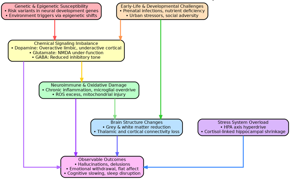
Figure.1 Neurobiological Pathways and Herbal Therapeutic Strategies in Schizophrenia

Table 2: Core Pathogenic Domains of Schizophrenia and Herbal Therapeutic Correlates

Pathogenic Domain

Mechanistic Insight

Herbal Correlates

REFRENCES

Neurodevelopment & Neurodegeneration

Early neurodevelopmental defects with later neurodegeneration during psychosis result in synaptic loss and cognitive deficits.

Withania somnifera, Centella asiatica support neuroplasticity and cognitive preservation.

[30]

Structural Brain Abnormalities

Reductions in grey/white matter in frontal, temporal, thalamic areas even before treatment onset.

Ginkgo biloba, Bacopa monnieri enhance cerebral circulation and support structural integrity.

[31]

Neurotransmitter Dysregulation

Dopaminergic imbalance, NMDA receptor hypofunction, and GABAergic dysfunction underlie core symptoms.

Valeriana officinalis, Rhodiola rosea regulate dopaminergic and GABAergic signaling.

[32]

Neuroinflammation & Oxidative Stress

Elevated cytokines, microglial activation, and ROS damage mitochondrial and synaptic function.

Curcuma longa, Panax ginseng possess anti-inflammatory and antioxidant mechanisms.

[33]

Gene–Environment & Epigenetic Modulation

Genetic predispositions interact with environmental insults, inducing maladaptive epigenetic changes.

Resveratrol, Curcumin modulate DNA methylation and histone acetylation.

[34]

HPA Axis Dysregulation & Chronic Stress

Chronic cortisol elevation from stress impairs hippocampus and neuroplasticity, worsening disease trajectory.

Ashwagandha, Rhodiola rosea reduce cortisol, modulate HPA axis, and build stress resilience.

[35]

SIGN AND SYMPTOMS

Table3: Symptom Clusters in Schizophrenia and Herbal Therapeutic Interventions – Mechanistic & Clinical Insights

Symptom Cluster

Core Clinical Features

Herbal Intervention

Mechanistic Action

DOI

Positive Symptoms

Hallucinations, paranoid delusions, conceptual disorganization

Withania somnifera (Ashwagandha)

Attenuates dopaminergic hyperactivity (mesolimbic pathway), modulates NMDA receptor function

[36]

Negative Symptoms

Avolition, anhedonia, affective flattening, social withdrawal

Panax ginseng

Enhances BDNF signaling and synaptic plasticity; augments dopaminergic and serotonergic tone in the prefrontal cortex

 [37]

Cognitive Impairment

Attention deficit, impaired working memory, reduced executive function

Bacopa monnieri

Facilitates acetylcholine transmission, protects hippocampal neurons from oxidative injury, supports learning and memory

 [38]

Affective Dysregulation

Depression, anxiety, irritability, low motivation

Rhodiola rosea

Modulates HPA axis; restores monoaminergic balance (serotonin, norepinephrine); reduces neuroinflammatory signaling

 [39]

Sleep Disturbances

Insomnia, disrupted circadian rhythm, non-restorative sleep

Valeriana officinalis (Valerian)

Potentiates GABAergic neurotransmission, enhances sleep onset latency and quality, reduces hyperarousal

[40]

Psychomotor Abnormalities

Motor retardation or agitation, catatonic features

Ginkgo biloba

Improves cerebral perfusion, scavenges reactive oxygen species, supports neuronal energy metabolism and mitochondrial function

 [41]

Mood Instability

Rapid mood fluctuations, dysphoria, lability

Curcuma longa (Curcumin)

Inhibits pro-inflammatory cytokines (e.g., TNF-α, IL-6), modulates serotonin pathways and NF-κB signaling

[42]

HERBAL MEDICINES OF SCHIZOPHRENIA

Schizophrenia is a psychiatric disorder that is characterized by chronicity and is characterized by the experience of delusions, hallucinations, cognitive, and social dysfunctions; therefore, it should be treated in the long-term using clinical management methods that are usually limited due to the adverse effects of the medication[43]. It is highlighted in recent scientific studies that herbal medicines hold vision in underlying therapeutic intension with regard to being used as an adjunct treatment that will cause greater effect coupled with antipsychotic induced side effects[44].

1. WITHANIA SOMNIFERA (ASHWAGANDHA)

The herb Withania somnifera (Ashwagandha) has been found to produce antipsychotic-like effects by regulating oxidative stress levels, GABAergic systems and neuroinflammation in animal models as well as clinical trials[45]. In one recent randomized, placebo-controlled and double-blind trial, it was shown that when Ashwagandha, an herbal supplement, was used as an adjunct treatment on patients with schizophrenia, measurable improvements in both magnitude and negative symptomatology was observed[45].

2. GINKGO BILOBA

Ginkgo biloba is a botanical species, commonly associated with large flavonoid and terpenoid content, which has been linked to cognitive improvement in human beings and loss of tardive dyskinesia signs in patients with schizophrenia[46]. Another 8-study meta-analysis randomized controlled trial shows that when taken together with antipsychotic drug, Ginkgo biloba has a significant positive outcome with regard to improvement of positive symptoms and also in ridding of overall psychopathology. The finding lends to the expanding body of knowledge pointing toward adjunctive Ginkgo biloba supplementation being a potentially efficacious adjunct to traditional, pharmacotherapy of schizophrenia[47].

3. PANAX GINSENG

Panax ginseng is an adaptogenic herb proven to have antipsychotic and neuroprotective effects in preclinical studies, including its ability to modulate dopamine as well as act as an antioxidant[48]. Based on the current evidence, it suggests that conventional Chinese formulas like Wendan decoction, and Anshen Dingzhi Wan are in the clinical trials to determine their effectiveness as drugs that can improve emotional regulation and reduce psychotic symptoms in those who already experienced a first episode[49]. Most often, herbal preparations trigger their pharmacological effects due to the modulation of neurotransmitter systems, namely dopaminergic, glutamatergic, and GABAergic pathways, as well as restore the neuroinflammatory process and promote neuroplasticity. Such features relate to the characteristics of schizophrenia pathophysiology [50]. Even though it has gathered strong empirical results, most studies are limited by their small sample size, lack of standardised formula and poor blinding guidelines. In turn, such large-scale and multicentric trials are of utmost importance in the development of evidence-based clinical guidelines[51].

4. BACOPA MONNIERI

 The allostatic stability has been found to be increased by bacopa monnieri (Brahmi) via its neuroprotective effects and support cognitive abilities, since it regulates the antioxidant defense against the antagonism and controls dopaminergic neurotransmission. This effect deserves more research with regards to cognitive impairment manifested in schizophrenia[52].

5. CURCUMA LONGA (TURMERIC)

Curcumin as the major component of curcuma longa (turmeric) exhibits anti-inflammatory, antioxidant as well as neuroprotective properties and have the capability to reduce the oxidative stress and neuroinflammation typical of schizophrenia[53].

6. RHODIOLA ROSEA

Rosea affects the mind with psychotropic effects that increase mental output, counter fatigue and modify stress mechanism mechanism by affecting serotoneric and dopaminergic transmissions making it a potentially relevant adjunctive agent in the treatment of mood-related symptoms in schizophrenias[54].

7. GASTRODIA ELATA

The traditional Chinese herb Gastrodia elata (Tian Ma) has neuroprotective and anticonvulsant properties and displays potent antioxidant properties mediated through changes in GABAergic pathways. They can be used in treatment of psychotic symptoms using such properties[55].

8. MELISSA OFFICINALIS (LEMON BALM)

Melissa officinalis ( Lemon Balm ) has both anxiolytic and mood-stabilizing properties that are mediated with help of cholinergic and GABAergic transmission. The mechanism has the potential to offer symptomatic support to anxiety and agitation that is related to schizophrenia[56].

9. ZIZIPHUS JUJUBA

Traditionally, Ziziphus jujuba (popularly referred to as jujube seed) has been used in the traditional medicine because of its anxiolytic, anti-anxiety, and cognitive-enhancing effects. Recent pharmacological studies suggest that at least part of those effects are due to activity on γ-aminobutyric acid (GABA) receptors and to the increased expression of neurotrophic factors[57].

10. SCUTELLARIA BAICALENSIS

Flavonoids presented in Scutellaria baicalensis (Chinese skullcap) including baicalin in particular have been shown to have neuroprotective and anti-inflammatory effects hence treatment in schizophrenia via the regulation of glutamate induced excitotoxicity[58].

CENTELLA ASIATICA (GOTU KOLA)

Gotu Kola- Centella asiatica is a demonstrated to carry strong neuro regenerative and cognitive enhancement functions through its positive association effects on BDNF expression, and antioxidant processes, both of which can be used to compensate the cognition shortcomings of Schizophrenia[59].

TABLE 4 : PROMISING HERBAL MEDICINES IN SCHIZOPHRENIA – MECHANISMS AND EVIDENCE-BASED FINDINGS

Herbal Medicine

Key Actions

Mechanisms Involved

Reference (DOI / PMID)

Withania somnifera (Ashwagandha)

Reduces oxidative stress, inflammation, and improves negative symptoms

GABAergic modulation, anti-inflammatory, antioxidant

[60]

Ginkgo biloba

Enhances cognition, reduces antipsychotic-induced side effects

Flavonoids & terpenoids; antioxidant and neuroprotective

[61]

Panax ginseng

Improves emotional regulation, antioxidant, antipsychotic-like effects

Dopaminergic modulation, neuroprotection

[62]

Bacopa monnieri

Enhances memory, cognition, and neuronal protection

Antioxidant activity, modulation of dopaminergic system

[63]

Curcuma longa (Turmeric)

Attenuates neuroinflammation and oxidative stress

Curcumin-mediated anti-inflammatory & antioxidant mechanisms

[64]

Rhodiola rosea

Alleviates stress-induced symptoms, improves fatigue and mood

Modulates serotonergic and dopaminergic pathways, adaptogenic effects

[65]

Gastrodia elata

Neuroprotective and anticonvulsant; enhances GABAergic activity

Enhances GABA transmission, antioxidant properties

[66]

Melissa officinalis (Lemon Balm)

Provides anxiolytic and mood-stabilizing effects

Cholinergic enhancement, GABA receptor agonism

[67]

Ziziphus jujuba (Jujube)

Improves cognition and reduces anxiety

GABA receptor activity, upregulation of neurotrophic factors

[68]

Scutellaria baicalensis

Reduces glutamate excitotoxicity, exhibits anti-inflammatory effects

Flavonoids (baicalin), NMDA receptor inhibition

[69]

Centella asiatica (Gotu Kola)

Enhances cognition and supports neurogenesis

Upregulates BDNF, antioxidant and neurotrophic modulation

[70]

Table 5 Marketed Formulation

Sr. No.

Product Name

Main Ingredient

Dosage Form

REFRENCES

1

KSM-66® Ashwagandha

Withania somnifera root extract

Capsule

 [71]

2

Tebonin®

Ginkgo biloba leaf extract (EGb 761)

Tablet

[72]

3

Ginsana®

Panax ginseng extract (G115)

Capsule

 [73]

4

Brahmi Mind®

Bacopa monnieri whole plant extract

Capsule

 [74]

5

CurcuWIN®

Curcuma longa (Curcumin) standardized extract

Capsule

 [75]

6

Rhodiolife®

Rhodiola rosea root extract

Capsule

 [76]

7

Tian Ma Wan®

Gastrodia elata tuber extract

Tablet

 [77]

8

Melissa Dream®

Melissa officinalis leaf extract

Tablet

 [78]

9

Jujube Seed Formula®

Ziziphus jujuba seed extract

Capsule

 [79]

10

Baicalin Plus®

Scutellaria baicalensis root extract

Capsule

 [80]

CONCLUSION

The now top-priority management of schizophrenia requires a multidimensional approach that would encompass the symptomatologic and long-term quality-of-life aspects. Nature remedies, which are based on the traditional healing paradigm, offer one path that needs further investigation and may become promising supplemental treatments that recruit a variety of neurobiological processes engaging in the condition. Empirical evidence suggests that special botanicals can complement the use of standard pharmacotherapies by improving cognitive performance, dampening negative symptoms, alleviating pharmacies-mediated adverse outcomes and enhancing stress resistance. However, integration into everyday clinical use is inhibited by the influence of variables that include variability in dosage prescribed, lack of regulatory supervision, and a comparative sparsity of bigger-scale experiments. Consistent formulas, high-quality clinical testing, and strict evaluation of the safety profiles will be the key requirements before the addition of the herbal treatment to the general psychiatric services. Current studies have a potential in the development of more patient-centered and holistic approach to treatment of schizophrenia with the use of herbal medicine.

REFERENCES

  1. D. B. Campbell, L. A. Lange, T. Skelly, J. Lieberman, P. Levitt, and P. F. Sullivan, “Association of RGS2 and RGS5 variants with schizophrenia symptom severity.,” Schizophr. Res., vol. 101, no. 1–3, pp. 67–75, Apr. 2008, doi: 10.1016/j.schres.2008.01.006.
  2. S. Zhu, Q. Li, Y. Du, X. Yang, J. Fan, and Z. Dong, “Toxicity of derivatives from semicarbazide-sensitive amine oxidase-mediated deamination of methylamine against Toxoplasma gondii after infection of differentiated 3T3-L1 cells.,” Toxicol. In Vitro, vol. 24, no. 3, pp. 809–14, Apr. 2010, doi: 10.1016/j.tiv.2009.12.014.
  3. M. S. Soyfoo, V. Gangji, and J. Margaux, “Successful treatment of SAPHO syndrome with ibandronate.,” J. Clin. Rheumatol., vol. 16, no. 5, p. 253, Aug. 2010, doi: 10.1097/RHU.0b013e3181e9be89.
  4. S. Paliwal, R. Chaudhuri, A. Agrawal, and S. Mohanty, “Regenerative abilities of mesenchymal stem cells through mitochondrial transfer.,” J. Biomed. Sci., vol. 25, no. 1, p. 31, Mar. 2018, doi: 10.1186/s12929-018-0429-1.
  5. X. Bai, C. Rao, and X. Wang, “Shape vocabulary: a robust and efficient shape representation for shape matching.,” IEEE Trans. Image Process., vol. 23, no. 9, pp. 3935–49, Sep. 2014, doi: 10.1109/TIP.2014.2336542.
  6. J. Miot, S. Bernard, M. Bourreau, F. Guyot, and A. Kish, “Experimental maturation of Archaea encrusted by Fe-phosphates.,” Sci. Rep., vol. 7, no. 1, p. 16984, Dec. 2017, doi: 10.1038/s41598-017-17111-9.
  7. E. Ghasemi, M. Sillanpää, and N. M. Najafi, “Headspace hollow fiber protected liquid-phase microextraction combined with gas chromatography-mass spectroscopy for speciation and determination of volatile organic compounds of selenium in environmental and biological samples.,” J. Chromatogr. A, vol. 1218, no. 3, pp. 380–6, Jan. 2011, doi: 10.1016/j.chroma.2010.12.005.
  8. K. M. Gaworecki, R. W. Chapman, M. G. Neely, A. R. D’Amico, and L. J. Bain, “Arsenic exposure to killifish during embryogenesis alters muscle development.,” Toxicol. Sci., vol. 125, no. 2, pp. 522–31, Feb. 2012, doi: 10.1093/toxsci/kfr302.
  9. Schizophrenia Working Group of the Psychiatric Genomics Consortium, “Biological insights from 108 schizophrenia-associated genetic loci.,” Nature, vol. 511, no. 7510, pp. 421–7, Jul. 2014, doi: 10.1038/nature13595.
  10. K. L. Davis, R. S. Kahn, G. Ko, and M. Davidson, “Dopamine in schizophrenia: a review and reconceptualization.,” Am. J. Psychiatry, vol. 148, no. 11, pp. 1474–86, Nov. 1991, doi: 10.1176/ajp.148.11.1474.
  11. J. Kesby, D. Eyles, J. McGrath, and J. Scott, “Dopamine, psychosis and schizophrenia: the widening gap between basic and clinical neuroscience,” Transl. Psychiatry, vol. 8, no. 1, p. 30, Jan. 2018, doi: 10.1038/s41398-017-0071-9.
  12. T. Paus, M. Keshavan, and J. N. Giedd, “Why do many psychiatric disorders emerge during adolescence?,” Nat. Rev. Neurosci., vol. 9, no. 12, pp. 947–57, Dec. 2008, doi: 10.1038/nrn2513.
  13. R. A. Watson and M. Sidor, “Bladder cancer in Native Americans and Alaskan Natives.,” Urology, vol. 72, no. 1, pp. 10–4, Jul. 2008, doi: 10.1016/j.urology.2007.12.087.
  14. Z. M. Brown et al., “Score Distributions of the Balance Outcome Measure for Elder Rehabilitation (BOOMER) in Community-Dwelling Older Adults With Vertebral Fracture.,” J. Geriatr. Phys. Ther., vol. 42, no. 3, pp. E87–E93, 2019, doi: 10.1519/JPT.0000000000000158.
  15. C. P. Taylor, T. Angelotti, and E. Fauman, “Pharmacology and mechanism of action of pregabalin: the calcium channel alpha2-delta (alpha2-delta) subunit as a target for antiepileptic drug discovery.,” Epilepsy Res., vol. 73, no. 2, pp. 137–50, Feb. 2007, doi: 10.1016/j.eplepsyres.2006.09.008.
  16. A. H. Glenngård, J. Hjelmgren, P. H. Thomsen, and T. Tvedten, “Patient preferences and willingness-to-pay for ADHD treatment with stimulants using discrete choice experiment (DCE) in Sweden, Denmark and Norway.,” Nord. J. Psychiatry, vol. 67, no. 5, pp. 351–9, Oct. 2013, doi: 10.3109/08039488.2012.748825.
  17. Y. G. Bachner, “Psychometric Properties of Responses to an Arabic Version of the Hamilton Depression Rating Scale (HAM-D6).,” J. Am. Psychiatr. Nurses Assoc., vol. 22, no. 1, pp. 27–30, 2016, doi: 10.1177/1078390316629959.
  18. C. Rioboo, R. Prado, C. Herrero, and A. Cid, “Population growth study of the rotifer Brachionus sp. fed with triazine-exposed microalgae.,” Aquat. Toxicol., vol. 83, no. 4, pp. 247–53, Aug. 2007, doi: 10.1016/j.aquatox.2007.04.006.
  19. E. Lasek-Nesselquist and J. P. Gogarten, “The effects of model choice and mitigating bias on the ribosomal tree of life.,” Mol. Phylogenet. Evol., vol. 69, no. 1, pp. 17–38, Oct. 2013, doi: 10.1016/j.ympev.2013.05.006.
  20. C. Tudor, L. Bumiller, T. Birkholz, P. Stockmann, J. Wiltfang, and P. Kessler, “Static and dynamic periosteal elevation: a pilot study in a pig model.,” Int. J. Oral Maxillofac. Surg., vol. 39, no. 9, pp. 897–903, Sep. 2010, doi: 10.1016/j.ijom.2010.05.005.
  21. L. J. Carriere and A. S. Kluck, “Appearance commentary from romantic partners: evaluation of an adapted measure.,” Body Image, vol. 11, no. 2, pp. 137–45, Mar. 2014, doi: 10.1016/j.bodyim.2013.12.003.
  22. K. S. Swapna and K. J. Rao, “Studies on effect of oat and cheese incorporation on sensory and textural quality of short-dough type biscuit.,” J. Food Sci. Technol., vol. 53, no. 3, pp. 1505–14, Mar. 2016, doi: 10.1007/s13197-015-2014-x.
  23. A. C. Chua, M. W. Knuiman, D. Trinder, M. L. Divitini, and J. K. Olynyk, “Higher concentrations of serum iron and transferrin saturation but not serum ferritin are associated with cancer outcomes.,” Am. J. Clin. Nutr., vol. 104, no. 3, pp. 736–42, Sep. 2016, doi: 10.3945/ajcn.115.129411.
  24. J. A. Lieberman, “Pathophysiologic mechanisms in the pathogenesis and clinical course of schizophrenia.,” J. Clin. Psychiatry, vol. 60 Suppl 1, pp. 9–12, 1999, [Online]. Available: http://www.ncbi.nlm.nih.gov/pubmed/10372603
  25. V. Nair, M. Dutta, S. S. Manian, R. K. S, and V. K. Jayaraman, “Identification of Penicillin-binding proteins employing support vector machines and random forest.,” Bioinformation, vol. 9, no. 9, pp. 481–4, 2013, doi: 10.6026/97320630009481.
  26. A. J. Clark, S. Burl, and C. Denning, “Genetic modification of sheep by nuclear transfer with gene-targeted somatic cells.,” Methods Mol. Biol., vol. 348, pp. 199–212, 2006, doi: 10.1007/978-1-59745-154-3_13.
  27. J. Guerra-Varela et al., “Susceptibility of Zebrafish to Vesicular Stomatitis Virus Infection.,” Zebrafish, vol. 15, no. 2, pp. 124–132, Apr. 2018, doi: 10.1089/zeb.2017.1499.
  28. L. Keyes-Elstein, E. Brittain, A. Pinckney, E. A. Goldmuntz, and K. M. Sullivan, “Safety and efficacy of HSCT for systemic sclerosis across clinical trials.,” Nat. Rev. Rheumatol., vol. 16, no. 11, p. 661, Nov. 2020, doi: 10.1038/s41584-020-0493-2.
  29. K. Aligizaki, P. Katikou, A. Milandri, and J. Diogène, “Occurrence of palytoxin-group toxins in seafood and future strategies to complement the present state of the art.,” Toxicon, vol. 57, no. 3, pp. 390–9, Mar. 2011, doi: 10.1016/j.toxicon.2010.11.014.
  30. R. Bruffaerts, A. Bonnewyn, and K. Demyttenaere, “The individual and societal effects of non-psychotic serious mental disorders on earnings in Belgium.,” Eur. Psychiatry, vol. 24, no. 4, pp. 207–13, May 2009, doi: 10.1016/j.eurpsy.2008.12.006.
  31. J. C. Blader, N. R. Schooler, P. S. Jensen, S. R. Pliszka, and V. Kafantaris, “Adjunctive divalproex versus placebo for children with ADHD and aggression refractory to stimulant monotherapy.,” Am. J. Psychiatry, vol. 166, no. 12, pp. 1392–401, Dec. 2009, doi: 10.1176/appi.ajp.2009.09020233.
  32. W. Jiang et al., “A Multi-Parameter Analysis of Cellular Coordination of Major Transcriptome Regulation Mechanisms.,” Sci. Rep., vol. 8, no. 1, p. 5742, Apr. 2018, doi: 10.1038/s41598-018-24039-1.
  33. A. Nayman, I. Yildiz, N. Koca, S. Deniz, M. Koplay, and L. Oguzkurt, “Risk factors associated with recanalization of incompetent saphenous veins treated with radiofrequency ablation catheter.,” Diagn. Interv. Imaging, vol. 98, no. 1, pp. 29–36, Jan. 2017, doi: 10.1016/j.diii.2016.06.003.
  34. K. Kamata, M. Takenaka, M. Tsurusaki, and M. Kudo, “Portal vein stenting for portal vein stenosis caused by bile duct cancer.,” Dig. Liver Dis., vol. 49, no. 11, p. 1282, Nov. 2017, doi: 10.1016/j.dld.2017.05.022.
  35. E. U. Selker, “Neurospora.,” Curr. Biol., vol. 21, no. 4, pp. R139-40, Feb. 2011, doi: 10.1016/j.cub.2011.01.006.
  36. A. Lin, L. Nguyen, L. M. Clotilde, J. A. Kase, I. Son, and C. R. Lauzon, “Isolation of Shiga toxin-producing Escherichia coli from fresh produce using STEC heart infusion washed blood agar with mitomycin-C.,” J. Food Prot., vol. 75, no. 11, pp. 2028–30, Nov. 2012, doi: 10.4315/0362-028X.JFP-12-157.
  37. C. R. Mahoney, J. Castellani, F. M. Kramer, A. Young, and H. R. Lieberman, “Tyrosine supplementation mitigates working memory decrements during cold exposure.,” Physiol. Behav., vol. 92, no. 4, pp. 575–82, Nov. 2007, doi: 10.1016/j.physbeh.2007.05.003.
  38. J. Y. Sacks, K. McKendrick, and S. Banks, “The impact of early trauma and abuse on residential substance abuse treatment outcomes for women.,” J. Subst. Abuse Treat., vol. 34, no. 1, pp. 90–100, Jan. 2008, doi: 10.1016/j.jsat.2007.01.010.
  39. R. Duncan, “Development of HPMA copolymer-anticancer conjugates: clinical experience and lessons learnt.,” Adv. Drug Deliv. Rev., vol. 61, no. 13, pp. 1131–48, Nov. 2009, doi: 10.1016/j.addr.2009.05.007.
  40. J. Van Borsel, C. Bontinck, M. Coryn, F. Paemeleire, and P. Vandemaele, “Acoustic features of palilalia: a case study.,” Brain Lang., vol. 101, no. 1, pp. 90–6, Apr. 2007, doi: 10.1016/j.bandl.2006.06.118.
  41. B. C. Ginsburg, H. L. Kimmel, F. I. Carroll, M. M. Goodman, and L. L. Howell, “Interaction of cocaine and dopamine transporter inhibitors on behavior and neurochemistry in monkeys.,” Pharmacol. Biochem. Behav., vol. 80, no. 3, pp. 481–91, Mar. 2005, doi: 10.1016/j.pbb.2005.01.004.
  42. S. Zafra-Stone, T. Yasmin, M. Bagchi, A. Chatterjee, J. A. Vinson, and D. Bagchi, “Berry anthocyanins as novel antioxidants in human health and disease prevention.,” Mol. Nutr. Food Res., vol. 51, no. 6, pp. 675–83, Jun. 2007, doi: 10.1002/mnfr.200700002.
  43. Z. Chen et al., “Establishment of Mini-pig Model of Ureteral Stricture by Electrocoagulation Under Ureteroscopy.,” Urology, vol. 167, pp. 67–72, Sep. 2022, doi: 10.1016/j.urology.2022.05.023.
  44. A. Schlegel, O. de Rougemont, R. Graf, P.-A. Clavien, and P. Dutkowski, “Protective mechanisms of end-ischemic cold machine perfusion in DCD liver grafts.,” J. Hepatol., vol. 58, no. 2, pp. 278–86, Feb. 2013, doi: 10.1016/j.jhep.2012.10.004.
  45. H. Nursoy, M. G. Ronquillo, A. P. Faciola, and G. A. Broderick, “Lactation response to soybean meal and rumen-protected methionine supplementation of corn silage-based diets.,” J. Dairy Sci., vol. 101, no. 3, pp. 2084–2095, Mar. 2018, doi: 10.3168/jds.2017-13227.
  46. A. V Taira et al., “Long-term outcome for clinically localized prostate cancer treated with permanent interstitial brachytherapy.,” Int. J. Radiat. Oncol. Biol. Phys., vol. 79, no. 5, pp. 1336–42, Apr. 2011, doi: 10.1016/j.ijrobp.2010.01.005.
  47. D. Green, “Interpreting coagulation assays.,” Blood Coagul. Fibrinolysis, vol. 21 Suppl 1, pp. S3-6, Sep. 2010, doi: 10.1097/01.mbc.0000388935.77612.d0.
  48. Z. Ordulu et al., “Intravenous leiomyomatosis: an unusual intermediate between benign and malignant uterine smooth muscle tumors.,” Mod. Pathol., vol. 29, no. 5, pp. 500–10, May 2016, doi: 10.1038/modpathol.2016.36.
  49. E. P. Morris et al., “Racial and ethnic differences in the relationship between financial worry and white matter hyperintensities in Latinx, non-Latinx Black, and non-Latinx White older adults.,” Neurobiol. Aging, vol. 129, pp. 149–156, Sep. 2023, doi: 10.1016/j.neurobiolaging.2023.05.008.
  50. J. Yim et al., “Incidentally identified left ventricular apical aneurysm in a patient with Fabry disease.,” Echocardiography, vol. 39, no. 8, pp. 1131–1137, Aug. 2022, doi: 10.1111/echo.15412.
  51. H.-C. Lin et al., “RSV pneumonia with or without bacterial co-infection among healthy children.,” J. Formos. Med. Assoc., vol. 121, no. 3, pp. 687–693, Mar. 2022, doi: 10.1016/j.jfma.2021.08.012.
  52. D. Heperkan and L. R. Beuchat, “International Journal of Food Microbiology. Preface.,” Int. J. Food Microbiol., vol. 167, no. 1, p. 1, Oct. 2013, doi: 10.1016/j.ijfoodmicro.2013.09.012.
  53. E. Nossek, “Occipital Microarteriovenous Malformation Resection, Strategy, and Nuances.,” World Neurosurg., vol. 108, p. 987.e3, Dec. 2017, doi: 10.1016/j.wneu.2017.09.087.
  54. J. Gao et al., “Increasing effect of Tangzhiqing formula on IRS-1-dependent PI3K/AKT signaling in muscle,” BMC Complement. Altern. Med., vol. 14, no. 1, p. 198, Dec. 2014, doi: 10.1186/1472-6882-14-198.
  55. B. C. N. Müller et al., “When Pinocchio acts like a human, a wooden hand becomes embodied. Action co-representation for non-biological agents.,” Neuropsychologia, vol. 49, no. 5, pp. 1373–1377, Apr. 2011, doi: 10.1016/j.neuropsychologia.2011.01.022.
  56. V. Kilambi, F. R. Johnson, J. M. González, and A. F. Mohamed, “Valuations of genetic test information for treatable conditions: the case of colorectal cancer screening.,” Value Health, vol. 17, no. 8, pp. 838–45, Dec. 2014, doi: 10.1016/j.jval.2014.09.001.
  57. G. Sowden, C. A. Mastromauro, J. L. Januzzi, G. L. Fricchione, and J. C. Huffman, “Detection of depression in cardiac inpatients: feasibility and results of systematic screening.,” Am. Heart J., vol. 159, no. 5, pp. 780–7, May 2010, doi: 10.1016/j.ahj.2010.02.029.
  58. R. Khanam et al., “Prenatal Environmental Metal Exposure and Preterm Birth: A Scoping Review.,” Int. J. Environ. Res. Public Health, vol. 18, no. 2, Jan. 2021, doi: 10.3390/ijerph18020573.
  59. D. van Diermen, A. Marston, J. Bravo, M. Reist, P.-A. Carrupt, and K. Hostettmann, “Monoamine oxidase inhibition by Rhodiola rosea L. roots.,” J. Ethnopharmacol., vol. 122, no. 2, pp. 397–401, Mar. 2009, doi: 10.1016/j.jep.2009.01.007.
  60. D. Puliti, D. Warther, C. Orange, A. Specht, and M. Goeldner, “Small photoactivatable molecules for controlled fluorescence activation in living cells.,” Bioorg. Med. Chem., vol. 19, no. 3, pp. 1023–9, Feb. 2011, doi: 10.1016/j.bmc.2010.07.011.
  61. D. B. Davalos, D. C. Rojas, and J. R. Tregellas, “Temporal processing in schizophrenia: Effects of task-difficulty on behavioral discrimination and neuronal responses,” Schizophr. Res., vol. 127, no. 1–3, pp. 123–130, Apr. 2011, doi: 10.1016/j.schres.2010.06.020.
  62. Y. Jiang, T. Zhang, Z. Yi, Y. Han, X. Su, and Y. Feng, “Diblock, Triblock and Cyclic Amphiphilic Copolymers with CO2 Switchability: Effects of Topology.,” Polymers (Basel)., vol. 12, no. 4, Apr. 2020, doi: 10.3390/polym12040984.
  63. W. Chen, Y. Lu, M. Gao, J. Wu, A. Wang, and R. Shi, “Anti-angiogenesis effect of essential oil from Curcuma zedoaria in vitro and in vivo.,” J. Ethnopharmacol., vol. 133, no. 1, pp. 220–6, Jan. 2011, doi: 10.1016/j.jep.2010.09.031.
  64. F. Olivieri et al., “MicroRNAs linking inflamm-aging, cellular senescence and cancer.,” Ageing Res. Rev., vol. 12, no. 4, pp. 1056–68, Sep. 2013, doi: 10.1016/j.arr.2013.05.001.
  65. I. La Mantia and C. Andaloro, “Effectiveness of intranasal sodium hyaluronate in mitigating adverse effects of nasal continuous positive airway pressure therapy.,” Am. J. Rhinol. Allergy, vol. 31, no. 6, pp. 364–369, Nov. 2017, doi: 10.2500/ajra.2017.31.4482.
  66. K. L. Shires, B. M. Da Silva, J. P. Hawthorne, R. G. M. Morris, and S. J. Martin, “Synaptic tagging and capture in the living rat.,” Nat. Commun., vol. 3, p. 1246, 2012, doi: 10.1038/ncomms2250.
  67. A. L. Ingram, O. Deparis, J. Boulenguez, G. Kennaway, S. Berthier, and A. R. Parker, “Structural origin of the green iridescence on the chelicerae of the red-backed jumping spider, Phidippus johnsoni (Salticidae: Araneae).,” Arthropod Struct. Dev., vol. 40, no. 1, pp. 21–5, Jan. 2011, doi: 10.1016/j.asd.2010.07.006.
  68. R. Echigo et al., “Trehalose treatment suppresses inflammation, oxidative stress, and vasospasm induced by experimental subarachnoid hemorrhage.,” J. Transl. Med., vol. 10, p. 80, Apr. 2012, doi: 10.1186/1479-5876-10-80.
  69. S. Ito and D. Yamamoto, “Identification of two bromocresol purple binding sites on human serum albumin.,” Clin. Chim. Acta., vol. 411, no. 19–20, pp. 1536–8, Oct. 2010, doi: 10.1016/j.cca.2010.05.044.
  70. T. Nijsten, J. Silverberg, P. Gisondi, C. Vestergaard, L. Hollestein, and M. Wakkee, “Considerations in association studies in dermatoepidemiology.,” Br. J. Dermatol., vol. 185, no. 1, pp. 1–2, Jul. 2021, doi: 10.1111/bjd.20393.
  71. C. Vaklavas, Y. S. Chatzizisis, and A. M. Tsimberidou, “Common cardiovascular medications in cancer therapeutics.,” Pharmacol. Ther., vol. 130, no. 2, pp. 177–90, May 2011, doi: 10.1016/j.pharmthera.2011.01.009.
  72. J. Gambrelle, C. Graswill, M. Mauget-Faysse, L. Kodjikian, B. Cochener, and J.-D. Grange, “[Treatment of circumscribed choroidal haemangiomas by photodynamic therapy: a review].,” J. Fr. Ophtalmol., vol. 33, no. 7, pp. 497–504, Sep. 2010, doi: 10.1016/j.jfo.2010.06.002.
  73. A. Persano, F. Quaranta, A. Taurino, P. A. Siciliano, and J. Iannacci, “Thin Film Encapsulation for RF MEMS in 5G and Modern Telecommunication Systems.,” Sensors (Basel)., vol. 20, no. 7, Apr. 2020, doi: 10.3390/s20072133.
  74. H. Mochizuki, Y. Ohnuki, and H. Kurosawa, “Effect of glucose concentration during embryoid body (EB) formation from mouse embryonic stem cells on EB growth and cell differentiation.,” J. Biosci. Bioeng., vol. 111, no. 1, pp. 92–7, Jan. 2011, doi: 10.1016/j.jbiosc.2010.09.001.
  75. “Reply by authors.,” J. Urol., vol. 190, no. 2, p. 812, Aug. 2013, doi: 10.1016/j.juro.2013.02.3246.
  76. A. Devereux-Fitzgerald, R. Powell, and D. P. French, “Conflating Time and Energy: Views From Older Adults in Lower Socioeconomic Status Areas on Physical Activity.,” J. Aging Phys. Act., vol. 26, no. 3, pp. 506–513, Jul. 2018, doi: 10.1123/japa.2017-0283.
  77. K. L. Shires, B. M. Da Silva, J. P. Hawthorne, R. G. M. Morris, and S. J. Martin, “Synaptic tagging and capture in the living rat,” Nat. Commun., vol. 3, no. 1, p. 1246, Dec. 2012, doi: 10.1038/ncomms2250.
  78. H. Zuo et al., “[Genetic analysis of a Chinese pedigree affected with Mucopolysaccharidosis type ?A].,” Zhonghua Yi Xue Yi Chuan Xue Za Zhi, vol. 40, no. 4, pp. 452–457, Apr. 2023, doi: 10.3760/cma.j.cn511374-20221107-00766.
  79. A. M. Jones, D. W. Ehrhardt, and W. B. Frommer, “A never ending race for new and improved fluorescent proteins.,” BMC Biol., vol. 10, p. 39, May 2012, doi: 10.1186/1741-7007-10-39.
  80. S. Ito and D. Yamamoto, “Identification of two bromocresol purple binding sites on human serum albumin,” Clin. Chim. Acta, vol. 411, no. 19–20, pp. 1536–1538, Oct. 2010, doi: 10.1016/j.cca.2010.05.044

Reference

  1. D. B. Campbell, L. A. Lange, T. Skelly, J. Lieberman, P. Levitt, and P. F. Sullivan, “Association of RGS2 and RGS5 variants with schizophrenia symptom severity.,” Schizophr. Res., vol. 101, no. 1–3, pp. 67–75, Apr. 2008, doi: 10.1016/j.schres.2008.01.006.
  2. S. Zhu, Q. Li, Y. Du, X. Yang, J. Fan, and Z. Dong, “Toxicity of derivatives from semicarbazide-sensitive amine oxidase-mediated deamination of methylamine against Toxoplasma gondii after infection of differentiated 3T3-L1 cells.,” Toxicol. In Vitro, vol. 24, no. 3, pp. 809–14, Apr. 2010, doi: 10.1016/j.tiv.2009.12.014.
  3. M. S. Soyfoo, V. Gangji, and J. Margaux, “Successful treatment of SAPHO syndrome with ibandronate.,” J. Clin. Rheumatol., vol. 16, no. 5, p. 253, Aug. 2010, doi: 10.1097/RHU.0b013e3181e9be89.
  4. S. Paliwal, R. Chaudhuri, A. Agrawal, and S. Mohanty, “Regenerative abilities of mesenchymal stem cells through mitochondrial transfer.,” J. Biomed. Sci., vol. 25, no. 1, p. 31, Mar. 2018, doi: 10.1186/s12929-018-0429-1.
  5. X. Bai, C. Rao, and X. Wang, “Shape vocabulary: a robust and efficient shape representation for shape matching.,” IEEE Trans. Image Process., vol. 23, no. 9, pp. 3935–49, Sep. 2014, doi: 10.1109/TIP.2014.2336542.
  6. J. Miot, S. Bernard, M. Bourreau, F. Guyot, and A. Kish, “Experimental maturation of Archaea encrusted by Fe-phosphates.,” Sci. Rep., vol. 7, no. 1, p. 16984, Dec. 2017, doi: 10.1038/s41598-017-17111-9.
  7. E. Ghasemi, M. Sillanpää, and N. M. Najafi, “Headspace hollow fiber protected liquid-phase microextraction combined with gas chromatography-mass spectroscopy for speciation and determination of volatile organic compounds of selenium in environmental and biological samples.,” J. Chromatogr. A, vol. 1218, no. 3, pp. 380–6, Jan. 2011, doi: 10.1016/j.chroma.2010.12.005.
  8. K. M. Gaworecki, R. W. Chapman, M. G. Neely, A. R. D’Amico, and L. J. Bain, “Arsenic exposure to killifish during embryogenesis alters muscle development.,” Toxicol. Sci., vol. 125, no. 2, pp. 522–31, Feb. 2012, doi: 10.1093/toxsci/kfr302.
  9. Schizophrenia Working Group of the Psychiatric Genomics Consortium, “Biological insights from 108 schizophrenia-associated genetic loci.,” Nature, vol. 511, no. 7510, pp. 421–7, Jul. 2014, doi: 10.1038/nature13595.
  10. K. L. Davis, R. S. Kahn, G. Ko, and M. Davidson, “Dopamine in schizophrenia: a review and reconceptualization.,” Am. J. Psychiatry, vol. 148, no. 11, pp. 1474–86, Nov. 1991, doi: 10.1176/ajp.148.11.1474.
  11. J. Kesby, D. Eyles, J. McGrath, and J. Scott, “Dopamine, psychosis and schizophrenia: the widening gap between basic and clinical neuroscience,” Transl. Psychiatry, vol. 8, no. 1, p. 30, Jan. 2018, doi: 10.1038/s41398-017-0071-9.
  12. T. Paus, M. Keshavan, and J. N. Giedd, “Why do many psychiatric disorders emerge during adolescence?,” Nat. Rev. Neurosci., vol. 9, no. 12, pp. 947–57, Dec. 2008, doi: 10.1038/nrn2513.
  13. R. A. Watson and M. Sidor, “Bladder cancer in Native Americans and Alaskan Natives.,” Urology, vol. 72, no. 1, pp. 10–4, Jul. 2008, doi: 10.1016/j.urology.2007.12.087.
  14. Z. M. Brown et al., “Score Distributions of the Balance Outcome Measure for Elder Rehabilitation (BOOMER) in Community-Dwelling Older Adults With Vertebral Fracture.,” J. Geriatr. Phys. Ther., vol. 42, no. 3, pp. E87–E93, 2019, doi: 10.1519/JPT.0000000000000158.
  15. C. P. Taylor, T. Angelotti, and E. Fauman, “Pharmacology and mechanism of action of pregabalin: the calcium channel alpha2-delta (alpha2-delta) subunit as a target for antiepileptic drug discovery.,” Epilepsy Res., vol. 73, no. 2, pp. 137–50, Feb. 2007, doi: 10.1016/j.eplepsyres.2006.09.008.
  16. A. H. Glenngård, J. Hjelmgren, P. H. Thomsen, and T. Tvedten, “Patient preferences and willingness-to-pay for ADHD treatment with stimulants using discrete choice experiment (DCE) in Sweden, Denmark and Norway.,” Nord. J. Psychiatry, vol. 67, no. 5, pp. 351–9, Oct. 2013, doi: 10.3109/08039488.2012.748825.
  17. Y. G. Bachner, “Psychometric Properties of Responses to an Arabic Version of the Hamilton Depression Rating Scale (HAM-D6).,” J. Am. Psychiatr. Nurses Assoc., vol. 22, no. 1, pp. 27–30, 2016, doi: 10.1177/1078390316629959.
  18. C. Rioboo, R. Prado, C. Herrero, and A. Cid, “Population growth study of the rotifer Brachionus sp. fed with triazine-exposed microalgae.,” Aquat. Toxicol., vol. 83, no. 4, pp. 247–53, Aug. 2007, doi: 10.1016/j.aquatox.2007.04.006.
  19. E. Lasek-Nesselquist and J. P. Gogarten, “The effects of model choice and mitigating bias on the ribosomal tree of life.,” Mol. Phylogenet. Evol., vol. 69, no. 1, pp. 17–38, Oct. 2013, doi: 10.1016/j.ympev.2013.05.006.
  20. C. Tudor, L. Bumiller, T. Birkholz, P. Stockmann, J. Wiltfang, and P. Kessler, “Static and dynamic periosteal elevation: a pilot study in a pig model.,” Int. J. Oral Maxillofac. Surg., vol. 39, no. 9, pp. 897–903, Sep. 2010, doi: 10.1016/j.ijom.2010.05.005.
  21. L. J. Carriere and A. S. Kluck, “Appearance commentary from romantic partners: evaluation of an adapted measure.,” Body Image, vol. 11, no. 2, pp. 137–45, Mar. 2014, doi: 10.1016/j.bodyim.2013.12.003.
  22. K. S. Swapna and K. J. Rao, “Studies on effect of oat and cheese incorporation on sensory and textural quality of short-dough type biscuit.,” J. Food Sci. Technol., vol. 53, no. 3, pp. 1505–14, Mar. 2016, doi: 10.1007/s13197-015-2014-x.
  23. A. C. Chua, M. W. Knuiman, D. Trinder, M. L. Divitini, and J. K. Olynyk, “Higher concentrations of serum iron and transferrin saturation but not serum ferritin are associated with cancer outcomes.,” Am. J. Clin. Nutr., vol. 104, no. 3, pp. 736–42, Sep. 2016, doi: 10.3945/ajcn.115.129411.
  24. J. A. Lieberman, “Pathophysiologic mechanisms in the pathogenesis and clinical course of schizophrenia.,” J. Clin. Psychiatry, vol. 60 Suppl 1, pp. 9–12, 1999, [Online]. Available: http://www.ncbi.nlm.nih.gov/pubmed/10372603
  25. V. Nair, M. Dutta, S. S. Manian, R. K. S, and V. K. Jayaraman, “Identification of Penicillin-binding proteins employing support vector machines and random forest.,” Bioinformation, vol. 9, no. 9, pp. 481–4, 2013, doi: 10.6026/97320630009481.
  26. A. J. Clark, S. Burl, and C. Denning, “Genetic modification of sheep by nuclear transfer with gene-targeted somatic cells.,” Methods Mol. Biol., vol. 348, pp. 199–212, 2006, doi: 10.1007/978-1-59745-154-3_13.
  27. J. Guerra-Varela et al., “Susceptibility of Zebrafish to Vesicular Stomatitis Virus Infection.,” Zebrafish, vol. 15, no. 2, pp. 124–132, Apr. 2018, doi: 10.1089/zeb.2017.1499.
  28. L. Keyes-Elstein, E. Brittain, A. Pinckney, E. A. Goldmuntz, and K. M. Sullivan, “Safety and efficacy of HSCT for systemic sclerosis across clinical trials.,” Nat. Rev. Rheumatol., vol. 16, no. 11, p. 661, Nov. 2020, doi: 10.1038/s41584-020-0493-2.
  29. K. Aligizaki, P. Katikou, A. Milandri, and J. Diogène, “Occurrence of palytoxin-group toxins in seafood and future strategies to complement the present state of the art.,” Toxicon, vol. 57, no. 3, pp. 390–9, Mar. 2011, doi: 10.1016/j.toxicon.2010.11.014.
  30. R. Bruffaerts, A. Bonnewyn, and K. Demyttenaere, “The individual and societal effects of non-psychotic serious mental disorders on earnings in Belgium.,” Eur. Psychiatry, vol. 24, no. 4, pp. 207–13, May 2009, doi: 10.1016/j.eurpsy.2008.12.006.
  31. J. C. Blader, N. R. Schooler, P. S. Jensen, S. R. Pliszka, and V. Kafantaris, “Adjunctive divalproex versus placebo for children with ADHD and aggression refractory to stimulant monotherapy.,” Am. J. Psychiatry, vol. 166, no. 12, pp. 1392–401, Dec. 2009, doi: 10.1176/appi.ajp.2009.09020233.
  32. W. Jiang et al., “A Multi-Parameter Analysis of Cellular Coordination of Major Transcriptome Regulation Mechanisms.,” Sci. Rep., vol. 8, no. 1, p. 5742, Apr. 2018, doi: 10.1038/s41598-018-24039-1.
  33. A. Nayman, I. Yildiz, N. Koca, S. Deniz, M. Koplay, and L. Oguzkurt, “Risk factors associated with recanalization of incompetent saphenous veins treated with radiofrequency ablation catheter.,” Diagn. Interv. Imaging, vol. 98, no. 1, pp. 29–36, Jan. 2017, doi: 10.1016/j.diii.2016.06.003.
  34. K. Kamata, M. Takenaka, M. Tsurusaki, and M. Kudo, “Portal vein stenting for portal vein stenosis caused by bile duct cancer.,” Dig. Liver Dis., vol. 49, no. 11, p. 1282, Nov. 2017, doi: 10.1016/j.dld.2017.05.022.
  35. E. U. Selker, “Neurospora.,” Curr. Biol., vol. 21, no. 4, pp. R139-40, Feb. 2011, doi: 10.1016/j.cub.2011.01.006.
  36. A. Lin, L. Nguyen, L. M. Clotilde, J. A. Kase, I. Son, and C. R. Lauzon, “Isolation of Shiga toxin-producing Escherichia coli from fresh produce using STEC heart infusion washed blood agar with mitomycin-C.,” J. Food Prot., vol. 75, no. 11, pp. 2028–30, Nov. 2012, doi: 10.4315/0362-028X.JFP-12-157.
  37. C. R. Mahoney, J. Castellani, F. M. Kramer, A. Young, and H. R. Lieberman, “Tyrosine supplementation mitigates working memory decrements during cold exposure.,” Physiol. Behav., vol. 92, no. 4, pp. 575–82, Nov. 2007, doi: 10.1016/j.physbeh.2007.05.003.
  38. J. Y. Sacks, K. McKendrick, and S. Banks, “The impact of early trauma and abuse on residential substance abuse treatment outcomes for women.,” J. Subst. Abuse Treat., vol. 34, no. 1, pp. 90–100, Jan. 2008, doi: 10.1016/j.jsat.2007.01.010.
  39. R. Duncan, “Development of HPMA copolymer-anticancer conjugates: clinical experience and lessons learnt.,” Adv. Drug Deliv. Rev., vol. 61, no. 13, pp. 1131–48, Nov. 2009, doi: 10.1016/j.addr.2009.05.007.
  40. J. Van Borsel, C. Bontinck, M. Coryn, F. Paemeleire, and P. Vandemaele, “Acoustic features of palilalia: a case study.,” Brain Lang., vol. 101, no. 1, pp. 90–6, Apr. 2007, doi: 10.1016/j.bandl.2006.06.118.
  41. B. C. Ginsburg, H. L. Kimmel, F. I. Carroll, M. M. Goodman, and L. L. Howell, “Interaction of cocaine and dopamine transporter inhibitors on behavior and neurochemistry in monkeys.,” Pharmacol. Biochem. Behav., vol. 80, no. 3, pp. 481–91, Mar. 2005, doi: 10.1016/j.pbb.2005.01.004.
  42. S. Zafra-Stone, T. Yasmin, M. Bagchi, A. Chatterjee, J. A. Vinson, and D. Bagchi, “Berry anthocyanins as novel antioxidants in human health and disease prevention.,” Mol. Nutr. Food Res., vol. 51, no. 6, pp. 675–83, Jun. 2007, doi: 10.1002/mnfr.200700002.
  43. Z. Chen et al., “Establishment of Mini-pig Model of Ureteral Stricture by Electrocoagulation Under Ureteroscopy.,” Urology, vol. 167, pp. 67–72, Sep. 2022, doi: 10.1016/j.urology.2022.05.023.
  44. A. Schlegel, O. de Rougemont, R. Graf, P.-A. Clavien, and P. Dutkowski, “Protective mechanisms of end-ischemic cold machine perfusion in DCD liver grafts.,” J. Hepatol., vol. 58, no. 2, pp. 278–86, Feb. 2013, doi: 10.1016/j.jhep.2012.10.004.
  45. H. Nursoy, M. G. Ronquillo, A. P. Faciola, and G. A. Broderick, “Lactation response to soybean meal and rumen-protected methionine supplementation of corn silage-based diets.,” J. Dairy Sci., vol. 101, no. 3, pp. 2084–2095, Mar. 2018, doi: 10.3168/jds.2017-13227.
  46. A. V Taira et al., “Long-term outcome for clinically localized prostate cancer treated with permanent interstitial brachytherapy.,” Int. J. Radiat. Oncol. Biol. Phys., vol. 79, no. 5, pp. 1336–42, Apr. 2011, doi: 10.1016/j.ijrobp.2010.01.005.
  47. D. Green, “Interpreting coagulation assays.,” Blood Coagul. Fibrinolysis, vol. 21 Suppl 1, pp. S3-6, Sep. 2010, doi: 10.1097/01.mbc.0000388935.77612.d0.
  48. Z. Ordulu et al., “Intravenous leiomyomatosis: an unusual intermediate between benign and malignant uterine smooth muscle tumors.,” Mod. Pathol., vol. 29, no. 5, pp. 500–10, May 2016, doi: 10.1038/modpathol.2016.36.
  49. E. P. Morris et al., “Racial and ethnic differences in the relationship between financial worry and white matter hyperintensities in Latinx, non-Latinx Black, and non-Latinx White older adults.,” Neurobiol. Aging, vol. 129, pp. 149–156, Sep. 2023, doi: 10.1016/j.neurobiolaging.2023.05.008.
  50. J. Yim et al., “Incidentally identified left ventricular apical aneurysm in a patient with Fabry disease.,” Echocardiography, vol. 39, no. 8, pp. 1131–1137, Aug. 2022, doi: 10.1111/echo.15412.
  51. H.-C. Lin et al., “RSV pneumonia with or without bacterial co-infection among healthy children.,” J. Formos. Med. Assoc., vol. 121, no. 3, pp. 687–693, Mar. 2022, doi: 10.1016/j.jfma.2021.08.012.
  52. D. Heperkan and L. R. Beuchat, “International Journal of Food Microbiology. Preface.,” Int. J. Food Microbiol., vol. 167, no. 1, p. 1, Oct. 2013, doi: 10.1016/j.ijfoodmicro.2013.09.012.
  53. E. Nossek, “Occipital Microarteriovenous Malformation Resection, Strategy, and Nuances.,” World Neurosurg., vol. 108, p. 987.e3, Dec. 2017, doi: 10.1016/j.wneu.2017.09.087.
  54. J. Gao et al., “Increasing effect of Tangzhiqing formula on IRS-1-dependent PI3K/AKT signaling in muscle,” BMC Complement. Altern. Med., vol. 14, no. 1, p. 198, Dec. 2014, doi: 10.1186/1472-6882-14-198.
  55. B. C. N. Müller et al., “When Pinocchio acts like a human, a wooden hand becomes embodied. Action co-representation for non-biological agents.,” Neuropsychologia, vol. 49, no. 5, pp. 1373–1377, Apr. 2011, doi: 10.1016/j.neuropsychologia.2011.01.022.
  56. V. Kilambi, F. R. Johnson, J. M. González, and A. F. Mohamed, “Valuations of genetic test information for treatable conditions: the case of colorectal cancer screening.,” Value Health, vol. 17, no. 8, pp. 838–45, Dec. 2014, doi: 10.1016/j.jval.2014.09.001.
  57. G. Sowden, C. A. Mastromauro, J. L. Januzzi, G. L. Fricchione, and J. C. Huffman, “Detection of depression in cardiac inpatients: feasibility and results of systematic screening.,” Am. Heart J., vol. 159, no. 5, pp. 780–7, May 2010, doi: 10.1016/j.ahj.2010.02.029.
  58. R. Khanam et al., “Prenatal Environmental Metal Exposure and Preterm Birth: A Scoping Review.,” Int. J. Environ. Res. Public Health, vol. 18, no. 2, Jan. 2021, doi: 10.3390/ijerph18020573.
  59. D. van Diermen, A. Marston, J. Bravo, M. Reist, P.-A. Carrupt, and K. Hostettmann, “Monoamine oxidase inhibition by Rhodiola rosea L. roots.,” J. Ethnopharmacol., vol. 122, no. 2, pp. 397–401, Mar. 2009, doi: 10.1016/j.jep.2009.01.007.
  60. D. Puliti, D. Warther, C. Orange, A. Specht, and M. Goeldner, “Small photoactivatable molecules for controlled fluorescence activation in living cells.,” Bioorg. Med. Chem., vol. 19, no. 3, pp. 1023–9, Feb. 2011, doi: 10.1016/j.bmc.2010.07.011.
  61. D. B. Davalos, D. C. Rojas, and J. R. Tregellas, “Temporal processing in schizophrenia: Effects of task-difficulty on behavioral discrimination and neuronal responses,” Schizophr. Res., vol. 127, no. 1–3, pp. 123–130, Apr. 2011, doi: 10.1016/j.schres.2010.06.020.
  62. Y. Jiang, T. Zhang, Z. Yi, Y. Han, X. Su, and Y. Feng, “Diblock, Triblock and Cyclic Amphiphilic Copolymers with CO2 Switchability: Effects of Topology.,” Polymers (Basel)., vol. 12, no. 4, Apr. 2020, doi: 10.3390/polym12040984.
  63. W. Chen, Y. Lu, M. Gao, J. Wu, A. Wang, and R. Shi, “Anti-angiogenesis effect of essential oil from Curcuma zedoaria in vitro and in vivo.,” J. Ethnopharmacol., vol. 133, no. 1, pp. 220–6, Jan. 2011, doi: 10.1016/j.jep.2010.09.031.
  64. F. Olivieri et al., “MicroRNAs linking inflamm-aging, cellular senescence and cancer.,” Ageing Res. Rev., vol. 12, no. 4, pp. 1056–68, Sep. 2013, doi: 10.1016/j.arr.2013.05.001.
  65. I. La Mantia and C. Andaloro, “Effectiveness of intranasal sodium hyaluronate in mitigating adverse effects of nasal continuous positive airway pressure therapy.,” Am. J. Rhinol. Allergy, vol. 31, no. 6, pp. 364–369, Nov. 2017, doi: 10.2500/ajra.2017.31.4482.
  66. K. L. Shires, B. M. Da Silva, J. P. Hawthorne, R. G. M. Morris, and S. J. Martin, “Synaptic tagging and capture in the living rat.,” Nat. Commun., vol. 3, p. 1246, 2012, doi: 10.1038/ncomms2250.
  67. A. L. Ingram, O. Deparis, J. Boulenguez, G. Kennaway, S. Berthier, and A. R. Parker, “Structural origin of the green iridescence on the chelicerae of the red-backed jumping spider, Phidippus johnsoni (Salticidae: Araneae).,” Arthropod Struct. Dev., vol. 40, no. 1, pp. 21–5, Jan. 2011, doi: 10.1016/j.asd.2010.07.006.
  68. R. Echigo et al., “Trehalose treatment suppresses inflammation, oxidative stress, and vasospasm induced by experimental subarachnoid hemorrhage.,” J. Transl. Med., vol. 10, p. 80, Apr. 2012, doi: 10.1186/1479-5876-10-80.
  69. S. Ito and D. Yamamoto, “Identification of two bromocresol purple binding sites on human serum albumin.,” Clin. Chim. Acta., vol. 411, no. 19–20, pp. 1536–8, Oct. 2010, doi: 10.1016/j.cca.2010.05.044.
  70. T. Nijsten, J. Silverberg, P. Gisondi, C. Vestergaard, L. Hollestein, and M. Wakkee, “Considerations in association studies in dermatoepidemiology.,” Br. J. Dermatol., vol. 185, no. 1, pp. 1–2, Jul. 2021, doi: 10.1111/bjd.20393.
  71. C. Vaklavas, Y. S. Chatzizisis, and A. M. Tsimberidou, “Common cardiovascular medications in cancer therapeutics.,” Pharmacol. Ther., vol. 130, no. 2, pp. 177–90, May 2011, doi: 10.1016/j.pharmthera.2011.01.009.
  72. J. Gambrelle, C. Graswill, M. Mauget-Faysse, L. Kodjikian, B. Cochener, and J.-D. Grange, “[Treatment of circumscribed choroidal haemangiomas by photodynamic therapy: a review].,” J. Fr. Ophtalmol., vol. 33, no. 7, pp. 497–504, Sep. 2010, doi: 10.1016/j.jfo.2010.06.002.
  73. A. Persano, F. Quaranta, A. Taurino, P. A. Siciliano, and J. Iannacci, “Thin Film Encapsulation for RF MEMS in 5G and Modern Telecommunication Systems.,” Sensors (Basel)., vol. 20, no. 7, Apr. 2020, doi: 10.3390/s20072133.
  74. H. Mochizuki, Y. Ohnuki, and H. Kurosawa, “Effect of glucose concentration during embryoid body (EB) formation from mouse embryonic stem cells on EB growth and cell differentiation.,” J. Biosci. Bioeng., vol. 111, no. 1, pp. 92–7, Jan. 2011, doi: 10.1016/j.jbiosc.2010.09.001.
  75. “Reply by authors.,” J. Urol., vol. 190, no. 2, p. 812, Aug. 2013, doi: 10.1016/j.juro.2013.02.3246.
  76. A. Devereux-Fitzgerald, R. Powell, and D. P. French, “Conflating Time and Energy: Views From Older Adults in Lower Socioeconomic Status Areas on Physical Activity.,” J. Aging Phys. Act., vol. 26, no. 3, pp. 506–513, Jul. 2018, doi: 10.1123/japa.2017-0283.
  77. K. L. Shires, B. M. Da Silva, J. P. Hawthorne, R. G. M. Morris, and S. J. Martin, “Synaptic tagging and capture in the living rat,” Nat. Commun., vol. 3, no. 1, p. 1246, Dec. 2012, doi: 10.1038/ncomms2250.
  78. H. Zuo et al., “[Genetic analysis of a Chinese pedigree affected with Mucopolysaccharidosis type ?A].,” Zhonghua Yi Xue Yi Chuan Xue Za Zhi, vol. 40, no. 4, pp. 452–457, Apr. 2023, doi: 10.3760/cma.j.cn511374-20221107-00766.
  79. A. M. Jones, D. W. Ehrhardt, and W. B. Frommer, “A never ending race for new and improved fluorescent proteins.,” BMC Biol., vol. 10, p. 39, May 2012, doi: 10.1186/1741-7007-10-39.
  80. S. Ito and D. Yamamoto, “Identification of two bromocresol purple binding sites on human serum albumin,” Clin. Chim. Acta, vol. 411, no. 19–20, pp. 1536–1538, Oct. 2010, doi: 10.1016/j.cca.2010.05.044

Photo
Amar Saxena
Corresponding author

Department of Pharmacy, IEC College of Engineering & Technology, Greater Noida, Uttar Pradesh, India

Photo
Shrestha Saxena
Co-author

Department of Arts, Lalit Kala Sansthan, Agra, Uttar Pradesh, India

Photo
Yash Saxena
Co-author

Department of Pharmacy, Anand College of Pharmacy, Agra, Uttar Pradesh, India

Photo
Pramod Kumar Mishra
Co-author

Faculty of Pharmacy, Goel Institute of Pharmaceutical Sciences Lucknow, Uttar Pradesh, India

Photo
Ashita Jain
Co-author

Faculty of Pharmacy, Agrawan Heritage University, Agra Uttar Pradesh, India

Photo
Ankit Kumar
Co-author

Faculty of Pharmacy, Agrawan Heritage University, Agra Uttar Pradesh, India

Photo
Sanjeev Kumar
Co-author

Faculty of Pharmacy, Agrawan Heritage University, Agra Uttar Pradesh, India

Photo
Prabhat Goyal
Co-author

Department of Pharmacy, College of Pharmacy, Agra Uttar Pradesh, India

Amar Saxena, Shrestha Saxena,Yash Saxena, Pramod Kumar Mishra, Ashita Jain, Ankit Kumar, Sanjeev Kumar, Prabhat Goyal, Neuroprotective Potential of Medicinal Plants in Schizophrenia: Mechanisms, Efficacy, and Future Pathways, Int. J. of Pharm. Sci., 2025, Vol 3, Issue 8, 2523-2539. https://doi.org/10.5281/zenodo.16932838

More related articles
Mucormycosis: Diagnosis, Treatment And Future Dire...
Pratik Borawake, Akash Doke, Siddhi Gavali , ...
Exploring the Link Between Eating Disorders and Me...
Rachit Shakya, Neetu Singh, Anu Ram Kailash Mishra, Prashant Saga...
Formulation And Evaluation of Face Serum...
Harshada Dhangar, Pooja Surve, Harsh Jadhav, Siddhi Jadhav, Shubh...
Related Articles
Nanosuspension: A Novel Approach for The Treatment of Hyperlipidaemia...
Mohammad Bakhatwar, Mamatha Kola, Dr. K. Swathi Priya, Dr Sumant Saini, Rajesh Khanna Kotrike, ...
Emulgel: A Revolution in Topical Drug Delivery...
Dr. Velenti Chauhan, Patel Hanee, ...
Buccal Films: A Review Of Therapeutic Application, Method Development ,Formulati...
Divya S. Gaikar , Maya Y. Gaikwad, Madhuri D. Sahane, ...
Phytopharmacological properties of Origanum vulgare: A Review ...
Sandhya verma, Dr. Sunil Kumar Jaiswal, Harsh Shukla, Riya Raj, ...
Mucormycosis: Diagnosis, Treatment And Future Direction...
Pratik Borawake, Akash Doke, Siddhi Gavali , ...
More related articles
Mucormycosis: Diagnosis, Treatment And Future Direction...
Pratik Borawake, Akash Doke, Siddhi Gavali , ...
Exploring the Link Between Eating Disorders and Mental Health: A Synthesis of Ex...
Rachit Shakya, Neetu Singh, Anu Ram Kailash Mishra, Prashant Sagar, ...
Formulation And Evaluation of Face Serum...
Harshada Dhangar, Pooja Surve, Harsh Jadhav, Siddhi Jadhav, Shubhangi Kedar, Chaitali Karande, Mehta...