Poona District Education Association’s Seth Govind Raghunath Sable College of Pharmacy, Saswad, Pune, Maharashtra, India.
Organ-on-chip technology has emerged as a promising microfluidic platform that aims to replicate the structural and functional characteristics of human organs in a controlled in vitro environment. Conventional experimental models such as two-dimensional cell culture systems and animal studies often fail to accurately mimic human physiological responses, which may result in unreliable predictions of drug efficacy and toxicity during preclinical evaluation. Organ-on-chip devices integrate living cells with micro engineered environments and dynamic fluid flow to simulate tissue–tissue interactions, mechanical forces and biochemical conditions similar to those present in human organs. These systems have been successfully applied to develop various organ-specific models including lung-on-chip, liver-on-chip, heart-on-chip and kidney-on-chip, enabling improved investigation of drug metabolism, toxicity assessment and disease mechanisms. In addition, organ-on-chip platforms provide significant advantages such as enhanced physiological relevance, reduced dependence on animal testing and the potential for real-time monitoring of cellular responses. Despite these advantages, several challenges remain, including fabrication complexity, standardization issues and regulatory acceptance for widespread pharmaceutical applications. Continuous advancements in microfabrication techniques, biomaterials and cell culture technologies are expected to further enhance the capability of organ-on-chip systems and expand their applications in drug discovery, disease modelling and personalized medicine. This review discusses the fundamental principles, components, fabrication techniques, different organ-specific models and the growing role of organ-on-chip technology in pharmaceutical research.
The process of drug discovery and development requires reliable experimental models that can accurately predict the physiological responses of the human body. Traditionally, two-dimensional (2D) cell culture systems and animal models have been widely used for studying disease mechanisms and evaluating drug efficacy and toxicity. However, these conventional models often fail to replicate the complex structural organization, biochemical environment and dynamic interactions that occur within human organs. As a result, many drug candidates that demonstrate promising results in preclinical studies may ultimately fail during clinical trials due to inaccurate prediction of human responses.[1]
Two-dimensional cell culture models, although easy to maintain and cost-effective, provide a simplified representation of biological systems. Cells grown in flat monolayers do not reproduce the three-dimensional architecture, mechanical stimuli and cell-to-cell interactions present in living tissues. Similarly, while animal models offer valuable insights into disease progression and pharmacological responses, significant physiological differences between animals and humans often limit their translational relevance. Ethical concerns associated with animal experimentation and increasing regulatory restrictions have also encouraged researchers to explore alternative in vitro models that more closely mimic human physiology.[1,2]
In recent years, advances in microfabrication and microfluidic technologies have led to the development of innovative platforms capable of simulating organ-level functions in vitro. Among these technologies, organ-on-chip systems have gained considerable attention as promising tools for biomedical research and pharmaceutical applications. These micro engineered devices integrate living cells within microfluidic channels that allow controlled fluid flow, thereby recreating important physiological parameters such as mechanical forces, biochemical gradients and tissue–tissue interactions.[2]
Organ-on-chip devices are designed to mimic the structural and functional characteristics of specific human organs by combining principles of tissue engineering, microfluidics and cell biology. By reproducing organ-like microenvironments, these platforms enable more accurate investigation of drug metabolism, toxicity and disease mechanisms compared with traditional in vitro models. Consequently, organ-on-chip technology has emerged as a powerful tool for improving drug screening, reducing reliance on animal experimentation and enhancing the predictive capability of preclinical studies in pharmaceutical research.[1,2]
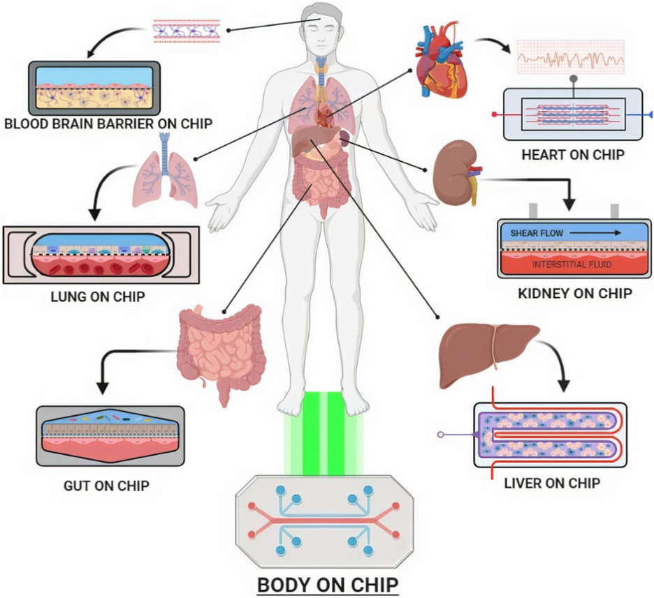
Figure 1: Schematic representation of major organ-on-chip platforms including blood–brain barrier-on-chip, lung-on-chip, heart-on-chip, kidney-on-chip, liver-on-chip and gut-on-chip, along with the concept of a body-on-chip system that integrates multiple organ models to simulate systemic physiological interactions.
2. CONCEPT AND PRINCIPLE OF ORGAN-ON-CHIP TECHNOLOGY
Organ-on-chip technology represents a rapidly evolving interdisciplinary platform that combines principles of microfluidics, tissue engineering, biomaterials science and cell biology to replicate key structural and functional features of human organs in a controlled in vitro environment. These micro engineered systems are designed to simulate the complex physiological microenvironment of living tissues by incorporating living cells within precisely fabricated microchannels through which culture medium continuously flows. The dynamic flow conditions within these microchannels enable the recreation of important physiological parameters such as nutrient transport, mechanical stimulation and biochemical signalling, thereby allowing the system to more closely mimic in vivo conditions compared with conventional static cell culture models.[1,3]
The fundamental concept of organ-on-chip devices is based on the ability to reconstruct organ-specific microenvironments at the microscale. Typically, these devices consist of transparent polymeric chips containing interconnected microfluidic channels that are lined with living human cells. These channels are designed to replicate the architecture and functional interfaces present in specific organs, such as epithelial–endothelial barriers, vascular networks or tissue compartments. Continuous perfusion of culture medium through the microchannels provides mechanical cues similar to blood flow, while also facilitating the removal of metabolic waste products and delivery of nutrients to the cultured cells.[3]
A key feature that distinguishes organ-on-chip platforms from traditional cell culture systems is their ability to reproduce dynamic mechanical forces that play an important role in regulating cellular behaviour. In many organs, cells experience various physical stimuli such as shear stress, cyclic stretching or pressure variations. Organ-on-chip devices can reproduce these forces using flexible membranes or controlled fluid flow systems, allowing researchers to investigate how mechanical signals influence cellular functions, tissue organization and disease progression. This capability significantly improves the physiological relevance of these models and enables more accurate evaluation of drug responses.[4]
Another important aspect of organ-on-chip technology is the integration of multiple cell types within a single device to simulate complex tissue interactions. In the human body, organs consist of multiple interacting cell populations that communicate through biochemical and mechanical signals. By co-culturing different cell types within spatially organized microenvironments, organ-on-chip platforms can reproduce these interactions and better represent the functional complexity of living tissues. This feature is particularly valuable for studying disease mechanisms, drug metabolism and tissue responses under physiologically relevant conditions.[3,4]
Overall, the principle of organ-on-chip technology lies in the creation of miniaturized, biomimetic systems that recreate essential aspects of human organ physiology. Through the integration of microfluidic engineering, biomaterials and living cells, these platforms provide an advanced experimental model capable of bridging the gap between conventional in vitro cell culture and in vivo studies. As a result, organ-on-chip devices are increasingly being explored as powerful tools for drug discovery, toxicity testing and disease modelling in modern biomedical and pharmaceutical research.[2,4]
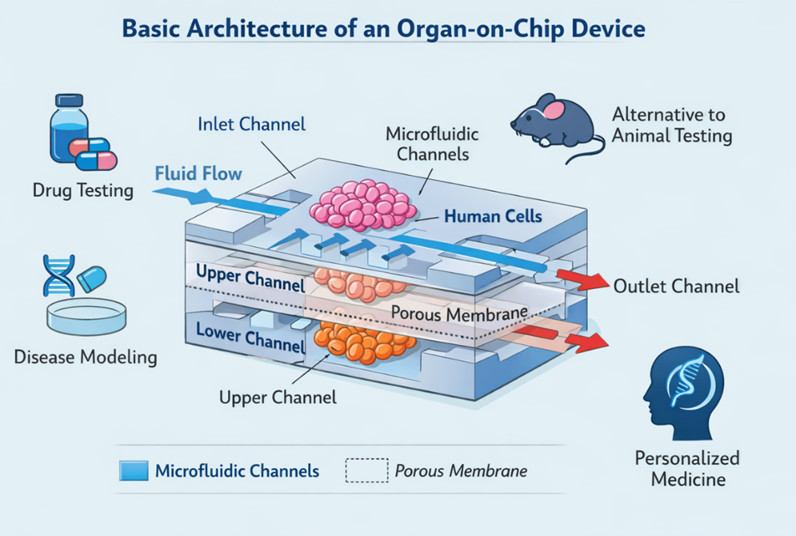
Figure 2: Schematic representation of a typical organ-on-chip device showing microfluidic channels, cell culture chambers and a porous membrane that enables interaction between different cell layers. Continuous fluid flow through the channels mimics physiological circulation and supports the transport of nutrients, metabolites and signalling molecules, thereby recreating a dynamic microenvironment for studying drug responses and disease mechanisms.
3. COMPONENTS OF ORGAN-ON-CHIP DEVICES
Organ-on-chip systems are composed of several integrated components that collectively recreate the microenvironment of human tissues. These devices combine microfluidic engineering, biomaterials and living cells to simulate the structural and functional characteristics of biological organs. The design of organ-on-chip platforms typically includes microfluidic channels, biomaterials used for device fabrication, appropriate cellular components and integrated sensing systems that enable monitoring of physiological responses. Each of these components plays a critical role in establishing a controlled environment that supports cell viability and replicates organ-specific functions.[3–5]
3.1 Microfluidic Channels
Microfluidic channels form the fundamental structural framework of organ-on-chip devices. These channels are typically fabricated at the micrometre scale and are designed to allow the controlled movement of fluids through the system. Continuous perfusion of culture medium through the microchannels enables the transport of nutrients, oxygen and signalling molecules to the cultured cells while simultaneously removing metabolic waste products. This dynamic fluid flow mimics physiological blood circulation and creates mechanical forces such as shear stress, which significantly influence cellular behaviour and tissue function.[3,5]
The geometry and arrangement of microfluidic channels are carefully designed to replicate the architecture of specific organs. For example, in lung-on-chip systems, parallel microchannels separated by a porous membrane simulate the interface between alveolar epithelial cells and vascular endothelial cells. Similarly, vascular networks within organ-on-chip platforms can reproduce capillary-like structures that facilitate realistic cellular interactions. The precise control of fluid flow also allows researchers to establish chemical gradients and simulate conditions such as inflammation or drug exposure within the device.[5]
3.2 Biomaterials Used in Device Fabrication
The selection of biomaterials is a critical factor in the construction of organ-on-chip systems because these materials must support cell growth while maintaining structural stability and optical transparency. One of the most widely used materials in organ-on-chip fabrication is polydimethylsiloxane (PDMS), a flexible silicone-based polymer that offers several advantages including biocompatibility, gas permeability and ease of microfabrication. PDMS-based devices can be rapidly produced using soft lithography techniques, making them suitable for experimental research applications.[5]
In addition to PDMS, other biomaterials such as hydrogels and thermoplastic polymers are increasingly being explored for organ-on-chip fabrication. Hydrogels can mimic the extracellular matrix environment by providing a three-dimensional scaffold that supports cellular attachment, proliferation and differentiation. Thermoplastic materials such as polymethyl methacrylate (PMMA) and polycarbonate are also used in some devices because they offer improved mechanical strength and scalability for potential commercial production.[6]
The choice of biomaterial also influences important device properties such as chemical absorption, mechanical flexibility and optical clarity. For instance, while PDMS is highly permeable to gases and suitable for cell culture applications, it may absorb certain hydrophobic drug molecules, which can affect drug testing results. Therefore, ongoing research focuses on developing alternative materials that maintain biocompatibility while minimizing these limitations.[6]
3.3 Cell Sources Used in Organ-on-Chip Systems
The cellular component of organ-on-chip devices is essential for replicating biological functions. Various types of cells can be incorporated into these systems depending on the target organ being modelled. Primary human cells, isolated directly from tissues, are commonly used because they retain many physiological characteristics of native cells. However, their limited availability and relatively short lifespan in culture can restrict their long-term use in experimental studies.[4,7]
To overcome these limitations, researchers often utilize stem cell-derived cells, including induced pluripotent stem cells (iPSCs), which can differentiate into multiple cell types. iPSC-derived cells provide an important advantage because they can be generated from patient-specific samples, enabling the development of personalized disease models and individualized drug testing platforms. Additionally, co-culture systems involving multiple cell types are frequently implemented in organ-on-chip devices to replicate complex tissue interactions and improve physiological relevance.[7]
3.4 Sensors and Monitoring Systems
Another important component of organ-on-chip devices is the integration of biosensors and monitoring systems that allow real-time analysis of cellular responses. These sensors can detect various biological parameters such as pH, oxygen concentration, metabolic activity and electrical signals generated by cells. By continuously monitoring these parameters, researchers can obtain valuable insights into tissue function, drug responses and disease progression within the micro engineered environment.[8]
Advanced organ-on-chip platforms may incorporate optical sensors, electrochemical sensors or microelectrode arrays that enable non-invasive measurement of cellular activity. For example, heart-on-chip systems often include electrical sensing systems to monitor cardiomyocyte contraction patterns, while liver-on-chip devices may measure metabolic biomarkers associated with drug metabolism. The integration of such sensing technologies enhances the analytical capability of organ-on-chip systems and supports their application in pharmaceutical research and toxicological studies.[8]
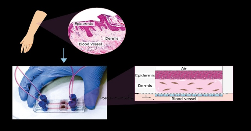
Figure 3: Skin-on-chip model illustrating the reconstruction of epidermal and dermal layers within a microfluidic device that mimics the physiological microenvironment of human skin for drug testing and dermatological research.
Table 1: Major Components of Organ-on-Chip Devices and Their Functions
|
Component |
Function |
|
Microfluidic channels |
Control fluid flow and simulate blood circulation |
|
Biomaterials (PDMS, hydrogels, polymers) |
Provide structural framework and support cell growth |
|
Cellular components |
Reproduce biological functions of specific organs |
|
Sensors and monitoring systems |
Enable real-time measurement of physiological parameters |
4. FABRICATION TECHNIQUES OF ORGAN-ON-CHIP DEVICES
The fabrication of organ-on-chip devices involves the use of advanced microengineering techniques that enable the precise construction of microscale structures capable of supporting living cells and controlled fluid flow. These fabrication methods are derived primarily from microelectronics and microfluidics technologies, where small-scale structures can be produced with high accuracy and reproducibility. The selection of an appropriate fabrication technique plays a critical role in determining the structural complexity, scalability and overall performance of the organ-on-chip system. Several fabrication strategies have been developed for constructing these devices, including soft lithography, photolithography, 3D printing and injection moulding, each offering distinct advantages depending on the intended application.[3,9]
4.1 Soft Lithography
Soft lithography is one of the most widely used fabrication methods for developing microfluidic devices, including organ-on-chip systems. This technique involves the use of elastomeric materials, typically polydimethylsiloxane (PDMS), to create microstructure Molds that replicate the desired microchannel architecture. The fabrication process generally begins with the preparation of a master Mold using photolithography, followed by casting liquid PDMS onto the Mold and curing it to form a flexible polymeric structure containing the microchannels.[5,9]
One of the major advantages of soft lithography is its ability to produce highly precise microstructures while remaining relatively cost-effective and simple to implement. The flexibility and optical transparency of PDMS also allow researchers to observe cellular behaviour directly under microscopy. Furthermore, PDMS exhibits gas permeability, which facilitates oxygen transport and supports long-term cell viability within the microfluidic device. Due to these characteristics, soft lithography has become a standard technique for prototyping organ-on-chip systems used in experimental research.[6,9]
However, despite its widespread use, soft lithography also presents certain limitations. For example, PDMS materials may absorb hydrophobic molecules, which can interfere with drug testing experiments. Additionally, the manual fabrication process may limit large-scale manufacturing and standardization of organ-on-chip devices for commercial applications.[6]
4.2 Photolithography
Photolithography is another important microfabrication technique that plays a foundational role in the development of microfluidic devices. In this method, a photosensitive material known as photoresist is applied to a substrate and exposed to ultraviolet (UV) light through a patterned mask. The exposed regions of the photoresist undergo chemical changes that allow selective removal during subsequent development steps, thereby creating microscale patterns on the substrate surface.[9]
This technique enables the production of highly precise and reproducible microstructures, which can subsequently be used as master Molds for fabricating microfluidic devices through soft lithography. Photolithography provides excellent control over microchannel dimensions and geometry, making it suitable for creating complex microfluidic architectures required in organ-on-chip platforms. Because of its high resolution and reliability, photolithography remains a fundamental step in the fabrication workflow of many organ-on-chip devices.[9,10]
4.3 Three-Dimensional (3D) Printing
Recent advances in additive manufacturing have introduced 3D printing as an emerging technique for the fabrication of organ-on-chip systems. Unlike traditional microfabrication approaches that rely on Molds or masks, 3D printing constructs structures layer by layer based on digital design models. This technique offers significant flexibility in device design and enables the rapid production of complex three-dimensional architectures that may be difficult to achieve using conventional fabrication methods.[10]
Various 3D printing technologies, such as stereolithography and extrusion-based printing, have been explored for creating microfluidic devices and biological scaffolds. One of the major advantages of 3D printing is its ability to integrate multiple components within a single fabrication process, including microchannels, cell culture chambers and support structures. Additionally, 3D printing facilitates rapid prototyping and customization, which is particularly useful for developing patient-specific models in personalized medicine research.[10]
Despite these advantages, the resolution of many 3D printing methods remains lower than that of traditional photolithography techniques. Furthermore, the availability of biocompatible printing materials suitable for long-term cell culture is still an area of active research.[10]
4.4 Injection Molding
Injection molding is a fabrication technique commonly used for the large-scale production of polymer-based microfluidic devices. In this process, molten thermoplastic materials are injected into precisely designed Molds where they solidify to form the final microstructure device. Unlike soft lithography, which is primarily used for laboratory-scale prototyping, injection molding offers significant advantages in terms of mass production, reproducibility and industrial scalability.[11]
Thermoplastic materials such as polycarbonate, polymethyl methacrylate (PMMA) and cyclic olefin polymers are often used in injection molding due to their mechanical stability and optical clarity. Devices fabricated using these materials exhibit improved chemical resistance and reduced absorption of small molecules compared with PDMS-based systems. Consequently, injection molding is increasingly being considered for the commercial manufacturing of organ-on-chip platforms intended for pharmaceutical testing and biomedical applications.[11]
Overall, the fabrication of organ-on-chip devices relies on the integration of advanced microengineering techniques that enable precise control over device architecture and functionality. Each fabrication strategy offers unique advantages depending on the intended application, whether for experimental prototyping or industrial-scale production. Continued improvements in microfabrication technologies are expected to enhance the reliability, scalability and accessibility of organ-on-chip platforms, thereby facilitating their broader adoption in drug discovery and biomedical research.[3,10,11]
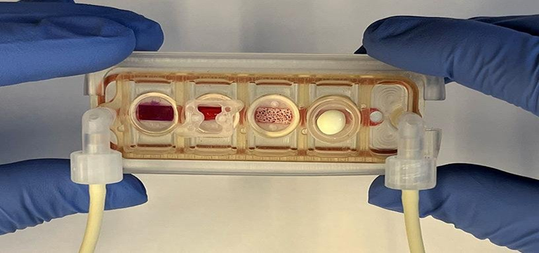
Figure 4: Representative microfluidic organ-on-chip device containing interconnected chambers and perfusion channels for culturing cells and simulating physiological fluid flow in vitro.
5. TYPES OF ORGAN-ON-CHIP MODELS
Organ-on-chip technology has enabled the development of a wide range of micro engineered platforms that replicate the structural and functional characteristics of individual human organs. These devices are designed to recreate the physiological microenvironment of specific tissues by integrating living cells, microfluidic channels and mechanical stimuli within a controlled microfabricated system. By mimicking essential organ-level functions, organ-on-chip platforms provide valuable tools for studying disease mechanisms, drug metabolism and toxicity responses in vitro. Over the past decade, numerous organ-specific models have been developed, including lung-on-chip, liver-on-chip, heart-on-chip, kidney-on-chip, brain-on-chip and gut-on-chip systems, each designed to reproduce unique biological functions of the respective organ.[4,12]
5.1 Single-Organ Chip and Multi-Organ Chip Systems
Organ-on-chip platforms can be broadly categorized into single-organ chips and multi-organ chip systems depending on the number of tissues represented within the device. Single-organ chips are designed to replicate the microenvironment of a specific organ by incorporating relevant cell types and physiological conditions. These systems are commonly used for studying organ-specific processes such as drug metabolism in liver-on-chip models or cardiotoxicity in heart-on-chip systems.[12]
In contrast, multi-organ chip systems, sometimes referred to as body-on-chip platforms, integrate multiple organ models within a single interconnected microfluidic network. These systems aim to simulate interactions between different organs, thereby providing a more comprehensive representation of human physiology. For example, a multi-organ chip may connect liver, heart and kidney tissues to study how drug metabolites generated in the liver influence cardiac or renal functions. Such integrated platforms are particularly useful for investigating systemic drug effects, pharmacokinetics and multi-organ toxicity.[13]
5.2 Lung-on-Chip
The lung-on-chip model is one of the earliest and most widely studied organ-on-chip systems. It was designed to replicate the alveolar–capillary interface of the human lung, where gas exchange occurs between air and blood. Typically, this device consists of two microfluidic channels separated by a flexible porous membrane. Human alveolar epithelial cells are cultured on one side of the membrane, while vascular endothelial cells are grown on the opposite side to simulate the air–blood barrier present in the lungs.[1,14]
A key feature of lung-on-chip devices is the ability to reproduce cyclic mechanical stretching that mimics breathing movements. This mechanical stimulation is achieved by applying vacuum pressure to flexible side chambers, causing the membrane to expand and contract rhythmically. By recreating both airflow and mechanical strain, lung-on-chip systems provide a physiologically relevant model for studying respiratory diseases, pulmonary drug delivery and inflammatory responses in the lung.[14]
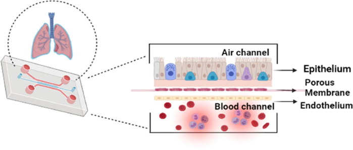
Figure 5: Schematic of lung on chip portraying two sections namely - air channel and blood channel which mimics the human lung
5.3 Liver-on-Chip
The liver plays a central role in drug metabolism, detoxification and biochemical homeostasis. Consequently, liver-on-chip models are widely used in pharmaceutical research to study drug metabolism and hepatotoxicity. These devices typically contain microfluidic chambers lined with hepatocytes and supporting cells such as Kupffer cells or endothelial cells, allowing the recreation of the complex cellular interactions that occur within hepatic tissue.[12]
Microfluidic perfusion within liver-on-chip systems maintains continuous nutrient supply and waste removal, thereby improving the viability and metabolic activity of hepatocytes compared with static culture conditions. This dynamic environment enables more accurate assessment of drug metabolism pathways, enzyme activity and toxicity responses. As a result, liver-on-chip models are increasingly used for predicting drug-induced liver injury during early stages of drug development.[15]
5.4 Heart-on-Chip
Heart-on-chip platforms are designed to reproduce the contractile behaviour and electrophysiological properties of cardiac tissue. These systems typically incorporate cardiomyocytes, often derived from induced pluripotent stem cells, cultured within micro engineered structures that support synchronized contraction and electrical signalling.[16]
One of the major applications of heart-on-chip technology is the evaluation of cardiotoxicity, which is a leading cause of drug withdrawal during clinical development. By monitoring parameters such as contraction frequency, electrical activity and calcium signalling, heart-on-chip models allow researchers to assess the effects of pharmaceutical compounds on cardiac function with greater physiological relevance than conventional cell culture models.[16]
5.5 Kidney-on-Chip
Kidney-on-chip devices are developed to replicate the filtration and transport functions of the renal system, particularly within the nephron, the functional unit of the kidney. These systems typically include microfluidic channels lined with renal epithelial cells that simulate the structure of renal tubules. Controlled fluid flow through the device recreates physiological shear stress conditions experienced by kidney cells in vivo.[12]
Kidney-on-chip models are particularly useful for studying drug-induced nephrotoxicity, which is a major concern during pharmaceutical development. By monitoring cellular responses under physiologically relevant flow conditions, these devices provide valuable insights into renal drug transport, filtration processes and toxicity mechanisms.[17]
5.6 Brain-on-Chip
The brain-on-chip model aims to replicate the complex microenvironment of the central nervous system, including the blood–brain barrier (BBB). This barrier plays a crucial role in regulating the transport of molecules between the bloodstream and brain tissue. Brain-on-chip systems typically incorporate neuronal cells, astrocytes and endothelial cells arranged within microfluidic channels to simulate neural tissue and vascular interactions.[18]
These platforms enable researchers to study neurological disorders, neuroinflammation and drug transport across the blood–brain barrier. Brain-on-chip models are also increasingly being used for investigating neurodegenerative diseases such as Alzheimer’s and Parkinson’s disease.[18]
5.7 Gut-on-Chip
The gut-on-chip model replicates the microenvironment of the intestinal epithelium, including peristaltic motion and microbial interactions within the gastrointestinal tract. These devices typically contain intestinal epithelial cells cultured within microfluidic channels that simulate nutrient flow and mechanical forces present in the digestive system.[19]
Gut-on-chip systems are valuable for studying drug absorption, intestinal barrier function and host–microbiome interactions. By recreating physiological conditions of the intestinal environment, these platforms provide improved models for investigating gastrointestinal diseases and evaluating oral drug delivery systems.[19]
Table 2: Examples of Organ-on-Chip Models and Their Pharmaceutical Applications
|
Organ Model |
Key Function Simulated |
Pharmaceutical Application |
|
Lung-on-chip |
Alveolar–capillary gas exchange |
Respiratory disease research and inhalation drug testing |
|
Liver-on-chip |
Drug metabolism and detoxification |
Hepatotoxicity screening |
|
Heart-on-chip |
Cardiac contraction and electrical activity |
Cardiotoxicity assessment |
|
Kidney-on-chip |
Filtration and tubular transport |
Nephrotoxicity evaluation |
|
Brain-on-chip |
Blood–brain barrier function |
Neurological drug delivery studies |
|
Gut-on-chip |
Intestinal absorption and microbiome interaction |
Oral drug absorption studies |
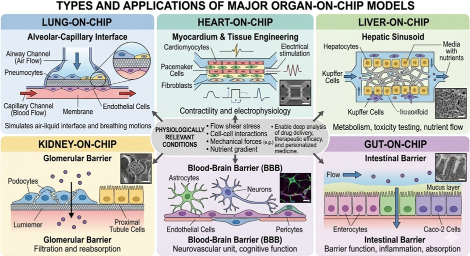
Figure 6: Representative organ-on-chip models including lung-on-chip, heart-on-chip, liver-on-chip, kidney-on-chip, brain-on-chip and gut-on-chip platforms used to simulate organ-specific physiological functions for biomedical and pharmaceutical research
6. APPLICATIONS OF ORGAN-ON-CHIP TECHNOLOGY IN PHARMACEUTICAL RESEARCH
Organ-on-chip technology has rapidly emerged as a powerful tool in pharmaceutical research due to its ability to reproduce physiologically relevant tissue microenvironments within a controlled in vitro system. By integrating microfluidic engineering with living human cells, these platforms provide advanced experimental models capable of simulating organ-level functions and dynamic biological processes. Consequently, organ-on-chip devices are increasingly being utilized for multiple pharmaceutical applications, including drug discovery, toxicity assessment, pharmacokinetic investigations and personalized medicine approaches.[12,20]
6.1 Drug Screening and Drug Discovery
One of the primary applications of organ-on-chip technology is in drug screening and early-stage drug discovery. Conventional drug screening methods often rely on two-dimensional cell cultures that lack the structural complexity and dynamic physiological conditions of living tissues. As a result, these models frequently fail to accurately predict drug responses in humans. Organ-on-chip platforms overcome these limitations by recreating tissue-specific microenvironments that allow more realistic evaluation of drug efficacy and cellular responses.[20]
Microfluidic drug screening systems enable controlled delivery of pharmaceutical compounds to cultured cells under physiologically relevant conditions. The continuous flow of culture medium within microchannels allows precise regulation of drug concentration gradients and exposure times. This dynamic environment closely resembles the circulation of drugs in the human body and facilitates more accurate assessment of pharmacological effects. Furthermore, organ-on-chip platforms allow high-content analysis of cellular responses, including gene expression, metabolic activity and morphological changes following drug treatment.[20,21]
Another advantage of organ-on-chip technology is the ability to perform high-throughput screening of drug candidates. Multiple microfluidic chambers can be incorporated within a single device, enabling simultaneous evaluation of numerous compounds under controlled experimental conditions. This capability significantly improves the efficiency of drug discovery pipelines and reduces the cost associated with large-scale screening programs.[21]
6.2 Toxicity Testing
Toxicity assessment is a critical component of drug development, as many pharmaceutical compounds fail during clinical trials due to unexpected toxic effects. Organ-on-chip systems provide improved models for studying drug toxicity because they replicate key physiological features of human tissues that are often absent in conventional cell culture systems. For example, liver-on-chip platforms can reproduce hepatic metabolic functions, allowing researchers to investigate drug-induced liver injury with greater accuracy.[12,15]
Similarly, heart-on-chip devices are widely used for evaluating cardiotoxicity, which is a major cause of drug withdrawal from the market. By monitoring parameters such as cardiomyocyte contraction patterns and electrical activity, these systems enable early detection of adverse drug effects on cardiac function. Kidney-on-chip models are also
valuable for assessing nephrotoxicity, as they replicate renal filtration and tubular transport mechanisms that influence drug clearance.[17]
The ability of organ-on-chip platforms to incorporate multiple cell types and dynamic fluid flow further enhances their relevance for toxicity testing. These systems can simulate interactions between tissues and circulating metabolites, thereby enabling more comprehensive evaluation of drug safety profiles during preclinical development.[12,21]
6.3 Pharmacokinetic and Pharmacodynamic Studies
Organ-on-chip systems have also been explored for investigating pharmacokinetic (PK) and pharmacodynamic (PD) processes, which describe how drugs are absorbed, distributed, metabolized and eliminated in the body. Multi-organ chip platforms, where several organ models are interconnected through microfluidic channels, allow simulation of systemic drug circulation and inter-organ interactions.[13]
For example, a multi-organ chip that integrates liver, heart and kidney models can be used to study how hepatic metabolism influences drug distribution and toxicity in other tissues. Such systems enable researchers to analyse drug transport, metabolic conversion and elimination pathways within a physiologically relevant context. By providing real-time monitoring of drug behaviour across multiple tissues, organ-on-chip platforms offer valuable insights into pharmacokinetic mechanisms that are difficult to capture using traditional experimental models.[21]
6.4 Personalized Medicine
Another promising application of organ-on-chip technology is in the field of personalized medicine. Advances in stem cell technology, particularly induced pluripotent stem cells (iPSCs), allow the generation of patient-specific cells that can be incorporated into organ-on-chip devices. These personalized models enable researchers to study how individual genetic backgrounds influence disease progression and drug responses.[7,22]
Patient-derived organ-on-chip systems can be used to evaluate the effectiveness and safety of therapeutic interventions before administering them to patients. This approach may improve treatment outcomes by enabling the selection of drugs that are most effective for a specific individual. Additionally, personalized organ-on-chip platforms have the potential to accelerate the development of targeted therapies for complex diseases such as cancer and neurodegenerative disorders.[22]
Table 3: Major Pharmaceutical Applications of Organ-on-Chip Technology.
|
Application Area |
Purpose |
Example Organ-on-Chip Model |
|
Drug discovery |
Screening and evaluation of new drug candidates |
Liver-on-chip, lung-on-chip |
|
Toxicity testing |
Detection of drug-induced organ damage |
Heart-on-chip, kidney-on-chip |
|
Pharmacokinetic studies |
Investigation of drug metabolism and distribution |
Multi-organ chip systems |
|
Personalized medicine |
Patient-specific drug response prediction |
iPSC-based organ-on-chip |
7. ADVANTAGES OF ORGAN-ON-CHIP SYSTEMS
Organ-on-chip technology offers several significant advantages compared with conventional experimental models such as two-dimensional cell cultures and animal models. By integrating microfluidic engineering, biomaterials and living human cells, these systems recreate physiologically relevant tissue microenvironments that enable more accurate investigation of biological processes and drug responses. As a result, organ-on-chip platforms have become increasingly valuable tools in pharmaceutical research, toxicology studies and disease modelling.[12,23]
Major advantages of organ-on-chip systems include:
Organ-on-chip platforms mimic the structural and functional characteristics of human tissues by recreating tissue–tissue interfaces, fluid flow and mechanical forces that regulate cellular behaviour in vivo.[3,4]
Because these systems often utilize human-derived cells, they provide more reliable prediction of human pharmacological responses compared with animal models that may exhibit species-specific differences.[12,20]
Organ-on-chip technology offers an alternative experimental platform that can reduce dependence on animal testing, thereby addressing ethical concerns and regulatory limitations associated with animal studies.[23]
Unlike conventional cell cultures, organ-on-chip devices allow controlled fluid flow that replicates physiological circulation, enabling accurate simulation of nutrient transport, drug delivery and mechanical stress.[3]
Integrated biosensors and imaging technologies enable continuous monitoring of cellular responses such as metabolic activity, electrical signalling and molecular biomarker expression.[8]
Organ-on-chip models provide advanced platforms for investigating complex disease processes by reproducing organ-specific microenvironments and cellular interactions.[12]
By incorporating patient-derived cells, these systems allow researchers to evaluate individual drug responses and develop patient-specific therapeutic strategies.[22]
High-throughput screening capability and more predictive experimental outcomes can reduce time and cost associated with drug development pipelines.[20]
Table 4: Comparison of Conventional Experimental Models and Organ-on-Chip Systems
|
Feature |
2D Cell Culture Models |
Animal Models |
Organ-on-Chip Systems |
|
Physiological relevance |
Limited representation of tissue structure |
Moderate but species differences exist |
High relevance using human cells |
|
Mechanical stimulation |
Absent |
Present but difficult to control |
Precisely controlled |
|
Cell–cell interactions |
Limited |
Present |
Recreated through co-culture |
|
Ethical concerns |
Minimal |
Significant |
Reduced animal use |
|
Drug response prediction |
Often inaccurate |
Variable |
More predictive for humans |
|
Real-time monitoring |
Limited |
Difficult |
Integrated sensors allow monitoring |
8. LIMITATIONS AND CHALLENGES OF ORGAN-ON-CHIP TECHNOLOGY
Although organ-on-chip technology offers significant advantages for biomedical research and pharmaceutical applications, several limitations and technical challenges still restrict its widespread implementation. These limitations arise from factors related to device fabrication, material properties, experimental standardization and regulatory acceptance. Addressing these challenges is essential for improving the reliability, scalability and practical application of organ-on-chip platforms in drug development and disease modelling.[12,24]
Major limitations and challenges of organ-on-chip systems include:
Development of organ-on-chip devices requires advanced microfabrication techniques such as photolithography and soft lithography. These procedures often involve specialized equipment and technical expertise, which may limit accessibility for many research laboratories.[24]
Polydimethylsiloxane (PDMS), commonly used in organ-on-chip fabrication, can absorb hydrophobic drug molecules. This property may alter drug concentrations within microfluidic systems and affect the accuracy of pharmacological experiments.[6,24]
Currently, there is significant variability in device design, fabrication methods and experimental protocols among different research groups. This lack of standardization makes it difficult to reproduce results and compare findings across studies.[24,25]
Most organ-on-chip devices simulate individual organs and may not fully capture the complex interactions between multiple organs within the human body. Although multi-organ chip systems are being developed, accurately replicating complete systemic physiology remains challenging.[13,25]
The fabrication of microfluidic devices and the integration of specialized sensors or biomaterials can increase research costs. These economic factors may limit large-scale implementation of organ-on-chip systems.[25]
Despite promising results, regulatory agencies have not yet fully integrated organ-on-chip platforms into standardized drug testing frameworks. Extensive validation studies are required before these systems can replace conventional preclinical testing methods.[24]
Table 5: Limitations of Organ-on-Chip Technology and Potential Solutions
|
Limitation |
Description |
Possible Solutions |
|
Fabrication complexity |
Requires advanced microfabrication techniques |
Development of simplified and automated manufacturing processes |
|
PDMS material limitations |
Absorption of hydrophobic drug molecules |
Use of alternative polymers or thermoplastic materials |
|
Lack of standardization |
Variability in device design and experimental methods |
Establish international guidelines and standardized protocols |
|
Incomplete physiological representation |
Difficulty replicating multi-organ interactions |
Development of integrated multi-organ or body-on-chip systems |
|
High cost |
Expensive materials and specialized equipment |
Scalable and cost-effective fabrication technologies |
|
Regulatory barriers |
Limited regulatory frameworks for new testing platforms |
Collaboration between researchers, industry and regulatory agencies |
9. FUTURE PERSPECTIVES OF ORGAN-ON-CHIP TECHNOLOGY
Organ-on-chip technology is rapidly evolving and is expected to significantly influence future biomedical research and pharmaceutical development. Continuous progress in microfabrication techniques, biomaterials and stem cell technologies is improving the physiological relevance and functional capabilities of these micro engineered systems. As organ-on-chip platforms become more advanced, they are increasingly being explored for applications in drug discovery, disease modelling and personalized medicine. Future research efforts are expected to focus on enhancing device integration, improving scalability and incorporating emerging technologies such as artificial intelligence and advanced sensing systems.[4,26]
One important direction in the evolution of organ-on-chip technology is the development of multi-organ or body-on-chip systems. These platforms integrate multiple organ models within a single microfluidic network, enabling researchers to simulate interactions between organs and study systemic physiological responses. Such integrated systems may provide valuable insights into drug metabolism, distribution and toxicity across different tissues, thereby improving the predictive accuracy of preclinical drug evaluation.[13,26]
Another promising advancement involves the integration of biosensors and real-time monitoring technologies. The incorporation of microelectronic sensors and imaging systems allows continuous measurement of physiological parameters such as oxygen concentration, metabolic activity and electrical signals generated by cells. These monitoring capabilities provide dynamic information about tissue responses and enhance the analytical potential of organ-on-chip platforms.[8,26]
The combination of organ-on-chip technology with artificial intelligence and computational modelling also represents a major emerging research direction. Machine learning algorithms can analyse complex biological datasets generated from organ-on-chip experiments, facilitating improved prediction of drug responses and identification of potential toxicity signals. AI-based tools may also assist in optimizing device design and accelerating drug discovery processes.[27]
Advancements in stem cell technology further expand the potential of organ-on-chip systems for personalized medicine. By using patient-derived induced pluripotent stem cells, researchers can generate individualized organ models that reflect specific genetic backgrounds and disease conditions. These personalized models may enable patient-specific drug screening and contribute to the development of precision therapeutic strategies.[22,27]
Key Future Directions of Organ-on-Chip Technology
CONCLUSION
Organ-on-chip technology represents a significant advancement in the development of physiologically relevant in vitro models for biomedical and pharmaceutical research. By integrating microfluidic engineering, biomaterials and living human cells, these micro engineered platforms are capable of replicating essential structural and functional characteristics of human organs. Such systems provide more realistic experimental environments compared with traditional two-dimensional cell cultures and offer improved predictive capability for studying drug efficacy, toxicity and disease mechanisms.
The development of various organ-specific models, including lung-on-chip, liver-on-chip, heart-on-chip, kidney-on-chip and brain-on-chip platforms, has demonstrated the potential of these systems to simulate complex biological processes under controlled laboratory conditions. These platforms have already shown considerable promise in pharmaceutical applications such as drug screening, toxicity assessment, pharmacokinetic analysis and disease modelling. In addition, the incorporation of patient-derived cells into organ-on-chip devices offers new opportunities for personalized medicine and individualized therapeutic evaluation.
Despite these promising advancements, several technical and regulatory challenges remain. Issues related to device fabrication complexity, material limitations, lack of standardization and regulatory validation must be addressed before organ-on-chip technology can be widely implemented in routine drug development processes. Continued interdisciplinary collaboration among engineers, biologists, clinicians and pharmaceutical scientists will therefore be essential to overcome these challenges and further improve the reliability and scalability of these systems.
Future developments in organ-on-chip technology are expected to focus on the integration of multi-organ systems, advanced biosensing technologies and artificial intelligence-based analytical tools. These innovations may significantly enhance the predictive accuracy of preclinical studies and reduce dependence on animal testing. As technological advancements continue, organ-on-chip platforms are likely to play an increasingly important role in transforming drug discovery, improving disease modelling and supporting the advancement of personalized healthcare strategies.
REFERENCES


Shruti Taware, Poonam Thite, Omkar Wable, Sarthak Unavane, Organ-On-Chip Technology: Bridging the Gap Between in Vitro Models and Human Physiology, Int. J. of Pharm. Sci., 2026, Vol 4, Issue 4, 1078-1096. https://doi.org/10.5281/zenodo.19460177
 10.5281/zenodo.19460177
