View Article

Abstract

Peptic ulcer disease (PUD) is a chronic gastrointestinal disorder caused by an imbalance between protective mucosal mechanisms and harmful factors such as gastric acid, pepsin, NSAIDs, alcohol, and Helicobacter pylori infection. Current therapies, including proton pump inhibitors, H? receptor antagonists, and prostaglandin analogs, are effective. Experimental assessment of anti-ulcer effects employs various in vivo models, including pylorus ligation, ethanol, histamine, NSAID, stress, and reserpine-induced ulcer models, alongside in vitro assays such as H?/K?-ATPase inhibition and titration-based neutralization tests. Every model provides additional insights into ulcer development and the effects of medication. Using in vivo and in vitro methods together may enhance the discovery of safer and more effective anti-ulcer medications, particularly from plant origins, offering appealing alternatives to conventional therapies.

Keywords

Peptic ulcer, Antiulcer drugs, Ulcer score, H. pylori, Screening models, NSAIDs

Introduction

A peptic ulcer (PU) is defined as a specific lesion, which consists of a loss of continuity of the proximal gastric and/or duodenal mucosa.[35]

Ulcerations are primarily crater-shaped sores or lesions that may occur on the stomach, colon, or oral mucosa. Once an ulcer forms, the area may bleed or release disagreeable substances, creating a painful sore that is prone to infection [2]. Patients experiencing peptic ulcer disease may not exhibit any symptoms at all, or they might experience heartburn, epigastric discomfort, anorexia, nausea, vomiting, and belching and bloating.

Stress, alcohol use, smoking, H. pylori infection, and drug and chemical ingestion are a few of the causes. Particularly, prolonged alcohol use, cigarette smoking, and long-term NSAID use are contributing factors to peptic ulcers. Free radicals are known to have a part in the pathophysiology of peptic ulcers caused by damage to the mucosa. [3] A gastric ulcer is an ulcer in the stomach, and a duodenal ulcer is an ulcer in the first segment of the intestine.

Factors that enhance acid secretion include HCl, pepsin, refluxed bile, NSAIDs, alcohol, pancreatic proteolytic enzymes, ingested irritants, bacterial toxins, and physicochemical damage.  Digestive factors include blood flow, mucus, bicarbonates, epithelium resolution, and the state of treatment. [1]. When defensive mechanisms and aggressive parameters are out of balance, mucosal integrity is disrupted, which eventually leads to the development of a peptic ulcer.

Antiulcer drugs aim to do more than only delay the disease's course. Additionally, it seeks to bring about remission as soon as possible and keep it going for a long time. Many traditional therapy techniques are being used to address PUD; however, it's important to keep in mind that each of these approaches has one or more unpleasant side effects. Currently known anti-ulcerative drugs aim to do more than just slow the progression of the condition. Additionally, it seeks to quickly establish and maintain a state of remission for a long time.

Table 1: The Anti-Ulcerative Effects of Diverse Plants in Preclinical Studies [2]

Medicinal Plants

Family

Phytoconstituents

Plant parts and solvents used

Animals used in preclinical studies

Reference

Annona muricata

Annonaceae

Acetogenins, Alkaloids, Flavonoids Tannins, Essential oil (trace)

Leaves; Maceration (Ethanol)

In vivo on male and female albino mice (Mus musculus),

Elizângela Beneval Bento et al 2018[11]

Arctium lappa

Asteraceae

Arctiin, Arctigenin, Inulin, Tannins, Polyacetylenes


Roots; Percolation (Ethanol 96%)

In vivo on female Wistar rats (Rattus norvegicus)

Luisa Mota da Silva et al 2018[12]

Artemisia absinthium

Asteraceae

Thujone , thujyl alcohol, cineole, Absinthin, anabsinthin, Flavonoids, Tannins, Organic acids

Aerial parts; Maceration (Water)

In vivo on female Wistar rats (R. norvegicus)

Thaise Boeing et al 2023[13]

Bryophyllum pinnatum

Crassulaceae

Bufadienolides, quercetin, kaempferol, Triterpenes, Alkaloids, Tannins,

Leaves; Maceration (Methanol

In vivo on Swiss mice (M. musculus)

Laure Brigitte Kouitcheu Mabeku

et al 2017 [14]

Casearia sylvestris

Salicaceae

Clerodane, diterpenes, Flavonoids, Tannins, Trace essential oil

Leaves; Infusion (Distilled Water)

In vivo on female Wistar rats (R. norvegicus)

Beatriz Monteiro Magalhães de Oliveira

et al 2022 [15]

Cinnamomum glanduliferum

Lauraceae

Cinnamaldehyde, Eugenol, Linalool, Safrole, Camphor

Leaves; Hydro distillation (Water)

In vivo on male Wistar albino rats (R. norvegicus)

Azab SS et al 2017[16]

Phyllanthus niruri

Phyllanthaceae

Phyllanthin, hypophyllanthin, Quercetin, kaempferol, Phyllantine, Tannins, Saponins, Terpenoids,

Leaves; Maceration (Methanol)

In vivo on female Swiss albino rats (R. norvegicus)

Mostofa R

et al 2017 [17]

Pathogenesis of ulcer [6]:
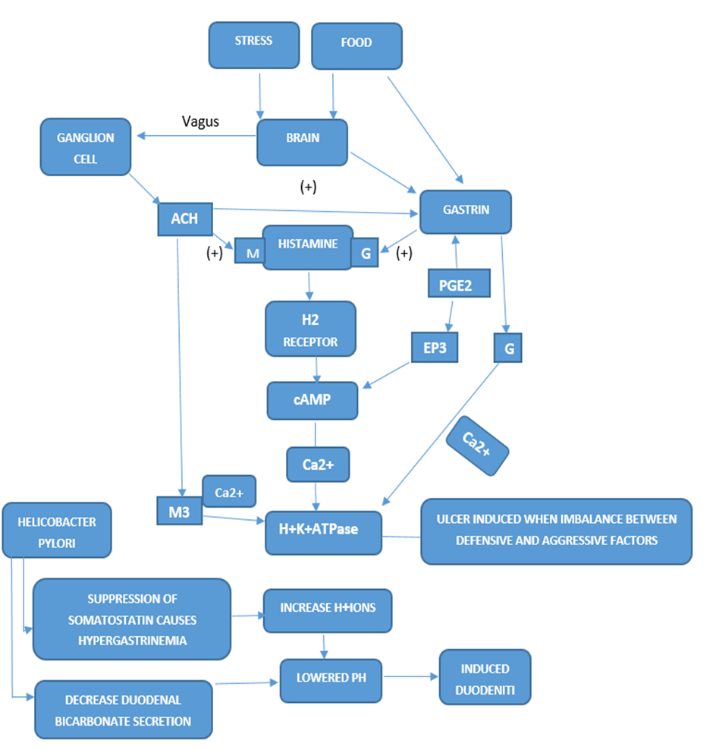
Figure no: I

Treatment strategies for peptic ulcer include [1]

Table No: II

Class

Subclass

Drugs

Gastric acid secretion inhibitors

Proton pump inhibitors

1. Omeprazole

2. Lansoprazole

3. Pantoprazole

4. Rabeprazole


H2 Receptor antagonist

1. Cimetidine

2. Ranitidine

3. Famotidine

4. Roxatidine


Prostaglandin analogues

1. Misoprostol

2. Enprostil

3. Rioprostil


Anticholinergics

1. Pirenzepine

2. Propanthelin

3. Oxyphenonium

Gastric acid neutralizers (Antacids)

Systemic

1. Sodium bicarbonate

2. Sodium citrate


Non systemic

1. Aluminium hydroxide

2. Magnesium hydroxide

Ulcer protective


1. Sucralfate

2. Colloidal bismuth subcitrate

Anti-H.Pylori


1. Amoxicillin

2. Clarithromycin

3. Metronidazole

4. Tinidazole

5. Tetracycline

Antiulcer screening methods include –

In vivo screening methods -

Pylorus ligated induced ulcer model (Shay rat method)

One of the oldest animal models for gastric ulcers [5]. The tying off of the pyloric region of the stomach results in the build-up of gastric acid within the stomach, leading to the formation of ulcers. [4] The autodigestion of the gastric mucosa causes the breakdown of the gastric mucosal barrier, resulting in ulcer formation, making the "Shay-rat" a useful method for assessing a drug molecule's anti-ulcer properties [6].

In this approach, Albino rats weighing 150-200 g are kept in separate cages and deprived of food for 24-36 hours before undergoing pyloric ligation. With light ether anaesthesia, a small midline incision is made in the abdomen just below the xiphoid process. The pyloric section of the stomach is tied off, ensuring no harm is done to its blood supply. The stomach is carefully replaced, and the abdominal wall is sutured closed with interrupted stitches. The medications are given subcutaneously right after pyloric ligation. The animals undergo a 48-hour fast during the postoperative phase and are euthanized 19 hours following the surgery. Stomachs are removed, and the contents are emptied into tubes. Then, the contents are analysed for pH, free acidity, and total acidity. The stomachs are subsequently incised along the greater curvature, and the inner surface is inspected for ulceration. The ulcer index is determined in this manner:

Ulcer Index = 10 / X,

where X = Total mucosal area / Total ulcerated area. [20], [21]

Ethanol-induced gastric ulcer model

Ethanol causes ulcer lesions by exposing the gastric mucosa to the hydrolytic and photolytic actions of HCl and pepsin [4]. These lesions are characterised by sub-epithelial haemorrhages, cellular exfoliation, inflammatory cell infiltration, and mucosal oedema [6].

Male Wistar rats (200–300 g) are used for this experiment. Animals are fasted for 18 hours with access to water. The test drug is given to animals orally. Administer the test drug or vehicle 30 minutes before ethanol. Absolute ethanol (95%–99%) is administered at a dose of 1mL/200g body weight to each animal, and after 1 hour, the animals are sacrificed by euthanasia with CO?, and their stomachs are dissected out.  The stomach is opened along the greater curvature, washed with warm water, and examined for ulcer scoring (tissue damage score 0 (least) – 3 (most)). [28, 29]

HR=CA−B(mmHg/h)

Where: HR = Healing Rate Equation, A = tensile strength (mmHg) at time-point C after puncture, B = tensile strength 30 min after puncture (avg. 143 mmHg), C = duration of experiment (h) [30, 31, 32, 33, 34]

Histamine-induced gastric ulcer model

Both increased stomach acid output and the vasospastic action of histamine influence the experimental induction of gastric ulcers in a number of animals [7]. Histamine's strong acid-stimulating and vasodilating properties cause increased vascular permeability, which is the mechanism by which it causes gastric ulcers. The histamine-induced ulcer model is based on these pharmacological actions of histamine, which makes it suitable for assessing the anti-secretory effects of possible ulcer-fighting medications and substances that act as H2-receptor antagonists. [34]

The experiment utilises male guinea pigs. Animals undergo a fasting period of 36 hours. A dosage of 50 mg of histamine acid sulphate is administered intraperitoneally. To avoid histamine toxicity, 5 mg of promethazine hydrochloride is administered intraperitoneally 15 minutes prior to and 15 minutes following the histamine injection. The test medication is given 30-45 minutes following the histamine injection. After 4 hours, the animals are killed, and the stomach is extracted and analysed. The assessment of ulcers is determined by the severity of the ulcers.

Assess ulcer severity

  • 0 - No abnormalities
  • 1 - Oedema of the mucosa
  • 2 - 1-5 tiny lesions (1-2 mm in diameter)
  • 3 - > 5 small to moderate (3-4 mm in diameter) lesions
  • 4 - ≥ 2 moderate lesions or 1 large (> 4 mm in size) lesion
  • 5 - Perforated wounds [27]

NSAIDs Induced Gastric Ulcer Model

NSAIDs lead to ulcers by blocking prostaglandin production via the inhibition of the cyclooxygenase enzyme in the COX pathway. Prostaglandins have a protective function by promoting the release of bicarbonates and mucus, preserving blood circulation, and controlling the regeneration and repair of mucus cells. A decrease in prostaglandin production may lead to inflammation and weaken the protective mucosal barrier in the stomach, potentially resulting in ulcer development.

In this approach, animals are subjected to fasting for 24 to 36 hours. An NSAID (either aspirin or indomethacin) is administered orally with an appropriate vehicle (water or 1% carboxymethylcellulose). The test drug is administered to animals one hour later. After four hours, the animals are euthanised, the stomach is removed, and the severity of the ulcer is evaluated. Amount of NSAIDs used to induce ulcers: Aspirin – 150 mg/kg of body weight, and Indomethacin – 40 to 100 milligrams for each kilogram of body weight. The maximum diameters of the lesions are measured and summed to give a total lesion score (in mm) for each animal, and the average score for each group is calculated. [18], [19]

Stress Ulcer

This form of ulcer is frequently observed in severely ill patients in the intensive care unit and occasionally leads to gastrointestinal bleeding, which is associated with significantly higher morbidity and mortality, even with intensive acid suppression therapy. [6] Stress ulcers are especially linked to heightened gastric acid secretion, decreased blood circulation to the stomach lining, and harm to the mucosal barrier of the gastrointestinal tract. The ways in which immobilization stress leads to stress ulcers are intricate and involve interactions among the nervous, endocrine, and immune systems. [22]

In this approach, 10 female Wistar rats, each weighing between 150 and 170 g, are randomly chosen per dose of the test medication and for control groups. Animals are denied food and water for 24 hours before the start of the experiment. Following oral or subcutaneous administration of the test compound/placebo, the animals are mildly anesthetized using appropriate anaesthesia such as ether. They are suspended horizontally in darkness at 20°C for 24 hours and ultimately euthanized using CO? anaesthesia. The stomach is excised and secured onto a cork plate, and the count and intensity of ulcers are recorded with a stereomicroscope using these scores: 0 = no ulcer, 1 = superficial ulcers, 2 = deep ulcers, 3 = perforation 

An ulcer index (UI) is calculated:

UI = UN + US + UP × 10?¹

  • UN = average number of ulcers per animal 
  • US = average severity score 
  • UP = percentage of animals with ulcers [34].

Reserpine-Induced Duodenal Ulcer

Reserpine is derived from the root of Rauwolfia serpentina, utilized for managing hypertension; however, it can lead to side effects such as gastric mucosal lesions (GMLs), sexual dysfunction, and depression due to excessive dosage and extended use. Reserpine causes gastric lesions by lowering sympathetic tone and raising cholinergic tone, leading to excessive secretion of gastric acid [6].

In this approach, rats deprived of food for 36 hours receive reserpine mixed in 10% Tween 80 (5–8 mg/kg, i.p.) as stated in [23]. Despite the model's dependence on acid, hypermotility appears to be more crucial than hypersecretion for causing gastric mucosal lesions. Typically, the substances or plant extracts under assessment are given to the test animals no less than 30 minutes prior to the administration of reserpine. The test animals are subsequently euthanized 24 hours later [7].

In vitro screening methods -

Preparation of H+/K+ - ATPase enzyme

The fresh Goat stomach was used to prepare the H+/ K+ - ATPase enzyme sample. The stomach of a goat was dissected open, washed, and the mucosa of the gastric fundus was removed, and the inner layer was scraped off to obtain parietal cells. Next, 10% Triton X-100 was added to a homogenate of parietal cells in 16 mM Tris buffer (pH 7.4), and the mixture was centrifuged at 5000xg for 10 minutes. The activity of the enzyme H+/K+- ATPase inhibitor was measured by isolating the supernatant. Bovine serum albumin was utilised as a reference reagent to calculate the protein concentration in the supernatant. The activity of H+K+ ATPase was measured in parietal cell extract. [9][10]

Using a Titration Method of Fortran’s Model for the Determination of Neutralizing Capacity 

A 250 ml beaker is used, and 90 ml of the prepared solution, along with the reference drug, is placed in separate beakers at 37°C. Continuous stirring is performed using a magnetic stirrer at 30 rpm to simulate stomach movements or the stomach environment. The prepared sample and standard reference are titrated individually with artificial gastric juice until the endpoint of pH 3 is reached. The volume of artificial gastric juice consumed is quantified as a reference drug. [24, 25, 26]

Neutralization Effects of Prepared Preparation on Artificial Gastric Acid

In this approach, we prepare laboratory acid, and the pH is modified to match stomach conditions, particularly between pH 1.2 and 2.5. This is a significantly acidic state where the mucosal layer starts to sustain damage. If this pH is not managed promptly, it leads to the development of gastric ulcers. The plant extract or formulated drug and reference drug are individually mixed with artificial gastric juice at a pH of 1.2 - 2.5, and subsequently, this combination is analysed for its neutralizing effect on artificial gastric juice using the titration method. During titration, surplus acid is neutralized, allowing us to find the precise quantity of the prepared solution that balances the artificial gastric juice. To prepare artificial gastric juice, dissolve NaCl (2 grams) and pepsin (3.2 mg) in distilled water (500 ml). Subsequently, hydrochloric acid (7.0 ml) is introduced, and an adequate quantity of water is added to raise the volume [24, 25].

CONCLUSION

In conclusion, a variety of anti-ulcer examination methods have been established worldwide, and we have a large number of experimental models accessible for assessing anti-ulcer therapies. Every model has a unique set of advantages and disadvantages.  This paper's main goal is to give a summary of the antiulcer models that are most commonly used by researchers in their gastrointestinal protection studies. Analysing antiulcer models, we discovered that the outlined models are frequently employed in accordance with treatment demands, and the timing and outcomes of the study impact the model selection.  Although in-vivo models are used more frequently compared to in-vitro models, our research indicates that combining the two could lead to better results and help find new pharmacological agents with better antiulcer capabilities.

REFERENCES

  1. Ghule PV, Deshmukh AR. A comprehensive review of the screening model for the pharmacological assessment of antiulcer drugs. EPRA Int J Multidiscip Res. 2024; 10(11):729–736. doi:10.36713/epra2013
  2. Muthusamy P, Jeyaraman R, Chinnathambi A, Balamuralikrishnan B, Thangavelu L, Chellan R, et al. Phytochemicals as potential antiulcer agents – A review. Journal of Applied Pharmaceutical Science. 2021;11(03):001–013. doi:10.7324/ JAPS.2021.1 10301
  3. Jain NK, Singh N, Kannojiya P, Garud N, Garud A, Tonpay SD. Pharmacological screening of antiulcer agents: A review. Int J Pharm Sci Res. 2010;1(9):29–37. Available from: https://www.ijpsr.com
  4. Meena DK, Jayanthi M. In-vivo models used for pre-clinical evaluation of anti-ulcer activity. Austin Pharmacol Pharm. 2018;3(2):1017.
  5. Parmar NS, Desai JK. A review of the current methodology for the evaluation of gastric and duodenal anti-ulcer agents. Indian J Pharmacol. 1993; 25:120–135.
  6. Parmar NS, Desai JK. A review of the current methodology for the evaluation of gastric and duodenal anti-ulcer agents. Indian J Pharmacol. 1993; 25:120-135
  7. Mishra AP, Bajpai A, Chandra S. A comprehensive review on the screening models for the pharmacological assessment of antiulcer drugs. Current Clinical Pharmacology. 2019;14:175-96 . doi: 10.2174/1574884714666190312143846.
  8. Adinortey MB, Ansah C, Galyuon I, Nyarko A. In Vivo Models Used for Evaluation of Potential Antigastroduodenal Ulcer Agents. Ulcers. 2013;2013:Article ID 796405. doi:10.1155/2013/796405.
  9. Singh R, Gupta A, Patel S. Pharmacological Screening Model and Its Treatment of Peptic Ulcer Disease. Journal for Research in Applied Sciences and Biotechnology. 2022;1(5):36-47. doi:10.55544/jrasb.1.5.4.
  10. Kumar R. Investigation of In- Vitro Method of Antiulcer Activity. Journal for Research in Applied Sciences and Biotechnology. 2023;2(1):264-7. doi:10.55544/jrasb.2.1.37.
  11. Bento EB, de B. Júnior FE, de Oliveira DR, et al. Antiulcerogenic activity of the hydroalcoholic extract of leaves of Annona muricata Linnaeus in mice. Saudi J Biol Sci. 2016;25(4):609-21. doi:10.1016/j.sjbs.2016.01.024
  12. da Silvada Silva LM, de M. Burci L, Crestani S, et al. Acid-gastric antisecretory effect of the ethanolic extract from Arctium lappa L. root: role of H+, K+-ATPase, Ca2+ influx and the cholinergic pathway. Inflammopharmacology. 2018;26(2):521-30. doi:10.1007/s10787-017-0378-5.
  13. Boeing T, de Souza J, Vilhena da Silva RC, Mariano LNB, Mota da Silva L, Gerhardt GM, et al. Gastroprotective effect of Artemisia absinthium L.: A medicinal plant used in the treatment of digestive disorders. J Ethnopharmacol. 2023;312:116488. Epub 2023 Apr 13. doi:10.1016/j.jep.2023.116488.
  14. Kouitcheu Mabeku LB, Eyoum Bille B, Tchouangueu TF, Nguepi E, Leundji H. Treatment of Helicobacter pylori infected mice with Bryophyllum pinnatum, a medicinal plant with antioxidant and antimicrobial properties, reduces bacterial load. Pharm Biol. 2017 Dec;55(1):603-10. doi:10.1080/13880209.2016.1266668. PMID: 27937097; PMCID: PMC6130447.
  15. de Oliveira BMM, Serpa PZ, da Costa Zanatta ME, et al. Gastroprotective and gastric healing effects of the aqueous extract of Casearia sylvestris in rodents: ultrasound, histological and biochemical analyzes. J Ethnopharmacol. 2022;298:115660. doi:10.1016/j.jep.2022.115660.
  16. Azab SS, Abdel Jaleel GA, Eldahshan OA. Anti-inflammatory and gastroprotective potential of leaf essential oil of Cinnamomum glanduliferum in ethanol-induced rat experimental gastritis. Pharm Biol. 2017;55(1):1654-61. doi:10.1080/13880209.2017 .1314512
  17. Mostofa R, Ahmed S, Begum MM, Rahman MM, Jahan S, Alam MA, et al. Evaluation of anti-inflammatory and gastric anti-ulcer activity of Phyllanthus niruri L. (Euphorbiaceae) leaves in experimental rats. BMC Complement Altern Med. 2017;17(1):10. doi:10.1186/s12906-017-1771-7
  18. Rainsford KD. The effects of 5-lipoxygenase inhibitors and leukotriene antagonists on the development of gastric lesions induced by nonsteroidal antiinflammatory drugs in mice. Agents Actions. 1987 Aug;21(3-4):316-9. doi: 10.1007/BF01966502. PMID: 3687587.
  19. Hayliar J, Bjarnason I. NSAIDs, COX-2 inhibitors, and the gut. The Lancet. 1995;346:521-2.
  20. Barret WE, Rutledge R, Plummer AJ, Yonkman FF. Inhibition of ulcer formation in the shay rat and reduction of gastric acidity in dogs by Antrenyl- (oxyphenonium) (BA5473) diethyl (2 hydroxy- ethyl) methyl-ammonium bromide an anti-cholinergic agent. J Pharmacol Exp Therap. 1953;108:3056.
  21. Adinortey MB, Ansah C, Galyuon I, Kwadwo NA. In vivo Models used for Evaluation of Potential Antigastroduodenal Ulcer Agents. Hindawi Publishing Corporation. 2013; 1-12.
  22. H. Kitagawa, M. Fujiwara, and Y. Osumi, “Effects of waterimmersion stress on gastric secretion and mucosal blood flow in rats,” Gastroenterology,vol.77,no.2,pp.298–302,1979.
  23. M. B. Gupta, K. K. Tangri, and K. P. Bhargava, “Mechanism of ulcerogenic activity of reserpine in albino rats,” European Journal of Pharmacology, vol. 27, no. 2, pp. 269–272, 1974.
  24. Adinortey MB, Ansah C, Galyuon I, Nyarko A. In-vivo models used for evaluation of potential antigastroduodenal ulcer agents. Ulcer 2013; 2013: 1-12.  [http://dx.doi.org/ 10.1155/2013/796405]
  25. Panda V, Khambat P. Evaluation of antacid activity of Garcinia Indica fruit rind by a modified artificial stomach model. BEPLS 2013; 2(June): 38-42.
  26. Fordtran JS, Morawski SG, Richardson CT. In-vivo and In-vitro evaluation of liquid antacid. NEng J M 1973; 288(18): 923-8
  27. Cho CH, Pfeiffer CJ. Gastrointestinal ulceration in the guinea pig in response to dimaprit, histamine, and H1- and H2-blocking agents. Dig Dis Sci 1981; 26(4): 306-11. [http://dx.doi.org/10.1007/BF01308370] [PMID: 6113104]
  28. Guslandi M. Effects of ethanol on the gastric mucosa. Dig Dis 1987; 5(1): 21-32. [http://dx.doi.org/10.1159/000171159] [PMID: 3297432]
  29. Laine L, Weinstein WM. Histology of alcoholic hemorrhagic “gastritis”: A prospective evaluation. Gastroenterology 1988; 94(6): 1254-62. [http://dx.doi.org/10.1016/0016-5085(88)90661-0] [PMID: 3258836]
  30. da Silva DT, Rodrigues RF, Machado NM, Maurer LH, Ferreira LF, SomacalS et.al. Natural deep eutectic solvent (NADES)-based blueberry extracts protect against ethanol-induced gastric ulcer in rats. Food Research International. 2020;138(A). https://doi.org/10.1016/j.foodres.2020.109718 PMid:33292963
  31. Zhou C, Chen J, Liu K, MaharajanK, Zhang Y, HouL at.al.Isoalantolactone protects against ethanol-induced gastric ulcer via alleviating inflammation through regulation of PI3K-Akt signaling pathway and Th17 cell differentiation. Biomedicine & Pharmacotherapy.2023;160.https://doi.org/10.1016/j.biopha.2023.114315 PMid:3671 6661
  32. Arab HH,SalamaSA, Omar HA, ArafaEl-ShaimaaA,andMaghrabi IA.Diosmin Protects against Ethanol-Induced Gastric Injury in Rats: Novel Anti-Ulcer Actions. PLoS One.2015;10(3).https://doi.org/10.1371/journal.pone.0122417 PMid:25821971 PM Cid: PMC4378914
  33. Hashim H, MughrabiFF, Mahmood A, KhalediH, Ali HM.Cytoprotective effect of benzyl N'-(5-chloro-indol-3-ylmethylidene)hydrazinecarbodithioate against ethanol-induced gastric mucosal injury in rats. Molecules 2012;17(8):9306-9320. https:// doi.org/10.3390/molecules17089306 PMid:22864239 PMCid:PMC6268369
  34. Satapathy T, Sen K, Sahu S, Pradhan B, Gupta A, Khan MA, Kumar D, Satapathy A, Yadav N. Experimental animal models for gastric ulcer / peptic ulcer: An overview. Journal of Drug Delivery and Therapeutics. 2024;14(1):182-192.
  35. Laucirica I, Iglesias PG, Calvet X. Peptic ulcer. Medicina Clínica (English Edition). 2023 Sep 29;161(6):260-6.

Reference

  1. Ghule PV, Deshmukh AR. A comprehensive review of the screening model for the pharmacological assessment of antiulcer drugs. EPRA Int J Multidiscip Res. 2024; 10(11):729–736. doi:10.36713/epra2013
  2. Muthusamy P, Jeyaraman R, Chinnathambi A, Balamuralikrishnan B, Thangavelu L, Chellan R, et al. Phytochemicals as potential antiulcer agents – A review. Journal of Applied Pharmaceutical Science. 2021;11(03):001–013. doi:10.7324/ JAPS.2021.1 10301
  3. Jain NK, Singh N, Kannojiya P, Garud N, Garud A, Tonpay SD. Pharmacological screening of antiulcer agents: A review. Int J Pharm Sci Res. 2010;1(9):29–37. Available from: https://www.ijpsr.com
  4. Meena DK, Jayanthi M. In-vivo models used for pre-clinical evaluation of anti-ulcer activity. Austin Pharmacol Pharm. 2018;3(2):1017.
  5. Parmar NS, Desai JK. A review of the current methodology for the evaluation of gastric and duodenal anti-ulcer agents. Indian J Pharmacol. 1993; 25:120–135.
  6. Parmar NS, Desai JK. A review of the current methodology for the evaluation of gastric and duodenal anti-ulcer agents. Indian J Pharmacol. 1993; 25:120-135
  7. Mishra AP, Bajpai A, Chandra S. A comprehensive review on the screening models for the pharmacological assessment of antiulcer drugs. Current Clinical Pharmacology. 2019;14:175-96 . doi: 10.2174/1574884714666190312143846.
  8. Adinortey MB, Ansah C, Galyuon I, Nyarko A. In Vivo Models Used for Evaluation of Potential Antigastroduodenal Ulcer Agents. Ulcers. 2013;2013:Article ID 796405. doi:10.1155/2013/796405.
  9. Singh R, Gupta A, Patel S. Pharmacological Screening Model and Its Treatment of Peptic Ulcer Disease. Journal for Research in Applied Sciences and Biotechnology. 2022;1(5):36-47. doi:10.55544/jrasb.1.5.4.
  10. Kumar R. Investigation of In- Vitro Method of Antiulcer Activity. Journal for Research in Applied Sciences and Biotechnology. 2023;2(1):264-7. doi:10.55544/jrasb.2.1.37.
  11. Bento EB, de B. Júnior FE, de Oliveira DR, et al. Antiulcerogenic activity of the hydroalcoholic extract of leaves of Annona muricata Linnaeus in mice. Saudi J Biol Sci. 2016;25(4):609-21. doi:10.1016/j.sjbs.2016.01.024
  12. da Silvada Silva LM, de M. Burci L, Crestani S, et al. Acid-gastric antisecretory effect of the ethanolic extract from Arctium lappa L. root: role of H+, K+-ATPase, Ca2+ influx and the cholinergic pathway. Inflammopharmacology. 2018;26(2):521-30. doi:10.1007/s10787-017-0378-5.
  13. Boeing T, de Souza J, Vilhena da Silva RC, Mariano LNB, Mota da Silva L, Gerhardt GM, et al. Gastroprotective effect of Artemisia absinthium L.: A medicinal plant used in the treatment of digestive disorders. J Ethnopharmacol. 2023;312:116488. Epub 2023 Apr 13. doi:10.1016/j.jep.2023.116488.
  14. Kouitcheu Mabeku LB, Eyoum Bille B, Tchouangueu TF, Nguepi E, Leundji H. Treatment of Helicobacter pylori infected mice with Bryophyllum pinnatum, a medicinal plant with antioxidant and antimicrobial properties, reduces bacterial load. Pharm Biol. 2017 Dec;55(1):603-10. doi:10.1080/13880209.2016.1266668. PMID: 27937097; PMCID: PMC6130447.
  15. de Oliveira BMM, Serpa PZ, da Costa Zanatta ME, et al. Gastroprotective and gastric healing effects of the aqueous extract of Casearia sylvestris in rodents: ultrasound, histological and biochemical analyzes. J Ethnopharmacol. 2022;298:115660. doi:10.1016/j.jep.2022.115660.
  16. Azab SS, Abdel Jaleel GA, Eldahshan OA. Anti-inflammatory and gastroprotective potential of leaf essential oil of Cinnamomum glanduliferum in ethanol-induced rat experimental gastritis. Pharm Biol. 2017;55(1):1654-61. doi:10.1080/13880209.2017 .1314512
  17. Mostofa R, Ahmed S, Begum MM, Rahman MM, Jahan S, Alam MA, et al. Evaluation of anti-inflammatory and gastric anti-ulcer activity of Phyllanthus niruri L. (Euphorbiaceae) leaves in experimental rats. BMC Complement Altern Med. 2017;17(1):10. doi:10.1186/s12906-017-1771-7
  18. Rainsford KD. The effects of 5-lipoxygenase inhibitors and leukotriene antagonists on the development of gastric lesions induced by nonsteroidal antiinflammatory drugs in mice. Agents Actions. 1987 Aug;21(3-4):316-9. doi: 10.1007/BF01966502. PMID: 3687587.
  19. Hayliar J, Bjarnason I. NSAIDs, COX-2 inhibitors, and the gut. The Lancet. 1995;346:521-2.
  20. Barret WE, Rutledge R, Plummer AJ, Yonkman FF. Inhibition of ulcer formation in the shay rat and reduction of gastric acidity in dogs by Antrenyl- (oxyphenonium) (BA5473) diethyl (2 hydroxy- ethyl) methyl-ammonium bromide an anti-cholinergic agent. J Pharmacol Exp Therap. 1953;108:3056.
  21. Adinortey MB, Ansah C, Galyuon I, Kwadwo NA. In vivo Models used for Evaluation of Potential Antigastroduodenal Ulcer Agents. Hindawi Publishing Corporation. 2013; 1-12.
  22. H. Kitagawa, M. Fujiwara, and Y. Osumi, “Effects of waterimmersion stress on gastric secretion and mucosal blood flow in rats,” Gastroenterology,vol.77,no.2,pp.298–302,1979.
  23. M. B. Gupta, K. K. Tangri, and K. P. Bhargava, “Mechanism of ulcerogenic activity of reserpine in albino rats,” European Journal of Pharmacology, vol. 27, no. 2, pp. 269–272, 1974.
  24. Adinortey MB, Ansah C, Galyuon I, Nyarko A. In-vivo models used for evaluation of potential antigastroduodenal ulcer agents. Ulcer 2013; 2013: 1-12.  [http://dx.doi.org/ 10.1155/2013/796405]
  25. Panda V, Khambat P. Evaluation of antacid activity of Garcinia Indica fruit rind by a modified artificial stomach model. BEPLS 2013; 2(June): 38-42.
  26. Fordtran JS, Morawski SG, Richardson CT. In-vivo and In-vitro evaluation of liquid antacid. NEng J M 1973; 288(18): 923-8
  27. Cho CH, Pfeiffer CJ. Gastrointestinal ulceration in the guinea pig in response to dimaprit, histamine, and H1- and H2-blocking agents. Dig Dis Sci 1981; 26(4): 306-11. [http://dx.doi.org/10.1007/BF01308370] [PMID: 6113104]
  28. Guslandi M. Effects of ethanol on the gastric mucosa. Dig Dis 1987; 5(1): 21-32. [http://dx.doi.org/10.1159/000171159] [PMID: 3297432]
  29. Laine L, Weinstein WM. Histology of alcoholic hemorrhagic “gastritis”: A prospective evaluation. Gastroenterology 1988; 94(6): 1254-62. [http://dx.doi.org/10.1016/0016-5085(88)90661-0] [PMID: 3258836]
  30. da Silva DT, Rodrigues RF, Machado NM, Maurer LH, Ferreira LF, SomacalS et.al. Natural deep eutectic solvent (NADES)-based blueberry extracts protect against ethanol-induced gastric ulcer in rats. Food Research International. 2020;138(A). https://doi.org/10.1016/j.foodres.2020.109718 PMid:33292963
  31. Zhou C, Chen J, Liu K, MaharajanK, Zhang Y, HouL at.al.Isoalantolactone protects against ethanol-induced gastric ulcer via alleviating inflammation through regulation of PI3K-Akt signaling pathway and Th17 cell differentiation. Biomedicine & Pharmacotherapy.2023;160.https://doi.org/10.1016/j.biopha.2023.114315 PMid:3671 6661
  32. Arab HH,SalamaSA, Omar HA, ArafaEl-ShaimaaA,andMaghrabi IA.Diosmin Protects against Ethanol-Induced Gastric Injury in Rats: Novel Anti-Ulcer Actions. PLoS One.2015;10(3).https://doi.org/10.1371/journal.pone.0122417 PMid:25821971 PM Cid: PMC4378914
  33. Hashim H, MughrabiFF, Mahmood A, KhalediH, Ali HM.Cytoprotective effect of benzyl N'-(5-chloro-indol-3-ylmethylidene)hydrazinecarbodithioate against ethanol-induced gastric mucosal injury in rats. Molecules 2012;17(8):9306-9320. https:// doi.org/10.3390/molecules17089306 PMid:22864239 PMCid:PMC6268369
  34. Satapathy T, Sen K, Sahu S, Pradhan B, Gupta A, Khan MA, Kumar D, Satapathy A, Yadav N. Experimental animal models for gastric ulcer / peptic ulcer: An overview. Journal of Drug Delivery and Therapeutics. 2024;14(1):182-192.
  35. Laucirica I, Iglesias PG, Calvet X. Peptic ulcer. Medicina Clínica (English Edition). 2023 Sep 29;161(6):260-6.

Photo
Rynu Tom Thykkaden
Corresponding author

Assosciate Proifessor, Caritas College of Pharmacy, Ettumanoor, Kottayam, Kerala

Photo
Fatima Mehrin
Co-author

Caritas College of Pharmacy, Ettumanoor, Kottayam, Kerala

Photo
Avani Santhosh
Co-author

Caritas College of Pharmacy, Ettumanoor, Kottayam, Kerala

Photo
Vrinda R
Co-author

Caritas College of Pharmacy, Ettumanoor, Kottayam, Kerala

Fatima Mehrin, Avani Santhosh, Vrinda R, Rynu Tom Thykkaden, Peptic Ulcer Disease and Screening Approaches: A Comprehensive Review, Int. J. of Pharm. Sci., 2025, Vol 3, Issue 12, 2923-2934. https://doi.org/10.5281/zenodo.18000619

More related articles
The Digital Garden: VR and AI in Pharmacognosy Edu...
Muppaneni Srikanth, Maddi Ramaiah, ...
Formulation and Evaluation of a Sunscreen Ingredie...
Kashmira Wagh, Dr. Sushma Singh, Dr. Paraag Gide, Kajal Deshmukh,...
Telepharmacy Knowledge, Attitude, And Experience A...
Binu K. M., Aniket Das Mahapatra , Keerthi J., Doddayya H., Rajes...
Gene Therapy for Inherited Retinal Diseases...
Sneha Patel, Vaidehi Pathak, Vidhi Patel, ...
Evaluating the Effectiveness of AI- Powered Physical Therapy Intervention for Pa...
Dr. Mohammed Aslam, Dr. Tripti Pandey, Dr. Sonia Gupta, ...
A Comprehensive Review on Phytochemistry and Pharmacological Potential of Annona...
Supriya Jadhav, Wadulkar R. D., Satpute K. L., Undare A. S., ...
Related Articles
Understanding the Impact of HIV-AIDS Awareness Classes on Secondary School Stude...
Ansu Sarah Koruthu, Christeena Mariam Thomas, V S Anjana, Anagha Sreekumar, S Aswathy, V V Visakh, ...
Evaluation of Combined Curcumin and Cassia fistula Therapy in the Management of ...
Deep Patel, Mahi Solanki, Krisha Prajapati, Nidhi Prajapati, Shreni Chaudhari, Sanket Kharwal, ...
More related articles
Formulation and Evaluation of a Sunscreen Ingredient...
Kashmira Wagh, Dr. Sushma Singh, Dr. Paraag Gide, Kajal Deshmukh, Dr. Roheit Dubepatil, ...
Telepharmacy Knowledge, Attitude, And Experience Among Pharmacists in North Karn...
Binu K. M., Aniket Das Mahapatra , Keerthi J., Doddayya H., Rajesh B., ...