
We use cookies to ensure our website works properly and to personalise your experience. Cookies policy
School of Pharmacy, Sharda University, plot no 32, 34, Knowledge Park III, Greater Noida, Uttar Pradesh 201310
Ocular drug delivery poses significant challenges due to multitude of physiological and anatomical barriers of the eye, such as the blood-aqueous barrier, corneal epithelium, and fast tear turnover, which makes ocular medication administration extremely difficult. Conventional formulations, such as eye drops, are ineffective for treating infections because of these obstacles, which significantly reduce drug penetration and retention duration. Particularly for ophthalmic applications, ethosomal carriers—which are made of phospholipids, ethanol, and water—offer a promising remedy by increasing medication permeability and bioavailability. This study offers an early examination through the development of herbal Ethosomes for ocular use, with a centered emphasis on the extraction and phytochemical assessments of selected medicinal herbs. The herbs chosen—Azadirachta indica (neem) and Curcuma longa (turmeric)—are widely recognized in traditional medicine for their potent antimicrobial, anti-inflammatory, and antioxidant properties. Standardized maceration techniques were used to hydroalcoholically extract these herbs, and the extracts were screened for the presence of key bioactive constituents such as flavonoids, alkaloids, phenolics, tannins, saponins, and terpenoids using qualitative phytochemical screening. These phytochemicals are ideal candidates for addressing ocular conditions such blepharitis, keratitis, and conjunctivitis because of their well-established antibacterial and wound-healing characteristics. By establishing the medicinal potential of the herbal extracts, this study offers a crucial basis even though the ethosomal formulation and further physicochemical assessments—such as vesicle size determination, zeta potential analysis, morphology studies, and antimicrobial activity testing—have not yet been completed. Following the incorporation of these extracts into ethosomal vesicles, a thorough assessment of formulation parameters and biological activity against gram-positive and gram-negative ocular pathogens, such as Streptococcus pneumoniae, Staphylococcus aureus, and S. epidermidis, will be conducted. This preliminary study demonstrates how combining herbal medicine with cutting-edge medication delivery technologies can result in safe, efficient, and patient-friendly treatments for eye conditions.
Lately there has been an increasing fascination, with the advancement of techniques, for the creation of methodologies that deliver drugs to the eyes surpassing the limitations faced by traditional eye drops in aqueous solutions. These limitations include effectiveness, clearance from the eyes and the need for frequent administration. Various strategies have been explored, such as using substances that increase viscosity enhancers that improve penetration implants placed directly in the eye and colloidal drug delivery systems. However one particular approach that shows promise is the utilization of micro-emulsions. Micro-emulsions offer advantages over colloidal drug delivery systems. They can sustain drug release and prolong contact with the cornea while enhancing penetration of hydrophobic compounds. Moreover micro-emulsions have shown potential in increasing drug residence time at the surface and improving long term tolerance. These advantages make them an appealing choice, for formulating eye drops aimed at enhancing delivery (Uner et al., 2023).
The development of eye drops is a method that utilizes ethosomes to improve the delivery of medication, to the eyes and maximize the therapeutic advantages of herbal ingredients. Ethosomes are lipid based vesicles containing concentrations of ethanol, which have shown potential as carriers for drugs due to their ability to increase skin permeability and enhance drug absorption. In the context of delivering drugs to the eyes ethosomes present a solution, for overcoming the limitations associated with eye drops. Their unique structure enables penetration of drugs through the surface of the eyes. Allows them to stay in the eye for a longer period.
Moreover, when it comes to eye drops using ethosomes can be advantageous as they can overcome the drainage and elimination of eye drops. This leads to absorption rates. Reduces the need, for frequent administration. By incorporating ethosomes into eye drop formulations we can effectively deliver the herbal components directly to the eyes. This innovative approach does not enhance the effectiveness of herbal eye drops. Also tackles the challenges associated with delivering medications to the eyes. By leveraging technology in formulating herbal eye drops we significantly improve how active ingredients are delivered to the eyes and ultimately enhance outcomes (Ahmed et al., 2021).
This innovative approach shows potential, in developing efficient herbal eye drops, which could revolutionize the way we deliver drugs to treat eye conditions. The primary objectives of formulating eye drops are to utilize ethosome technology to enhance the delivery of compounds into the eyes ultimately improving their effectiveness in treating ocular diseases.
To achieve this, we can incorporate extracts into lipid based nanocarriers called ethosomes, which consist of phospholipids and ethanol. Research has demonstrated that ethosomes significantly enhance the penetration of drugs through tissues and improve drug release resulting in absorption and prolonged presence, in the eyes (Mbah et al., 2014).
Ethosomes are type of vesicles that are lipid based and have applications, in both cosmetic formulations. These vesicles have attracted attention in years due to their distinct characteristics and potential uses. Our laboratorys discovery, ethosomes stands out as carriers that possess the skin has the capability to allow different substances to enter its layers. This advanced approach utilizes a combination of phospholipids and alcohol resulting in ethosomes having elasticity and efficiency in encapsulating substances (Nainwal et al., 2019). Consequently, ethosomes is a choice for drug delivery systems effectively transporting drugs or active ingredients through the skin. Additionally compared to liposomes. Another used type of lipid vesicle. Ethosomes demonstrate higher transdermal flux. Their ease of preparation and high encapsulation efficiency for an array of molecules make ethosomes both practical and versatile for drug delivery purposes. In summary ethosomes represent a category of lipid based vesicles that offer enhanced drug delivery capabilities and improved skin penetration, in pharmaceuticals and cosmetics formulations.
Ethosomes offer a groundbreaking solution, for delivering drugs through the skin. Their ability to easily penetrate the skin and carry compounds to layers makes them highly promising for transdermal drug delivery (Dewi et al., 2018). Furthermore, ethosomes have demonstrated flux compared to liposomes making them an appealing choice for drug delivery systems. Using ethosomes as a drug delivery system brings advantages over liposomes. Firstly, the substantial amount of ethanol in ethosomes renders them more flexible and malleable than liposomes. This enhanced flexibility enables ethosomes to navigate openings in the skin facilitating their penetration into layers and enhancing drug delivery. Additionally, ethosomes exhibit stability at room temperature ensuring the integrity and effectiveness of encapsulated drugs or active ingredients (Paiva et al., 2021). Moreover, the ease of preparation and high encapsulation efficiency make ethosomes an option for formulating drugs. The unique properties and advantages of ethosomes over liposomes have garnered attention in the field of drug delivery. These modified liposomes, with an ethanol content are now recognized as a dermal drug delivery system that exhibits exceptional efficacy (Aute et al., 2021).
The herbal eye solution is a remedy, for maintaining eye health. It is specifically formulated to enhance alleviate itching, redness, burning sensations and excessive tearing in the eyes (Thakur et al., 2018). With its effects on allergic eye conditions the herbal eye solution is a dependable and effective choice for individuals seeking natural remedies using traditional herbal medicines for ocular diseases.
The effectiveness of medicines in treating ocular diseases is gradually gaining recognition in modern scientific research. Researchers are now acknowledging the effectiveness of herbal medicine as a remedy, in treating eye conditions (Abdul et al., 2010). These herbal medicines, like the Unani eye drops offer inflammatory and antihistaminic effects. With ingredients such as Berberis aristata DC plant (Daruhaldi) and Cassia absus Linn. (Chaksu) the Unani eye drops have been proven to provide relief improve eye health and demonstrate efficacy within the domain of herbal medicine.The increasing understanding of the efficacy of medicines in treating ocular diseases represents a significant advancement in modern scientific research. Researchers and scientists are progressively recognizing the potential of these remedies, in addressing various ocular ailments.
The research carried out by Biswas and colleagues showcased the impact of using an eye drop formula, on different eye related ailments (Horng & Shieh 2021). These findings indicated enhancements in conditions like cataracts and presbyopia. It is worth mentioning however that the study did not provide information about the ingredients of the herbal medication or detailed instructions on its usage. This discovery also emphasizes the significance of offering guidance and information regarding the utilization of remedies, for treating eye diseases.
In this study we will be exploring the benefits of using plant leaves and turmeric rhizomes (curcuma longa) for eye health. According to (Gupta et al., 2019) the remarkable properties of neem and turmeric cannot be overlooked when it comes to maintaining eyes. Both neem and turmeric have been found to possess properties, which make them effective, against infections in the eyes. Additionally, they also exhibit inflammatory, antioxidant and anti-neoplastic properties that contribute to their potential therapeutic advantages for overall eye health (Tomar et al., 2009). Moreover, previous research studies have shown that neem. Turmeric are capable of inhibiting the growth of bacteria. These findings emphasize the potential of neem and turmeric as alternatives, to antibiotics for treating eye infections.
Neem contains azadirachtin as its component along, with other important constituents such as nimbolinin, nimbin, nimbidin, nimbidol, sodium nimbinate, gedunin, salannin and quercetin. The neem herbal eye drop is an solution for various eye infections and problems such as redness, itching and watering. It is formulated using neem a herb known for its properties and ability to support overall eye health. Including the eye drop, in your regular eye care routine can be immensely beneficial as it utilizes the potency of neem to combat bacterial infections and provide relief from common eye ailments (Trivedi et al., 2019).

Fig 1.: Neem tree (Azadirachta indica A. Juss)
CHEMICAL CONSTITUENT PRESENT NEEM
Neem, scientifically known as Azadirachta indica possesses a range of chemical components, with characteristics. Some notable chemical constituents present in neem include.
1. Azadirachtin; This known compound acts as an insecticide. Is a major element in neem oil contributing to its effectiveness against pests.
2. Nimbin and Nimbidin; These triterpenoid compounds have inflammatory and antipyretic properties making them beneficial for addressing inflammation and fever.
3. Quercetin; A flavonoid renowned for its inflammatory properties.
4. Beta sitosterol; A phytosterol that potentially aids in lowering cholesterol levels adding to the health benefits of neem.
5. Azadirone; A compound with reported cancer properties.
6. Nimbolide; A compound that has demonstrated anti-cancer, anti-inflammatory and antifungal activities.
7. Vitamin E; Neem oil contains vitamin E, which acts as an antioxidant.
8. Essential Oils; Neem leaves and seeds contain oils comprising constituents such, as limonoids and terpenoids which contribute to the plant’s medicinal properties.
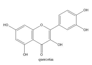
Fig 2.: Quercetin
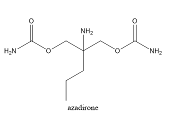
Fig 3.: Azadirone

Fig 4.: Nimbin
Fig 5.: Azadirachtin
Fig 6.: Nimbolide
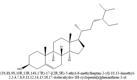
Fig 7.: Beta-sitosterol
Turmeric the plant discussed, contains three curcuminoids; curcumin (which gives turmeric its color) demethoxycurcumin and bisdemethoxycurcumin. It also contains oils such, as tumerone, atlantone and zingiberene that have effects. Turmeric herbal eye drops are an effective solution for promoting eye health and addressing eye related problems. These eye drops include turmeric, a known spice known for its inflammatory and antioxidant properties. It aids in improving while reducing inflammation, itching, redness, burning sensation and excessive tearing of the eyes (Thakur et al., 2018). Scientific studies have confirmed that turmeric oil is an agent offering a reliable alternative to traditional antibiotics, for treating eye infections (Jaiswal & Naik 2021).
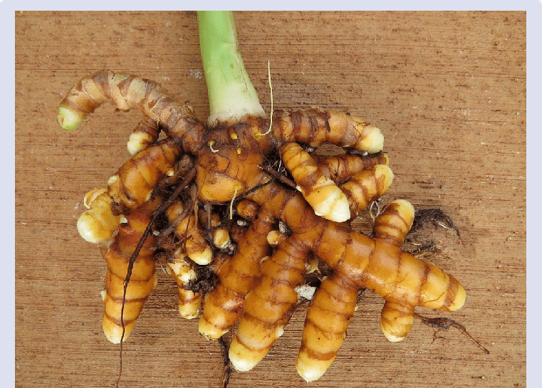
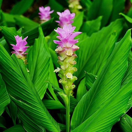
Fig 8.: Turmeric plant (leaves and rhizome)
CHEMICAL CONSTITUENT PRESENT IN TURMERIC
1. In addition, to curcumin turmeric contains compounds called curcuminoids such as demethoxycurcumin and bisdemethoxycurcumin. These compounds play a role in the plants properties.
2. There are turmerones and curcuma turmerones in turmeric, which have shown anti inflammatory and antioxidant properties.
3. Turmeric shares compounds with ginger, such, as gingerols (found in Zingiber officinale). These compounds also contribute to the inflammatory and antioxidant effects of turmeric.
4. Essential oils found in turmeric include terpenes that contribute to its aroma and potential health benefits.
5. Polysaccharides found in turmeric are carbohydrates that possess modulating properties.
6. Turmeric also contains proteins and resins that contribute to its chemical composition.
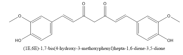
Fig 9.: Curcumin
Fig 10..: Turmerones
Fig. 11. Gingerol
PURPOSE AND OBJECTIVES OF RESEARCH
SIGNIFICANT AND RELEVANCE OF THIS RESEARCH
LITERATURE REVIEW
In the realm of delivering drugs to the eye scientists have been exploring methods to enhance the effectiveness and efficiency of drug delivery. One particularly promising method under investigation is the utilization of ethosomes which're carriers made up of lipids for drug delivery. Ethosomes, a type of lipid vesicles has demonstrated potential, in improving drug delivery to tissues. These lipid carriers, developed by (Touitou et al., 2000), It was discovered that the application of this method improved the transportation of medications to the skin without producing any effects as evidenced by studies conducted in laboratories and, on animals. As a result, there is growing interest in utilizing ethosomes for delivering drugs into the eye. Previous research has highlighted the advantages of lipid based drug delivery systems like liposomes and niosomes, for drug delivery. However there hasn't been an explanation regarding using lipid carriers specifically known as ethosomes for delivering drugs into tissues.
Advancements, in the field of drug delivery have opened doors for innovative approaches. One such approach gaining popularity is the use of formulations, which offer advantages over traditional methods. By combining extracts with cutting edge techniques like liposomes, ethosomes, phytosomes, emulsions, microspheres and solid lipid nanoparticles the therapeutic impact of these plant based formulations has been greatly enhanced. These advanced delivery systems allow for targeting of the medication to areas of the body increasing its effectiveness while also improving its availability in the system. As a result, these modern drug delivery methods improve the stability of ingredients within formulations ensure controlled release over time protect against toxicity risks and ultimately enhance therapeutic outcomes. The main objective behind developing these drug delivery technologies is to make drug administration more efficient and safer for patients while offering convenience. This research paper provides an overview of advancements, based on herbal formulations, explored in drug delivery systems (Chaturvedi et al., 2011).
In a study conducted by (Uner et al., 2023), they discussed the utilization of Timolol maleate (TML) as a medication for managing glaucoma. However traditional eye drops face limitations due to factors. Researchers have come up with a solution to tackle these challenges by developing ethosomes that contain TML, which helps in reducing pressure (IOP). These ethosomes were created using a method called film hydration and through the Box approach an optimal formulation was identified. Studies, on the characteristics of the formulation confirmed that its components are compatible. The particle size was measured at 88.23 ± 1.25 nm with a zeta potential of, around 28.7 ± 2.03 mV and an encapsulation efficiency of 89.73 ± 0.42%. The drugs release in vitro exhibited Korsmeyer Peppas kinetics, with a high correlation coefficient (R2 = 0.9923). As for the findings, from the Hens Egg Test–Chorioallantoic Membrane model (HET CAM), the demonstration supported the suitability of this formulation for applications. Importantly when compared to the eye drop regimen taken three times a day there was no significant difference (p > 0.05) in intraocular pressure (IOP) measurements when using this optimal formulation only once a day. Overall, these findings suggest that TML loaded ethosomes offer an effective alternative, for treating glaucoma.
The research conducted by Vijayakumar in 2010 aimed to evaluate a developed carrier called ethosomal, which contains Diclofenac Potassium. The goal was to investigate its potential, for delivering drugs. The study discovered that increasing the concentration of phospholipid and ethanol resulted in a percentage of drug entrapment efficiency. Out of the formulations that were tested F16 showed the entrapment efficiency. In particular when we used membranes, we observed that higher concentrations of both phospholipid and ethanol did not increase the efficiency of drug entrapment but also enhanced the release of the drug, in laboratory conditions. Among all the formulations we tested the ethosomal formulation with 4% w/v phospholipon 90 and 40% v/v ethanol showed promising results in terms of drug release. When comparing the skin permeation using formulations such, as drug solution and phosphate buffer saline (pH; 7.4) drug solutions it was found that the ethosomal formulation exhibited significantly higher cumulative drug permeation.
The order of drug release and skin absorption occurred in the sequence; starting with the formulation followed by a hydroethanolic solution of the drug then the liposomal formulation and a phosphate buffer saline (pH 7.4) solution containing the drug. The confirmation of ethosomes structure was observed through Scanning Electron Microscope images. Furthermore, particle size analysis indicated that as the concentration of phospholipids increased the vesicles grew larger. Among all the formulations it was observed that liposomal vesicles were relatively larger compared to formulations.
After analyzing the infrared (IR) studies it was found that there is no interaction, between the membrane and the drug. The stability experiments indicated that ethosomes successfully maintained the drugs integrity at a temperature of 4°C compared to 25°C. As for the gel formulation, with Diclofenac Potassium ethosomes it had a composition. When evaluating its pharmacodynamics, it was found that the gel exhibited heightened inflammatory activity compared to a commercially available product. These findings suggest that ethosomes hold promise in enhancing the effects of Diclofenac Potassium while minimizing any side effects.
To summarize this study highlights how ethosomes hold promise as tools, to improve the effectiveness of drug delivery systems by allowing drugs to penetrate deeper and maximizing their capabilities. The technology could as well be utilized for developing formulations containing anticancer drugs for treating tumors located within the body since ethosomes have deep penetration capabilities. Furthermore, ethosomes can also be employed for preparing drug formulations requiring action as they enhance availability through transdermal administration.
Research carried out by (Ahmed et al., 2021), aimed to enhance effectiveness of Ketoconazole (KET), in treating eye infections. KET faces challenges due to its solubility in water and weight which make it difficult for the medication to effectively penetrate the cornea. To overcome this challenge the researchers focused on developing an eye formulation that contains optimized ethosomal vesicles specifically designed to improve KETs corneal penetration, antifungal activity, speed of elimination from the eye and its short elimination half-life. To achieve their goal the researchers conducted a study where they optimized four factors that influence the characteristics of these ethosomal vesicles; their size, zeta potential (which indicates their electrical charge) entrapment efficiency (how well they retain KET) and flexibility. The resulting optimal formulation underwent rigorous evaluation through examination and effectiveness testing against infections. Moreover, different ophthalmic formulations incorporating these optimized vesicles were. Evaluated. The study also investigated irritation caused by these vesicles as well as their ability to permeate the cornea in live subjects. The findings showed that various elements, such, as the drug employed the proportion of drug, to phospholipid the amount of edge activator the percentage of ethanol and the concentration of stearyl amine influenced the properties of these vesicles. Following optimization, the vesicles exhibited a shape and an average size measuring 151.34 ± 8.73 nm. Additionally, they exhibited a zeta potential of around +34.82 ± 2.64 mV. The entrapment efficiency was quite impressive, at a rate of 94.97 ± 5.41%. Additionally, the flexibility was remarkable at 95.44 ± 4.33%. Moreover, when these optimized vesicles were used there was an improvement in the activity of KET as shown by the study findings. It is important to mention that the gel formulations used in the eye did not cause any irritation and the trans-ethosomal vesicles were able to enter the eye segment without causing any effects.
Fluconazole, an agent, with a molecular weight bis triazole structure (306 Da) is known for being hydrophilic and the protein binding of this substance is low. While it can be used as eye drops to treat mycoses a cause of blindness, in developing nations its effectiveness is limited due, to its duration of action (15-30 minutes) and low lipid solubility (log P value of 0.25). These factors often result in patient compliance. Restricted usage. To overcome these challenges researchers, have ingeniously incorporated fluconazole into a type of vesicular system called spanlastics, which is based on sorbitan. The aim is to prolong the drugs duration and enhance its effects. Like liposomes in Transfersomes spanlastics show potential, for optimized drug delivery (Kaur et al., 2012).
In a study conducted by (Kakkar and Kaur, 2011), they investigated a drug delivery technique called "spanlastics." This approach involves using surfactants to transport drugs that are applied to the back of the eye. The formulation they used consisted of Span 60 and an edge activator known as Tween 80. One of the drugs they examined was ketoconazole, which's lipophilic has a weight of 531.44 Da and exhibits limited solubility (0.04 mg/ml). Ketoconazole usually faces challenges, in penetrating the cornea making it difficult to develop formulations. However, the spanlastics developed in this study have nanoscale size and elasticity resulting in improved permeation (p ≤ 0.001) compared to similar niosomal formulations.
The stability of these spanlastics was evaluated over two months under refrigerated conditions with results indicating their safety. Several tests were conducted according to OECD guidelines to assess genotoxicity using the Ames test cytotoxicity on human gingival fibroblasts using the MTT assay, acute tests to assess the irritation or corrosion potential, on the skin and eyes as chronic tests to evaluate long term eye irritation or corrosion. All these tests confirmed the safety of spanlastics despite their reliance, on surfactants (spans plus edge activators) highlighting their nature as demonstrated by Indian Patent Application numbers 2390/DEL/2008 and 1447/DEL/2010.
Furthermore, the research employed glowing sacs marked, test was conducted to determine the potential, for irritation or corrosion, on the skin and eyes. Additionally, test was performed to evaluate the long term effects of eye irritation or corrosion, within the internal tissues of the eye for a duration of up to 2 hours after application. These discoveries provide evidence that spanlastics have promising capabilities, in delivering medications to the part of the eye.
RESEARCH METHODOLOGY
Azadirachta indica leaves were collected from Meenakshi pi 1 Acchier market. The plant leaves was, in good health free from any infections and in good condition. The authenticity and verification of the plants were confirmed.
The sample of leaves was thoroughly washed to remove any impurities. Afterwards excess moisture was eliminated by placing the leaves in a sieve. They were then transferred to a tray drier ensuring that the temperature did not exceed 85 degrees Celsius to preserve the components of the leaves.
Once dried the neem leaves weighed 96.59g. To prepare for extraction they were ground using a mortar and pestle followed by grinding with an electric grinder until they formed a fine powder. This powdered form was carefully stored in an airtight container in preparation, for the extraction process.
96.59 grams of dried leaf powder. Added water. The mixture was then subjected to hydro distillation at a temperature of 85 degrees Celsius, for a duration of 7 hours. Throughout the process it was under close management.
After completing the extraction process, sieve was used to strain the extract, filtration was done using sterile filter paper. Finally, the extract was stored in a sealed sterile volumetric flask in the refrigerator, for analysis.
Azadirachta indica leaves were collected from Meenakshi pi 1 Acchier market. The plant leaves was, in good health free from any infections and in good condition. The authenticity and verification of the plants were confirmed.
The leaf sample was carefully washed to remove any impurities and put into a sieve to get rid of moisture. Next the leaves were crushed using a mortar and pestle before being placed in the hydro distillation apparatus along, with water. The hydro distillation process was conducted at a temperature of 85 degrees Celsius for a duration of 8 hours with monitoring at intervals.
Once the extraction was finished a sterile sieve was employed to filter the extract, which was then further purified using filter paper. Finally the purified extract was stored in a volumetric flask with an airtight seal. Kept in the refrigerator, for future analysis.
Fresh turmeric rhizomes were purchased from pi 3 market at greater Noida, India. It was ensured that the rhizomes were healthy, uninfected and in perfect condition.
The rhizomes were cleansed to remove any substances. Then thinly sliced for optimal drying. These slices were placed on a sterilized Petri dish. Subjected to an oven at a temperature of 105°C, for about of 3 hours. Afterward, rhizomes were taken out from the oven. Dried under a fan to achieve the possible outcome. They were then finely ground using a mortar and pestle. Sifted through no.80 to obtain very fine particles. The resulting extract was carefully stored in an air tight container for analysis. Next 21 grams of powder was. Placed inside a thimble, which was subsequently inserted into a Soxhlet apparatus. The apparatus was filled with 150 milliliters of 99% ethanol and the extraction process was conducted at 60°C temperature for duration of 8 hours. Once the extraction process was complete the ethanol was separated from the extract through evaporation. The resulting extract was then transferred into a flask. Stored in a refrigerator, for further analysis.
Fresh turmeric rhizomes were purchased from pi 3 market at greater Noida, India. It was ensured that the rhizomes were healthy, uninfected and in perfect condition.
The rhizomes were carefully washed to remove any particles before being sliced for optimal drying. These sliced rhizomes were placed onto a sterilized Petri dish. Then placed in the oven at 105°C, for 3 hours. Once removed from the oven the rhizomes were dried under a fan to achieve the results. They were further processed by grinding them with a mortar and pestle and then passed through a sieve with mesh size no.80 to obtain particles. The resulting extract was stored in an air tight container for analysis.
Next the powdered rhizomes were placed into the thimble of the soxhlet apparatus. Filled with water. The extraction process was carried out at a temperature for 7 hours while regular monitoring occurred at intervals.
Once the extraction process was completed the extract was carefully sieved using a sieve. Subsequently filtered through filter paper. Finally it was transferred to a sterile volumetric flask with sealing. Stored in the refrigerator, for further analysis.
TEST FOR FLAVONOIDS:
SHINODA TEST:
Requirements: Fresh Azadirachta indica leaves aqueous extract, dried Azadirachta indica leaves -aqueous extract, Ethanol, Magnesium turnings, Conc.HCl
Test: Two test tubes were taken, Added fresh neem leaves water extract to one and dried neem leaves water extract to the other. Then add drops of 95% ethanol into each tube along, with an amount of magnesium turnings. After that add drops of hydrochloric acid (HCl) and heat the solution until it reaches it boiling point.
NAOH TEST:
Requirements: Fresh Azadirachta indica leaves aqueous extract, dried Azadirachta indica leaves -aqueous extract, 10% aqueous NaOH, dil.HCl
Test: Take 2ml of fresh neem leaves extract in a test tube and a separate test tube containing dried neem leaves water extract. Add 10% solution of sodium hydroxide (NaOH), to both solutions. This resulted in the appearance of a color followed by adding of hydrochloric acid (HCl).
STEROIDS TEST:
Requirements: Fresh Azadirachta indica leaves aqueous extract, dried Azadirachta indica leaves -aqueous extract, Chloroform, Conc. H2SO4
Test: Fresh neem leaves water extract, dried neem leaves -water extract were placed in two test tubes, add 10ml of chloroform and the same amount of sulfuric acid (H2SO4), to the test tubes.
ALKALOIDS TEST:
Requirements: Fresh Azadirachta indica leaves aqueous extract, dried Azadirachta indica leaves -aqueous extract, Picric acid solution
Test: 2ml of Fresh neem leaves water extract, dried neem leaves -water extract was dispensed into a test tube, followed by the addition of picric acid solution.
TANNINS:
Requirements: Fresh Azadirachta indica leaves aqueous extract, dried Azadirachta indica leaves -aqueous extract, 1% lead acetate
Test: 2ml of Fresh neem leaves water extract, dried neem leaves -water in a test tubes, add small quantity of a 1% lead acetate solution.
SAPONINS TEST:
Requirements: Azadirachta indica powder, Distilled water
Test: Measure 1g of the sample. Put it in a flask. Then I added 10ml of water to the flask and boiled it for 5 minutes. After that I filtered the mixture. pour 2.5 ml of the liquid into a test tube. Added 10ml of distilled water. I sealed the test tube with a stopper. Gave it a shake, for about 30 seconds.
TEST FOR REDUCING SUGARS:
Requirements: Fresh Azadirachta indica leaves aqueous extract, dried Azadirachta indica leaves -aqueous extract, Fehling’s solution A&B
Test: A solution of plant extracts measuring 0.5ml was mixed, with 1ml of water. Then 5-8 drops of Fehling’s solution were added. The mixture was gently heated using a water bath.
TEST FOR TERPENOIDS:
Requirements: Fresh Azadirachta indica leaves aqueous extract, dried Azadirachta indica leaves -aqueous extract Acetic acid, Chloroform, Conc. H2SO4
Test: Four milligrams of the extract with 0.5 ml of acid and 0.5 ml of chloroform. Then we slowly added a solution of sulfuric acid.
TEST FOR ALKALOID
Test: The mixture was stirred with 5.0 ml of hydrochloric acid (HCl) solution containing 2% concentration heated on a steam bath and then filtered. Afterward we added Wagners reagent (a solution containing iodine and potassium iodide) to 1 ml of the filtrate we obtained.
TEST FOR SAPONIN
Test: One milliliter of the filtrate was mixed with an equal volume of water and vigorously agitated.
TEST TO TANNINS
Test: Five milliliters of the extract were combined with 2.0ml of a 1% hydrochloric acid (HCl) solution.
STEROL TEST
Test: 1ml of the extract was dissolved in the test tube containing 2.0 ml of chloroform. Then I added 1 ml of sulfuric acid (H2SO4) to it.
FLAVONOIDS TEST
Test: 1.0ml of extract, add 1.0 ml of sodium NaOH) that was diluted. If a precipitate is observed it suggests the presence of flavonoids.
STEP 1-WASHING OF THE EYE DROP CONTAINERS:
The glass vails were first washed with detergents and hot distilled water and it was soaked in the hot distilled water for a few minutes.
STEP 2-STERILIZATION OF GLASS WARES:
To avoid contamination of the eye drops and risk of ocular infection, the glass ware used in the preparation of the ophthalmic formulation were sterilized using autoclave, the glass wares were sterilized by moist heat under pressure at temperature 126 degree centigrade.
-STEP 3:EXRACTION AND FILTRATION OF HERBS
Fresh neem leaves, dried neem leaves and rhizomes of curcuma longa were procured and processed for preparation of the extract. The neem extract was done in 2 ways; dried neem aqueous extract by hydro distillation and fresh neem leaves (aqueous) by hydro distillation. The turmeric was powdered and extracted using 94% ethanol and another extraction was carried out using distilled water. These contents were filtered. The extracts were stored in the refrigerator for carrying out further experiments.
STEP 4: WEIGHING THE EXTRACTS ACCURATELY ON A SENSITIVE WEIGHING BALANCE
The extracts were weighed on the weighing balance for accurate measurement
STEP 5: USE OF DISTILLED WATER
Distilled water was used in the formulation of the eye drops
STEP 6: PREPARATION OF THE SOLUTION IN THE LAMINAR HOOD
The herbal extract was accurately weighed, poured in a beaker and dissolve in the required volume of autoclaved distilled water under aseptic conditions in the laminar hood.
The drug was mixed and the solution was stirred occasionally until proper mixing was achieved, the beaker and the test tubes were covered with aluminium foil.
-STEP7: ADDITION OF VISCO-ELASTIC SUBSTANCE
Visco-elastic substance of 0.5% was weighed and prepared and it was added to different ratios of the formulation and the formulations were covered using aluminium foil which was later stored in the refrigerator for further use.
Table 2: Different formulations were created in various ratios to explore and evaluate different ranges and variations.
DRUG A: fresh neem leaves + fresh turmeric + visco-elastic agent+ distilled water.
DRUG B: dried neem leaves+ fresh turmeric+ visco-elastic agent+ distilled water
DRUG C: fresh neem leaves +solvent turmeric +visco-elastic agent +distilled water
DRUG D: dried neem leaves +solvent turmeric+ viscoelastic agent + distilled water
|
FORMULATION A
|
FORMULATION B |
FORMULATION C |
FORMULATION D |
FORMULATION E |
|
Neem extract – 0.03ml (30 drops). Turmeric extract- 0.02ml (20 drops). Visco-elastic agent -0.02ml (20 drops). Distilled H20- 9.9ml |
Neem extract -0.02ml (20 drops) Turmeric extract-0.001ml (1 drop) Visco-elastic agent- 0.03ml (30 drops) Distilled water-9.9ml
|
Neem extract -0.01ml (10 drops) Turmeric extract-0.005ml (5 drops) Viscoelastic agent- 10 drops Distilled water-9.9ml
|
Neem extract -0.04ml (40 drops) Turmeric extract-0.003ml (3 drops) Visco-elastic agent- 0.015ml (15 drops) Distilled water-9.9ml
|
Neem extract -0.02ml (20 drops) Turmeric extract-0.002ml (2 drops) Visco-elastic agent- 20 drops Distilled water-9.9ml
|
RESULT AND DISSCUSION
This chapter meticulously examines and interprets the results obtained from the conducted experiments, aiming to elucidate the key findings and their significance within the context of the research objectives, Table 3.
|
NEEM EXTRACT |
|
|
|
ANALYSIS |
OBSERVATION |
INFERENCE |
|
FLAVONOIDS: SHINODA TEST: Two test tubes were taken, Added fresh neem leaves water extract to one and dried neem leaves water extract to the other. Then add drops of 95% ethanol into each tube along, with an amount of magnesium turnings. After that add drops of hydrochloric acid (HCl) and heat the solution until it reaches it boiling point. |
Dried leaves extract: No change in coloration, original Colour remain on heating.
Fresh leaves extract:
No change in coloration, original Colour remain on heating. |
Absent
Absent |
|
NAOH TEST: 2ml of Fresh neem leaves water extract, dried neem leaves -water extract were separately placed into two different test tubes. To these solutions, a 10% aqueous solution of sodium hydroxide (NaOH) was added. yellow coloration was observed and then add dilute HCl
|
Dried leaves extract: This results in a yellow coloration, but upon the addition of diluted hydrochloric acid (dil. HCl), the yellow color gradually fades away, signaling the potential presence of flavonoids.
Fresh leaves extract: This results in a yellow coloration, but upon the addition of diluted hydrochloric acid (dil. HCl), the yellow color gradually fades away, signaling potential presence of flavonoids. |
Present
Present |
|
TEST FOR STEROIDS: Fresh neem leaves water extract, dried neem leaves -water extract were placed in two test tubes, add 10ml of chloroform and the same amount of sulfuric acid (H2SO4), to the test tubes.
|
Dried leaves extract: There's a distinct separation observed between the brick-red layer, existing between the upper and lower layers. After mixing, this separation results in the formation of two distinct layers. The lower layer appears as a dark red color, while the upper layer remains transparent, signifying the presence of oil.
Fresh leaves extract:
There's a distinct separation observed between the brick-red layer, existing between the upper and lower layers. After mixing, this separation results in the formation of two distinct layers. The lower layer appears as a dark red color, while the upper layer remains transparent, signifying oil is present. |
Absent
Absent |
|
TEST FOR ALKALOIDS: 2ml of Fresh neem leaves water extract, dried neem leaves -water extract was dispensed into a test tube, followed by the addition of picric acid solution.
|
Dried leaves extract: Yellow colour was observed on addition of picric acid
Fresh leaves extract: Yellow colour was observed on addition of picric acid
|
Absent
Absent |
|
TANNINS: 2ml of Fresh neem leaves water extract, dried neem leaves -water in a test tubes, add small quantity of a 1% lead acetate solution.
|
Dried leaves extract: The yellow appearance of the substance suggested that tannins were present. Fresh leaves extract: The yellow appearance of the substance suggested that tannins were present.
|
Present
Present |
|
TEST FOR SAPONINS: Measure 1g of the sample. Put it in a flask. Then I added 10ml of water to the flask and boiled it for 5 minutes. After that I filtered the mixture, add 2.5 ml of the liquid to a test tube. Combined it with 10ml of distilled water. Then I sealed the test tube tightly with a stopper. Shook it vigorously for 30 seconds. |
The foam, on the honeycomb suggests that there are saponins. |
Present |
|
TEST FOR REDUCING SUGARS: A solution of plant extracts measuring 0.5ml was mixed, with 1ml of water. Then 5-8 drops of Fehling’s solution were added. The mixture was gently heated using a water bath.
|
FEHLING’S A Dried leaves extract: Observation of light green color after heating in the water bath. Fresh leaves extract: Observation of light green color after heating in the water bath.
FEHLING’S B Dried leaves extract: Observation of mustard color after heating in the water bath Fresh leaves extract: Observation of mustard color after heating in the water bath |
Absent
Absent
Absent
Absent |
|
TEST FOR TERPENOIDS: Add four milligrams of the extract to a mixture consisting of 0.5 ml of acid and 0.5 ml of chloroform. Then carefully pour, in a solution of sulfuric acid ensuring it is added slowly. |
Dried leaves extract: Colorless under, the upper layer was the normal color of the extract, on addition of dil. acetic acid. 2layers are there, light yellow at the upper and the down layer was white with touch of oil. On addition of sulphuric acid, red violet color at the bottom layer. Fresh leaves extract: Colorless under, the upper layer was the normal color of the extract, on addition of dil. acetic acid. 2layers are there, light yellow at the upper and the down layer was white with touch of oil. On addition of sulphuric acid, red violet color at the bottom layer.
|
Present
Present |
|
pH TEST Few quantity of the extract was tested using pH meter |
Dried leaves extract: 8.87 Fresh leaves extract: 8.96 |
Slightly alkaline
Slightly alkaline |
|
TURMERIC EXTRACT |
|
|
|
ANALYSIS |
OBSERVATION |
INFERENCE |
|
TEST FOR ALKALOID The mixture was stirred with 5.0 ml of hydrochloric acid (HCl) solution containing 2% concentration heated on a steam bath and then filtered. Then we proceeded by introducing Wagner’s reagent, which consists of iodine and potassium iodide into a volume of 1 ml, from the filtrate that was obtained. Afterwards we repeated the process by adding Wagner’s reagent to another 1 ml portion of the filtrate. |
Oil extract: Extract formed 2 layers on addition of HCL; upper layer has a yellow color and the lower layer gave a lighter yellow color. And finally gave a yellow color on addition of Wagner’s reagent. Solvent extract: When we added HCL we noticed a shade, along, with the formation of a solid substance. Eventually after adding Wagner’s reagent the solution turned reddish brown and another precipitate formed. Aqueous extract: On addition of HCl gave pale yellow color and the color remained unchanged on the addition of Wagner’s reagent.
|
Absent
Present
Absent |
|
TEST FOR SAPONIN One milliliter of the filtrate was mixed with an equal volume of water and vigorously agitated.
|
Oil extract: Formation of froth after vigorous agitation Solvent extract: Gave a palm oil like staining on glass and absence of froth even after vigorous agitation Aqueous extract: Presence of froth after agitation |
Present
Absent
Present |
|
TEST TO TANNINS Different amounts of the extract were mixed with volumes of a 5% solution of hydrochloric acid (HCl) and concentrated hydrochloric acid. |
5% HCL Oil extract: 2.5ml of extract and 1ml of HCL were mixed there was a separation of layer causing them to be immiscible. Solvent extract: 0.5ml of the extract and 0.2ml of HCL were added to a test tube and a deposition of red precipitate was observed Aqueous extract: 5ml of extract, with 2ml of HCL. There was no change that could be seen. CONC. HCL Oil extract: 2.5ml of extract and 1ml of HCL were mixed there was a separation of layer causing them to be immiscible. Solvent extract: 0.5ml of the extract and 0.2ml of HCL were added to a test tube and a deposition of red precipitate was observed Aqueous extract: 5ml of extract, with 2ml of HCL. There was no change that could be seen. |
Absent
Present
Absent
Absent
Present
Absent |
|
STEROL TEST Dissolved 1ml of the extract in a test tube that already had 2.0 ml of chloroform. After that I added 1 ml of sulfuric acid (H2SO4) to the mixture.
|
Oil extract: On addition of chloroform to extract, light yellow color was observed and gave a dark brown interface on addition of concentrated sulphuric acid.
Solvent extract: Extract with chloroform gave a dark red layer at interface and on addition of conc. sulphuric acid, dark purple color was observed with presence of precipitate. Aqueous extract: On addition of chloroform to the extract there was a separation of two layers where the extract was found on the top and chloroform on the bottom. Rapid bubbling and a brown layer at interface were found on addition of concentrated sulphuric acid |
Present
Present |
|
TEST FOR FLAVONOIDS 1.0ml of extract, add 1.0 ml of sodium NaOH) that was diluted. If a precipitate is observed it suggests the presence of flavonoids.
|
Oil extract: The solution was gelatinous in yellow. 2 layers were formed on settling; the upper was yellow and the bottom was pale yellow. Solvent extract:-Dark brown was observed. Aqueous extract:-There was no visible change in the solution. |
Absent
Absent
Absent |
CONCLUSION
The framework for the creation of a unique herbal ethosomal formulation meant to treat eye infections has been established by this initial inquiry. We concentrated on the phytochemical characterisation and extraction of two well-known therapeutic herbs, Curcuma longa (turmeric) and Azadirachta indica (neem), in the first part of our investigation. Because of their well-established antibacterial, anti-inflammatory, and antioxidant qualities—all of which are especially pertinent in the context of ocular therapeutics—these botanicals were considered. To assure the most effective potential extraction of active ingredients, established maceration processes were used to create the hydroalcoholic extracts of turmeric rhizomes and neem leaves. Numerous bioactive substances, such as flavonoids, tannins, phenolic acids, terpenoids, saponins, and alkaloids, were found in these extracts according to the phytochemical study. These results confirm the extracts' therapeutic potential and lend credence to their application in creating a safe, efficient ocular medication delivery system.
This work represents an important milestone in the formulation process, even though we have not yet reached the point of final ethosomal formulation and in vitro efficacy testing. Ethosomes' enhanced capacity to pass through ocular barriers, including the corneal epithelium and conjunctiva, as a result of their high ethanol concentration and vesicular shape, is the basis for the idea of employing them as carriers for herbal extracts. Ethosomes provide a tactical edge over traditional eye drops by improving the permeability of encapsulated medications as well as their retention duration in ocular tissues. In future times, researchers desire to employ the previously identified botanical extracts in ethosomal vesicles. Clarity testing, optical microscopy to view early vesicle formation, and dynamic light scattering (DLS) for particle size analysis are some of the crucial evaluations that will take place throughout the preformulation phase. These experiments are essential for determining the ethosomal formulation's basic characteristics and refining the circumstances for large-scale production.
Assuming acceptable development, the ethosomes will undergo a thorough physicochemical and microbiological analysis. Given how susceptible the ocular region is to microbial contamination, sterility testing will be of utmost importance. The stability and homogeneity of the vesicles will be evaluated by measuring the particle size and zeta potential. The form and surface properties of the ethosomes will be revealed through morphological characterisation utilizing transmission electron microscopy (TEM) or scanning electron microscopy (SEM).
To assess drug–carrier interactions and guarantee compatibility between the herbal extracts and ethosomal components, sophisticated analytical techniques will be used. Any chemical interactions or changes in functional groups after encapsulation will be detected with the aid of Fourier Transform Infrared Spectroscopy (FTIR). The ethosomal system's thermal behavior will be examined using Differential Scanning Calorimetry (DSC), and the formulation's crystalline or amorphous nature—which may affect drug release kinetics—will be revealed by X-ray diffraction (XRD) examination.
The formulated herbal ethosomes' biological effectiveness will be evaluated against clinically relevant ocular infections, such as Streptococcus pneumoniae, Staphylococcus aureus (Gram-positive), and Staphylococcus epidermidis (Gram-negative). Conjunctivitis and other exterior eye infections are frequently caused by these bacteria. The antibacterial potential of pure herbal extracts will be compared to that of their ethosomal counterparts using in vitro antimicrobial assays, including the minimum inhibitory concentration (MIC) determination and the agar well diffusion method. Ex vivo or in vivo studies utilizing appropriate models will be required to evaluate the formulation's ocular safety and irritation potential in addition to antibacterial testing. Important information regarding the herbal ethosomal system's biocompatibility and tolerability for extended ocular usage will be supplied by these investigations.
This comprehensive method, including formulation, biological evaluation, and phytochemical validation, shows how traditional herbal treatment can be combined with contemporary drug delivery technologies based on nanotechnology. The results of this study support the growing interest in ethosomal systems for better ocular drug administration, in addition to confirming the effectiveness of Azadirachta indica and Curcuma longa as therapeutic agents for eye infections.
In conclusion, this research has effectively created a scientific justification for additional development, even though it is still in the preformulation and phytochemical analysis stage. One intriguing approach to improving ocular medicine delivery and overcoming the drawbacks of traditional treatments is the integration of herbal bioactives into ethosomal carriers. Future research will concentrate on refining the ethosomal formulation, carrying out in-depth antibacterial and physicochemical analyses, and advancing toward in vivo efficacy and safety assessments. In the end, this line of inquiry may result in a novel, all-natural, and patient-friendly substitute for the efficient treatment of ocular infections.
REFERENCES

Fadilullahi Opeyemi Ibiyemi, Preeti Singh, Priya Sharma*, Preliminary Investigation of Herbal Ethosomes for Ocular Applications: Focus on Phytochemical Analysis, Int. J. of Pharm. Sci., 2025, Vol 3, Issue 6, 1424-1445. https://doi.org/10.5281/zenodo.15614505
 10.5281/zenodo.15614505