
We use cookies to ensure our website works properly and to personalise your experience. Cookies policy
Department of Pharmaceutics, Arvind Gavali College of Pharmacy Jaitapur, Satara.
The purpose of this review on hollow microballoon with a special focus on the novel different technologies and technique which are used for the preparation of hollow microballoon. The hollow microballoon, as versatile carriers for controlled drug delivery, have garnered significant attention in pharmaceutical research because they facilitate drug release kinetics and increase therapeutic efficacy. For medications with site-specific absorption, microballoon-based drug delivery devices have proven to be more significant in regulating release rate. These low density microballoons buoyant remain in the stomach enhance the availability of drug with narrow absorption windows. The microballoon prepared using different techniques and technologies such as solvent evaporation, solvent diffusion, spray-drying and hot-melt method’s. The evaluation of prepared hollow microballoon using such method like particle size, bulk density, tapped density etc,.
Traditional oral dose form includes different type of formulation offer certain mass of drug present in the systemic circulation that dosage form not release at a steady rate over an extended time. This type of dosage form maximizes therapeutic effect of drug by regulating lower and less frequent dosage of its release into the body. It accomplishes this by delivering medication release either locally or systematically at a predetermined, predictable rate for the desired amount of time.[1]. Short-half-life medications that are easily absorbed from the digestive system are quickly eliminated from the bloodstream. controlled release formulations have been create to prevent this issue The drug will be progressively released into the gastrointestinal tract and sustain a steady medication mass in the serum for longer time. To clear benefits of these systems are patient acceptance and ease of administration [2].A hollow microsphere, often known as a microballoon, is defined technically as a spherical, empty particle without a core3]. One of the most promising buoyant systems is thought to be hollow microspheres or microballoons2]. Microballoons are non-effervescent medication delivery devices that are gastro-retentive. Strictly speaking, microballoons are spherical, empty particles. These microballoons, which are typically smaller than 200 micrometres, are powders composed of synthetic polymers or proteins that flow freely.7]An enlarge in stomach holding duration and enhanced administration of variations drug plasma concentrations which are the outcomes of this. Additionally, the systems are employed for medications that are unstable in digestive fluids or poorly soluble. Numerous methods, including the Emulsion Solvent Evaporation Method, Emulsion Solvent Diffusion Method, Single Emulsion Method, Double Emulsion Method, Polymerization Method, Wax Coating, and Hot Melt, are used to create microballoons.
Mechanism of microballoon[8]
When gastric juice comes into touch with microballoons, polysaccharides, Polymers and gel formers hydrate to create a barrier of gel that is colloidal regulates the fluid entering rate into the apparatus and subsequent deliver of medication. The gel dissolves as The form of dosing's outer surface layer is preserved by the nearby hydrocolloid layer's hydration.
The enlarged polymer provides the microballoons decreases their density and buoyancy. But a
smallest amount of stomach content required to achieve buoyancy. The following are some possible mechanisms for drug release from microballoon.
Diffusion: Water diffuses into the particle's interior when it comes into touch with aqueous fluids in the GIT. Drug dissolution takes place, and the drug solutions spread to the outside of the release coat.
Erosion: Certain coatings have the ability to dissolve gradually over time, releasing the medication that is stored within the particle.
Osmosis: When water is allowed to enter a particle under certain conditions, an osmotic pressure can develop inside the particle. The coating pushes the medication out of the particle and onto the outside.18]
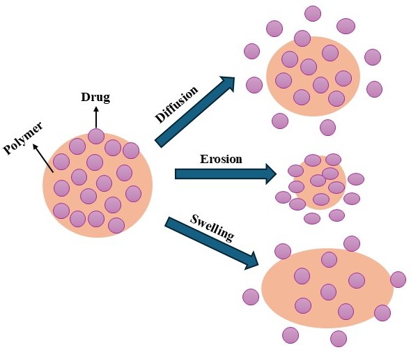
Figure. 1: Mechanism of release of Microballoon
Disadvantages
Method of preparation
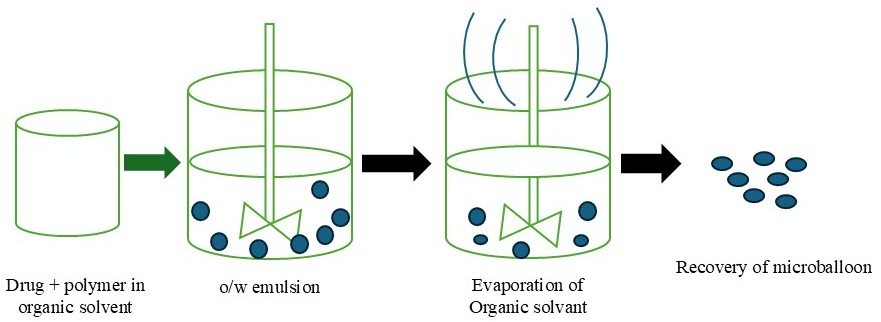
Figure. 2: Solvent Evaporation Technique
drugs and organic solvents have a better affinity in this technique. Although the organic solvent's miscibility, the medication dissolves in it, resulting in a formulated mixture that disperses in a water-based solvent to produce emulsion droplets. Gradually, the organic solution diffuses into the surrounding aqueous phase from the Liquid droplets and the medication crystallizes when the aqueous phase spreads into the droplets. The combination of drugs and polymer is added. dropwise to the polyvinyl alcohol solution after being dissolved in the ethanol: dichloromethane solution. This combination is agitated at different temperatures for one hour at 1500 rpm. Microballoons with different medication contents can be made by adjusting the concentration of polymer in the co-solvent and the ethanol-to-dichloromethane ratio.
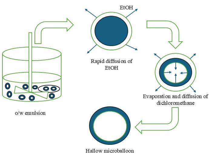
Figure. 3: Solvent Diffusion Method
Figure.4: preparation method of microballoon

Figure.5: Single emulsion Technique
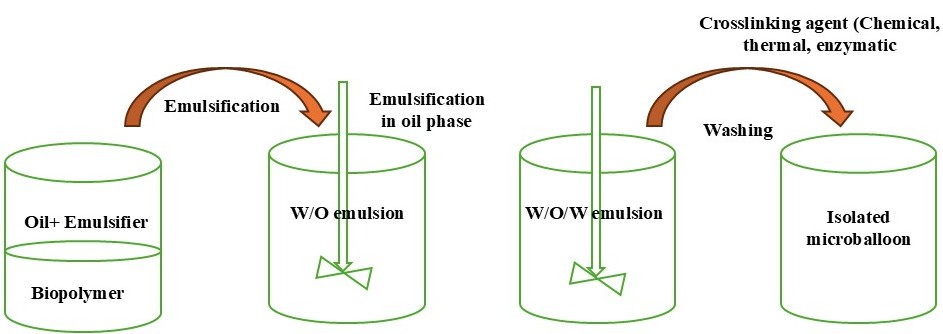
Figure.6: Double emulsion technique
Vaccines, peptides, proteins, and drugs that dissolve in water are the perfect candidates for the double emulsion technique, which produces single emulsions or double emulsions of type w/o/w while creating microballoon. Both natural and synthetic polymers can be prepared using this methods.
The polymerization methods commonly used to create the micrballoons are primarily categorized as
Many methods, including such as suspension polymerization, precipitation polymerization, emulsion polymerisation, processes, are used to accomplish it. In bulk, polymerization is often started by monomer heating or a combination of monomers with the catalyst or initiator. The resulting polymer can be shaped into a microballoon. [11]
This process, additionally known such as bead polymerization technique require heating or mixture of monomers with an active principle that disperses droplets in a aqueous phase. The droplets may also contain an initiator and other substances.[11]
An initiator is present and diffuses of aqueous phase to the emulsion globule surface, which sets it apart from suspension polymerization.[11]
This method develops a film of polymer that successfully encloses the distributed medium by polymerizing at the stage where the two immiscible liquid phases mix. While one of the two reactive monomers was distributed in to continuous medium where the secondary monomer was emulsified, the other was mixed in a continuous phase. Depending on whether the formulated polymer was soluble in the globules of emulsion, two conditions were raised: The polymer was dissolved in the globules if the formulation was monolithic; if it was insoluble in the droplet, it was capsular.
This process involves dissolving the polymer at a high temperature in an appropriate organic phase .The active ingredients is included into a mixture. Phase separation results by cooling the mixture. Microballoons are gathered, cleaned, and dried.
Polymer is mixed into organic solvent i.e. dichloromethane for formation of thick paste, which is then dispersed in drying chamber. A density gradient develops inside a small droplet, and the highest concentration is maintained at the droplet's surface because the solute's absorption is stronger than the solvent's from the droplets evaporating during the drying process. The solid shell-like microballoons are separated from the gases using a cyclone separator, and any remaining solvent is removed by vacuum drying.
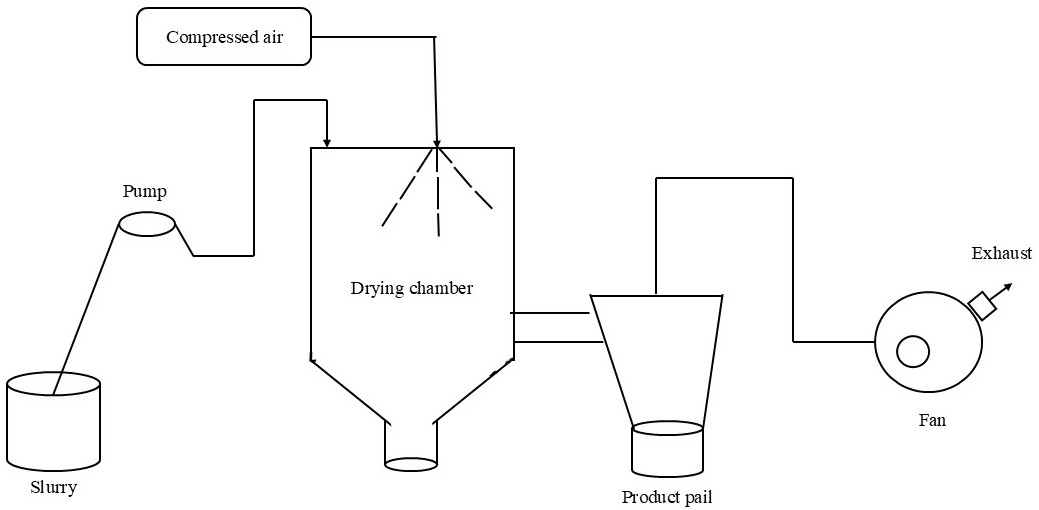
Figure.7: Spray Drying
This technique depends on the decrease of polymer solubility in the organic medium, which produces coacervation, a phase rich in polymers. In order to achieve this, the medication is added to a polymer solution, which causes the polymer to undergo its initial phase separation before to the drug particles being absorbed.

To create a microballoon, the polymer is spread out in the proper medium and cooled gradually. Low melting point polymers should be utilized. The medication is distributed throughout the molten wax, which is employed to create the particle's core and coat. The wax suspension is quickly mixed with a cold solution, such as liquid paraffin. After an hour of agitation, the continuous phase was decanted, and microballoons were gathered. After that, the microballoon was allowed to air dry. When compared to other ways, this one is less expensiv, and the medicine is released more quickly. To obtain the appropriate properties, a combination of beeswax and carnauba wax can be employed as coating materials.

The potential of polyelectrolytes to create a cross-link when counterions to form beads is the base of ionotropic gelation. Since the encapsulation process, ionotropic gelation has been widely used of medications and even cells that use carboxymethyl cellulose, alginates, gellan gum, and chitosan. Despite possessing an potential to cover main ingredients and function as retardants of release rate, natural polyelectrolytes have specific anions in their chemical composition. These anions often combine with the cations that are polyvalent to form a web-like arrangement after binding to the anion blocks to produce gelation. A drug-filled polymeric mixture is tossed into a water-based mixture of polyvalent cations to create the gel-like pellets.
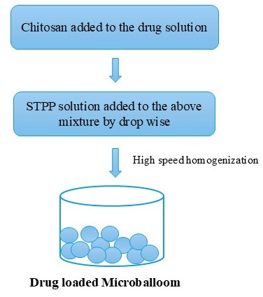

Figure. 8: Ionic Gelation Technique
Evaluation Parameters For Microballoons
Using the optical microscopic method and a calibrated eye piece micrometer for particle size analysis was done. The average diameter was calculated by determining the size of roughly 100 particles.
The bulk density is measured by dividing the Mass of powder divided by the Bulk volume. A 10gm sample of granules is accurately weighed and inserted into a 25 ml measuring cylinder. Bulk density is determined using the eq. (values must stated in gm/cm3) and Without disrupting the cylinder, the volume occupied by the granules was measured.
Bulk density = Weight of the sample/Volume of the sample.
It's the ratio of the blend's mass to the tapped volume. It was determined using a digital tap densitometer that assessed the amount of powder occupied after 100 standard tappings.
Tapped Density = Mass of microballoon / Volume of microballoon after tapping
Morphological examination of the surface and internal structure of the microballoon was performed by using a scanning electron microscope (SEM). For examination of the internal structure of the microballoon, they were cut in half with a steel blade.21]
The % Yield of prepared hollow microballoon was calculated by using following formula. Percentage Yield=M/Mo x 100 Where M = weight of beads Mo = total expected weight of drug and polymer.
The molecular properties of swollen polymers are computed using the swelling index. Dissolution equipment, optical microscopy, and other advanced methods, such as H1NMR imaging, Confocal laser scanning microscopy (CLSM), Cryogenic scanning electron microscopy (Cryo-SEM), Light scattering imaging (LSI), etc., are used to determine swelling. The following formula is used to compute the swelling studies using the dissolution equipment (USP dissolution apparatus USP-24)
Swelling ratio = Weight of wet formulation / Weight of formulations
A beaker holding 300 ml of 0.1N HCL, pH 1.2, at 370 C was filled with microballoons weighing 100 mg. After that, a stirrer was used to agitate the liquid at 100 rpm for six hours, and the floating time was noted.
The invitro release studies was performed by USP-II method. Microballoons like to 100mg of drug was studied in simulated stomach buffer (900ml) pH 1.2 for a period of 2 hrs. and subsequently in simulated phosphate buffer pH 6.8 maintained at 37c and 100rpm. The aliquot samples were withdrawn at frequent intervals, suitably diluted and assayed spectrophotometrically at 273nm
By distributing 50 mg of the medication in 10 ml of ethanol and stirring with a magnetic stirrer for 12 hours to break down the polymer and collect the drug, the drug content of the microballoon was measured. The concentration of drug in the ethanol medium was measured spectrometrically at 273 nm following filtration by using a 5 mcg (Millipore). In these circumstances, Eudragit RL100 did not interfere. This is how the proportion of drug entrapment was determined.
% entrapment efficiency = Calculated drug concentration/ Theoretical drug content X100
Applications
CONCLUSION
In recent review concluded that the prepared hollow microballoon show controlled released delivery system, promises to be a potential approach for gastric retention. To optimise key parameter such as particle size analysis, tapped density, bulk density, drug entrapment efficiency, and drug delivery profile. Microballoon based drug delivery system which can improve therapeutic effect and patient care.
REFERENCE

Shubham D. Karne*, Onkar S. Kale, Vishal D.Yadav , Vasant Y. Lokhande , Recent Avenue in Different Technique or Technologies for Microballoons , Int. J. of Pharm. Sci., 2026, Vol 4, Issue 2, 61-622. https://doi.org/10.5281/zenodo.18490671
 10.5281/zenodo.18490671