Vidya Niketan Institute of Pharmacy and Research Center, Bota.
Liposomes are widely recognized and essential nano-sized drug delivery systems. They are phospholipid vesicles composed of cell membrane components and have been used as artificial cell models to mimic the structure and functions of cells, making them highly valuable in various biological analyses. Liposomes offer significant advantages and provide a wide range of applications as effective drug carriers in both pre- clinical and clinical trials. This review focuses exclusively on scalable techniques for liposome preparation and examines their strengths and limitations concerning industrial applicability. It also discusses recent advancements in biomedical applications, highlighting key development in commercially available formulations, clinical trials, and patents from recent years. Additionally, the review provides brief information on the classification, composition, and characterization of liposomes.
Liposomes are tiny, self-made packages made from lipids that form a single layer (uni-lamellar) or several layers (multi-lamellar) around a water-filled centre. They range in size from 30 nanometers to the micrometer level, and the lipid layer itself is about 4 to 5 nanometers thick. The study of liposomes was started by a British scientist named Alec Bangham and his team at Babraham, Cambridge, in the mid-1960s. They first described the structure of liposomes in 1964. Since then, liposomes have been studied a lot as a way to deliver medicines, proteins, genetic material, and imaging tools. Different ways of giving liposomes, like through injection, inhalation, mouth, skin, eyes, and nose, have been developed to make treatments more effective and easier for patients to follow. They are also widely used in food and beauty products. As drug carriers, liposomes possess exceptional properties such as protecting the encapsulated substances from physiological degradation, extending the drug's half-life, controlling the release of drug molecules, and offering excellent biocompatibility and safety. Additionally, liposomes can selectively deliver their payload to the diseased site through passive and/or active targeting, thus reducing systemic side effects, increasing the maximum-tolerated dose, and enhancing therapeutic benefits. A significant advantage of systemic liposomes as drug formulations is their high biocompatibility, low immunogenicity, biodegradability, increased efficiency, prolonged drug half-life, targeted delivery, reduced systemic toxicity, protection of sensitive molecules, and improved pharmacokinetics. The utmost advantage of systemic liposomes is the simultaneous incorporation and release of two different materials with different solubility. Reports from various studies have shown that different types of liposomes are classified based on the number of bilayers, size, and liposomal composition, and are discussed in further sections. Various publications focus on conventional methods, biomedical applications, and recent advances in liposomal methodologies. In this review, we broadly focus on inventive ideas in methods of preparation and commercially available liposomal formulations with different routes of administration, characteristics, and their applications to overcome the limitations of conventional preparations. In addition, this review discusses a broad range from conventional methods to recent advancements in preparation techniques and new innovation technologies in liposomal preparation, along with the mechanism of formation wherever possible, with a mention of specific advantages and limitations of each liposomal methodology. Furthermore, ongoing research on clinical trials and patents approved in recent years is well detailed. Therefore, we anticipate this resource can offer an overall pathway for researchers to choose an optimal method with up-to-date knowledge on various biomedical applications, along with an idea on current research in clinical trials and patents, providing a pathway for liposomes from pre-clinical research to production and clinical use.
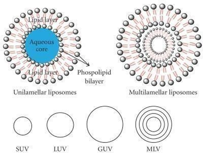
Fig 1: Schematic representation of liposomes.
Drug molecules, and exhibiting excellent biocompatibility and safety. Moreover, liposomes can selectively deliver their payload to the disease site through passive and/or active targeting, reducing systemic side effects, increasing the maximum tolerated dose, and enhancing therapeutic benefits. A significant advantage of systemic liposomes as drug formulations is their high biocompatibility, low immunogenicity, biodegradability, increased efficacy, prolonged drug half-life, targeted delivery, reduced systemic toxicity, and protection of sensitive molecules, with improved pharmacokinetics. The greatest advantage of systemic liposomes is their ability to incorporate and release two different materials with varying solubility simultaneously. Reports from various studies have classified different types of liposomes based on the number of bilayers, size, and liposomal composition, which are discussed briefly in further sections. Various publications have focused on conventional methods, biomedical applications, and recent advances in liposomal methodologies. In this review, we broadly focus on innovative ideas in methods of preparation and commercially available liposomal formulations with different routes of administration, characteristics, and their applications to overcome the limitations of conventional preparations. Furthermore, this review discusses a wide range from conventional methods to recent advancements in preparation techniques and new innovative technologies in liposomal preparation, along with the mechanism of formation wherever possible, with a mention of specific advantages and limitations of each liposomal methodology. Additionally, ongoing research on clinical trials and patents approved in recent years is well detailed. Therefore, we expect this resource to provide an overall pathway for researchers to choose an optimal method with up-to-date knowledge on various biomedical applications, along with an idea on current research in clinical trials and patents, paving the way for liposomes from pre-clinical research to production and clinical use.
A:1,2-Dipalmitoyl-sn-glycero-3-phosphorylethanolamine (DPPE): This is a type of glycerophospholipid, specifically a phosphatidylethanolamine, where the glycerol backbone is esterified with two palmitic acid chains and a phosphate group linked to ethanolamine.
B:1,2-Dipalmitoyl-sn-glycero-3-phosphatidic acid sodium salt (DPPA): This is also a glycerophospholipid, specifically phosphatidic acid, where the glycerol backbone is esterified with two palmitic acid chains and a phosphate group. The "sodium salt" indicates the counterion for the negatively charged phosphate group.
C: 1,2-Dipalmitoyl-sn-glycero-3-phosphoglycerol sodium salt (DPPG): This structure is a glycerophospholipid, specifically a phosphatidylglycerol, where the phosphate group is linked to another glycerol molecule. Like DPPA, it's shown as a sodium salt.
D: 1,2-Dipalmitoyl-sn-glycero-3-phosphocholine (DPPC): This is a glycerophospholipid. specifically a phosphatidylcholine, where the phosphate group is linked to a choline molecule. DPPC is a common component of cell membranes.
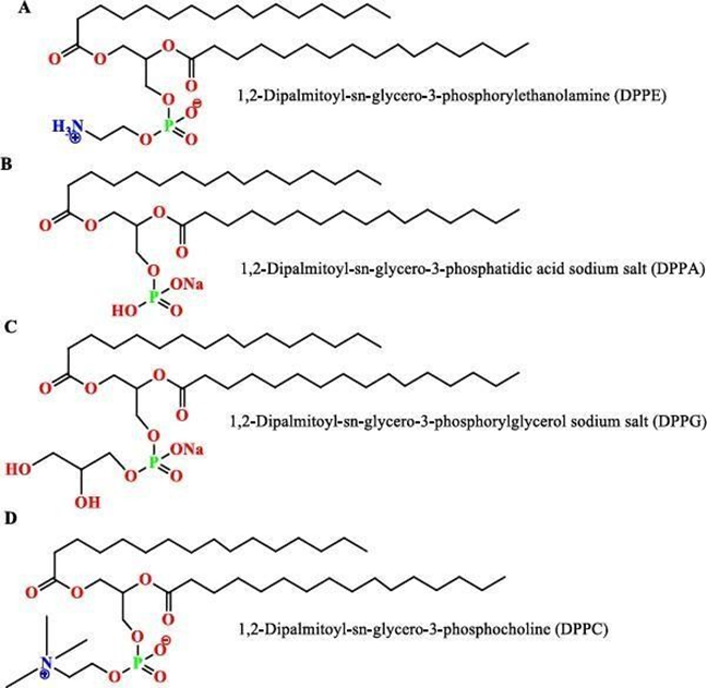
Fig2:-Palmitic acid-based synthetic phospholipids: A) 1,2-Dipalmitoyl-sn-glycero-3-phosphoethanolamine, B) 1,2-Dipalmitoyl-sn- glycero-3-phosphatidic acid sodium salt, C) 1,2-Dipalmitoyl-sn-glycero-3-phosphorylglycerol sodium salt, andD)1,2-Dipalmitoyl-sn- glycero-3-phosphocholine(DPPC).
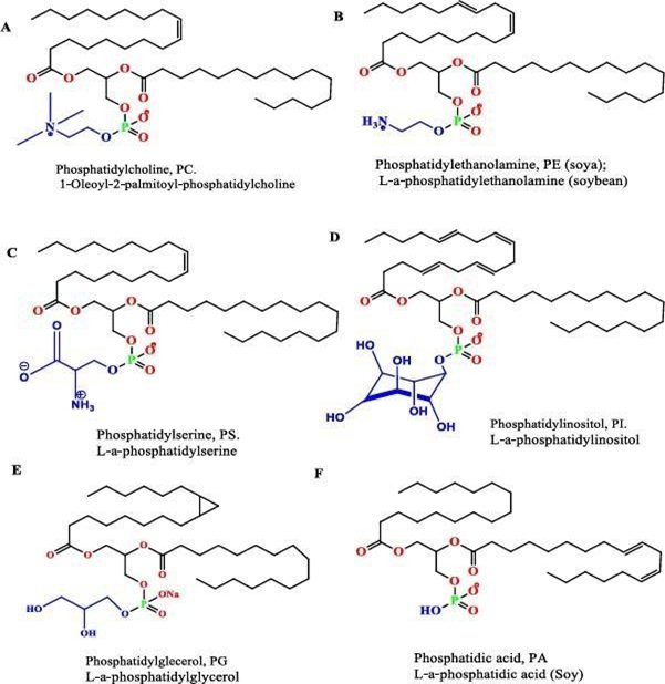
Fig3:-Natural phosphatides most commonly used in liposome production include: A) Phosphatidylcholine, B) Phosphatidylethanolamine, C) Phosphatidylserine, D) Phosphatidylinositol, E) Phosphatidylglycerol, and F) Phosphatidic acid.
Mechanism of Formation of Liposomes:
Liposomes perform their movement through four distinct mechanisms:
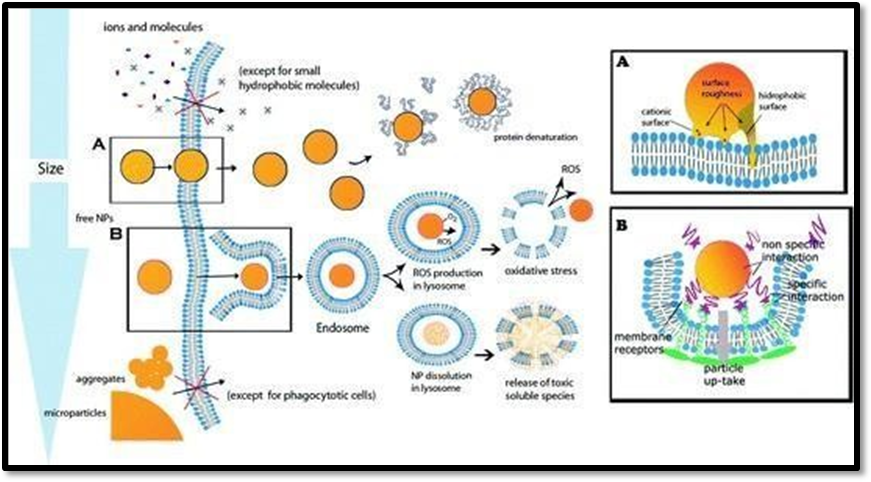
Fig 4: Mechanism of formation of Liposomes.
Classification of Liposomes: -
|
Basis |
Types |
Examples/Features |
|
Size & Lamellarity |
-SUV (Small Unilamellar Vesicles)
- LUV (Large Unilamellar Vesicles) -MLV (Multilamellar Vesicles) |
20–100 nm, single bilayer >100 nm to 1 µm, single bilayer, higher encapsulation 0.5–5 µm, multiple concentric bilayers (“onion- like”) |
|
Composition |
Conventional Liposomes Stealth (PEGylated) Liposomes Cationic Liposomes Stimuli-sensitive Liposomes Immunoliposomes |
Natural/synthetic phospholipids + cholesterol Surface coated with PEG → long circulation, immune evasion Positively charged, used in gene delivery pH-sensitive or temperature- sensitive, drug release under trigger Antibody/ligand attached for targeted delivery |
Conventional Methods for the Preparation of Liposomes:
The main goals of a method for making liposome Nano-formulations are to create particales that are all the same size ( with a narrow size range ), have the desired number of layers, include the drugs efficiently, and stay stable over time. In traditional methods, liposomes are first dissolved in volatile organic solvent and then mixed with an aqueous solution. Using an organic solvent can affect the chemical properties of the active ingredients included or may impact the stability or toxicity of final Nano-formulation. The conventional methods for making liposomes usually include the following meain steps:-
Depending on the specific formation method, the hydration of the lipid (step 3) may occur prior to the removal of the organic solvent from the lipid solution (step 2).
The thin-film hydration technique, commonly known as the Bangham method, is the oldest, most widely used, and simplest approach for preparing multilamellar vesicles (MLVs). To achieve a uniform mixture, the main phospholipid components are first dissolved in an organic solvent such as dichloromethane, chloroform, ethanol, or a chloroform-methanol mixture. Subsequently, the solvent is removed under reduced pressure using a vacuum pump at a temperature of 45–60°C. For small volumes (<1 mL), the solvent can be evaporated using a dry nitrogen or argon stream within a fume hood until the residual solvent is entirely removed, while</mark> rotary evaporation is typically used for larger volumes. After the solvent is removed, a homogeneous, dry, thin-lipid film composed of stacked bilayers is formed. The final step involves hydrating the lipid film using an appropriate aqueous solution (buffer).that, for the pharmaceutical formulation, it may consist of a solution of simple distilled water or a normal (phosphate) saline buffer at pH 7.4. The hydration process, which typically lasts 1–2 hours, is generally carried out at a temperature of 60–70 °C, and in any case, above the phase-transition temperature of the component lipids. During this stage, agitation (stirring) may assist in detaching the (swelling) lipids' lamellae from the internal vessel surface. To ensure full lipid hydration, the final liposome suspension is then left overnight at a temperature of T = 4 °C. During the hydration stage, the lipid becomes swollen and hydrated, resulting in the formation of a MLV suspension that is highly heterogeneous in size.
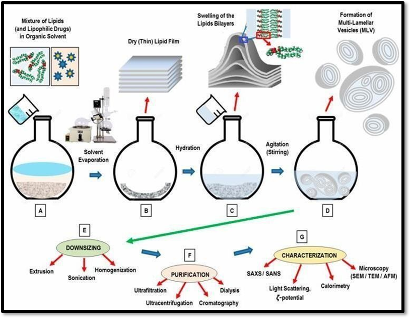
Fig 5: - Thin-Film Hydration (TFH) Technique
Schematic representation of the primary steps involved in the thin-film hydration method for liposome preparation. The main lipid components, along with lipophilic drugs or macromolecules, are dissolved in an organic solvent (A). Following the removal of the solvent, a dry (thin) lipid film is formed (B). The lipid film is then rehydrated in a saline buffer (which may contain hydrophilic drugs to be encapsulated), leading to the swelling of the lipid bilayer stacks (C). Continuous agitation or stirring of the sample promotes the formation of (polydisperse) multilamellar vesicles (D). The final steps in the production process involve downsizing the liposomes (E), purification (F), and characterization (G).
Advantages: Avoids toxic organic solvents and is relatively straightforward.
Advantages: Can achieve high encapsulation rates and uniform liposome sizes.
Advantages: Non-toxic, recyclable, and can enhance drug encapsulation efficiency.
Liposomes function as a vital delivery system, especially for targeted drug delivery. They are particularly valuable in the treatment of diseases such as cancer and antiviral conditions. Stealth liposomes are spherical vesicles with a phospholipid bilayer membrane and are employed to transport drugs or genetic materials into cells. A study conducted by Luoetal. investigated the drug release effectiveness of Doxorubicin encapsulated within porphyrin-phospholipid stealth liposomes. Near-infrared light, capable of penetrating tissues, serves as an external stimulus for drug release, enabling precise spatial and temporal regulation. The research indicated that Dox-loaded stealth PoP liposomes exhibited a prolonged circulation half-life in mice, lasting up to 21.9 hours and maintaining stability for months. A single chemophototherapy treatment using these liposomes at a dose of 5-7 mg/kg of Dox effectively eradicated tumors, outperforming conventional chemo- or photodynamic therapies. Thus, stealth liposomes present significant potential in the delivery of drugs for cancer treatment. Recent developments emphasize notable therapeutic improvements through the use of corticosteroid-loaded liposomes in experimental arthritis models. A key element of liposome drug delivery is the encapsulation of drugs into the liposomes, which can occur passively during formation or actively afterward. Hydrophobic drugs such as amphotericin B, taxol, or annamycin can be directly incorporated during liposome creation, and their uptake and retention depend on drug-lipid interactions. Water-soluble drugs with protonizable amine groups can be actively encapsulated using pH gradients. The advantages of drug loading in liposomes include enhanced solubility of lipophilic and amphiphilic drugs, passive and active targeting to immune cells, site-specific targeting, and increased transfer of hydrophilic and charged molecules. Evaluation of liposomes involves pharmacokinetics, in-vitro testing, and efficacy assessment. A study by Wang describes the preparation and in-vitro evaluation of an acidic environment-responsive liposome for paclitaxel tumor targeting. This study employed cholesteryl hemisuccinate (CHEMS) to improve drug accumulation at the tumor site. The in-vitro release characteristics were analyzed using dynamic dialysis, demonstrating acid sensitivity and sustained release properties. In comparison to free paclitaxel, the liposomes showed higher cytotoxicity and improved cellular uptake, making them suitable for targeted cancer therapy with paclitaxel. Microscopic imaging plays a crucial role in analyzing the structural and morphological properties of liposomes. A study by S. Bibi et al. discusses the use of various microscopic imaging techniques to evaluate liposome structure. Larger vesicles such as multilamellar and giant unilamellar vesicles can be observed using light microscopy. Common techniques include light, fluorescence, confocal microscopy, and electron microscopy methods like transmission, cryo, freeze fracture, and environmental scanning electron microscopy. In transmission electron microscopy, a small amount of hydrated specimen is placed on a grid, and a negative stain like Uranyl Acetate or Osmium Tetroxide is used to visualize the vesicles against a stained background. Fluorescent microscopy tracks particulate delivery systems in biological environments and provides information about the structure of bilayer vesicles, allowing the assessment of various parameters since probes can be placed in both the aqueous and bilayer compartments. A review by Klang et al. discusses electron microscopic techniques for pharmaceutical systems. In scanning electron microscopy, images are formed by scanning a focused electron beam across the surface of a solid specimen. One major advantage of SEM is its pronounced depth of focus combined with image formation, where projecting areas cast shadows and recessed areas appear dark, allowing easy interpretation of the information. Another advantage is the absence of sample preparation for solid samples, enabling the investigation of large areas with high depth of focus. However, a major disadvantage is the time required to collect data one pixel at a time, leading to extended exposure to the electron beam.
One may conclude that, at present, the term "liposomes" encompasses not only phospholipid-based vesicles but also other vesicular structures with properties identical or similar to those of classical, natural phospholipid-based liposomes. In the early 70's, the use of liposomes as a drug carrier system was proposed by Gregoriadis & Ryman. Since this initial report, liposomes have been developed as an advanced drug delivery vehicle. They are generally considered non-toxic, biodegradable, and non-immunogenic (Osborne D. W. et al.). Associating a drug with liposomes significantly alters its pharmacokinetics and reduces systemic toxicity; furthermore, the drug is protected from early degradation and/or inactivation after introduction into the target organism (Gabizon A. et al.). The use of liposomes, or in general, vesicular structures for the delivery of various active compounds is related to the water solubility of the compound. When the compound is water soluble, the size and volume of the aqueous compartment of the vesicle are crucial (Daan J. A. et al.). In contrast, hydrophobic compounds will prefer incorporation into the lipid (amphiphile) layer that forms the vesicle. In such a case, the size of the aqueous compartment is not important.
Liposomes are a promising and innovative drug delivery system with a broad range of applications in the pharmaceutical field. Over the years, extensive research has demonstrated their potential to address various limitations of traditional drug delivery methods. They have emerged as a promising class of drug delivery systems that offer significant benefits in improving the therapeutic efficacy and safety of various drugs. Although challenges persist, the ongoing innovation and improvement of liposomal technologies hold significant promise for the future of drug delivery in the pharmaceutical industry. Liposomes represent an exciting and versatile approach to drug delivery, with the potential to revolutionize the pharmaceutical industry by enhancing drug efficacy, minimizing side effects, and enabling precise therapeutic targeting. Further advancements in liposomal technology are expected to expand their application across a wide range of medical uses.
REFERENCE

Radhika Deshpande*, Roopa Sangvikar, Antidiabetic Activity of Artocarpus heterophyllus Leaf Extract: In vitro Assessment and Mechanistic Insights, Int. J. of Pharm. Sci., 2025, Vol 3, Issue 10, 16-27 https://doi.org/10.5281/zenodo.17240097
 10.5281/zenodo.17240097
