Department of Pharmaceutics, Krupanidhi College of Pharmacy, Bengaluru.
Niosomes are vesicular systems based on non-ionic surfactants that have gained recognition as a promising alternative to liposomes for drug delivery and targeted therapy. Their different features, including improved stability, cost efficiency, and the ability to encapsulate both hydrophilic and lipophilic drugs, make them highly attractive for improving therapeutic outcomes. This review critically examines the potential of niosomes to transform drug delivery systems. It thoroughly discusses aspects such as formulation, characterization, and the functional benefits of niosomes. Additionally, the review covers their uses in areas like cancer therapy, antimicrobial treatment, and vaccine delivery. It also addresses the challenges and limitations of niosomal systems, such as scalability, drug release kinetics, and regulatory issues. Despite these hurdles, niosomes are a versatile platform that offers innovative solutions to enhance bioavailability, enable targeted delivery, and provide controlled drug release. This overview highlights the current research landscape of niosomes and their promising role in advancing modern drug delivery technologies.
In recent years, the field of drug delivery has made impressive strides toward enhancing how medicines behave in the body—with aims like increasing absorption, improving stability, and boosting effectiveness. Among the standout developments are niosomes—tiny, self-assembled vesicles made from non-ionic surfactants and cholesterol. They are often seen as a strong alternative to liposomes because they combine flexibility in design, affordability, straightforward production, and excellent stability. This makes them ideal for carrying both water-soluble and fat-soluble drugs.(1)
Niosomes bring more benefits to the table. They maintain drug stability better than liposomes, they don’t require harsh storage conditions, and they can steadily release drugs over time. Their structure allows both hydrophilic and lipophilic drugs to be encapsulated effectively, and they can be tuned for targeted, site-specific delivery—potentially reducing side effects and improving treatment outcomes. These vesicles have applications in therapeutic fields like cancer treatment, vaccine delivery, and fighting infections. Their skilfulness allows for controlled release and targeted delivery, which can limit systemic exposure and help tackle issues like drug resistance.(2)
Despite their promise, niosomal systems face difficulties in moving from the lab to real-world use. Scaling up production can be tricky, with issues like vesicle aggregation, drug leakage, and inconsistent loading efficiency that can arise. The processes often involve specialized techniques and equipment, which will lead to costs and complicate mass manufacturing.
This review aims to provide a complete overview of the current landscape of noisome research by examining how they have been formulated, how they work, and key practical applications. By exploring the innovations and strategies, we hope to highlight how niosomes could transform drug delivery approaches in the future.(3)
Mechanism of Drug Release from Niosomes
The mechanism of drug release from niosomes is a critical aspect that determines the efficiency and therapeutic outcomes of niosomal drug delivery systems. Niosomes are non-ionic surfactant-based vesicular carriers, and their drug release characteristics depend on several factors, including the type of drug, the surfactant composition, the nature of the membrane, and the environmental conditions. Below are the key mechanisms by which drugs are released from niosomes. (4),(5)
1. Diffusion Mechanism (5)(6)
2. Osmotic Drug Release (7)
3. Membrane Permeability (Leakage)
4. Erosion and Degradation of Niosomal Membrane
5. pH-Sensitive Drug Release
6. Thermally-Triggered Drug Release
7. Enzyme-Mediated Drug Release
8. Active Targeting and Triggered Release
Structure of Niosomes
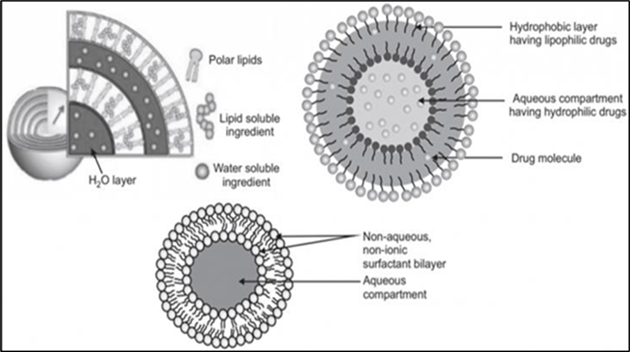
Niosomes are vesicular structures made with non-ionic surfactants in a bilayering arrangement, with both hydrophilic and hydrophobic ends out and inside, respectively. They trap an aqueous core that can load hydrophilic drugs. Surfactants, such as Span and Tween, and may be mixed with cholesterol to improve stability. Niosomes are flexible at the unilamellar level and can be large multilamellar structures. Their composition enables them to deliver both hydrophilic and lipophilic drugs effectively and with a higher stability than liposomes.(13)(14)
1.2. Advantages
1.3. Disadvantages
2. Composition of Niosomes (13)(15)
The three main components used in the preparation of Niosomes are
2.1. Non-ionic surfactants
Non-ionic surfactants play an important part in the medication of Niosomes. They're the introductory factors. They correspond of a polar head and a hydrophobic tail. They arrange themselves in a bilayer with their hydrophilic head towards waterless media and hydrophobic or non-polar tail facing outside. In order to attain thermal stability, they fold inwards to form a vesicle. They're banal because they don't carry any charge. They beget lower hemolysis in the apkins. They beget lower vexation. They enhance permeability, ameliorate the solubility, and also they're also good wetting down agents and used as emulsifiers. They enhance immersion and targeting because they inhibit p-glycoprotein. Selection of non-ionic surfactants depends on HLB value, liquid transition temperatures, and CPP (critical packaging parameter). High HLB value surfactants aren't suitable for medication of Niosomes. Loftiest ruse effectiveness is set up with an HLB value of 8.
Examples:
2.2. Cholesterol
Cholesterol also acts as an important part of Niosomes. Cholesterol isn't needed for the expression of Niosomes, but it provides several parcels to the expression. Cholesterol imparts several characteristics to the expression, like an increase in stability, ruse effectiveness, permeability, rehydration of dried Niosomes, etc. Stability of the vesicle is increased if cholesterol is used with low HLB value surfactants, and helps in the conformation of a bilayer of vesicles if the HLB value is lesser than 6.
2.3. Charged molecules
They're added to help the coalescence of Niosomes and increase their stability. Only 2.5–5 spook attention is tolerable. However, it inhibits the conformation of Niosomes if the concentration of charged moieties is high in the expression. Niosomes having a composition of cholesterol, Span 20, diacetyl phosphate (10101, 15151, and 20201) were prepared and estimated for vaccine encapsulation effectiveness using the HI test. The values attained from the different rates were analogous, and the loftiest encapsulation was 50.
Examples:
3. Methods of Preparation (16)(17)
Since the medication styles impact the number of bilayers, size, size distribution, and ruse effectiveness of the waterless phase and the membrane permeability of the vesicles, the medication styles should be chosen grounded on the use of the Niosomes.
3.1. Ether injection method
This process involves sluggishly introducing a result of surfactant dissolved in diethyl ether into warm water, which is maintained at 60°C. The surfactant admixture in ether is fitted into a waterless result through a 14-hand needle. Latterly, the ether is wracked, which results in the conformation of single-layered vesicles. The periphery of the vesicles may be around 50–1000 nm, depending on the conditions.
3.2. Hand shaking method / thin film hydration method
An admixture of surfactant and cholesterol is dissolved in an unpredictable organic detergent like chloroform or methanol, or diethyl ether, in a round-bottom beaker. Organic detergent is removed at room temperature at 20°C using a rotary evaporator, which leaves a thin subcaste of solids deposited on the walls of RBF. This dried surfactant film is rehydrated with a waterless phase at 0–60°C with agitation. This forms multilamellar vesicles.
3.3. Sonication method
A medicine in the buffer result is added to the surfactant admixture. This admixture is taken into a 10 ml glass vial. This vial containing a medicine and surfactant admixture is sonicated for 10 twinkles at 60°C using an inquiry sonicator with a titanium inquiry to produce multilamellar niosomes. Also, these Niosomes are ultrasonicated by bath sonicator or inquiry sonicator to produce unilamellar niosomes.
3.4. Reverse phase evaporation method
Surfactant and cholesterol are taken in a 1:1 rate and dissolved in an admixture of chloroform and ether. A medicine in waterless result is added to the below admixture. As a result, 2 phases are formed. These are sonicated at 5°C after adding a small quantum of phosphate-softened saline. Under low pressure, the organic phase is removed at 40°C. The performing thick suspense of Niosomes is adulterated using PBS and hotted in a water bath for 10 twinkles at 60°C to form Niosomes.
3.5. Transmembrane pH gradient (Inside Acidic) Drug Uptake Process (Remote Loading)
In this system, surfactant and cholesterol are dissolved in chloroform. Under reduced pressure, the detergent is faded to get a thin film on the wall of round-bottom beaker. This film is doused with 300 mM citric acid of pH 4. The multilamellar Niosomes formed are sonicated. To this suspense of Niosomes, a waterless result of 10 mg medicine is added and vortexed and the pH is raised to 7.0–7.2 with 1 M disodium phosphate. This performing admixture is hotted at 60°C for 10 min to give Niosomes.
3.6. Membrane extrusion method
An admixture of surfactant and cholesterol and diacetyl phosphate in chloroform is made into a thin film by evaporation. The film is doused with a waterless medicine polycarbonate membrane with a mean severance size of 0.1 µm. The result and the attendant suspense is extruded which are placed in series for up to 8 passages. This is a good system for controlling the size of niosomal vesicles.
3.7. Microfluidization
This is a recent fashion that's used to prepare unilamellar vesicles of a particular size. This system is grounded on the submerged spurt principle. In this system, the two fluidized aqueducts interact with high rapidity in micro-channels within the commerce chamber. The result is also passed through the cooling circle to remove the heat produced during the process. This system produces Niosomes of lower size with great uniformity and better reproducibility.
3.8. Bubble method
This is a new fashion for single-step medication of Niosomes and liposomes without using organic detergents. The washing unit consists of a round-bottomed beaker with 3 necks kept in the water bath to control the temperature. Water-cooled influx and the thermometer are deposited in the first and alternate neck independently and nitrogen gas is supplied through the 3rd neck. Surfactant and cholesterol are dispersed in the buffer result of pH 7.4 at 70°C. The attendant dissipation is mixed in a high shear homogenizer for 15 seconds and incontinently gurgled using nitrogen gas at 70°C.
4. Evaluation of Niosomes (13)(17)
There are several characterization techniques for Niosomes
4.1. Size, morphology, and size distribution\
Numerous ways like light microscopy, photon correlation spectroscopy, electron bitsy analysis, SEM (surveying electron microscope), TEM (transmission electron microscope), light scattering, and zeta sizer can be used to study their morphology and determine the size of Niosomes. Flyspeck size measured by the transmission electron microscope is lower than the dynamic light scattering (DLS) system because of the different dimension principles used by the two ways.
4.2. Transmission Electron Microscopy (TEM)
TEM is used in the determination of the shape, size, and lamellarity of Niosomes. The set suspense is mixed with 1 phosphotungstic acid (in the needed quantum), and also a drop of this attendant was used on a carbon-carpeted grid, and also the grid was observed after draining off the excess. The images are taken under suitable exaggeration under TEM after drying fully.
4.3. Optical Microscopy Technique
This fashion is also used for the observation of the shape and size of the Niosomes. The flyspeck size is determined by taking nearly 100 Niosomes are used for flyspeck size determination. The size of the Niosomes is also determined by coinciding the stage micrometer and eyepiece micrometer and calculating the measures.
4.4. Entrapment efficiency
Entrapment efficiency can be calculated by subtracting the amount of unloaded drug from the total amount of drug added. The unloaded drug can be determined using techniques such as filtration, exhaustive dialysis, centrifugation, or gel chromatography. The concentration of loaded drugs can be calculated by dissolving the Niosome in 50% n-propanol or 0.1% Triton X–100 and the resulting solution can be assayed using any specific method. The following equation can be used to calculate the % entrapment efficiency.
4.5. Charge on Niosome and zeta potential
The charge present on the Niosomes causes them to repel each other. This electrostatic aversion prevents aggregation and emulsion and keeps them stable. The face charge on Niosomes is determined using zeta eventuality. Zeta eventuality is determined by the zeta sizer, master size, and DLS instrument.
5. Applications (18) (19) (20)
5.1. Ophthalmic Drug Delivery
Niosomes formulated for ophthalmic medicine delivery show dragged medicine release. The most effective gentamicin Niosomes for extending medicine release from the optical delivery system were those made of Tween 60, cholesterol, and DCP, according to an in-vitro comparison of gentamicin Niosomes and gentamicin result.
5.2. Transdermal Delivery of Drugs
Medicines like Lidocaine and estrogen derivations like estradiol, cyclosporine, etc. are used for topical operation and transdermal medicine delivery systems by formulating them as Niosomes.
5.3. Antiviral Drug Delivery
Niosomes are used in the delivery of numerous antiviral medicines. Ruckman and Sankar prepared Niosomes containing zidovudine and set up the ruse effectiveness and sustainability of medicine release. The Niosomes were made up of Tween 80, Span 60, and cholesterol in colorful proportions. Niosomes, which are prepared using Tween 80, displayed lesser ruse of the medicine zidovudine. The addition of diacetyl phosphate to Niosomes bettered the medicine release for a longer duration.
5.4. Anticancer Drug Delivery
Chemotherapy is the current treatment for cancer. Poor penetration into apkins and side goods on other healthy cells limit the remedial efficacity of numerous anti-cancer medicines. The application of Niosomes as a new medicine delivery system is one of the multitudinous approaches that have been taken to overcome these limitations.
5.5. To Improve Oral Bioavailability
It has been reported that the oral bioavailability of the niosomal expression of acyclovir and griseofulvin was increased than the medicine alone.
5.6. To Modify the Physicochemical Properties of Drugs
Studies showed that the physicochemical parcels of the free medicine were bettered by using non-ionic surfactants.
5.7. Improvement of Stability of Peptide Drugs
The stability of peptide medicines was set up to be increased after formulating as Niosomes. The Niosomes prepared using Span 60-prepared are resistant to proteolytic enzymes and stable at storehouse temperature.
5.8. Other Applications Include
REFERENCES


Shreyas H M, Dr. Eswar Gupta Maddi, Revolutionizing Drug Delivery: The Potential of Niosomes in Modern Therapeutics, Int. J. of Pharm. Sci., 2025, Vol 3, Issue 8, 3022-3031. https://doi.org/10.5281/zenodo.16993779
 10.5281/zenodo.16993779
