
We use cookies to ensure our website works properly and to personalise your experience. Cookies policy
1Amity Institute of Pharmacy, Amity University, Uttar Pradesh, Lucknow Campus India, 226010
2Babu Sunder Singh college of Pharmacy, Nigohan, Lucknow, India, 226063
Human safety incidents during trials come at a high cost. By following safety testing protocols, many of these can be avoided. Unfortunately, toxicity problems in the early phases of a drug development program can still be caused by a variety of variables. The zebrafish model can be continuously improved to start the creation of novel medications and methods for treating illnesses. Numerous scientific questions can be effectively addressed using the vertebrate model. It has been extensively employed in the research of disease biology and medication toxicity. Numerous methods frequently employed in zebrafish breeding labs have been recognized for their positive effects on the physiology and wellbeing of the animals. Nonetheless, information sharing amongst many institutions can increase the reproducibility of these findings. The ranges of husbandry parameters that are most likely to be suitable and successful for zebrafish are provided by standardization protocols. The model's benefits and drawbacks are also covered in the review. It examines the model's many facets and possible applications.
Zebrafish, scientifically known as Danio rerio, are freshwater fish that are members of the Order Cypriniformes and Family Minnow. Originally from South Asia, these fish are commonly kept in aquariums. This vertebrate is widely utilized in scientific research, such as in medication development and pre-clinical testing [1]. Their capacity for regeneration makes them valuable [2], and the researcher can alter them to create a variety of transgenic strains [3]. Numerous drug treatments that have recently entered the clinic or clinical trials have their genesis in zebrafish. The zebrafish's capacity to experience a range of pharmacological effects makes it an ideal animal model for drug screening. Zebrafish bioassays contain features that make them appropriate for drug screening, in contrast to mouse embryos. They can be produced more quickly and at a lower cost. Numerous facets of biology and human health can be studied with it. Zebrafish bioassays can be used to assess apoptosis, angiogenesis, or toxicity. They are similar to the findings of research on mice [4, 5]. The term "pre-clinical trial" or "pre-clinical development" refers to the stage of research that starts prior to clinical trials, which involve testing the drug on humans. Iterative testing, feasibility, and drug safety data are gathered during this time, usually on laboratory animals. Finding the initial safe dosage for the first human research and evaluating the drug's possible toxicity are the objectives of this pre-clinical trial. For example, out of every 5,000 compounds that enter the discovery stage, only one gets authorized as a medication [6]. Mice, hamsters, guinea pigs, rats, rabbits, dogs, and monkeys are frequently employed in pre-clinical experiments. Because of their strong sensitivity and affordability, zebrafish can be used as pre-clinical animal models for a number of pathological conditions. Cancer, diabetes, obesity, epilepsy, cardiovascular diseases, immune system disorders, and infectious diseases are a few medical research topics that involve zebrafish. Zebrafish are known for their contributions to developmental biology, and because of their highly conserved and clinically relevant illness features, aetiology, and progression, as well as their molecular mechanisms, they have recently become a potent preclinical model for human disease. Zebrafish respond to drug treatments and small molecules at physiologically relevant dose ranges. When paired with gene editing technologies and cell-specific or tissue-specific reporters, drug activity can be examined at single-cell resolution across tissues, over a long period of time, and within the complexity of an entire animal. These characteristics allow for the creation of new drug classes, the repurposing of existing medications for compassionate and individualized usage, and high-throughput and high-content phenotypic drug screening. Drugs and drug leads investigated in zebrafish frequently have an inter-organ mechanism of action that would be missed by specific screening methods. Here, we go over why zebrafish are a valuable model for drug discovery, how these discoveries are made, and potential future uses for zebrafish in medical research [7].
2. Taxonomy
The Brachydanio family includes the zebrafish. It is connected to both the Devario genus and the Danio aesculapii, creating a sister group relationship. Although the zebrafish has been called Danio rerio [16], molecular research indicates that it should actually be a member of the Brachydanio genus.
2.1. Areas found in
Throughout South Asia, this species is indigenous to freshwater environments. Its range extends throughout India's river basins, with the South Himalayas serving as its limit. In Myanmar, it has been reported to reside. In the United States, zebrafish have been brought to Florida, California, Connecticut, and New Mexico. The survival of the others and whether they were released on purpose are unknown [17].
2.2. Habitat
These fish typically live in shallow-depth ponds, ditches, and streams. They typically grow vegetation that is submerged or overhanging the banks, and they feed on algae that may be left on the tank's bottom. Additionally, they favor living in silty or sandy water. The temperature of the water varied from 16 to 34 degrees Celsius in colder regions. Despite the exceptionally low temperatures at both locations, the fish seemed to be in good health. At 1,576 meters above sea level, they were among the greatest densities of these animals known to exist [18].
3. Housing and Breeding
The existing body of scientific literature is either inconclusive or has only been examined in small quantities without considering the impact on future generations. In these situations, we chose to draw attention to the need for more work rather than offer a recommendation.
Preventing the introduction of potentially harmful organisms into the main facility is the first step. To reduce the possible risk of disease spread, quarantined animals should be closely monitored [8]. Establishing a quarantine unit with its own water circulation is typically ideal for labs with fewer than a few tanks. In a quarantine unit, only specialized equipment should be utilized [8].
Zebrafish may be kept in the majority of commercially available tank systems. Filter systems, light control units, UVC, and other characteristics are typically integrated into these systems. Tank capacities range from 1 to 10 liters. Standard configurations often use a recirculating water system. Moreover, flow-through systems are frequently employed. Because of the constant heat and humidity in recirculating systems, they use more energy. Additionally, they need a lot of water to move about [9].
One tropical species that may be categorized as poikilothermic is the zebrafish. It has undergone developmental and physiological phases that are impacted by temperature. A fish's physiological and developmental stages are frequently assessed based on how quickly it produces hpf. In most cases, recirculating systems are not necessary to add oxygen. They can, however, be kept at temperatures ranging from 24 to 29 °C. We are aware of the significance of controlling the temperature. Zebrafish are known to tolerate a wide range of temperatures [10]. Lower temperatures may be employed in specific experimental settings, such as those requiring the creation of vaccinations against cold-water viruses. Higher temperatures have been used in other studies, though [10].
The fish are given a steady dark-light cycle (D–L) in modern lab setups, which might change based on the time of year and the weather. Fish physiological functions may be impacted by this cycle. A published paper's section should include the light cycle parameter. Light levels at the entrance of the tank and across tanks should be consistent and set between 334 and 54 lux [11].
For recirculating systems, some labs can use tap water. To be safe for people, chlorine must still be eliminated. It is necessary to correct the high content of tap water hardness in most facilities before using them. This can be accomplished by adding salts or by combining low-pH deionized water. Softened water must be treated with RO in order to eliminate the salt that is present. The two most important variables that affect the system's conductivity are the hardness of the water and the carbonate. Depending on the environment and the facility, the conductivity range can vary significantly. A range of 150 to 1700 µS/cm, for example [12]. For systems that use recirculating water, the biofilter is perfect. It has a filtration capacity of up to 7. The proper concentrations of ammonia, nitrate, and nitrite in the water can be maintained by a well-designed biofilter. To maintain the water's low nitrate content, it is customary to replace it with fresh supplies every day.
After hatching, embryos spend roughly five days floating on the bottom of a tank or Petri plate. They are typically prepared to depart once their swim bladder has grown sufficiently to permit swimming. The function of density in the establishment of male and female sex differentiation is one significant but frequently disregarded component. Higher density can encourage male reproduction, according to studies. Studies have also demonstrated that the presence of environmental enrichment has no effect on the ability of newborn fish to reproduce [13]. Standard stocking densities also depend on ensuring water quality. Increased numbers may also have an impact on the fish's physiological factors. Separating the individual fish is also crucial. Time spent in isolation is frequently required to keep a fish from becoming unwell. Limiting the amount of time the animal spends alone is crucial, though.
It is commonly acknowledged that a mix of live and dry feeds enhances quality of life, development, and reproduction. Enhancing fish welfare is also suggested by the use of processed dry meals. Many species, including rotifers, Artemia nauplii, and Paramecium caudatum, are frequently utilized as live feed. Either a paramecium caudatum or a static rotifer can feed them. Live feed should be given to larvae at least twice daily. This guarantees that when they develop, they will get the right nutrients. Additionally, the feed must be automated and clearly specified. It's critical that staff members routinely check the tanks while automatic feeding is being used. It should also be emphasized how important it is to visually inspect the fish's tanks [14].
The impacts of inbreeding on the population can be lessened if a colony contains the right amount of fish strains and lines. Inbreeding can be avoided by strictly adhering to the guidelines. In a zebrafish facility, managing embryos and larvae correctly is crucial. They are housed with up to 100 embryos in the stock medium. The range of the temperature is 28.5 to 0.5 degrees Celsius. Once the eggs have hatched, the remnants should be taken out. It is important to keep adult larvae in a clean medium that is changed on a regular basis. Around 120 hpf, the swim bladder is inflated and the anus has formed. Upon reaching sexual maturity, the juveniles are moved to a water-exchange tank. It usually takes two to four months to get to this point. The fish's name, the date of fertilization, the quantity of fish, any molecular alterations, and genetic data are the bare minimum needed to track and observe the health of a zebrafish system [15, 16].
Keeping the environment clean is essential to upholding good standards for animal welfare. Avoiding cross-contamination during normal treatments is absolutely essential. With chemical residue, safe cleaning techniques are typically impractical. Heating to at least 60C (220 degrees Fahrenheit) for at least an hour is another technique. Large volumes of liquids are typically impractical for this procedure. Equipment utilized for other purposes at the main facility should be kept separate from quarantine equipment. Prior to beginning work in the quarantine units, it is equally crucial that the main facility be cleaned and disinfected. For fish to grow properly, an environment with healthy algae is essential. Additionally, it can act as a marker for biofilms, which may contain dangerous bacteria [17, 18].
The zebrafish, or Danio rerio, typically takes three months to mature. Under ideal circumstances, spawning can occur successfully even on a daily basis [19]. The embryonic development starts as soon as it is released. Almost instantly after fertilization, eggs turn translucent. Within 36 hours after birth, the zebrafish embryo's precursors start to show. On top of the yolk, it begins as a single cell. After then, it divides in half and keeps on doing so until it has hundreds of little cells. As the fish matures, the tail grows and separates from the body. Then, as it grows, the yolk gets smaller. It serves as food for the fish. Researchers utilize a fish tank with a movable bottom addition to entice zebrafish to spawn. In order to allow the animals to reach the beach, this technique lowers the pool's depth. Up to 300 eggs can be laid in a single morning by a single pair of adult fish. In order to initiate a mating match, male zebrafish are known to react to specific marks on females. Commonly found in plastic products, diisononyl phthalate can disrupt sexual function and have an impact on the reproductive system [20].
4. Other scientific research happening on Zebrafish
The zebrafish, or D. rerio, is sometimes used as a model in science to study vertebrate development and genetics. George Streisinger of the University of American State undoubtedly invented it around the 1970s. The genetic information of the zebrafish is contained in a specific online database. Its global resource center also serves as an allele repository. His research has led to breakthroughs in a wide range of disciplines, including genetics, environmental sciences, teratology, cancer, biological process biology, toxicity, and muscular dystrophies.
Scientists can benefit greatly from using the zebrafish as a model biological system. Both its embryos and its fully sequenced genome are readily apparent and testable. Its capacity to produce a two-celled embryo, which enables the application of staining techniques, is another benefit. Additionally, it has a diurnal sleep cycle, just like people.
Zebrafish larvae have the ability to rebuild their heart and hair cells by the time they reach that stage [21], [22]. The British Heart Federation started a marketing effort in 2011 to investigate the potential applications of this skill in people. After an injury, zebrafish can repair damaged photoreceptor cells and other neurotrophic neurons, according to research. The cells' dedifferentiation mediates the process. Australian researchers discovered in 2012 that zebrafish may repair damaged spinal cords by using a protein called fibroblast growth factor. After embryonic disturbance, this protein can also be utilized to restore hair cells [23]. Researchers are examining how hereditary conditions including muscular dystrophy and epilepsy can impact central nervous system function using zebrafish. They are also investigating the potential effects of a gene called Hedgehog on human cell growth.
4.3.1. Background genetics
Anaemia in biomedical research may arise from genetic diversity among wild-type lines at various research facilities, even though inbred strains have not been produced for use in laboratory zebrafish.
4.3.2. Gene expression
Zebrafish are frequently utilized in reverse genetics research because of their enormous clutch numbers and brief life spans. Certain genes can have their expression altered or decreased by using these animals. Only cells originating from the 32-cell stage can have their gene expression reduced by Morpholino Oligonucleotide (MO). Diffusion between cells can occur during the first few days of development because bigger molecules can pass through cells that are interpermeable.
4.3.3. Genome sequencing
The National Centre for Biotechnology Information has the whole genome sequence of the 2001-sequenced zebrafish reference strain Tuebingen. The genome of a wild zebrafish was sequenced in 2009, according to Indian researchers. The organism possesses roughly 1.7 billion genetic letters, according to the statistics. In 2013, the fish's reference genome was also made public.
4.3.4. Mitochondrial DNA
The full mitochondrial DNA sequence of D. rerio was published by researchers in 2001. This animal has the same gene order as vertebrates and is only 18 pairs longer than a fish. The light strand replication sequence shown in this work is similar to that of vertebrates. In addition to a noncoding area that explains the heavy strand's origin, it contains 13 protein-coding gene sequences [24].
4.3.5. Pigmentation genes
The zebrafish mutation nacre was discovered in the mammalian MITF transcriptome in 1999. Pigment loss and eye abnormalities are caused by this mutation. The gene behind the golden strain's peculiar coloring was discovered in 2005. Then, in humans, the nacre mutation was identified to distinguish between darker-skinned Africans and fair-skinned Europeans. Zebrafish with transparent skin and no melanophores were subsequently bred using this mutation.
4.3.6. Transgenesis
This method entails creating a functional transposon gene in zebrafish using the Tol2 transposon platform. One completely functional transposase that can initiate transposition in the zebrafish germ lineage is the Tol2 element.
4.3.7. Transparent adult bodies
Casper, a new strain of zebrafish with translucent skin, was created in 2008. It made it possible for scientists to see different cellular processes. A hybrid between a T-cell-deficient strain and a prkdc-/- strain that resulted in immunodeficient offspring was reported by researchers in 2019. A fish can be warmed to 37 degrees Celsius with this strain. In 2013, Japanese researchers succeeded in creating a visible glow in a translucent zebrafish specimen's brain. In order to identify the presence of oestrogen in the environment, Chinese scientists introduced genetically altered zebrafish into the eggs of fertile females in 2007. When the altered fish came into contact with contaminated water, they became greenish.
4.3.8. RNA splicing
According to study conducted in 2015, 10% of zebrafish genes do not depend on the protein U2AF2, which is necessary for RNA splicing. Rather, they start the process with repeating base sequences. They found that the human splicing process does not require the U2AF2 proteins. This implies that humans became dependent on the protein for the process as a result of an evolutionary shift that took place in tetrapods.
4.3.9. Inbreeding depression
Offspring may suffer negative consequences when a close related mate induces inbreeding depression. The homozygotic expression of the harmful alleles results in inbreeding depression. the effects on zebra fish embryo development of exposure to chemicals used in agriculture and human medicine, as well as water stress. Due to these impacts, the viability of embryos decreased, and inbred males tended to have fewer offspring.
One intriguing model for drug research and discovery is the zebrafish. It has the ability to forecast human health and illness, and its rapid development and tiny size make it a perfect platform for larger-scale studies. For scientists looking to pinpoint the genes that might be responsible for human illness, the zebrafish model is an effective tool. Additionally, it can be applied to the development of novel medication candidates [25].
4.4.1. Drug screening
Zebrafish drug screens are capable of detecting new types of substances with biological effects. Additionally, they can be used to repurpose already-approved medications for new use. At least one of the 65 small-molecule screens that have been carried out so far resulted in a clinical trial. Nonetheless, the field of drug research still faces numerous technical obstacles [26].
4.4.2. Toxico- or Pharmacokinetics
Understanding how medications affect the body's internal drug exposure is the aim of this research. Because of their small size, zebrafish make it extremely difficult to analyze the concentration-effect connections that can be inferred from this data. Researchers are looking toward a technique that can take several blood samples to describe a drug's concentration over time [27].
4.4.3. Computational data analysis
Human-relevant pharmacological and pathophysiological processes can be found with the aid of clever data analysis tools. The study of the different pharmacological processes that result in a particular reaction in both healthy and pathological conditions is the aim of pharmacometrics. It is possible to forecast which medications will cause a reaction and which won't by researching these processes. The measurement and forecasting of processes associated with a drug's interaction with a biological system are the main goals of systems pharmacology. According to the computational approaches, higher vertebrate exposure levels are linked to the clearance of paracetamol from zebrafish larvae.
5. Reasons to use Zebrafish in pre-clinical trials
A variety of transgenic cancer models have been created using zebrafish. When these animals get past the human body's resistance to p53 usage, they develop melanomas. They display the traits of human illness. In 2011, the function of certain genes known to overexpress in human melanoma was investigated using the melanoma model [28]. The study emphasized the significance of SETDB1, a novel oncogene that has been found to play a key role in the development of melanoma tumors. It is well recognized that SETDB1 is essential for controlling epigenetic regulation. In the development of tumor cell biology, this is a significant milestone. According to a study, the formation of the neural crest cells that ultimately give rise to melanoma can be halted by chemically screening for the DHODH protein. This strategy seeks to target the melanoma cell's identity rather than a specific genetic defect. Patients with melanoma may benefit from this treatment.
5.2. Repairing retinal damage
The four types of cone cells present in zebrafish can supplement human cells that are sensitive to UV light. Its color spectrum is very broad. The zebrafish is also used to study how the cone cells in the eye mature. Cone cell arrangement is remarkably fine-tuned in this fish [29]. Researchers at University College London cultivated a particular kind of stem cell in fish eyes in 2007 such that it could differentiate into retinal neurons. Conditions like Alzheimer's disease and blindness might be treated with this. The cells were successfully transformed into different kinds of human retinal neurons by the researchers. Additionally, they were able to exploit the cells' properties by reproducing them in the lab
5.3. Cardiovascular disease
Studies on heart failure and the development of human blood arteries have made use of zebrafish. Human heart damage is frequently caused by myocardial infarction. It causes scar tissue to form and the myocardium cells to be destroyed. Congestive heart failure may result from the absence of a replacement heart [31]. The teleost zebrafish has the ability to mend a damaged heart, while humans are unable to do it. This animal is a useful model for researching human disease because of its capacity for regeneration. Regarding the creation of novel heart regeneration models, these animals have proven their skills. The regulation of the healing process after a heart injury may be better understood in light of the formation of new cardiomyocytes. As a muscle, the heart requires oxygen and nourishment continuously. If the heart's supply is cut off, some parts of the heart may suffer from "cardiac ischemia." When the blood behind the heart lacks oxygen, this disease develops. Heart tissue death may result from cardiac ischemia if treatment is not received. We call it a MI, or heart attack. The left cardiac muscle is typically affected by this ailment. After the ventricular wall is injured, non-contractile fibrotic scar tissue develops. Additionally, it may result in congestive heart failure. The information sheds fresh light on the processes that underlie zebrafish epicardial cell regeneration. They might make it possible to determine these cells' biomarkers and adjust their levels accordingly. Numerous studies have demonstrated that many biological components, including growth hormones, proteins, and nucleic acids, can initiate the molecular pathways involved in the establishment of cardiomyocyte proliferation after injury. The dual specificity phosphatase Dusp6 activates the MAPK pathway. This process raises the blood's oxygen content, which promotes the growth of healthy cardiac cells. In the absence of intact hearts, the proliferation of BCI and BCI215 was elevated following a cardiac amputation. This implies that the effects of these substances can be injury-dependent.
5.4. Immune system
Scientists have been able to investigate the genetic mechanisms underlying acute inflammation and create novel medications by using a zebrafish model. One model organism that can be utilized to investigate adaptive immunity is the zebrafish's innate immunity. Bacterial infections can be suppressed by its strong phagocytic activity [29].
5.5. Infectious diseases
Studying infectious diseases in people is made possible by the comparatively conserved immune systems of zebrafish and humans. It is possible to investigate host-pathogen interactions using the transparent early life stages of zebrafish. The pathophysiology of tuberculosis can be fundamentally understood using the zebrafish model. It has also been applied to drug resistance screening [30].
5.6. Muscular dystrophies
Muscular dystrophies are uncommon hereditary conditions that result in muscle atrophy and weakening. They typically result in early death. For instance, Duchenne muscular dystrophy's orthologue is the sapje protein mutation. The lack of the MBNL splicing factor has been demonstrated to be the cause of myotonic dystrophy type 1. This model was created to investigate how the expression of the CUG repeat affects the onset of DM1 illness. Additionally, it is a great animal model for researching how laminin 2 affects muscular dystrophies.
5.7. Bone physiology and pathology
It is known that these animals participate in the following processes: osteogenesis, resorption, tissue turnover, and bone metabolism. Human bone problems have also been studied with them. Mineralization of the bone elements begins as early as four days after birth, during the early stages of embryonic development. In order to detect and cure complicated age-related bone disorders, adult zebrafish are being researched. Osteoblasts produce these scales, which serve as the fish's primary calcium storage organ. To investigate novel medications and their impact on bone metabolism, they can be cultivated ex vivo in a multi-well plate.
5.8. Diabetes
The pancreas of zebrafish and mammals are extremely similar. It regulates its endocrine activity using a range of cells. When researching endocrine-related disorders, knowledge of the pancreas' anatomy and function is crucial. Since zebrafish can identify diabetes and other disorders that can be brought on by glucose intolerance, the creation of glucose tolerance tests in this species has been justified.
5.9. Obesity
Both hereditary and overnutrition-induced obesity have been studied by researchers using the zebrafish model system. The model showed that weight gain results from abnormal lipid metabolism. Some people are more likely to develop hereditary obesity than others, which can
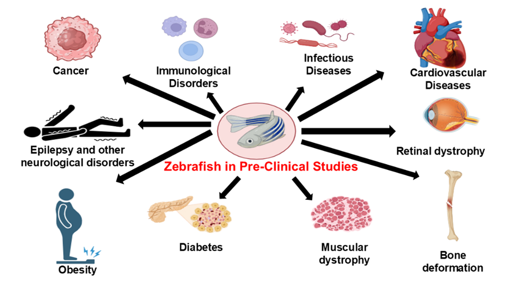
Fig.1 Utilization of Zebrafish in various pathological conditions
be explained by research on genetic obesity in zebrafish. Additionally, the fact that some persons become obese even in the absence of hereditary abnormalities may be explained by their capacity to acquire resistance to endocrine disrupting medicines. These studies could help find possible genetic origins of human obesity, even if zebrafish and human genes are not the same. Overfeeding and high-fat diets cause the same metabolic problems, but they can also promote hepatosteatosis, hypertriglyceridemia, and adipose deposition. According to this study, humans could benefit from treatment for these disorders [30].
5.10. Epilepsy and other Neurological Conditions
Zebrafish are becoming more and more popular as a screening tool since they are acknowledged as an appropriate in vivo model for epilepsy research. The findings of a few preclinical investigations are compiled in this review to offer reliable data for the future creation of efficient screening techniques for plant-derived antiseizure/antiepileptic medications utilizing zebrafish models. Many herb preparations and medications derived from plants have been shown to have anticonvulsant and/or antiepileptogenic qualities in preclinical research on animal models of seizures and epilepsy. In fact, traditional ethnomedicine has made use of some of the plants under study. Nevertheless, there aren't enough solid preclinical study findings to unbiasedly support or refute their prospective application as ASMs. It is also employed as a multiple sclerosis animal model [31, 32].
6. Use of Zebrafish to understand regeneration after heart injury:
Loss of cardiomyocytes cannot be replaced by the human heart. As a result, patients die sooner and have a lower quality of life. Patients with MI are now better managed thanks to modern treatment. But without a cardiac transplant, it remains incurable. To increase the survival rate of MI patients, a number of therapy approaches have been investigated and improved. One of the main challenges in the development of new cardiac models is the creation of engineered tissue architecture that can support a microvascular circulation. Understanding its healing qualities and molecular pathways is equally crucial. The creation of functionally designed heart tissue will be made possible by the understanding of myocardial biology [33]. Previously, it was thought that the creation of new cardiac myocytes could be the cause of cardiac hypertrophy. Nevertheless, research revealed that abnormal cardiac expansion was not a result of larger cardiomyocytes. The traditional understanding of this mechanism has been altered by discoveries made in the past ten years. These findings demonstrate that in animals, a limited number of cardiac celllets can regenerate into adulthood. In mature human hearts, the rate of cardiomyocyte renewal following a MI is insufficient to make up for the myocardium's loss. The field of cell replacement treatment has expanded when it was discovered that adult human heart cells can contribute to cardiac healing and balance. Nevertheless, the clinical trials have been quite subpar in spite of the positive outcomes. The presence of a diverse set of cells in the heart does not necessarily mean that these cells have a particular function in the body, therefore even while c-Kit expression has been used to detect stemness markers, it is insufficient to characterize CSCs. The production of multipotent cells from the c-kit positive population may be inhibited by the negative sorting of CD45 and CD31. According to this study, these cells may hold the secret to creating novel medications that target the heart's capacity for regeneration. Researchers are now concentrating on the function of natural models in heart regeneration. Their objective is to create treatment approaches that are effective in regenerating human hearts [34]. Since the 1960s, the zebrafish has been a significant animal model in the study of organ regeneration. It is a smart choice for researching human genetics because of several of its properties. Zebrafish have many benefits for studying human diseases. Its capacity to investigate the physiological organ regeneration that takes place during human illness is one of these. Compared to the human heart, the zebrafish's adult heart is smaller and less complex. It resembles the structure of the heart in mammals. The gold standard for studying the development of vertebrates has been established as a result of zebrafish embryos' capacity to survive for up to five days after hatching. Zebrafish can also be utilized to study conditions including cardiovascular development. New genes that are necessary for healthy development are also found using this technique. Zebrafish are not suitable for the development of human-based treatments due to their evolutionary separation from humans. In their adult state, they may also regenerate several organs. Depending on the organ, several mechanisms govern regeneration. For example, the regeneration of the fin is triggered by the creation of a dedifferentiated cell structure called blastema. In a similar manner, the telencephalon can produce a blastema when the Notch gene is activated [34]. They demonstrated how an animal's heart may use its own blood supply to rebuild itself. The ventricles' size and shape return to normal after 60 days. The heart's ability to contract also gets better. Where does the regenerated tissue come from cellularly? Regenerated tissue in humans can only be produced following a heart damage. It is well known that heart regeneration is influenced by other variables [35]. Fibroblasts produce collagen, which can help prevent a heart attack. However, heart failure may eventually result from a non-contractile scar that remains in the heart. Abnormal heart function may also occur as a result of this scar. A zebrafish model that replicates the pathophysiological mechanism of a human heart attack was developed in 2011. The model demonstrates that following a cryoinjury, the heart regenerates. The cells in zebrafish die after a cardiac arrest, and the inflammatory infiltration causes myofibroblasts to develop and ECM components to be secreted. The presence of matrix metallopeptidases led to the degradation of ECM components. MMPs are essential for tissue remodeling and post-injury repair. The two most important MMPs that are known to contribute to post-MI remodeling are MMP9 and MMP2. However, the interactions between several MMPs confound their roles. Developing new treatment targets that can stop or reduce the effects of MMPs may be made easier with an understanding of their pathophysiological activities. The development of cardiac remodeling and fibrosis in mammals is also known to be regulated by the Smad3-dependent TGFb signaling pathway. Zebrafish's post-injury scar resolution mechanism is comparable to that of the heart of a newborn mouse. Following cryoinjury, the injured heart's inability to produce new cardiomyocytes causes collagen to build up and the scar to deteriorate. In the course of the heart's regeneration after an injury, the mechanical forces decrease. Eventually, the scar is removed as a result of the downregulation of collagen synthesis. Studies have demonstrated that as people age, the dynamics of the intraventricular mechanical stresses change due to changes in tissue composition. Defective collagen production can also emerge as a result of additional mechanical signaling and the presence of ECM crosslinking density. Sartore and Ausoni (2009) suggested that the zebrafish heart's capacity for regeneration might be explained by the absence of fibroblasts. According to several writers, the proliferation of cardiomyocytes during heart regeneration also depends on the presence of cardiac fibroblast cells in the heart following injury. ECM deposition from cardiac fibroblasts and epicardiums is primarily responsible for the healing of heart function in mice after MI. The lack of endocardial cells results in a distinct kind of cardiac fibrosis. The absence of a full fibroblast-type phenotype in the endocardial cells indicates that they do not fully go through the epithelial-to-microbe transition (EMT). After MI, the ECM in the zebrafish heart degrades, in contrast to mammals. Fibrosis regression results from fibroblasts' reduced ability to produce extracellular matrix [34]. They pointed out that zebrafish's lack of fibrotic response may restrict the growth of cardiomyocytes. In a species with innate regenerative capability, our study sheds light on how fibrosis may impact regeneration. Anti-fibrotic medications may be more effective in people than those that target inactivating fibroblasts. However, it is still unclear how the heart regenerates in zebrafish. To seal the wound following a ventricular damage, a blood clot forms. EMT and epicardial cells are initiated within a few hours of the damage. These elements can encourage the migration and proliferation of epicardial cells and aid in the healing process of the damaged region. The heart's outer layer, the epicardium, is essential to the body's development and renewal. It can promote the growth of heart cells by secreting soluble growth factors. Additionally, the epicardium can retain the physiological characteristics of organs and promote the formation of heart tissue. The formation of adult cardiomyocytes during post-injury cardiac remodeling is known to be regulated by epicardium. Mammals are capable of cardiac healing, however the mechanism is somewhat restricted. They may be able to accomplish this by identifying signaling pathways that can promote the growth of heart tissue. A molecule involved in cell adhesion, neuRegulin 1 is essential for the growth of the heart and nervous system. It is known to promote the heart's capacity for healing. BCI may potentially be utilized to improve Nrg1 signaling in mammals. Nonetheless, it has been demonstrated that Nrg1 can promote the development of tumors in mice. The capacity of adult cardiomyocytes varies significantly between lower vertebrates and mammals. Gaining an understanding of these elements may aid in the creation of successful plans that promote the growth of these cells. The Hippo/Yap/Taz pathway is known to contribute to the development of the heart and is a key element in cardiac regeneration. It is also recognized to be essential for cardiomyocyte proliferation and scar formation. The buildup of reactive oxygen species-mediated DNA damage in mature cardiomyocytes may set off the cycle. New cardiomyocyte development and the control of their cardiac renewal may result from this. It is well recognized that miRNAs are important for the growth of cardiomyocyte proliferation. By blocking or suppressing the cell cycle, they can also cause it to start. Future research on cardiac regeneration in zebrafish may reveal chemicals that control the process. Even while there is proof that heart injury and regeneration are related, it is still unclear how and why this process is controlled following a cardiac injury. Utilizing zebrafish as a model to investigate cardiac plasticity may aid in the discovery of novel therapeutic targets that can lessen the harm that MI causes [35].
7. Significance of Zebrafish in various toxicological studies:
7.1 Embryo toxicity
Zebrafish embryos are increasingly being used for screening for developmental toxicity. Acute fish toxicity is already being assessed using the embryos. In vivo and ex vivo toxicity evaluations are not thought of as the standard method for creating toxicity testing, according to a 2015 statement. These evaluations might, however, be taken into consideration for regulatory approval under specific conditions. For evaluating how different stages of organogenesis affect an entire vertebrate creature, the zebrafish embryo is a great instrument. This is also useful for researching how various medications affect various organs. By enabling in-house toxicity assessments, this approach can boost the productivity of chemical and pharmaceutical businesses. Additionally, it might lessen the number of animal experiments that follow the 3rd Rs principle [36]. The inconsistent findings in the zebrafish developmental toxicity test could be caused by several factors. These consist of variations in species, metabolism, and chemical concentrations. Chorion elimination can still be problematic for certain chemicals, even though it doesn't seem to affect uptake in the majority of small molecules. Without examining the internal concentrations, the concentration of the media that can influence the zebrafish embryo's growth may be overstated. Efforts have been made to enhance the prediction of the concentrations of embryotoxic media in order to overcome this problem. It is generally acknowledged that zebrafish have a low intrinsic metabolic capacity at 72 hpf with respect to the biotransformation capability of embryos. The findings of the zebrafish embryo experiment served as the foundation for this conclusion. Additionally, when evaluating the toxicity of zebrafish eggs, the method of action of substances can be taken into account. For example, it has been demonstrated that ribavirin results in defects in mammalian development. The organogenesis of zebrafish is well known, however there are only a few morphological endpoints that can be assessed using a conventional assay. This is mostly because the existing protocols don't provide a complete set of endpoints [37-39].
7.2. Intestine, pancreas, and hepatobiliary toxicity
The post-esophageal digestive system of zebrafish consists of several organs. Although it is similar to that of mammals, it has differences in terms of structure, function, and toxicity.
7.2.1. Intestine - The intestinal bulb is an anterior section of the intestine found in zebrafish. It serves as a food store where nutrients can be consumed. The zebrafish's nervous system regulates its enteroendocrine cells and villi, just like in mammals. Additionally, the intestinal lining cells differ. The intestinal crypts, the submucosal layer, and the Paneth cells are among the characteristics that zebrafish lack. The animal instead displays smooth muscles that connect to the mucosal layer. Despite the rarity of using zebrafish for drug development, research on gastrointestinal toxicity has been published. It was discovered that the intestinal contractility of fish larvae varied. It might be brought on by the different ways that some medications affect the stomach. According to the researchers, this variability might be brought about by the different ways that different medications affect the gut. According to the reports, false positive findings are less often than false negative results. The absence of toxicity detection in zebrafish could be the cause of these results. Compounds that can be safely evaluated for human consumption can be found using detailed studies on the intestinal toxicities of zebrafish [38].
7.2.1. Pancreas - In the pancreas, the endocrine and exocrine compartments regulate the basic function. Enzymes that enter the digestive tract are produced in the exocrine compartment. Chemicals secreted by the endocrine compartment may have an impact on blood sugar management. The development of diabetes in zebrafish has not been positively correlated with the expression of cells that produce polypeptides. Researchers are examining the harmful impact of environmental contaminants on the development of the zebrafish exocrine pancreass [38].
7.2.3. Liver - Given how many medications might harm the liver, hepatitis is an enormous concern for the pharmaceutical business. The use of in vitro human tissue models, like 3D models, is expanding as a result of their growing popularity. These models enable us to investigate the impact of specific medications on an individual by offering comprehensive details about the different cellular components of a tissue. In many in vitro and in vivo models, exposure to hepatotoxins caused gene alterations, indicating that zebrafish embryos had characteristics similar to those found in life. Zebrafish embryos and humans have comparable metabolic pathways. In contrast to mammals, zebrafish have a less ordered liver, with cells arranged in tubules rather than bilayered plates. It is attached to the gall bladder by tiny bile channels. An appealing model for researching how poisons affect the liver is the zebrafish. Its versatile and fast development makes it a useful instrument for examining the impact of numerous chemicals on the liver. A large pharmaceutical company's study demonstrated the predictive utility of combining high-content cellular toxicity assays with zebrafish liver toxicity assays. The same study also shown that DILI may be predicted by the usage of the same medications. According to recent research, substances based on hepatotoxins may safeguard liver health [38].
7.2.4. Gall bladder - Zebrafish's lipid transport and metabolism can be seen using a lipophilic dye. A reporter dye called PED6 is used to track the toxicity of substances present in zebrafish. Toxins in gall bladder cells have been identified using PED6 and other vertebrates. The lack of reports at this time demonstrates the effectiveness of this tactic [38].
7.3. Ocular toxicity
The human eye has a great degree of conservation. It is therefore a great model for researching the consequences of ocular toxicity. Zebrafish and the human eye have shown notable anatomical similarities. The main cell types are divided into distinct strata. Axons divide the inner and outer plexiform portions, whereas nerve fibers and photoreceptors divide the inner and outer nuclear cell types, respectively. Other structural distinctions between zebrafish and mammals include the fact that zebrafish have more cones than rods and have radiation-sensitive double cones. They also have compartments that are sensitive to red and green. Glial cells with the ability to differentiate into diverse tissue types give rise to progenitor cells that can develop into the retina's primary neurons. The inner and outer layers of the zebrafish eye are not vascularized, in contrast to human eyes. Rather, their distribution depends on the eye's surface vessels. These findings demonstrate that drug-induced ocular reactions in zebrafish are comparable to those in humans. The authors discovered oculartoxic compounds by testing medications that have no known effects on humans. Additionally, they pointed out that the tests were sensitive to specific medications and that the results' usefulness was shown. The response of the optokinetic and optomotor To assess vision in both juvenile and adult zebrafish, 137 tests are frequently employed. The former gauges how light affects the eyes, and the latter describes how the fish react to movement. The first kind evaluates overall mobility using a stimulus, or a second. The second kind is employed to measure response and makes use of video recording [40].
7.4. Nephrotoxicity
Since the kidney's job is to filter harmful substances out of the bloodstream, it is especially susceptible to harmful substances. The goal of toxicology research has been to create useful indicators for tracking kidney toxicity. A straightforward and sophisticated model for researching nephrotoxicity is the zebrafish pronephros. It is a desirable candidate for nephrotoxicity research due to its straightforward anatomical form and resemblance to the cellular makeup of the mammalian metanephro. Fenestrated cells and cells that produce podocytes are found in the glomerulus of the zebrafish. Additionally, the nephron tubules are lined with polarized epithelial cells. Proteinuria and podocyte cell effacement resulted from puromycin or podicin treatment. Additionally, they presented a fluorescent tracer known as 70 kDa dextran. It has been demonstrated that certain medications that are known to cause tubulitis in people can alter the morphology and physiology of zebrafish larvae. Several biomarkers were used to identify the impact of these medications [41].
7.5. Endocrine Toxicity
The hypothalamus, which regulates the activity of the adenohypophyseal, is primarily responsible for the action of the endocrine system. Neuroendocrine peptides secreted by the hypothalamus cause the adenohypophyseal to react to peripheral organs. The hypothalamicpituitaryadrenal axis, the hypothalamicthyroid axis, and the hypothalamicpituitarygonadal axis are the three subdivisions of the hypothalamic-pituitary-adrenal axis. Vertebrates have a well-preserved endocrine system. The regulation of numerous physiological processes is under the control of this system. Zebrafish, on the other hand, lack both the hypothalamus and neurosecretory fibers. After entering the adenohypophyseal cell, these fibers release their hormones. The development of the interrenal and adrenal glands depends on both the mammalian NR5A1 and its zebrafish counterpart, nr5a1a. In zebrafish, the endocrine system's development coincides with the start of puberty. Chemicals that alter hormones control the thyroid gland's output. Both human and wildlife development and reproduction depend heavily on these substances. Research on zebrafish endocrine disruption has concentrated on how these substances affect the fish's growth and ability to reproduce. Induction of a protein precursor called vitellogenin is one of the most commonly measured responses. Exposure to estrogenic radiation causes the liver to generate this protein. Numerous biological impacts of endocrine disrupting chemicals (EDCs) on zebrafish embryos have been identified through the use of gene expression analysis. For instance, in 96 hpf embryos, treatment to fadrozole reduced the amounts of brain aromatase and vitellogenin. According to the study, steroidal drug exposure caused the androgen-pathway genes cyp2k22 and sult2st3 to be activated. Similarly, exposure to perfluorinated water changed the expression of numerous important genes associated with the HPT axis in zebrafish embryos. Triazole fungicides, polyphenyl ethers (PBEs), and other polycyclic aromatic ethers (PAEs) are endocrine disruptors that can be used to track endocrine activity. Under the regulation of the brain aromatase gene, this line displays GFP in a concentration-dependent manner. Both synthetic and natural estrogens can affect the model. In Europe, the EASZY assay is a method used to track the endocrine activity of surface waters and wastewater. It was created to determine the target tissues and gauge the entire fish's reaction. High-quality and reasonably priced screening may be possible with the use of reporter lines for endocrine disruptor identification [42].
7.6. Hematologic Toxicity
These consist of T cells, B cells, neutrophils, and red blood cells. The thrombocyte is no different. It is possible to assess the toxicity of hematopoietic stem cells and view them in various hues. Adult fish exposed to radiation can also be used to produce hematopoietic cells. Numerous zebrafish mutations display hematopoietic abnormalities. 26 complementation groups—now referred to as new genes—were found on the initial screen. There have been investigations into the hematologic toxicity of zebrafish. Numerous methods can be used to conduct different types of research on the hematopoietic system [43].
7.7. Cardiovascular Toxicity
Zebrafish and human physiology are conserved at distinct levels. Zebrafish physiology is similarly affected by a number of human cardiovascular medications. Drug impacts on human heart health have been studied using the animal. To find bradycardia in zebrafish, the authors used a high-throughput test. They discovered that nearly half of the medications frequently prescribed to treat QT prolongation also induce the syndrome in people. Only one creature exhibits the effects of a given medication on the metabolism of a second substance. Research on zebrafish offers a valuable chance to examine how harmful substances affect the creatures' physiological states. Repolarization toxicity reporting has a 75% specificity in people. The amount of poorly absorbed medications was 96%, and the sensitivity was 80%. Based on the compounds' capacity to block the zERG channel and their impact on QT prolongation, a validation of the platform using known negative and positive compounds was conducted. These assays solely evaluate the drugs' affinity for the hERG channel, even if the channel itself is the target. Additionally, the zebrafish's little atrial arrhythmia allows for the identification of substances that inhibit other ion channels. In vitro data sets do not support data showing how chemicals affect zebrafish. They might be brought on by substances that act on a target that in vitro models do not recommend. It is still uncertain whether zebrafish have the ability to heal injured heart muscle. Gaining insight into the several elements influencing its capacity to mend could aid in the creation of novel techniques for people [44].
CONCLUSION
Zebrafish are primarily admired for their small size, rapid development, and transparent young, despite some significant contrasts with mammals. Additionally, this animal is a great substitute for toxicological research. Zebrafish are well-equipped to carry out the same fundamental tasks as humans and share many of the same organs. A typical aquarium can accommodate them. After mating, hundreds of embryos grow quickly. By early development, the majority are nearly finished. The pancreas, liver, and gut reach full development at 76 hpf. Additionally, the GI tract develops. A basic handheld platform can be used to do toxicology on hundreds of whole organisms. An excellent setting for researching how harmful substances affect organ development is offered by young zebrafish. Cell-specific reporter assays can be easily manipulated due to their bodies' transparency. The ability to transfer discoveries from the lab to people was demonstrated by the advancement of eight zebrafish-discovered compounds into clinical trials. Zebrafish have long been employed in a variety of scientific fields, including environmental toxicology, cell biology, and genetics. Thus, zebrafish are a strong choice for pre-clinical testing due to all of these similarities with mammals.
REFERENCES

Rudrakshri Kaushik, Vivek Srivastava*, Alok Kumar Shukla, The Emerging Role of Zebrafish in Pre-Clinical Research, Int. J. of Pharm. Sci., 2025, Vol 3, Issue 5, 2942-2960. https://doi.org/10.5281/zenodo.15453414
 10.5281/zenodo.15453414