Ideal Institute of Pharmacy, Posheri, Wada.
Background: Noisome are nanometric vesicular systems made of cholesterol and non-ionic surfactants that were created to enhance drug delivery through reduced toxicity, increased bioavailability, and tailored therapy. Their structural resemblance to liposomes, together with their increased stability and affordability, have sparked interest in their application in a number of therapeutic fields. Method: The scientific literature from databases including PubMed, ScienceDirect, and Google Scholar was systematically evaluated to create this review. Comprehensive data on noisome composition, preparation methods, characterization techniques, drug targeting mechanisms, and pharmaceutical uses were gathered through an analysis of research publications, clinical studies, and reviews. Results: The results demonstrate that noisome can encapsulate medicines that are hydrophilic, lipophilic, or amphiphilic, providing enhanced targeting efficiency and controlled release. Numerous preparation techniques, including sonication, micro fluidization, and thin-film hydration, have an impact on the stability, size, and drug loading capability of vesicles. Since they are biocompatible and less immunogenic, noisome have shown promise in peptide, anticancer, ophthalmic, and transdermal drug delivery systems. Conclusion: By combining enhanced therapeutic results with formulation flexibility, noisome offer a viable platform for sophisticated drug delivery. Despite issues with sterilizing and physical stability, more study and improvement can realize their full promise in vaccine administration, diagnostics, and pharmaceuticals.
Back in 1909, Paul Ehrlich came up with a groundbreaking idea: targeted drug delivery. He envisioned a system that could send medications straight to the sick cells, leaving healthy ones untouched [1]. This concept, which we now call drug targeting, focuses on delivering drugs right where they're needed, reducing the risk of affecting healthy tissues. One exciting way to make this happen is through noisome [2]. These are tiny vesicles created by mixing non-ionic surfactants with water, and sometimes adding cholesterol or other lipids. They look a lot like liposomes and can hold both fat-loving and water-loving drugs, making them a fantastic option for precise and effective drug delivery [3]. The cosmetics industry was the first to use noisome [4]. Because of their lower irritating power—which falls in the order of cationic > anionic > ampholytic > non-ionic non-ionic surfactants are recommended. As seen in Fig. 1, non-ionic surfactants are made up of polar and non-polar segments with strong interfacial activity that, when hydrated, create a bilayer to entrap both hydrophilic and hydrophobic medications [2]. Noisome can be used for a variety of medication delivery methods, including topical, ophthalmic, targeted, parental, and more [1]. Recent years have seen a great deal of research on noisome' potential as a vehicle for the transfer of hormones, antigens, medications, and other bioactive substances. In addition, noisome have been employed to address the issues of drug instability, insolubility, and fast degradation [5].
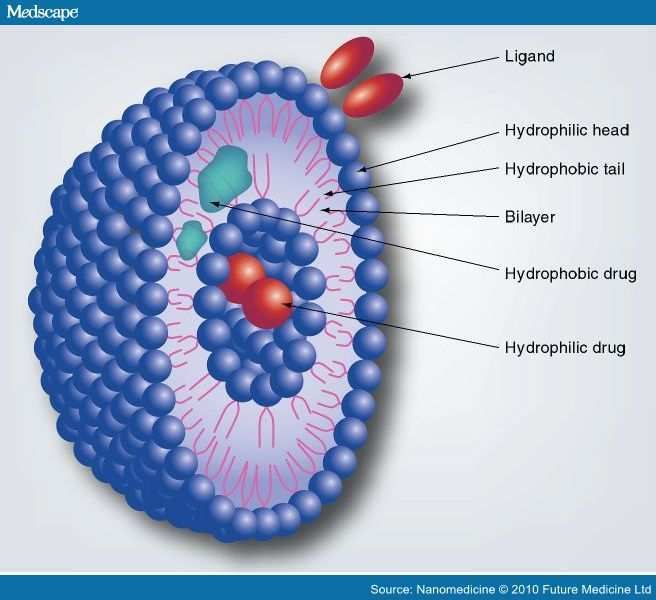
Fig No.1. Noisome structure [11]
Definition
Noisome are tiny vesicles, just a few nanometres in size, crafted from a blend of cholesterol and non-ionic surfactants. This unique combination gives them better structural stability than liposomes. Plus, because they use non-ionic surfactants, noisome are less toxic, making them a safer and more stable option for pharmaceutical uses. Noisome have particle sizes ranging from 20 nm to 100 nm-1[6].
STRUCTURAL FEATURES AND COMPOSITION OF NOISOME
A typical noisome vesicle would have a vesicle-forming amphiphile, or non-ionic surfactant like Span860, which is typically stabilized by adding cholesterol, along with a tiny quantity of anionic surfactant, such diacetyl phosphate, which also aids in vesicle stabilization [7]. Selecting a surfactant for noisome synthesis primarily depends on the surfactant's HLB value, which ranges from 4 to 8. The procedure utilized to prepare the noisome determines whether its structure is uni-lamellar or multi-lamellar. All different kinds of medicines, including hydrophilic, lipophilic, and amphiphilic ones, can be included into the noisome structure. Figure 1 displays the locations of every drug kind in the noisome structure [6].
The noisome is mostly composed of the following elements:[8]
Noisome production is significantly influenced by surfactants. When preparing noisome, the following non-ionic surfactants are typically utilized.
For instance,
spans (60, 40, 20, 85, 80)
Brij’s (Brij 30, 35, 52, 58, 72, 76)
Tweens (tween 20, 40, 60, 80).
Non-ionic surfactants have a unique structure made up of two different parts: a hydrophilic head that loves to mingle with water and a hydrophobic tail that prefers to stay away from it [11].
Cholesterol is employed to give the noisome preparations stiffness and the right shape and conformity [6].
To prevent noisome aggregation, a charge molecule is introduced to the noisome formulation. e.g. Molecules with a negative charge- Di hexadecyl phosphate, lipoamino acid, phosphatidic acid, and diethyl phosphate. Molecule with a positive charge- Stearyl pyridinium chloride and stearyl amine [8].
Phosphate buffer is the most widely utilized hydration medium while making noisome. At different pHs, these phosphate buffers are employed. The pH level of the hydration medium can change based on how well the encapsulated drug dissolves in the solution [10].
TYPES OF NOISOME [12]
Noisome can be categorized into three main types based on their size and structural features:
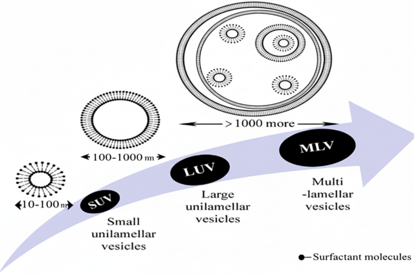
Fig No.2.Schematic structure of SUVs, LUVs and MLVs
Table No:1- Comparison with liposomes [6]
|
Sr. no |
Noisome |
Liposome |
|
1 |
Surfactant |
Phospholipid |
|
2 |
Size 10-100nm |
Size 10-3000nm |
|
3 |
Inexpensive |
Expensive |
|
4 |
Not required special storage condition |
Required special storage condition |
|
5 |
Less toxic |
More toxic |
|
6 |
Cholesterol present |
Not contain cholesterol |
Table No:2- Advantages and Disadvantages of Noisome
|
Advantages |
Disadvantages |
|
Compared to liposomes, noisome have a longer storage period, are chemically stable, and are osmotically active [12] |
Physical instability [19] |
|
When you compare oil-based formulations to aqueous vesicular suspensions, you'll often find that patients tend to tolerate the latter much better. This leads to higher compliance rates [1]. |
Combination [15]
|
|
can improve a drug's skin penetration [4] |
Entrapped medication leakage [16] |
|
Noisome have a better bioavailability than traditional dosage formulations [5]. |
Encapsulated medication hydrolysis that reduces the dispersion's shelf life [11] |
|
The surfactants are non-immunogenic, biodegradable, and biocompatible [14]. |
Fusion [17] |
Salient features [10]
PREPARATION METHOD OF NOISOME
The preparation techniques should be selected based on the noisome' intended application, as they affect the aqueous phase's entrapment efficiency, size distribution, number of bilayers, and membrane permeability of the vesicles. The benefits and drawbacks of noisome preparation techniques are described in Table 3. The method of preparation of noisome is shown in below,[6]
1)Sonication
2)Micro fluidization
3)Multiple membrane extrusion method
4)Ether injection method
5)Reverse phase evaporation technique
6)Bubble method
7) Thin film hydration technique (Hand shaking method)
8) Trans membranes pH gradient method (inside acidic)
9) Formulation of noisome from proniosome
Table No:3- Preparation Methods
|
Sr. No |
Types of noisome |
Preparation technology |
Advantages |
Disadvantages |
|
1 |
Small unilamellar vesicles [18,20] |
Sonication |
Green technique; no need for organic solvents |
High energy consumption and costly equipment |
|
Micro fluidization |
high level of consistency and reproducibility
|
Not suitable for heat-sensitive medications that are readily hydrolysed and/or oxidized |
||
|
Multiple membrane extrusion method |
Less polydispersity
|
Higher drug losses
|
||
|
2 |
Large unilamellar vesicles [6,2,20] |
Ether injection method |
Basic technology.
|
Not suitable for use with heat-sensitive medications or organic solvent residue |
|
Reverse phase evaporation technique (REV) |
High efficiency of encapsulating |
Possible organic solvent residues |
||
|
Bubble method |
Organic solvents are not used. |
Low stability during long-term storage |
||
|
3 |
Multilamellar vesicles [20,3] |
Thin film hydration technique (Hand shaking method) |
Basic technology
|
Organic solvents are challenging to eliminate.
|
|
Trans membranes pH gradient method (inside acidic) |
High encapsulation efficiency and stability
|
Organic solvents have a high polydispersity index, heterogeneity, and are challenging to remove. |
||
|
Formulation of noisome from proniosome |
High levels of chemical and physical stability during extended storage
|
complicated and time-consuming procedure
|
Formulation of noisome from proniosome [3]
The pro-noisome can be converted into a noisome by adding an aqueous component, such water, to the pro-noisome. Figure 3 illustrates how the pro-noisome and noisome develop. It took a brief period of agitation at a temperature higher than the surfactant's typical transition phase temperature for noisome to develop from pro-noisome.
T>Tm.
were
T=temperature.
Tm = mean temperature at phase transition.
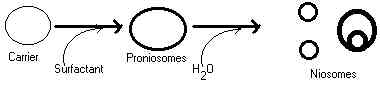
Fig No.3: Formation of noisome from proniosome [1]
Trans membranes pH gradient method [12]
In chloroform, cholesterol and surfactant dissolve. To create a thin layer on the flask's round bottom wall, the solvent is subsequently evaporated at lower pressure. 300 mM citric acid (pH 4.0) is added to the film via vortex mixing. The multilamellar vesicles go through three rounds of freezing and thawing, and then they’re sonicated. After that, this noisome dispersion is mixed with an aqueous drug solution that has 10 mg/ml of the active ingredient, using vortex mixing to ensure everything blends well. Finally, the pH of the mixture is fine-tuned to sit between 7.0 and 7.2 by adding a 1M solution of disodium phosphate. Noisome are then produced by heating this mixture for ten minutes at 60°C.
Fig No.4: Protocol for trans membranes pH gradient method [12]
Sonicator [21,30]
This approach initially disperses the mixture of cholesterol and surfactant in the aqueous phase. Multilameller vesicles (MLV) are created when this dispersion is probe sonicated for ten minutes at 60 °C. These MLVs undergo additional ultrasonication using a bath or probe sonicator, which causes unilameller vesicles to develop.
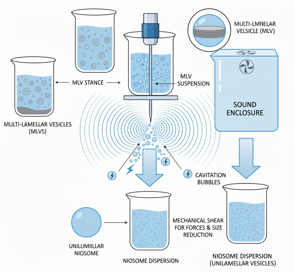
Fig No.5: Sonication
Micro fluidization [22]
Using this technique, two fluidized streams—one carrying a medication and the other a surfactant—interact at extremely high speeds in carefully crafted microchannels inside the interaction chamber so that the energy delivered to the system stays in the region where noisome form. We refer to this as the submerged jet principle. Better homogeneity, reduced size, and reproducibility in noisome formulation are the outcomes.
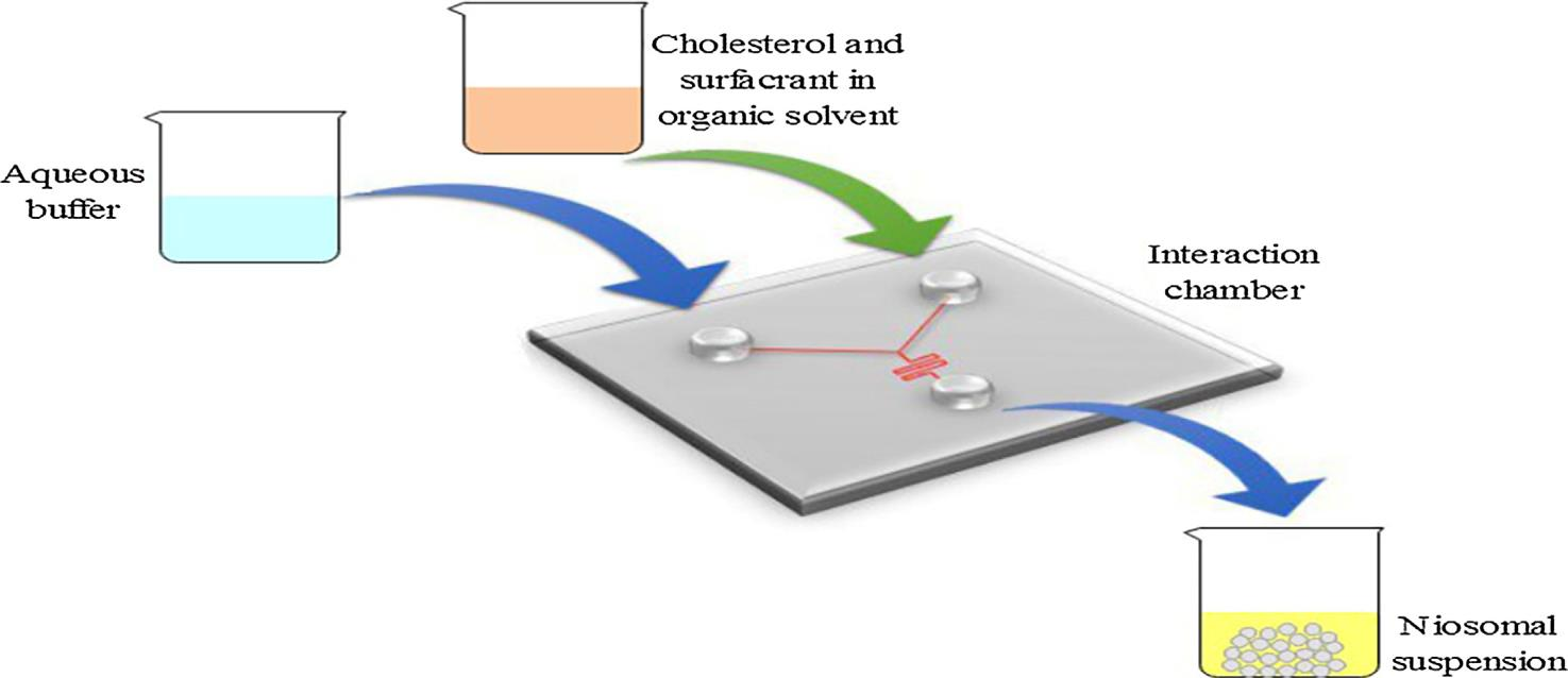
Fig No.6: The protocol for micro-fluidization [31]
The multiple membrane extrusion method [23]
To prepare the formulation, a mixture of surfactant, cholesterol, and diacetyl phosphate dissolved in chloroform is evaporated to form a thin lipid film. This film is then hydrated using an aqueous drug solution, and the resulting suspension is passed through polycarbonate membranes to ensure uniformity in vesicle size. The film is then positioned in sequence for up to eight passages. It's a useful technique for managing noisome size.
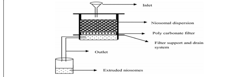
Fig No.7: Protocol for multiple membrane extraction method [23]
Ether injection method [12]
This method involves gradually adding a surfactant that’s been dissolved in diethyl ether into water that’s kept at 60°C. This process helps in creating noisome. A 14-gauge needle is used to inject a combination of ether-based surfactants into a solution of water. When the ether is vaporized, it leads to the formation of single-layered vesicles. Vesicle diameters can vary quite a bit, ranging from 50 to 1000 nanometres, depending on the specific conditions during the formulation process. One downside of this method is that it might leave behind tiny traces of ether in the vesicle suspension, making it tricky to eliminate completely.
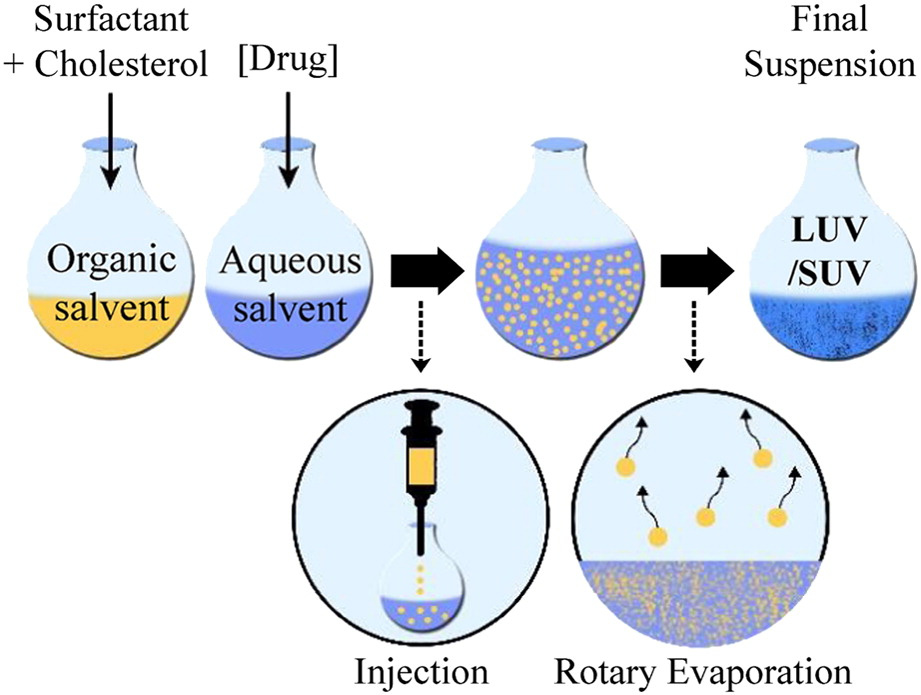
Fig No.8: Protocol for ether injection method [32]
Thin film hydration technique (Hand shaking method) [12]
When using the hand shaking method, a round-bottom flask is used to dissolve cholesterol and a non-ionic surfactant in a volatile organic solvent (such as methanol, diethyl ether, or chloroform). A thin coating of solid mixture is left on the flask wall after the organic solvent is eliminated using a rotary evaporator set to room temperature (20°C). The drug-containing aqueous phase is added to the dry surfactant film at 50–60°C while being gently stirred. This process creates multilamellar noisome.
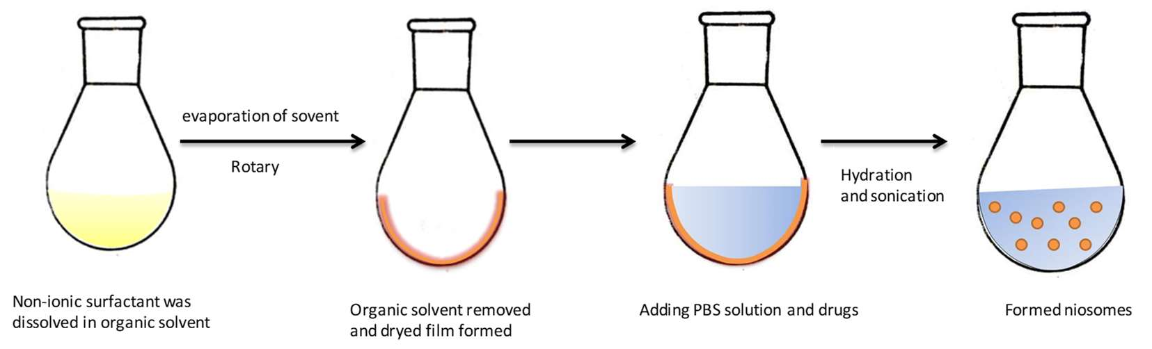
Fig No.9: Protocol for thin film hydration technique [27]
Bubble method [10]
The bubble method is a brand-new process that produces noisome without the use of organic solvents. This method involves a bubbling setup where we control the temperature using a three-necked round-bottom flask that sits in a water bath. One neck connects to a water-cooled reflux condenser, another holds a thermometer, and the last one is for introducing nitrogen gas. We mix surfactant and cholesterol in a buffer solution with a pH of 7.4 and heat it up to 70°C. After that, we subject the mixture to high shear homogenization for 15 seconds, and then we bubble nitrogen gas through it right away while keeping it at the same temperature.
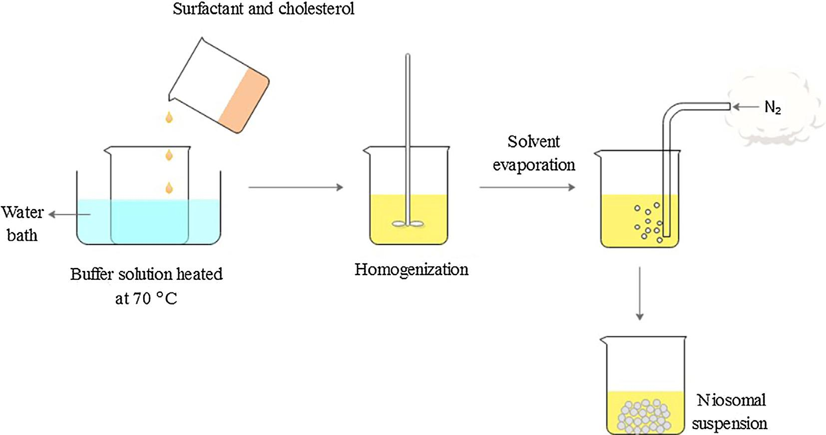
Fig No.10: Protocol for bubble method [31]
CHARACTERIZATION TECHNIQUES
The shape, size distribution, charge and zeta potential, entrapment efficiency, drug release curve, lamellarity, rigidity, stability, viscosity, conductivity, and homogeneity are all included in the physicochemical characterization and analysis of noisome.[32]
Table No:4- Method for Characterization of Noisome
|
Noisome parameter |
Measurement |
|
Particle size |
DLS, SEM, AFM, STM, CLS [18,24] |
|
Zeta potential |
DLS, Electrophoretic mobility [12] |
|
Encapsulation efficiency |
Encapsulation efficiency = Encapsulated amount/ total amount × 100% The amount of the loaded drug is determined by HPLC, UV/VIS, Fluorescence [18] |
|
Stability testing |
DLS (determine size and zeta potential in 37 ?C, or in serum to mimic the in vivo situation), Leaky of the loaded drugs [11] |
Abbreviations: - DLS (Diameter Laser Scatter), SEM (Scanning Electron Microscope), AFM (Atomic Force Microscope), STM (Scanning Tunnelling Microscope), HPLC (High Performance Liquid Chromatography)
Particle size and Zeta potential [18,24,12]: -
Noisome are spherical in shape, and Table 4 summarizes the several methods that can be used to determine their size. The size of vesicles can vary from about 20 nm to over 50 μm. Zeta potential analysis is employed to ascertain the prepared formulations' colloidal characteristics.
Encapsulation efficiency [18]: -
The unentrapped medication can be separated via gel filtration, centrifugation, or dialysis to increase the noisome dispersion's entrapment efficiency. The drug's ability to stay trapped in noisome is assessed by spectrophotometrically evaluating the sample after full vesicle destruction with 50% n-propanol or 0.1% Triton X-100.
Where,
percentage of entrapment= Total drug - diffused drug / total drug
Stability testing [11]: -
The ideal batch of noisome was kept in airtight, sealed vials at various temperatures to assess their stability.
In vitro drug release [34]: -
The dialysis bag method is a popular technique for assessing drug release in vitro. To get started, the dialysis tubing needs to be cleaned and soaked in distilled water for about half an hour. After that, the noisome formulation containing the drug is placed inside the bag. This bag is then submerged in a buffer solution, which is maintained at either 25°C or 37°C, and it’s continuously shaken to ensure consistent conditions. At specific time intervals, samples are taken from the surrounding buffer and replaced with fresh buffer of the same volume. Finally, the amount of drug that has been released into the buffer is measured using a suitable assay technique.
MECHANISM OF DRUG TARGETING
Table No:5-Passive vs. Active Targeting [25]
|
Passive targeting |
Active targeting |
|
modifies how drugs are biodistributed within the body |
enhances target cells' absorption of drugs
|
|
Based on the Enhanced Permeability and Retention (EPR) effect in tumours, targeting
|
The basis for targeting is the molecular interactions between cancer receptors and NP's ligands. |
|
Limited efficacy and modest specificity Limited in application More toxicity and adverse effects |
High effectiveness and specificity in use incredibly adaptable Reduced toxicity and adverse consequences |
|
Reduced engineering and synthesis effort
|
It is more difficult to synthesize NPs with ligands attached while preserving their capacity to connect with target receptors in order to accomplish the desired activity (active targeting). |
Passive Targeting [26]
The term "passive targeting" typically describes drug delivery methods that aim to deliver the medication to the systemic circulation. Passive targeting takes advantage of the natural interactions between the body’s environment and the physical and chemical properties of a drug or its delivery system. This process helps the drug to gather at the intended site (see Figure). For instance, Zhang and his team successfully targeted breast cancer and cancer stem cells by using salinomycin-loaded micelles through passive targeting.
Active Targeting [26]
Active targeting works by attaching specific ligands to the surface of a drug delivery system, allowing them to bind to receptors found on target cells. These ligands can be things like albumin, antibodies, or bio adhesive non-ionic surfactants. There are three levels of active targeting, as illustrated in the figure: first-order targeting focuses on organs, second-order targeting hones in on specific cells, and third-order targeting goes even deeper, targeting within cells. For example, Zwicke et al. used folate receptors to effectively deliver anticancer drugs.
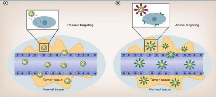
Fig No.11.Mechanisam of active targeting and passive targeting [33]
APPLICATIONS
Table No:6-Application in Pharmaceutical Delivery
|
Application |
Components |
Drugs |
|
Anticancer drug delivery [6,28,10] |
Span 60, Cholesterol, DCP |
Doxorubicin |
|
Ophthalmic drug delivery [5,28,29] |
Span 20, Span 60, Cholesterol |
Pilocarpine hydrochloride |
|
Transdermal drug delivery [3,5,10] |
a, w-Hexadecyl-bis-(1-aza)18-crown-6(bola), Span 80, Cholesterol Brij 96, Cholesterol |
Ammonium glycyrrhiinate |
|
Nasal drug delivery [28] |
Span20, Span40, Span80, Span85, Cholesterol, Phosphate buffered saline |
Nefopam |
|
Peptide drug delivery [3,5,10] |
Span20, Span40, Span60, Tween20, Tween80, Cholesterol |
Insulin |
|
Pulmonary drug delivery [5,28] |
Span40, Span60, Span85, Tween60, Cholesterol |
Zanamivir |
|
Diagnostic imaging [5,28] |
N-Palmitoyl-glucosamine (NPG), Polyethylene glycol (PEG)-4400 |
Gadobenate |
|
For anti-inflammatory effect [27,6] |
Cholesterol (CH), Diacetyl phosphate (DCP) and Surfactants (Tween 85, Pluronic F108) |
Diclofenac sodium |
|
For brain targeting [5,28] |
N-Palmitoyl glucosamine (NPG), Span 60, Cholesterol, Solulan C24 |
Vasoactive Intestinal Peptide |
TOXICITY [12,23]
Noisome' non-ionic composition is likely to contribute to their low toxicity. In actuality, non-ionic surfactants are less harmful and more compatible than their anionic, amphoteric, or cationic equivalents. On the other hand, non-ionic surfactant segregation could be harmful. Additionally, the location and concentration of released drug provide another rationale for toxicity when noisome are involved in lowering and targeting pharmacological side effects. Noisome also have the ability to target, which lessens the negative effects of the medication; but, in certain situations, the excessive concentration and improper placement of the released drug may still be harmful.
BIOCOMPATIBILITY [12]
The numerous benefits of noisome, including their biodegradability, non-immunogenicity, bioavailability, and ability to effectively modify drug release parameters, have garnered significant interest in controlled drug delivery systems. Furthermore, due to their non-ionic structure and remarkable biodegradability, noisome have demonstrated low toxicity and great biocompatibility. Therefore, there are many chances for future advancements in drug delivery due to noisome' diverse biological functions, such as their low immunogenicity.
FUTURE PERSPECTIVES
Noisome are a promising method of medication delivery. Noisome can be used to boost the bioavailability and targeting capabilities of hazardous medications, such as antiviral, anti-AIDS, and anti-cancer medications, by encapsulating them. Noisome do not require particular handling or storage conditions. Use the noisome technology to prepare the cosmetics. The two main barriers to noisome' prospective use as drug delivery vehicles are their physical stability and sterilizing.
CONCLUSION
Noisome drug delivery is a successful approach of creating novel medication delivery. Noisome are mostly made up of cholesterol and non-ionic surfactants. Noisome, which have a structure similar to liposomes, offer a versatile vesicular system capable of encapsulating a diverse array of drugs within their multilayered design. For drug delivery, noisome are thought to be superior to liposomes for a number of reasons, including stability and affordability. Noisome are highly promising for targeted administration of anti-inflammatory, anti-cancer, and anti-infective drugs as well as transdermal drug delivery. More recently, they have been used as adjuvants in vaccines and as diagnostic agents.
REFERENCE


Tejal V. Mhaskar*, Maithili Patil, Lokesh Vyas, Sonali Uppalwar, The Noisome Revolution: Redefining Pharmaceutical Delivery, Int. J. of Pharm. Sci., 2025, Vol 3, Issue 9, 3207-3222 https://doi.org/10.5281/zenodo.17221811
 10.5281/zenodo.17221811
