View Article

Abstract

AI has truly transformed the area of cancer imaging and made radical changes in the improved diagnosis, therapy approaches, and patient outcomes. The applications, advantages, and difficulties of AI in cancer imaging are highlighted in this review paper. Applications of AI in lung, breast, and prostate cancers are discussed, along with the potential to enhance evaluation, diagnosis, and treatment strategy; issues and constraints with AI in cancer imaging; precision and prognostication; and the significance of clinician education and training. Lastly, we explore future directions of AI in cancer imaging, such as multimodality data and more transparent and interpretable AI algorithms.

Keywords

Artificial Intelligence (AI), Cancer Imaging, Machine Learning (ML), Deep Learning (DL), Computer-Aided Detection (CAD), Imaging Analysis, Diagnosis, Treatment Planning, Lung Cancer, Breast Cancer, Prostate Cancer.

Introduction

Although our understanding of the molecular causes of cancer has improved significantly, patients, researchers, and doctors are still baffled by this self-reliant and adaptable disease that dynamically connect with its environment [1]. This makes it more challenging to correctly distinguish between preneoplastic and neoplastic lesions, predict infiltrative tumor margins during surgical treatment, monitor tumor progression and possible acquired resistance to treatments over time, and assess tumor aggressiveness, metastatic pattern, and recurrence [2]. These problems in cancer monitoring, treatment, and detection may be resolved by technological advancements in minimally invasive biomarkers and medical imaging. Yet, the processing of the enormous amount of data produced by these innovations poses several new possible challenges [3]. The daily lives of most individuals are influenced by AI and ML. Indeed, digital health care is expected to contribute significantly to disease diagnosis and treatment in the future. The technology has evolved such that independent diagnostic tools for the illness have been designed according to the need for humongous databases to enable them to take care of issues related to future development in the detection of human diseases, especially at the initial stages of cancer disease [4]. The ability of recent developments in artificial intelligence to decode radiographic patterns automatically in medical images has demonstrated remarkable achievement. One particularly useful technique is machine learning, a subfield of artificial intelligence that has been demonstrated to perform better than humans in task-specific applications and can automatically learn feature representations from example photos. Among other things, deep learning has shown a notable level of robustness against noise in the ground truth labels, even though it requires vast data sets for training [5]. Examples of how AI's automated capabilities open up new avenues for qualitative improvement of clinical judgment include: conversion of subtleties in intertumoral phenotype to consequences for genotype, parallel monitoring of lesions, precision tumor size volumetric demarcation with time, and outcomes predicted by connecting each tumor to a database of possibly infinitely similar instances [6]. In addition, compared to other diseases and imaging modalities, deep learning has great promise for generalization, noise resilience, and reduced inaccuracy. These factors will ultimately lead to early interventions, which are critical for enhancing diagnostics and therapeutic care. Medical imaging uses these AI techniques to treat prevalent tumors, including prostate, breast, lung, and brain cancer [7].  We draw attention to the clinical challenges associated with cancer detection and treatment, how new advancements attempt to address these challenges, and how these challenges may influence the potential future course of artificial intelligence [8].

Artificial intelligence in medicine

Additionally, it has enabled computers and robots to replicate human intelligence and behavior, create medical statistical datasets, assist with robotic surgery and clinical diagnostics, formulate pharmaceuticals, and comprehend the molecular makeup of human disorders like cancer. The impact of AI on medicine is both tangible and intangible [9]. In addition to helping the doctor make wise judgments, the DL information management technologies-reliant virtual component has the ability to evaluate the electronic health record information dataset. DL enhances experience-based learning by using a mathematical method. Nonetheless, the physical system of AI may be useful for applications such as tailored drug delivery through robotic surgery and nano-robotic surgery [10]. AI has been employed in robotic surgery for the correction of heart valves, gynecological illnesses, prostatectomy-related procedures, and is believed to have an significant part in the future battle against cancer. AI can already solve intricate problems, including complicated biological ones [11]. Medicine's potential ability to transform healthcare through increased speed, accuracy, and safety is offered by AI. To investigate the medical consequences of artificial intelligence in radiography for medical purposes, extensive datasets have been developed and are frequently updated [12]. Scotland's "National health service based on the DL NHS 111 algorithm, is presently undergoing medical evaluations to help the public having a little health concern via telephone call at home [13]. Semantic web technology is being used by "Babylon Health," another online healthcare provider, to provide beneficial digital services that improve clinical outcomes. Enabling machine reading of online data is the goal of the semantic web. Create clinical LDGs (Linked Data Graphs) to connect different bioinformatic-based biomedical data and employ AI-based medical services [14].

AI's current function in radiology

Traditional artificial intelligence (AI), a type of machine learning, was introduced in the 1980s to diagnostic imaging. The users are required to predefine first the specific characteristics and features of the imaging using their knowledge [15]. Considering the number of data entries available, training typically employs part of them, and testing employs the rest. A specific learning algorithm is chosen to learn the features for the training process. These algorithms are CNN, SVM, PCA, etc. The trained algorithm must identify the attributes and classify a given test image [ 16]. The fact that the user must identify the characteristics that specify the class to which the image belongs is one of the challenges with machine learning. This might, however, ignore some of the relevant elements. To identify a lung tumor, for example, the user needs to be able to segment the tumor's location as structural parts [17]. In order to gradually extract higher level information from raw image input, deep learning employs numerous layers. It identifies the features that can boost performance and aids in deciphering the abstractions [18]. It has only gained relevance in the past ten years due to advancements in hardware, specifically graphics processing units (GPU), and the creation of thousands of medical images. However, machine learning and its importance increased over time, and even the GPU was partly overtaken. But as the day went on, machine learning and its significance grew, and even the GPU was somewhat surpassed [19].

Current role of AI in radiation oncology

AI has been applied to radiation oncology imaging studies. in image registration, segmenting organs and lesions, fiducial radiomics, markers identification, etc. Like imaging, it began with Using deep learning and conventional AI. In the current edition of the journal Medical Physics (May 2019, Volume 46) 5, 16 of 51 imaging-related studies were based on machine learning studies. As we all know, medical imaging is only one area of radiation oncology research. The majority of the the quantity of released research articles on deep learning imaging demonstrates the significant contribution artificial intelligence is making to the subject [20].

Automatically dividing the organs at danger in order to plan therapy is the main goal of organ and lesion segmentation. Deep learning methods have been used to divide the brain, lung, prostate, kidney, pelvis, and head and neck organs. The rectum, lymph nodes, brain, liver, lung, bone, head and neck are among the areas where lesion segmentation is used. The data collection, deep learning methods, segmentation object, and related performance have all been compiled by Sahiner et al. [21]. One often used algorithm was U-net. Unlike classical AI, U-nets consist of several convolution layers, deconvolution layers, and links between the opposing convolution and deconvolution components. As a result, the network may provide segmentation likelihood maps quickly by looking at the complete image during training [22]. When receiving radiation therapy, a patient must go through a number of steps, such as immobilization and positioning, CT acquisition planning, radiation (i.e., figuring out the best dose, time, and fractionation regimen), beam position optimization with the best dose coverage and preservation of healthy tissues, radiation delivery, and follow-up after therapy. AI solutions are specifically made to support and enhance this approach. It has been suggested that machine learning be used for automatic organ segmentation, error avoidance, and therapy preparation [23].

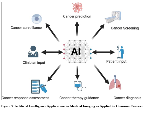
AI Applications in Cancer Imaging

Artificial intelligence is one of the several advancements being employed to improve the efficacy of therapeutic care. Clinical workflow optimization and simplification are becoming more and more important due to the growing need for medical care and the vast amounts of data produced from multiple sources. AI has the potential to make image interpretation less subjective and qualitative and more quantitative and reproducible due to its exceptional capacity to identify intricate designs in images. Furthermore, AI could improve clinical decision-making by quantifying information from photographs that humans are unable to detect [24].  AI is quite beneficial in cancer imaging for three primary clinical duties: tumor monitoring, characterization, and detection. Computer-aided detection (CADe) is the term used to describe the localization of items of interest in radiography. AI-based detection technologies can be employed as a first line of defense against errors of omission and to minimize observational oversights. In a pattern-recognition context, the reader is shown areas with dubious imaging features highlighted [25]. CADe has been used to detect missed tumors in low-dose CT screening, discovered microcalcification clusters in screening mammography as an indicator of early breast carcinoma, and more. It is also used as an auxiliary aid to increase radiological interpretation time while preserving good detection sensitivity, identify brain metastases in MRIs [26]. Apart from imaging, a number of microsurgical biomarkers are created for long-term illness monitoring and cancer detection. Liquid biopsies, which analyze circulating tumor DNA (ctDNA) released from tumor cells, are especially notable because they offer a window into the real time and dynamic state of a cancer, track the disease's progression, and enable near real-time reporting for the emergence of cancer mutations linked to resistance or targetability [27]. Therefore, by enabling Real-time disease monitoring and more precise prognostic evaluation using noninvasive characterization of cancer biology for precision medicine, the combination of liquid biopsies and radiomics profiling may greatly enhance cancer treatment [28]. It is expected that the aforementioned AI treatments will complement their existing standard-of-care equivalent in clinical settings. In addition to giving physicians helpful information, a number of projects have shown the usefulness of AI in the medical decision-making stages of the process. AI-powered image-based analysis combined with pathology and molecular data will enhance the results' intelligence and ultimately lead to better decision-making [29].

Lung Cancer Imaging

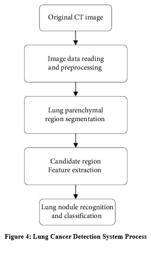
Lung cancer is among the top killers of cancer deaths in both women and men worldwide. Lung cancer is lagging behind despite the advances in survival over the last few decades made by other forms of cancers. This is due primarily to the fact that lung cancer tends to be advanced when it is diagnosed, and at that point, there are not a lot of options available. The late stage at diagnosis can be the cause of the fact that the majority of people suffering from lung cancer would die of their disease [30] Medical diagnosis and artificial intelligence are expected to greatly enhance the early identification and description of lung cancer by distinguishing between harmful and non-cancerous nodules. The early stages are mostly treatable, thereby significantly improving patient outcome, reducing overtreatment, and saving lives in some cases. AI will also add value to the classification and staging of lung cancer for treatment selection and treatment response monitoring [31]. Among the leading causes of death from cancer for both men and women worldwide is lung cancer. However, over the last few decades, progress has been seen in many cancers, but lung cancer has lagged mostly because the disease was detected quite late, and by the time of discovery, no treatment has been available. It could be the one of the primary causes death from malignant diseases is still lung cancer [32]. From diagnosis to tracking response to treatment, imaging is essential to the management of lung cancer. Creation of imaging-related artificial intelligence (AI) models may potentially aid in personalized treatment planning and early detection. The paradigm of early illness detection through screening programs has shifted as a result of computer-aided lung nodule detection. AI models that combine imaging features with clinical and laboratory data demonstrate extremely encouraging outcomes in terms of forecasting patient outcomes, response to certain treatments, and the likelihood of hazardous reactions [33]. In order to inform radiologists and clinicians about the potential for the majority of patients to die from lung cancer in very late-stage cases, the review highlights the current work on major artificial intelligence imaging tools developed in the field of lung cancer screening, such as automated lesion identification, characterization and segmentation, and forecasting the result and reaction to treatment [34].


Breast Cancer Imaging

In the US, breast cancer is the second leading cause of cancer-related death among women is also the most prevalent malignant disease among women. Since the 1980s, the five-year survival rate for breast cancer has significantly improved, in breast cancer treatment and greater use of mammography screening [35]. Because breast cancer is heterogeneous, each tumor has a different source, prediction, and outcome to treatment. The estrogen receptor's existence may establish etiologic subgroups and is crucial for responsiveness to particular treatments and for prognostic outcomes that are worse for those with ER-negative disease [36]. Lack of ER, PR, and HER2 is a hallmark of triple-negative breast cancer. They have lower five-year survival rates, identified as interval and advanced cancers, and do not exhibit the usual indicators of malignancy on conventional mammography. Because of the rapid improvements in computer and imaging technologies, the usage of AI has become more prevalent in breast imaging tasks like risk assessment, detection, diagnosis, prognosis, and therapeutic response dramatically [37].

Prostate Cancer Imaging

It helps to diagnosis around prostate cancer manifestation, which characterizes highly the aggressive biological phenotype of the disease differing according to treatment options available. Evaluation in clinics, TRUS or CT-based staging of prostate cancer is significantly improved by considering MRI since it displays simultaneous involvement of pelvic, periprostatic, and prostate architecture [38]. The location, size, and extent of cancer can be more accurately assessed with MRI or dynamic computer-enhanced MRI with MR spectroscopic imaging as well as it provides indications on aggression at clinical practice. Implementation of minimally invasive, patient-specific therapy is dependent on pretreatment understanding of these prognostic factors [39]. Treatment of prostate cancer remains plagued by many challenges. Subclinical illness and clinically severe prostate cancer cannot be distinguished by screening testing, which is due to very early [40]. The whole idea of screening for prostate cancer on an uninhibited population scale is yet to be the subject of heated debate, even with evidence that the vast majority of cancers (85% to 90% of all) identified by prostate cancer screening programs constitute genuinely clinically important disease and not a harmless deviation from it and are being identified at a more premature stage of disease. Further, it is very expensive to screen all men above 50 years, and the available funds for healthcare cannot cover this expense. The second barrier hinders planning for treatment[41].  The capacity of currently available tumor prognostic markers to distinguish between Indolent and aggressive illness is limited. At the extremes, tumor prognostic might be able to forecast how the illness will behave [42]. However, prognostic variables for most cancers may fall somewhere in the center, making it very challenging to distinguish between malignancies that are likely to advance and those that are suited for surveillance. The accuracy with which currently used tumor prognostic variables can categorize the risks and treatment strategy of such individuals is a topic of intense discussion. [43]. Over the next ten years, the main goals of prostate cancer imaging will be to accurately characterize the illness by combining anatomical, functional, and molecular imaging data. There is no agreement on the evaluation of primary prostate cancers using imaging [44]. Superparamagnetic nanoparticle magnetic resonance imaging is limited to research settings and has shown better sensitivity and specificity in imaging lymph node metastases, despite the fact that there are currently no published guidelines for its application [45]. CT is especially useful to evaluate advanced illness. While its application in evaluating primary disease is restricted, combined PET/CT is becoming more widely accepted for monitoring prostate cancer treatment outcomes [46].

Challenges of AI in imaging

Before AI is widely utilized in clinical settings, a number of restrictions and challenges must be resolved, regardless of how much more acceptance there is in the field of cancer imaging. Continuous structures that generate data are presented by the rising need for CT and MRI imaging [47]. The application of CAD to enhance radiologists' performance in lung and mammography imaging has shown promise, but the high rate of false positives still makes it difficult for the health sector to adopt AI [48]. Errors in CAD software often cause normal structures to appear odd. Radiologists find it extremely difficult to distinguish between these real aberrant lesions and fictitious prompts, which hinders their effort in terms of recall rates, throughput, and expenses. CAD FPs range from roughly 1 to 2.2 each mammography test and from 4.6 to 11 per chest CT scan. In real-world situations, Such elevated FP rates negatively affect patient recall rates and make CAD initiatives unsustainable [49]. There are more needless invasive treatments performed when recall rates are higher. However, since false positives are no longer an issue in the future, improvements in study of CAD and image processing during the last ten years have led to a growing decrease in false positives. Such recent advances illustrate the rapid evolution of the algorithms and technology that AI is based on, giving credence to the idea that the CAD application will soon become easy and very efficient [50]. Although AI is initial promising medical imaging research, there are still hurdles to cross before it is more dependable and common in the clinic. A shortcoming of AI is the paucity of appropriately high-volume, high-quality longitudinal outcomes information. The same disease site may be photographed using the same technique, but various imaging modalities and scenarios are sometimes utilized in the clinic. A specific set of images will be linked with a specific clinical scenario [51]. It may become difficult for anyone organization to utilize any type of AI algorithm to deal with the various clinical situations that could arise and the various parts of labor that could be included in each picture. Different patient cohorts are associated with different clinics [52].  Every clinic operates differently too. One of the largest challenges for AI and medical imaging research is putting the data generated by the wide variety of activities into a consistent format. Medical imaging data sorting could be a worth-while area of research [53].

The future of AI in Imaging

The AI program only has a high false positive rate, which is fairly high but is gradually improving, and it appears to be a suitable second reader. IBM spent $1 billion on an algorithm called the Watson Health Project currently undergoing testing for the healthcare industry, is a clear sign of the company's trust in the imaging AI future [54]. Watson will be able to learn from 30 billion images thanks to this investment, which will permit its algorithm to access the most extensive knowledge source available. It will also benefit from patient supporting data, such as medical histories, blood tests, genetic sequences, and prior imaging [55] AI events may recognize incidental findings that are clinically significant, but conversely, may be clinically insignificant. Unless properly put in the right clinical context, they can cause significant added patient anxiety, negative medical costs, and at times unforeseen side effects from treatment [56]. Most incidental findings, though, will normally be evaluated by humans on the consideration of whether to treat them as being clinically significant or not, as when they were discovered by humans, in this initial stage of AI, Human specialists will proceed to predominate AI's output through responsible gatekeeping [57]. Like for primary lesions, such findings could have given rise to the development of AI systems into ordinary parts of data assessment and reporting [58]. Moreover, imaging is not a reliable predictor of disease on its own. More and more people are realizing how cancer genetic fingerprints, incorporating tumor, socioeconomic, and even social network noninvasive blood biomarkers affect the prognosis of cancer patients. Data sources are also expanding rapidly outside of direct medical testing [59]. These include data from social media, smartphones, wearable technologies, disorganized electronic health records, as well as other elements of the virtual era. AI is ideal to progressively combining biological, demographic, and social data streams to enhance patient outcome prediction models that are assessed and communicated within the clinical context of their patients [60]. As the strength and potential of AI become clearer, there are several ways its clinical application is expanding. To displace clinician workflows with AI approaches to imaging analysis, more work will be required to enhance their predictive value and accuracy than demonstrating they can generate output comparable to or superior to human control in well-controlled studies [61]. Certain conditions hold promise initially, yet additional clinical utility evidence from prospective research and education for physicians, technologists, and physicists might be required before they become generally available [62]. Indeed, while looking at outcomes produced by AI, there will always be this "black box" from the perspective of the human expert. Conversely, there are already many data visualization tools available that will enable a basic understanding of how algorithms truly make judgments [63].

CONCLUSION

By increasing the precision, speed, and effectiveness of picture interpretation, AI's application to cancer imaging holds the ability to completely transform the industry. However, several challenges and limitations must be addressed, including data quality and availability, algorithmic bias, regulatory frameworks, and clinical validation. However, challenges regarding data quality, availability, and standardization must be resolved in order to avoid bias in the training of models created on insufficiently diverse datasets, resulting in poor predictive quality. In addition, there is a need for algorithms that are not biased, regulatory frameworks that are robust, and clinical validation which remain significant barriers to adoption in large scale. The directions for the future would include progress in liquid biopsies, imaging genomics, radiomics, and imaging informatics, which will create great prospects in improving cancer diagnosis and prognosis toward a situation in the future where AI becomes readily useful in clinical practice.

REFERENCES

        1. Bi WL, Hosny A, Schabath MB, Giger ML, Birkbak NJ, Mehrtash A, Allison T, Arnaout O, Abbosh C, Dunn IF, Mak RH. Artificial intelligence in cancer imaging: clinical challenges and applications. CA: a cancer journal for clinicians. 2019 Mar;69(2):127-57.
        2. Souhami R, Tobias JS. Cancer and its management. John Wiley & Sons; 2008 Apr 15.
        3. Pulumati A, Pulumati A, Dwarakanath BS, Verma A, Papineni RV. Technological advancements in cancer diagnostics: Improvements and limitations. Cancer Reports. 2023 Feb;6(2):e1764.
        4. Dandotiya AS, Gupta SK, Dandotiya N, Sharma MP. AI IN EVERYDAY LIFE: TRANSFORMING SOCIETY. Navi International Book Publication house; 2024 Sep 18.
        5. Baduge SK, Thilakarathna S, Perera JS, Arashpour M, Sharafi P, Teodosio B, Shringi A, Mendis P. Artificial intelligence and smart vision for building and construction 4.0: Machine and deep learning methods and applications. Automation in Construction. 2022 Sep 1;141:104440.
        6. Najjar R. Redefining radiology: a review of artificial intelligence integration in medical imaging. Diagnostics. 2023 Aug 25;13(17):2760.
        7. Jiang X, Hu Z, Wang S, Zhang Y. Deep learning for medical image-based cancer diagnosis. Cancers. 2023 Jul 13;15(14):3608.
        8. Shreve JT, Khanani SA, Haddad TC. Artificial intelligence in oncology: current capabilities, future opportunities, and ethical considerations. American Society of Clinical Oncology Educational Book. 2022 Jun 10;42:842-51.
        9. Subramanian M, Wojtusciszyn A, Favre L, Boughorbel S, Shan J, Letaief KB, Pitteloud N, Chouchane L. Precision medicine in the era of artificial intelligence: implications in chronic disease management. Journal of translational medicine. 2020 Dec;18:1-2.
        10. Sarker IH. Deep learning: a comprehensive overview on techniques, taxonomy, applications and research directions. SN computer science. 2021 Nov;2(6):420.
        11. Shastry KA, Sanjay HA. Cancer diagnosis using artificial intelligence: a review. Artificial Intelligence Review. 2022 Apr;55(4):2641-73.
        12. Carini C, Seyhan AA. Tribulations and future opportunities for artificial intelligence in precision medicine. Journal of Translational Medicine. 2024 Apr 30;22(1):411.
        13. Holzinger A, Langs G, Denk H, Zatloukal K, Müller H. Causability and explainability of artificial intelligence in medicine. Wiley Interdisciplinary Reviews: Data Mining and Knowledge Discovery. 2019 Jul;9(4):e1312.
        14. Iqbal MJ, Javed Z, Sadia H, Qureshi IA, Irshad A, Ahmed R, Malik K, Raza S, Abbas A, Pezzani R, Sharifi-Rad J. Clinical applications of artificial intelligence and machine learning in cancer diagnosis: looking into the future. Cancer cell international. 2021 May 21;21(1):270.
        15. Hosny A, Parmar C, Quackenbush J, Schwartz LH, Aerts HJ. Artificial intelligence in radiology. Nature Reviews Cancer. 2018 Aug;18(8):500-10.
        16. Mun SK, Wong KH, Lo SC, Li Y, Bayarsaikhan S. Artificial intelligence for the future radiology diagnostic service. Frontiers in molecular biosciences. 2021 Jan 28;7:614258.
        17. Bhandari A. Revolutionizing Radiology With Artificial Intelligence. Cureus. 2024 Oct 29;16(10).
        18. Panayides AS, Amini A, Filipovic ND, Sharma A, Tsaftaris SA, Young A, Foran D, Do N, Golemati S, Kurc T, Huang K. AI in medical imaging informatics: current challenges and future directions. IEEE journal of biomedical and health informatics. 2020 May 29;24(7):1837-57.
        19. Reyes M, Meier R, Pereira S, Silva CA, Dahlweid FM, Tengg-Kobligk HV, Summers RM, Wiest R. On the interpretability of artificial intelligence in radiology: challenges and opportunities. Radiology: artificial intelligence. 2020 May 27;2(3):e190043.
        20. Huynh E, Hosny A, Guthier C, Bitterman DS, Petit SF, Haas-Kogan DA, Kann B, Aerts HJ, Mak RH. Artificial intelligence in radiation oncology. Nature Reviews Clinical Oncology. 2020 Dec;17(12):771-81.
        21. Parkinson C, Matthams C, Foley K, Spezi E. Artificial intelligence in radiation oncology: A review of its current status and potential application for the radiotherapy workforce. Radiography. 2021 Oct 1;27:S63-8.
        22. Netherton TJ, Cardenas CE, Rhee DJ, Court LE, Beadle BM. The emergence of artificial intelligence within radiation oncology treatment planning. Oncology. 2021 Dec 22;99(2):124-34.
        23. Deig CR, Kanwar A, Thompson RF. Artificial intelligence in radiation oncology. Hematology/Oncology Clinics. 2019 Dec 1;33(6):1095-104.
        24. Bi WL, Hosny A, Schabath MB, Giger ML, Birkbak NJ, Mehrtash A, Allison T, Arnaout O, Abbosh C, Dunn IF, Mak RH. Artificial intelligence in cancer imaging: clinical challenges and applications. CA: a cancer journal for clinicians. 2019 Mar;69(2):127-57.
        25. Mendes J, Domingues J, Aidos H, Garcia N, Matela N. AI in breast cancer imaging: A survey of different applications. Journal of Imaging. 2022 Aug 26;8(9):228.
        26. Koh DM, Papanikolaou N, Bick U, Illing R, Kahn Jr CE, Kalpathi-Cramer J, Matos C, Martí-Bonmatí L, Miles A, Mun SK, Napel S. Artificial intelligence and machine learning in cancer imaging. Communications Medicine. 2022 Oct 27;2(1):133.
        27. Coccia M. Deep learning technology for improving cancer care in society: New directions in cancer imaging driven by artificial intelligence. Technology in Society. 2020 Feb 1;60:101198.
        28. Obuchowicz R, Strzelecki M, Piórkowski A. Clinical applications of artificial intelligence in medical imaging and image processing—A review. Cancers. 2024 May 14;16(10):1870.
        29. Iqbal MJ, Javed Z, Sadia H, Qureshi IA, Irshad A, Ahmed R, Malik K, Raza S, Abbas A, Pezzani R, Sharifi-Rad J. Clinical applications of artificial intelligence and machine learning in cancer diagnosis: looking into the future. Cancer cell international. 2021 May 21;21(1):270.
        30. Hollings N, Shaw P. Diagnostic imaging of lung cancer. European Respiratory Journal. 2002 Apr 1;19(4):722-42.
        31. Panunzio A, Sartori P. Lung cancer and radiological imaging. Current radiopharmaceuticals. 2020 Dec 1;13(3):238-42.
        32. Schaefer-Prokop C, Prokop M. New imaging techniques in the treatment guidelines for lung cancer. European Respiratory Journal. 2002 Feb 1;19(35 suppl):71s-83s.
        33. Bruzzi JF, Munden RF. PET/CT imaging of lung cancer. Journal of thoracic imaging. 2006 May 1;21(2):123-36.
        34. Purandare NC, Rangarajan V. Imaging of lung cancer: Implications on staging and management. Indian Journal of Radiology and Imaging. 2015 Apr;25(02):109-20.
        35. Karellas A, Vedantham S. Breast cancer imaging: a perspective for the next decade. Medical physics. 2008 Nov;35(11):4878-97.
        36. Morris EA. Breast cancer imaging with MRI. Radiologic Clinics. 2002 May 1;40(3):443-66.
        37. Bartella L, Smith CS, Dershaw DD, Liberman L. Imaging breast cancer. Radiologic Clinics. 2007 Jan 1;45(1):45-67.
        38. Fuchsjäger M, Shukla-Dave A, Akin O, Barentsz J, Hricak H. Prostate cancer imaging. Acta radiologica. 2008 Feb;49(1):107-20.
        39. Kyle KY, Hricak H. Imaging prostate cancer. Radiologic Clinics of North America. 2000 Jan 1;38(1):59-85.
        40. Hricak H, Choyke PL, Eberhardt SC, Leibel SA, Scardino PT. Imaging prostate cancer: a multidisciplinary perspective. Radiology. 2007 Apr;243(1):28-53.
        41. Wibmer AG, Burger IA, Sala E, Hricak H, Weber WA, Vargas HA. Molecular imaging of prostate cancer. Radiographics. 2016 Jan;36(1):142-59.
        42. Kelloff GJ, Choyke P, Coffey DS. Challenges in clinical prostate cancer: role of imaging. American journal of roentgenology. 2009 Jun;192(6):1455-70.
        43. Turkbey B, Albert PS, Kurdziel K, Choyke PL. Imaging localized prostate cancer: current approaches and new developments. American Journal of Roentgenology. 2009 Jun;192(6):1471-80.
        44. Wallitt KL, Khan SR, Dubash S, Tam HH, Khan S, Barwick TD. Clinical PET imaging in prostate cancer. Radiographics. 2017 Sep;37(5):1512-36.
        45. Thompson J, Lawrentschuk N, Frydenberg M, Thompson L, Stricker P, Usanz. The role of magnetic resonance imaging in the diagnosis and management of prostate cancer. BJU international. 2013 Nov;112:6-20.
        46. Jung AJ, Westphalen AC. Imaging prostate cancer. Radiologic Clinics of North America. 2012 Nov 1;50(6):1043-59.
        47. Bi WL, Hosny A, Schabath MB, Giger ML, Birkbak NJ, Mehrtash A, Allison T, Arnaout O, Abbosh C, Dunn IF, Mak RH. Artificial intelligence in cancer imaging: clinical challenges and applications. CA: a cancer journal for clinicians. 2019 Mar;69(2):127-57.
        48. Sharma P, Suehling M, Flohr T, Comaniciu D. Artificial intelligence in diagnostic imaging: status quo, challenges, and future opportunities. Journal of thoracic imaging. 2020 May 1;35:S11-6.
        49. Panayides AS, Amini A, Filipovic ND, Sharma A, Tsaftaris SA, Young A, Foran D, Do N, Golemati S, Kurc T, Huang K. AI in medical imaging informatics: current challenges and future directions. IEEE journal of biomedical and health informatics. 2020 May 29;24(7):1837-57.
        50. Prevedello LM, Halabi SS, Shih G, Wu CC, Kohli MD, Chokshi FH, Erickson BJ, Kalpathy-Cramer J, Andriole KP, Flanders AE. Challenges related to artificial intelligence research in medical imaging and the importance of image analysis competitions. Radiology: Artificial Intelligence. 2019 Jan 30;1(1):e180031.
        51. Cheung HM, Rubin D. Challenges and opportunities for artificial intelligence in oncological imaging. Clinical radiology. 2021 Oct 1;76(10):728-36.
        52. Armato SG, Drukker K, Hadjiiski L. AI in medical imaging grand challenges: translation from competition to research benefit and patient care. The British Journal of Radiology. 2023 Oct 1;96(1150):20221152.
        53. Ozcan BB, Patel BK, Banerjee I, Dogan BE. Artificial intelligence in breast imaging: challenges of integration into clinical practice. Journal of Breast Imaging. 2023 May 1;5(3):248-57.
        54. Fazal MI, Patel ME, Tye J, Gupta Y. The past, present and future role of artificial intelligence in imaging. European journal of radiology. 2018 Aug 1;105:246-50.
        55. Panayides AS, Amini A, Filipovic ND, Sharma A, Tsaftaris SA, Young A, Foran D, Do N, Golemati S, Kurc T, Huang K. AI in medical imaging informatics: current challenges and future directions. IEEE journal of biomedical and health informatics. 2020 May 29;24(7):1837-57.
        56. Sharma P, Suehling M, Flohr T, Comaniciu D. Artificial intelligence in diagnostic imaging: status quo, challenges, and future opportunities. Journal of thoracic imaging. 2020 May 1;35:S11-6.
        57. Ai M. Future of imaging technology. Sensors and Actuators A: Physical. 1996 Aug 1;56(1-2):31-8.
        58. Alexander A, Jiang A, Ferreira C, Zurkiya D. An intelligent future for medical imaging: a market outlook on artificial intelligence for medical imaging. Journal of the American College of Radiology. 2020 Jan 1;17(1):165-70.
        59. Lewis SJ, Gandomkar Z, Brennan PC. Artificial Intelligence in medical imaging practice: looking to the future. Journal of Medical radiation sciences. 2019 Dec;66(4):292-5.
        60. Saboury B, Morris M, Siegel E. Future directions in artificial intelligence. Radiologic Clinics. 2021 Nov 1;59(6):1085-95.
        61. Li X, Zhang L, Yang J, Teng F. Role of artificial intelligence in medical image analysis: A review of current trends and future directions. Journal of Medical and Biological Engineering. 2024 Apr;44(2):231-43.
        62. Fuchsjäger M. Is the future of breast imaging with AI?. European Radiology. 2019 Sep 1;29(9):4822-4.
        63. Asha P, Srivani P, Ahmed AA, Kolhe A, Nomani MZ. Artificial intelligence in medical Imaging: An analysis of innovative technique and its future promise. Materials Today: Proceedings. 2022 Jan 1;56:2236-9..

Reference

  1. Bi WL, Hosny A, Schabath MB, Giger ML, Birkbak NJ, Mehrtash A, Allison T, Arnaout O, Abbosh C, Dunn IF, Mak RH. Artificial intelligence in cancer imaging: clinical challenges and applications. CA: a cancer journal for clinicians. 2019 Mar;69(2):127-57.
  2. Souhami R, Tobias JS. Cancer and its management. John Wiley & Sons; 2008 Apr 15.
  3. Pulumati A, Pulumati A, Dwarakanath BS, Verma A, Papineni RV. Technological advancements in cancer diagnostics: Improvements and limitations. Cancer Reports. 2023 Feb;6(2):e1764.
  4. Dandotiya AS, Gupta SK, Dandotiya N, Sharma MP. AI IN EVERYDAY LIFE: TRANSFORMING SOCIETY. Navi International Book Publication house; 2024 Sep 18.
  5. Baduge SK, Thilakarathna S, Perera JS, Arashpour M, Sharafi P, Teodosio B, Shringi A, Mendis P. Artificial intelligence and smart vision for building and construction 4.0: Machine and deep learning methods and applications. Automation in Construction. 2022 Sep 1;141:104440.
  6. Najjar R. Redefining radiology: a review of artificial intelligence integration in medical imaging. Diagnostics. 2023 Aug 25;13(17):2760.
  7. Jiang X, Hu Z, Wang S, Zhang Y. Deep learning for medical image-based cancer diagnosis. Cancers. 2023 Jul 13;15(14):3608.
  8. Shreve JT, Khanani SA, Haddad TC. Artificial intelligence in oncology: current capabilities, future opportunities, and ethical considerations. American Society of Clinical Oncology Educational Book. 2022 Jun 10;42:842-51.
  9. Subramanian M, Wojtusciszyn A, Favre L, Boughorbel S, Shan J, Letaief KB, Pitteloud N, Chouchane L. Precision medicine in the era of artificial intelligence: implications in chronic disease management. Journal of translational medicine. 2020 Dec;18:1-2.
  10. Sarker IH. Deep learning: a comprehensive overview on techniques, taxonomy, applications and research directions. SN computer science. 2021 Nov;2(6):420.
  11. Shastry KA, Sanjay HA. Cancer diagnosis using artificial intelligence: a review. Artificial Intelligence Review. 2022 Apr;55(4):2641-73.
  12. Carini C, Seyhan AA. Tribulations and future opportunities for artificial intelligence in precision medicine. Journal of Translational Medicine. 2024 Apr 30;22(1):411.
  13. Holzinger A, Langs G, Denk H, Zatloukal K, Müller H. Causability and explainability of artificial intelligence in medicine. Wiley Interdisciplinary Reviews: Data Mining and Knowledge Discovery. 2019 Jul;9(4):e1312.
  14. Iqbal MJ, Javed Z, Sadia H, Qureshi IA, Irshad A, Ahmed R, Malik K, Raza S, Abbas A, Pezzani R, Sharifi-Rad J. Clinical applications of artificial intelligence and machine learning in cancer diagnosis: looking into the future. Cancer cell international. 2021 May 21;21(1):270.
  15. Hosny A, Parmar C, Quackenbush J, Schwartz LH, Aerts HJ. Artificial intelligence in radiology. Nature Reviews Cancer. 2018 Aug;18(8):500-10.
  16. Mun SK, Wong KH, Lo SC, Li Y, Bayarsaikhan S. Artificial intelligence for the future radiology diagnostic service. Frontiers in molecular biosciences. 2021 Jan 28;7:614258.
  17. Bhandari A. Revolutionizing Radiology With Artificial Intelligence. Cureus. 2024 Oct 29;16(10).
  18. Panayides AS, Amini A, Filipovic ND, Sharma A, Tsaftaris SA, Young A, Foran D, Do N, Golemati S, Kurc T, Huang K. AI in medical imaging informatics: current challenges and future directions. IEEE journal of biomedical and health informatics. 2020 May 29;24(7):1837-57.
  19. Reyes M, Meier R, Pereira S, Silva CA, Dahlweid FM, Tengg-Kobligk HV, Summers RM, Wiest R. On the interpretability of artificial intelligence in radiology: challenges and opportunities. Radiology: artificial intelligence. 2020 May 27;2(3):e190043.
  20. Huynh E, Hosny A, Guthier C, Bitterman DS, Petit SF, Haas-Kogan DA, Kann B, Aerts HJ, Mak RH. Artificial intelligence in radiation oncology. Nature Reviews Clinical Oncology. 2020 Dec;17(12):771-81.
  21. Parkinson C, Matthams C, Foley K, Spezi E. Artificial intelligence in radiation oncology: A review of its current status and potential application for the radiotherapy workforce. Radiography. 2021 Oct 1;27:S63-8.
  22. Netherton TJ, Cardenas CE, Rhee DJ, Court LE, Beadle BM. The emergence of artificial intelligence within radiation oncology treatment planning. Oncology. 2021 Dec 22;99(2):124-34.
  23. Deig CR, Kanwar A, Thompson RF. Artificial intelligence in radiation oncology. Hematology/Oncology Clinics. 2019 Dec 1;33(6):1095-104.
  24. Bi WL, Hosny A, Schabath MB, Giger ML, Birkbak NJ, Mehrtash A, Allison T, Arnaout O, Abbosh C, Dunn IF, Mak RH. Artificial intelligence in cancer imaging: clinical challenges and applications. CA: a cancer journal for clinicians. 2019 Mar;69(2):127-57.
  25. Mendes J, Domingues J, Aidos H, Garcia N, Matela N. AI in breast cancer imaging: A survey of different applications. Journal of Imaging. 2022 Aug 26;8(9):228.
  26. Koh DM, Papanikolaou N, Bick U, Illing R, Kahn Jr CE, Kalpathi-Cramer J, Matos C, Martí-Bonmatí L, Miles A, Mun SK, Napel S. Artificial intelligence and machine learning in cancer imaging. Communications Medicine. 2022 Oct 27;2(1):133.
  27. Coccia M. Deep learning technology for improving cancer care in society: New directions in cancer imaging driven by artificial intelligence. Technology in Society. 2020 Feb 1;60:101198.
  28. Obuchowicz R, Strzelecki M, Piórkowski A. Clinical applications of artificial intelligence in medical imaging and image processing—A review. Cancers. 2024 May 14;16(10):1870.
  29. Iqbal MJ, Javed Z, Sadia H, Qureshi IA, Irshad A, Ahmed R, Malik K, Raza S, Abbas A, Pezzani R, Sharifi-Rad J. Clinical applications of artificial intelligence and machine learning in cancer diagnosis: looking into the future. Cancer cell international. 2021 May 21;21(1):270.
  30. Hollings N, Shaw P. Diagnostic imaging of lung cancer. European Respiratory Journal. 2002 Apr 1;19(4):722-42.
  31. Panunzio A, Sartori P. Lung cancer and radiological imaging. Current radiopharmaceuticals. 2020 Dec 1;13(3):238-42.
  32. Schaefer-Prokop C, Prokop M. New imaging techniques in the treatment guidelines for lung cancer. European Respiratory Journal. 2002 Feb 1;19(35 suppl):71s-83s.
  33. Bruzzi JF, Munden RF. PET/CT imaging of lung cancer. Journal of thoracic imaging. 2006 May 1;21(2):123-36.
  34. Purandare NC, Rangarajan V. Imaging of lung cancer: Implications on staging and management. Indian Journal of Radiology and Imaging. 2015 Apr;25(02):109-20.
  35. Karellas A, Vedantham S. Breast cancer imaging: a perspective for the next decade. Medical physics. 2008 Nov;35(11):4878-97.
  36. Morris EA. Breast cancer imaging with MRI. Radiologic Clinics. 2002 May 1;40(3):443-66.
  37. Bartella L, Smith CS, Dershaw DD, Liberman L. Imaging breast cancer. Radiologic Clinics. 2007 Jan 1;45(1):45-67.
  38. Fuchsjäger M, Shukla-Dave A, Akin O, Barentsz J, Hricak H. Prostate cancer imaging. Acta radiologica. 2008 Feb;49(1):107-20.
  39. Kyle KY, Hricak H. Imaging prostate cancer. Radiologic Clinics of North America. 2000 Jan 1;38(1):59-85.
  40. Hricak H, Choyke PL, Eberhardt SC, Leibel SA, Scardino PT. Imaging prostate cancer: a multidisciplinary perspective. Radiology. 2007 Apr;243(1):28-53.
  41. Wibmer AG, Burger IA, Sala E, Hricak H, Weber WA, Vargas HA. Molecular imaging of prostate cancer. Radiographics. 2016 Jan;36(1):142-59.
  42. Kelloff GJ, Choyke P, Coffey DS. Challenges in clinical prostate cancer: role of imaging. American journal of roentgenology. 2009 Jun;192(6):1455-70.
  43. Turkbey B, Albert PS, Kurdziel K, Choyke PL. Imaging localized prostate cancer: current approaches and new developments. American Journal of Roentgenology. 2009 Jun;192(6):1471-80.
  44. Wallitt KL, Khan SR, Dubash S, Tam HH, Khan S, Barwick TD. Clinical PET imaging in prostate cancer. Radiographics. 2017 Sep;37(5):1512-36.
  45. Thompson J, Lawrentschuk N, Frydenberg M, Thompson L, Stricker P, Usanz. The role of magnetic resonance imaging in the diagnosis and management of prostate cancer. BJU international. 2013 Nov;112:6-20.
  46. Jung AJ, Westphalen AC. Imaging prostate cancer. Radiologic Clinics of North America. 2012 Nov 1;50(6):1043-59.
  47. Bi WL, Hosny A, Schabath MB, Giger ML, Birkbak NJ, Mehrtash A, Allison T, Arnaout O, Abbosh C, Dunn IF, Mak RH. Artificial intelligence in cancer imaging: clinical challenges and applications. CA: a cancer journal for clinicians. 2019 Mar;69(2):127-57.
  48. Sharma P, Suehling M, Flohr T, Comaniciu D. Artificial intelligence in diagnostic imaging: status quo, challenges, and future opportunities. Journal of thoracic imaging. 2020 May 1;35:S11-6.
  49. Panayides AS, Amini A, Filipovic ND, Sharma A, Tsaftaris SA, Young A, Foran D, Do N, Golemati S, Kurc T, Huang K. AI in medical imaging informatics: current challenges and future directions. IEEE journal of biomedical and health informatics. 2020 May 29;24(7):1837-57.
  50. Prevedello LM, Halabi SS, Shih G, Wu CC, Kohli MD, Chokshi FH, Erickson BJ, Kalpathy-Cramer J, Andriole KP, Flanders AE. Challenges related to artificial intelligence research in medical imaging and the importance of image analysis competitions. Radiology: Artificial Intelligence. 2019 Jan 30;1(1):e180031.
  51. Cheung HM, Rubin D. Challenges and opportunities for artificial intelligence in oncological imaging. Clinical radiology. 2021 Oct 1;76(10):728-36.
  52. Armato SG, Drukker K, Hadjiiski L. AI in medical imaging grand challenges: translation from competition to research benefit and patient care. The British Journal of Radiology. 2023 Oct 1;96(1150):20221152.
  53. Ozcan BB, Patel BK, Banerjee I, Dogan BE. Artificial intelligence in breast imaging: challenges of integration into clinical practice. Journal of Breast Imaging. 2023 May 1;5(3):248-57.
  54. Fazal MI, Patel ME, Tye J, Gupta Y. The past, present and future role of artificial intelligence in imaging. European journal of radiology. 2018 Aug 1;105:246-50.
  55. Panayides AS, Amini A, Filipovic ND, Sharma A, Tsaftaris SA, Young A, Foran D, Do N, Golemati S, Kurc T, Huang K. AI in medical imaging informatics: current challenges and future directions. IEEE journal of biomedical and health informatics. 2020 May 29;24(7):1837-57.
  56. Sharma P, Suehling M, Flohr T, Comaniciu D. Artificial intelligence in diagnostic imaging: status quo, challenges, and future opportunities. Journal of thoracic imaging. 2020 May 1;35:S11-6.
  57. Ai M. Future of imaging technology. Sensors and Actuators A: Physical. 1996 Aug 1;56(1-2):31-8.
  58. Alexander A, Jiang A, Ferreira C, Zurkiya D. An intelligent future for medical imaging: a market outlook on artificial intelligence for medical imaging. Journal of the American College of Radiology. 2020 Jan 1;17(1):165-70.
  59. Lewis SJ, Gandomkar Z, Brennan PC. Artificial Intelligence in medical imaging practice: looking to the future. Journal of Medical radiation sciences. 2019 Dec;66(4):292-5.
  60. Saboury B, Morris M, Siegel E. Future directions in artificial intelligence. Radiologic Clinics. 2021 Nov 1;59(6):1085-95.
  61. Li X, Zhang L, Yang J, Teng F. Role of artificial intelligence in medical image analysis: A review of current trends and future directions. Journal of Medical and Biological Engineering. 2024 Apr;44(2):231-43.
  62. Fuchsjäger M. Is the future of breast imaging with AI?. European Radiology. 2019 Sep 1;29(9):4822-4.
  63. Asha P, Srivani P, Ahmed AA, Kolhe A, Nomani MZ. Artificial intelligence in medical Imaging: An analysis of innovative technique and its future promise. Materials Today: Proceedings. 2022 Jan 1;56:2236-9..

Photo
Neha Chavan
Corresponding author

Department of Pharmaceutical Chemistry/Ashokrao Mane college of pharmacy, Peth-Vadgaon / Shivaji University 416112, Maharashtra, India

Photo
Manasi Kadam
Co-author

Department of Pharmaceutical Chemistry/Ashokrao Mane college of pharmacy, Peth-Vadgaon / Shivaji University 416112, Maharashtra, India

Photo
Ankita Dhere
Co-author

Department of Pharmaceutical Chemistry/Ashokrao Mane college of pharmacy, Peth-Vadgaon / Shivaji University 416112, Maharashtra, India

Photo
Supriya Shete
Co-author

Department of Pharmaceutical Chemistry/Ashokrao Mane college of pharmacy, Peth-Vadgaon / Shivaji University 416112, Maharashtra, India

Photo
Tejashree Khamkar
Co-author

Department of Pharmaceutical Chemistry/Ashokrao Mane college of pharmacy, Peth-Vadgaon / Shivaji University 416112, Maharashtra, India

Neha Chavan*, Manasi Kadam, Ankita Dhere, Supriya Shete, Tejashree Khamkar, Unveiling the Future of Cancer Imaging: The Rise of AI, Int. J. of Pharm. Sci., 2025, Vol 3, Issue 4, 934-946 https://doi.org/10.5281/zenodo.15176411

More related articles
Recent Trends in The Diagnosis and Pharmacotherapy...
Jayshri Sawarkar , Onkar Ramanshetti , Shivani Rathod , Yogesh Sh...
Ultrasonography in dentistry - An overview...
K.SANTHIYA, D.PAVITHRA, T.SARUMATHI, M.KAVITHA, A.I.SAMUFATHIMA, ...
The Pathogenesis, Diagnosis, Treatment and Future ...
Sabafarin Shaikh, Prachi Akolkar, Manisha Ravate, Monika Gaikwad,...
Pancreatic Cancer: A Review Of Epidemiology, Pathophysiology, Risk Factor, Clini...
R.Mahaprbu, S.Karpagam Kumara Sundari, K S.A. Mohammed Shafeeq, ...
Emerging Technologies: Application Of Artificial Intelligence and Machine Learni...
Atharva Dumbre , Saurabh Dushing , Shejal Dongare , Sanjana Gadekar, Rina Gaikwad , S.D.Mankar , ...
Related Articles
An Overview Of The Background, Causes, Diagnosis, Pathophysiology, Screening Met...
Deepak Venkataraman N, Charumathi P, Bharadwaj J, Lakshminarasimman S, Purushothaman S, Sudharsan S,...
Understanding The Role of Molecular Targeted Therapeutic Drugs In Non Small Cell...
Himanshu Singh, Arunee Garg, Nency Parihar, Vedant Patel, Ranjan Mani Tripathi, ...
Leveraging Artificial Intelligence for Enhanced Pharmacy Practice: A Comprehensi...
Abhijit S. Dongardive, Salman A. Khan, Sagar Sirsat, Trupti Kokane, Akshay Jagtap, ...
Recent Trends in The Diagnosis and Pharmacotherapy of Stroke...
Jayshri Sawarkar , Onkar Ramanshetti , Shivani Rathod , Yogesh Shendage , Dr. Ashok Giri, ...
More related articles
Recent Trends in The Diagnosis and Pharmacotherapy of Stroke...
Jayshri Sawarkar , Onkar Ramanshetti , Shivani Rathod , Yogesh Shendage , Dr. Ashok Giri, ...
Ultrasonography in dentistry - An overview...
K.SANTHIYA, D.PAVITHRA, T.SARUMATHI, M.KAVITHA, A.I.SAMUFATHIMA, MALLOLU ANTHONY SANJANA VIJAY, ...
The Pathogenesis, Diagnosis, Treatment and Future Prospectives in Pharmaceutical...
Sabafarin Shaikh, Prachi Akolkar, Manisha Ravate, Monika Gaikwad, Rohini A. Satdive, Renuka K. Sagne...