
We use cookies to ensure our website works properly and to personalise your experience. Cookies policy
1M. Pharm Scholar, Department of Pharmaceutical Quality Assurance, Sinhgad Institute of Pharmacy, Narhe, Pune, Maharashtra, India.411041
2M. Pharm Graduate, Department of Pharmaceutics, BVDU Poona College of Pharmacy, Pune-38, Maharashtra, India. 411038
One method that is frequently used to examine the atomic and molecular structures of crystalline materials is X-ray diffraction (XRD). This overview examines X-ray crystallography's foundations, tools, and developments. Because of Bragg's Law, which controls X-ray scattering, crystal shapes may be precisely determined. New developments such as synchrotron radiation, liquid metal jet sources, and microfocus X-ray tubes have improved efficiency and resolution. XRD is used in forensic analysis, materials science, nanotechnology, and medicines. Because of its crucial function in structural determination, XRD remains an essential instrument in scientific study, supporting the creation of novel materials, medications, and industrial advancements.
In 1912, Max von Laue and colleagues found that crystalline materials function as three-dimensional diffraction gratings at X-ray wavelengths that are comparable to the distances between the planes in a crystal lattice. Nowadays, a popular method for examining atomic spacing and crystal structures is X-ray diffraction. Monochromatic X-rays and a crystalline sample constructively interfere to produce X-ray diffraction. A cathode ray tube produces these X-rays, which are then pointed toward the sample after being collimated to concentrate them and filtered to produce monochromatic radiation. As they travel through matter, the energy of the alpha-beta and gamma rays released during radioactive decay is transferred to the atom, molecule, or ions they collide with. There are numerous x-ray procedures and techniques in use. However, we will group all approaches into the three primary types. These are talked about as follows:
X-ray absorption methods
These are comparable to absorption techniques used in other electromagnetic spectrum regions. These techniques involve letting an X-ray beam pass through the sample and measuring the attenuation, or the percentage of X-ray photons absorbed, to determine the concentration of the absorbing material. Only in some situations, like as elemental analysis and thickness studies, are X-ray absorption techniques useful. These are the least popular X-ray techniques when compared to others.
X-ray diffraction methods
These techniques are far more significant than X-ray absorption and X-ray fluorescence techniques.
Theory
• X-rays are a type of electromagnetic radiation that is also known as X-radiation.
• The majority of x-rays have wavelengths between 0.01 and 10 nanometres’, which correspond to energies between 100 eV and 100 KeV and frequencies between 30 petahertz and 30 hexahertz.
• The most effective range for analytical purposes is between 0.07 and 0.2 manometers.
• X-ray wavelengths are usually longer than gamma ray wavelengths and shorter than ultraviolet ray wavelengths.
When high-velocity electrons strike a metal object, X-rays are produced.
• The scattered X-ray experiences both constructive and destructive interreference in materials having crystalline structures. This is the diffraction process.
• Bragg's law describes how crystals deflect x-rays. In fig no. -1
2d sinθ =nλ
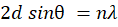
here, d is the space between the diffracting planes,
θ (theta) = incident angle,
n = integer
λ= beam wavelength
Bragg's Law reflection (Figure 1).
Reflection of x-ray from two different planes of a crystal
We may compute information about the crystal structure using the maximum intensity criterion found in Bragg's equation above. or to ascertain the wavelength of the x-ray’s incident upon the crystal if the crystal structure is known.
X-ray Crystallography:
Different types of crystal structure (Bravais lattice structure)
When describing the periodic arrangement of points in a crystal, the Bravais lattice system is a classification of the various three-dimensional lattice configurations that are feasible. Understanding the general symmetry and organization of crystal formations is made easier by these lattice systems. Based on the relationships between the lattice points and their lattice parameters, there are 14 different varieties of Bravais lattices. There is distinct crystal systems linked to each Bravais lattice.
1. Primitive Cubic (P):
In a primitive cubic lattice, each lattice point is positioned at the corners of a cube, meaning that each unit cell shares its corner atoms with adjacent cells. This structure represents the simplest and least dense form of the Bravais lattices. It belongs to the triclinic crystal system and has a coordination number of six, indicating that each lattice point is directly connected to its six nearest neighbours. This arrangement results in a relatively low packing efficiency compared to other crystal structures.
2. Body-centred Cubic (BCC):
The corners of a cube have lattice points in a body-centred cubic (BCC) lattice, whereas the cube's centre has an extra lattice point. With a coordination number of eight, this structure is a member of the cubic crystal system, and each lattice point is directly coupled to its eight nearest neighbours. Although the BCC arrangement is less dense than a face-centred cubic (FCC) structure, it offers a higher packing efficiency than a plain cubic lattice.3. Face-Cantered Cubic (FCC).
Lattice points are arranged in a structured and orderly manner at the corners of a rectangular prism in a primitive tetragonal lattice. This lattice has a coordination number of four, which indicates that every lattice point is directly related to its four nearest neighbours. It is a member of the tetragonal crystal system. One axis is longer or shorter than the other two in the primitive tetragonal structure, which is a variant of the cubic system and produces a unique geometric arrangement.
5. Primitive Orthorhombic (P):
Lattice points are arranged in a systematic and structured manner at the corners of a rectangular box in a primitive orthorhombic lattice. With a coordination number of eight, this lattice is a member of the orthorhombic crystal system, and each lattice point is directly coupled to its eight nearest neighbours. Because each of the three axes in the orthorhombic structure has a different length, it has a different geometric configuration than the cubic system.
6. Primitive Rhombohedral (P):
Lattice points are placed at the corners of a rhombohedron, which is a six-faced parallelepiped with equal length on all sides but not necessarily 90-degree angles between them, in a basic rhombohedral lattice. With a coordination number of twelve, this structure is a member of the trigonal crystal system, which means that every lattice point is physically related to its twelve nearest neighbours. In contrast to other crystal systems, the rhombohedral arrangement offers a distinctive geometric configuration.
7. Primitive Hexagonal (P):
Lattice points are placed at the corners of a hexagonal prism to create a repeating geometric pattern in a primitive hexagonal lattice. With a coordination number of twelve, this structure is a member of the hexagonal crystal system, and each lattice point is physically coupled to its twelve nearest neighbours. An effective packing structure produced by the hexagonal arrangement is frequently observed in a variety of natural and manufactured materials.
Instrumentation
The instrumentation used in crystallography consists of following components.as per figure no.-2
Instrumentation of X-ray diffraction
Source:
Two major sources used to obtain x-radiation are as follows
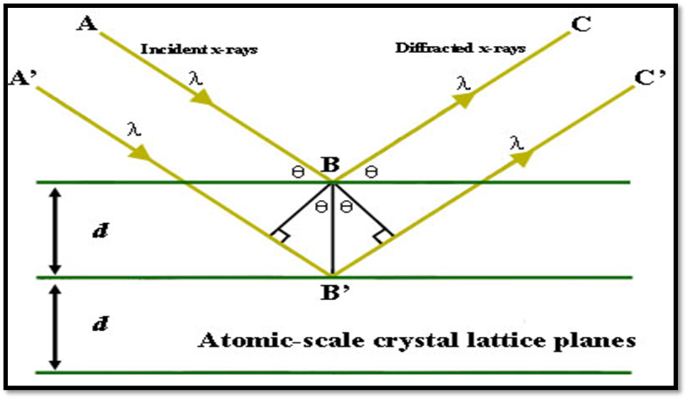
The most prevalent source of X-ray radiation is the X-ray tube, as per figure no.-3 sometimes referred to as a Coolidge tube. It is made up of an evacuated glass tube that has an anode, and a cathode attached to it. Electrons are released from the cathode, which is a tungsten filament heated by a high voltage. The anode, a heavy copper block coated with target elements like tungsten, copper, molybdenum, rhodium, silver, iron, or cobalt, is where these electrons are accelerated. To avoid overheating, some systems circulate cool water around the anode. The accelerating potential, or positive voltage given to the anode, controls the force with which the electrons contact the target metal, whereas the cathode's emission of electrons is determined by the voltage applied for heating. This in turn influences the energy and wavelength of the X-rays that are released, with larger accelerating forces resulting in X-rays that are more energetic and have shorter wavelengths. A beryllium window installed on one side of the tube allows the generated X-rays to exit, making the Coolidge tube a popular and effective X-ray source. Synchrotron/FEL X-rays for structural research, liquid metal jets for brightness, CNT tubes for efficiency, laser-driven sources for contrast, and microfocus tubes.
Figure- 2 Instrumentation of X-ray Crystallography
Production of x-ray using radioisotopes
Some radioactive materials are useful sources of X-ray radiation because they naturally release X-rays as a byproduct of their radioactive decay process. Various processes, depending on the kind of radioactive decay, can produce these X-rays. Electron capture or K-pressure is the main method by which elements like iron-55 (²?Fe??), cobalt-57 (²?Co??), cadmium-109 (??Cd¹??), and iodine-125 (?¹I¹²?) produce X-rays. This process results in the emission of distinctive X-rays. However, isotopes such as tritium (¹H³) and lead-210 (?²Pb²¹?) make X-rays by releasing high-energy electrons through the beta (β) emission mechanism, which results in secondary X-ray radiation. These X-ray sources that arise naturally are important for many scientific and medicinal purposes.
Wavelength selector
Certain tools, including filters, can be used to isolate a limited range of wavelengths from the entire X-ray spectrum. X-ray photometers are devices that make use of these filters. X-ray filters are composed of thin metallic strips or particular materials with a specified thickness that are intended to absorb particular wavelengths only. For example, a molybdenum target yields two distinctive lines from 0.62 Å to 0.75 Å and a continuous spectrum from 0.4 to 0.6 Å. A pure Kα line can be isolated when X-rays from this target travel through a thin strip of zirconium because the filter absorbs the Kβ line and the majority of the continuous energy. In order to create monochromatic X-ray beams for a variety of applications, this selective filtration process is crucial.
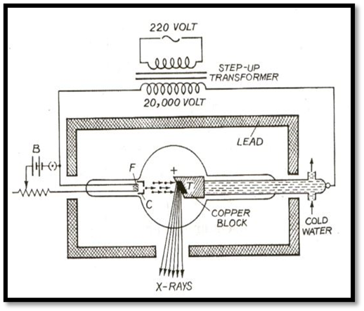
Figure -3 X-ray tube or Coolidge tube
2.Monochromator
Spectrophotometers are devices that choose wavelengths by using a monochromator. In essence, an X-ray monochromator is a crystal that separates wavelengths from a wider spectrum by diffracting X-rays according to their wavelengths. A crystal's lattice spacing determines how well it can diffract X-rays, hence determining the useful wavelength range that may be acquired from it. Mounting the crystal on a goniometer or revolving table enables controlled modifications to the diffraction angle, resulting in exact wavelength selection. Bragg's equation, which explains the connection between the angle of diffraction, the interatomic spacing of the crystal lattice, and the wavelength of incident X-rays, forms the basis of a crystal monochromator's operation. Certain wavelengths can be isolated for use in a variety of analytical applications, including crystallography and X-ray spectroscopy, by carefully choosing the crystal type and orientation. This technique is essential for getting high-resolution data and improving X-ray analysis accuracy. (figure no-4)
Figure-4 Crystal monochromator
Sample holder
The sample crystal is held in place during analysis by the sample holder, also called a crystal mount, which is a revolving table. To guarantee consistent exposure to X-rays, the sample crystal is placed in the middle of the mount, which rotates at a predetermined speed. The precision of diffraction measurements made possible by this controlled rotation improves the precision of data collection in crystallography and X-ray spectroscopy. In order to provide in-depth structural research, the crystal mount is essential in ensuring that the sample interacts with X-rays at the best angles.
X-ray detector
Three types of detectors are used for x-ray detection.
A metal tube filled with an inert gas, such as argon, krypton, or xenon, makes up a gas-filled detector, a form of X-ray detection apparatus. X-rays can flow through the tube's two transparent windows, which are positioned on opposing sides and are composed of materials like mica, beryllium, aluminum, or mylar. The tube's lower side, which is negatively charged, serves as the cathode, while the tube's center has an anode in the shape of a thin wire. Excitation and the loss of an outer electron result from interactions between the X-rays and the inert gas's atoms when they enter through one of the windows. An electric signal is produced by the free electrons produced by this ionization process moving in the direction of the anode. The voltage applied to the anode determines how many electrons arrive at the anode for every incoming X-ray photon. Minimal ionization takes place if the potential is less than 200 volts. The detector functions in the ionization chamber zone, where primary ionization is monitored, when the potential is between 200 and 400 volts. Secondary ionization intensifies the signal in the proportionate counter zone, which has a potential between 800 and 1000 volts and is proportional to the X-ray photon's energy. The detector operates in the Geiger counter zone at even higher potentials, between 1100 and 1600 volts, where a massive electron avalanche is created, producing a consistent pulse for every photon detected, independent of energy. Gas-filled detectors, which provide many modes of operation depending on the applied voltage and the particular requirements of the measurement, are essential in X-ray spectroscopy and radiation detection.(figure no – 5)

Figure 5 -Gas filled detector
Scintillation counters/ Detectors
A translucent, cylindrical sodium iodide (NaI) crystal that is roughly 3–4 inches long and wide makes up a scintillation counter, a radiation detection instrument. A tiny amount (0.2%) of thallium iodide is added to the sodium iodide crystal to increase its effectiveness by promoting the activation of electrons inside the crystal structure. The crystal's electrons are energized when X-rays hit it, and as they return to their initial energy levels, photons are released. A fluorescent substance inside the system detects the visible scintillations produced by this process. A photomultiplier tube then gathers and intensifies the released photons, creating a cascade of electrons that produces a sizable electrical signal. Scintillation energy.
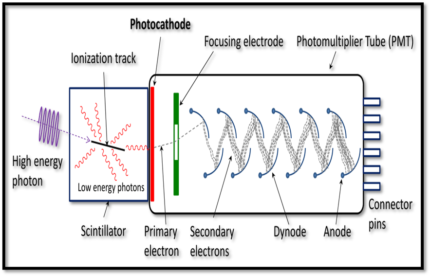
Figure 6 - Scintillation counter/detector
is the quantity of energy produced by electrons as they go from an excited state back to their ground state. The intensity of the X-rays striking the surface precisely correlates with the amplitude of the accompanying electric current, which is created by further converting this energy into electrical energy. The scintillation counter, which provides high sensitivity and precision in radiation detection and measurement, is essential to X-ray and gamma-ray spectroscopy. (figure no.- 6)
3. Semiconductor detector
Since semiconductor detectors offer high-resolution X-ray photon detection, they are important in X-ray crystallography. In this discipline, the lithium-drifted silicon detector (Si (Li)) and the lithium-drifted germanium detector (Ge(Li)) are the two main types of semiconductor detectors. When subjected to X-rays, the principle of charge collection within a semiconductor material is the basis for these detectors' operation. One p-type semiconducting layer, one central intrinsic layer, and one n-type semiconducting layer make up the Si (Li) detector's three primary layers. The p-type semiconductor's exterior is covered in a thin coating of gold for electrical contact, which is followed by an X-ray-transparent beryllium window. It is the intrinsic lithium-drifted silicon region beneath this layer that interacts with X-ray photons to gather charges. To guarantee correct electrical contact, a small coating of aluminium is applied externally to the n-type semiconductor, which is found at the end of the intrinsic layer. Before the signal is sent to an amplifier and recorder for additional processing, this aluminium sheet is first linked to a preamplifier, which strengthens it. With the main exception of using germanium as the semiconductor material rather of silicon, the Ge (Li) detector has the same structural design as the Si (Li) detector. Detecting X-rays with wavelengths shorter than 0.3 Å is made easier with Ge (Li) detectors because of the better stopping power of germanium for high-energy photons. High-purity germanium (HPGe) detectors, which do not require lithium drifting and provide improved resolution and efficiency, are the result of recent developments. High-precision X-ray crystallography, synchrotron radiation investigations, and X-ray fluorescence (XRF) spectroscopy all make extensive use of semiconductor detectors such as Si (Li) and Ge (Li) because of their exceptional energy resolution, which enables accurate elemental and structural analysis.
Rotating crystal technique
In X-ray diffraction analysis, a crystal set on a graded turntable is exposed to an X-ray beam with a known wavelength. The X-rays experience diffraction in accordance with Bragg's Law as they contact with the crystal lattice. After entering a recorder's ionization chamber, the diffracted rays ionize the air within, creating an electric current that flows between the
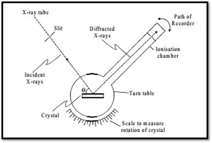
Figure 7 - Rotating crystal method
chamber walls and an electrode that is positioned within. An electrometer that monitors the ionization current's intensity is linked to this electrode. The intensity of the diffracted X-rays is directly proportional to the electrometer measurement. The scale records the angles at which the maximum X-ray intensity occurs when the recorder and crystal spin. These measured angles are used to calculate the separations between the parallel lattice planes inside the crystal structure, and they correspond to various diffraction orders (n = 1, 2, 3, etc.). X-ray diffraction is a basic method in crystallography for examining the atomic arrangement of materials because it offers precise measurement of interplanar spacings, which yields important structural information.
X-ray diffraction methods
Through its important insights into both organic and inorganic substances, X-ray diffraction (XRD) has made a substantial contribution to our understanding of atomic groupings and interatomic spacing in crystalline materials. This method has proven useful in many different sectors, such as material science, biomedical research, industry, science, and technology. Drug research and molecular engineering have been made easier in the pharmaceutical and biochemical industries by XRD's ability to clarify the structures of complicated natural products like steroids, vitamins, and antibiotics. Furthermore, by improving our knowledge of the characteristics of metals, alloys, minerals, polymers, and other solid materials, XRD has improved the use of these materials in engineering and industrial operations. The production of X-rays in an X-ray tube and their subsequent direction toward a sample form the basis of the basic idea of X-ray diffraction. Diffraction is the result of these rays interacting with the material's atomic planes; structural features are ascertained by collecting and analysing the diffracted beams. When utilizing Bragg's Law to calculate interatomic distances, the angle between the incident and diffracted X-rays is a crucial component of diffraction research. Phase transitions, strain effects, and nanomaterial structures can now be analysed in real time because to recent developments in XRD technology, including high-resolution synchrotron XRD, in situ diffraction research, and computational modelling. With these advancements, X-ray diffraction remains a vital analytical technique in structural biology, crystallography, and materials science, facilitating ground-breaking discoveries and breakthroughs in a variety of fields.
Single-crystal x-ray diffraction
One effective method for precisely analyzing the atomic structure of crystalline materials is single-crystal X-ray diffraction. The regular arrangement of atoms in a single crystal causes X-rays to scatter when they impact it, creating a characteristic diffraction pattern. These dispersed beams create a pattern of spots when they arrive on a detector, either an electrical sensor or a piece of photographic film. The strengths and angles of these beams are methodically recorded as the crystal is slowly rotated, yielding thorough structural information. Every reflection, or spot, in the diffraction pattern represents the reflection of X-rays from a particular group of equally spaced atomic planes inside the crystal lattice. X-ray diffraction data may accurately identify chemical bond lengths within a few thousandths of an angstrom and bond angles within a few tenths of a degree if the crystal is sufficiently regular and pure. Moreover, atoms inside a crystal oscillate around their equilibrium positions, usually by a few tenths of an angstrom, as revealed by X-ray crystallography. Scientists can determine the strength of these oscillations by examining diffraction data, which offers important information about material characteristics and atomic mobility. This method is still crucial for accurately characterizing molecular and crystalline structures in disciplines including chemistry, materials science, and structural biology.
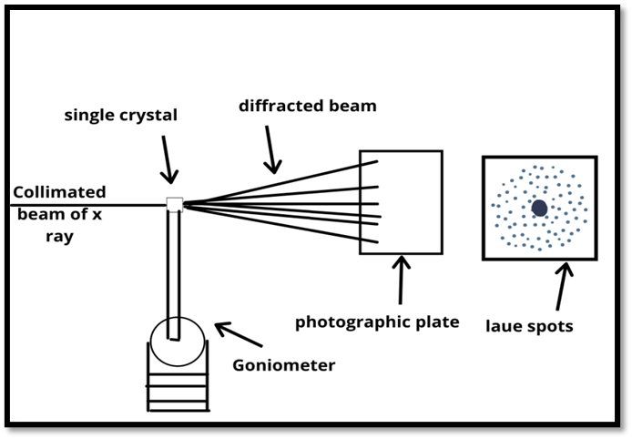
Figure 8-Single crystal X-ray diffraction
Procedure
There are three basic processes in the single-crystal X-ray crystallography process. A suitably large crystal (usually larger than 0.1 mm in all dimensions), one that is pure in composition, structurally regular, and free of major internal flaws like cracks or twinning is necessary for the first step. To get precise diffraction data, the crystal's quality is essential. The crystal is subjected to a powerful monochromatic X-ray beam in the second step, which results in a highly organized pattern of reflections. Every compound has a distinct diffraction pattern, and the angles and intensities of the diffracted X-rays are measured. Reflections that were previously seen vanish when the crystal is slowly turned, and new ones appear. Each diffraction spot's intensity is recorded at several orientations, and in order to guarantee thorough coverage, it would be necessary to gather several data sets. Tens of thousands of distinct reflections are typically included in each dataset, which typically records just over half of a full rotation of the crystal. In the third and last stage, a detailed model of the atomic arrangement within the crystal is created by computationally processing the gathered data and combining it with complimentary chemical information. For future study and scientific reference, this improved structural model—also referred to as the crystal structure—is thereafter kept in open databases. Because it makes it possible to accurately determine molecular and atomic structures, single-crystal X-ray crystallography is still a vital technique in structural biology, chemistry, and materials research.
Powder diffraction method
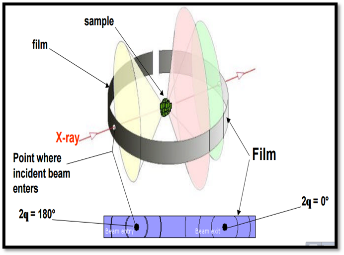
Figure 9- Powder diffraction method
A very useful analytical approach for both qualitative and quantitative investigation of solid samples is the powder diffraction method. By using a finely powdered crystalline material, the powder approach gets around the need for a single, undisturbed crystal, which is a requirement of the rotating crystal method. This method exposes the sample to an incident X-ray beam while it is usually in a capillary tube or on a flat holder. To make sure that every potential crystallographic orientation contributes to the diffraction pattern, the sample is then rotated by a motor. A fraction of the powdered sample's numerous randomly oriented microcrystals will always be orientated at the ideal angle to meet Bragg's law, resulting in the simultaneous production of diffracted X-rays from several lattice planes. The intensity and location of these reflected X-rays, which create distinctive diffraction rings or peaks, reveal vital details about the crystal structure. Precise measurement of the diffraction angle (2θ) enables the identification of distinct phases in the material and the calculation of interplanar spacings. The precision and breadth of structural analysis have been greatly enhanced by contemporary developments in powder diffraction, including high-resolution X-ray diffraction (HRXRD), synchrotron radiation, and Rietveld refinement methods. This technique is a vital tool for describing crystalline materials, identifying impurities, and researching phase transitions since it is extensively used in materials science, geology, medicines, and nanotechnology.
APPLICATION
X-ray diffraction is a powerful analytical technique used to study the structure of materials at the atomic or molecular level. It involves shining X-rays on a crystalline sample and observing how the X-rays are scattered as they interact with the atomic arrangement within the crystal. This scattering pattern provides valuable information about the structure of the material. Here are some important applications of X-ray diffraction with references:
1. Determining Crystal Structures: X-ray diffraction is widely used to determine the atomic or molecular structure of crystalline materials. One of the most famous examples is the discovery of the double helix structure of DNA by James Watson and Francis Crick in 1953. Their work, "Molecular Structure of Nucleic Acids: A Structure for Deoxyribose Nucleic Acid," published in the journal Nature, is a seminal reference in this field.(1)
2. Material Characterization: X-ray diffraction is used to study various materials, including metals, ceramics, polymers, and pharmaceuticals. Researchers use it to identify crystal structures, defects, and phases in these materials.(2)
3. Protein Crystallography: X-ray crystallography is a fundamental tool in structural biology. It has been used to determine the three-dimensional structures of proteins, which is crucial for understanding their functions and designing targeted drug therapies. The reference here can be a publication on the structure of a specific protein, like the structure of myoglobin determined by J.C. Kendrew in Nature in 1958.(3)
4. Mineralogy and Geology: X-ray diffraction is vital for identifying and characterizing minerals and geological samples. Researchers use it to determine the composition and crystal structures of minerals in rocks and ores. (4)
5. Pharmaceuticals: X-ray diffraction is used to analyse the crystalline structure of pharmaceutical compounds. Understanding the structure is critical for formulation and drug development.(5)
6. Thin Film Analysis: In materials science and electronics, X-ray diffraction is used to characterize thin films, such as those used in semiconductors.(6)
7. Studying Polymers: X-ray diffraction is applied to investigate the structure of polymers, helping to understand their crystallinity and other properties.(7)
8. Forensic Science: X-ray diffraction is utilized in forensic science for the analysis of trace evidence, such as gunshot residue and unknown powders.(8)
9. Archaeology and Art Conservation: X-ray diffraction is used to analyse historical and cultural artifacts, helping to identify the composition of materials and to aid in restoration efforts. (9)
10. Nanomaterials and Nanotechnology: X-ray diffraction is used to investigate the structures of nanomaterials, which have unique properties due to their small size. (10)
11. Metallurgy and Materials Engineering: XRD is crucial for characterizing metals, alloys, and ceramics. It is used to determine phase compositions, crystal structures, and residual stress in manufactured components. In the automotive and aerospace industries, XRD is applied to assess the quality and integrity of materials.(11)
12. Mineral Processing and Mining: The mining industry uses XRD to identify and quantify mineral phases in ore samples. This information is essential for optimizing ore processing and determining the economic viability of mining operations.(12)
13. Cement and Construction Materials: XRD is used to analyse the mineral composition of cement and construction materials, ensuring their quality and performance. It helps in the development of high-strength and durable concrete and cementitious materials.(13)
14. Petrochemical and Energy: In the petrochemical industry, XRD is employed for the analysis of catalysts, zeolites, and other materials used in refining and catalysis processes. It aids in improving catalyst performance and extending their lifespan.(14)
15. Electronic and Semiconductor Manufacturing: XRD is used to analyze the crystal structures and defects in semiconductor materials, such as silicon and gallium arsenide. It ensures the quality and performance of semiconductor devices and integrated circuits.(15)
16. Pharmaceutical Formulation: XRD helps in the formulation of drug products, particularly in the development of solid dosage forms like tablets. It ensures the stability and uniformity of the drug formulations.(16)
17. Food Industry: XRD is used to analyse the crystalline structure of food ingredients and additives. This is important for understanding the texture, shelf-life, and processing properties of food products.(17)
18. Agriculture: XRD can be used to analyse the mineral content and crystal structures of soil samples, helping to assess soil quality and determine the best agricultural practices for crop growth.(18)
19. Quality Control and Failure Analysis: XRD is applied in quality control to ensure that materials meet the required specifications and standards. It is also used for failure analysis to understand the cause of material failures and defects.(19)
20. Art and Cultural Heritage Conservation: XRD is used in museums and conservation laboratories to analyse the composition and structure of historical artifacts and artworks. This information guides restoration efforts and helps preserve cultural heritage.(20)
21. Environmental Analysis: XRD is used to analyse environmental samples, such as air particulates, soil, and sediments, to determine the presence of minerals and pollutants.(21)
REFRENCES


Tejas Ghadge, Poonam Tharkude*, X-Ray Crystallography and Diffraction: A Review of Theory and Practical Applications, Int. J. of Pharm. Sci., 2025, Vol 3, Issue 3, 1693-1708. https://doi.org/10.5281/zenodo.15046804
 10.5281/zenodo.15046804