1,2,4,5,6Soniya Education Trust’s College of Pharmacy, Dharwad, Karnataka, India.
3Department of Pediatrics, Karnataka Medical College and Research Institute, Hubballi, Karnataka, India.
A 12-year-old boy, the eldest child of a non-consanguineous couple from a lower socio-economic background, was brought with a history of fever for four days, headache for five days, and four episodes of vomiting associated with giddiness over the last three days. His immunizations were up to date, and his developmental milestones were normal. There was no relevant antenatal, perinatal, or past medical history. He was initially managed at Haveri District Hospital and later referred to Karnataka Medical College and Research Institute for further evaluation. Family history was unremarkable, and his nutritional intake was reported to be adequate. On admission, his vital parameters were stable. General examination showed no gross abnormalities, though anthropometry suggested undernutrition. Neurological assessment revealed meningeal signs, including neck stiffness, positive Kernig’s sign, and positive Brudzinski’s sign. Other systemic examinations were within normal limits. Laboratory investigations demonstrated leukocytosis on complete blood count, while renal and liver function tests and serum electrolytes were normal. Cerebrospinal fluid analysis showed lymphocytic predominance, and confirmatory testing established the diagnosis of Japanese Encephalitis.
Japanese encephalitis (JE) continues to pose a major health challenge across Asia [1]. The disease is caused by the Japanese encephalitis virus (JEV), a mosquito-borne flavivirus that has been recognized as the leading cause of viral encephalitis in the region [2,3]. Historical records show that outbreaks were noted as early as the late 19th century in Japan, and in 1935, the Nakayama strain of JEV was first isolated from a fatal case, later becoming the standard reference strain [4]. JEV belongs to the Flavivirus genus, which also includes important human pathogens such as dengue, yellow fever, Murray Valley encephalitis, West Nile virus, Zika virus, St. Louis encephalitis, and tick-borne encephalitis viruses [5,13].
Globally, JE is responsible for a significant burden of illness and mortality. WHO has estimated that tens of thousands of cases occur each year, with mortality rates ranging between 20–30%. In 2015, the number of reported cases exceeded 100,000, with up to 30,000 deaths [6]. Children under 15 years of age remain the most vulnerable group, particularly newborns and young children, who are more likely to develop severe neurological complications [7]. Clinical presentation often includes fever, headache, muscle pain, and altered mental status, with seizures observed in the majority of patients [14].
As there is no antiviral treatment specifically for JE, vaccination remains the cornerstone of prevention and control [8]. The most widely used vaccines are based on JEV genotype III and have been deployed across endemic areas with proven effectiveness [9]. However, surveillance data suggest that the true burden of disease is underestimated due to misdiagnosis, a problem common to several rare neurological and metabolic disorders [11]. At present, it is estimated that more than two billion people live in areas at risk of JEV transmission, and increasing mosquito populations may further expand its geographic range.
Transmission occurs primarily through the bite of infected female Culex tritaeniorhynchus mosquitoes [10]. The main amplifying hosts are pigs and certain bird species, particularly herons and egrets from the Ardeidae family [12].
Case report
Demographic details and chief complaints
A 12-year-old male child, the first-born of a non-consanguineously married couple, presented to the Karnataka Medical College and Research Institution (KMCRI), Hubballi, and was admitted on 15th February 2024 with the following chief complaints:
The child was apparently well until 5 days prior to admission, when he developed a diffuse, continuous headache of moderate to severe intensity. The headache was not relieved by over-the-counter analgesics and progressively worsened over the following days. It was associated with nausea and multiple episodes of non-projectile, non-bilious vomiting, occurring 3–4 times per day.
A low-grade intermittent fever developed on the fourth day of illness, without chills or rigors. It was not associated with rash, seizures, or altered sensorium. The child also began to complain of giddiness, described as a feeling of light-headedness and imbalance while walking, leading to general discomfort and reduced physical activity.
There was no history of trauma, visual disturbances, ear discharge, neck stiffness, loss of consciousness, or any recent travel. There is no known history of tuberculosis exposure, and no significant past medical or surgical history. Immunizations are up to date for age.
Family history is non-contributory, and there is no known consanguinity between the parents.
Past History:
Prior to admission at KMCRI, the child was evaluated and admitted at a district hospital in Haveri for the same complaints—headache, vomiting, fever, and giddiness. Due to persistent symptoms and lack of significant clinical improvement, he was referred to Karnataka Medical College and Research Institution, Hubballi, for further evaluation and management.
Past Medical History:
During his stay at the district hospital, the child was treated empirically for a suspected infection. The following medications were administered:
A CT scan of the brain was performed, which was reported as normal, ruling out gross structural abnormalities, intracranial hemorrhage, or space-occupying lesions at that point.
There is no prior history of chronic illness, hospitalization, neurological disorders, or known drug allergies. Growth and developmental milestones have been age-appropriate.
Family history
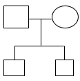
Figure:1
The patient is the first-born child of a non-consanguineously married couple. The family consists of two male children—the patient, aged 12 years, and a younger sibling aged 6 years. The second child, an 11-year-old male, is reported to be in good health with no significant medical history. There is no known history of hereditary or genetic disorders, neurological illnesses, or chronic diseases in the immediate or extended family.
Birth and Perinatal History:
The patient was delivered at full term through normal vaginal delivery (FTVD) with a birth weight of 2.5 kg. The antenatal, perinatal, and intrapartum periods were uneventful, with no maternal or fetal complications reported. The child did not require neonatal intensive care, and the neonatal period was smooth. There was no history of birth asphyxia, delayed cry, or neonatal seizures.
Clinical findings
Vital Signs:
On examination, the child was alert and oriented. Vital parameters were assessed as follows:
Overall, vital signs were stable at the time of examination, with mildly elevated body temperature being the only notable finding.
Systemic Examination
Head-to-Toe Examination:
Neck:
Anthropometry
Table: 1 Anthropometric Measurements of the Patient
|
Parameter |
Observed Value |
Expected Value |
Centile |
Interpretation |
|
Weight |
21 kg |
39.0 kg |
< 3rd |
Underweight |
|
Height |
101 cm |
114.4 cm |
3rd–10th |
Stunted growth (mild) |
|
BMI |
11.1 kg/m² |
17.7 kg/m² |
< 3rd |
Severe undernutrition |
Overall Interpretation: The child demonstrates chronic undernutrition, with weight and BMI both below the 3rd centile and height between the 3rd–10th centile, predisposing to increased susceptibility and complications from infections like Japanese Encephalitis.
Systemic Examination (continued):
Respiratory System:
Cardiovascular System:
Central Nervous System Examination:
Neurological Examination
On admission, a detailed neurological evaluation was performed:
Developmental History:
Diet history
Table: 2 Diet history
|
Timimgs |
Food |
Calories |
Protein |
|
Morning |
Milk Idli Sambar |
60 41 162 |
6 2 4 |
|
Afternoon |
Chapati Curry Egg |
54 172 60 |
3 3 6 |
|
Evening |
Milk |
60 |
6 |
|
Night |
Chapati Rice Curry |
54 162 172 |
3 4 3 |
|
|
Required Deficient |
937 10% |
40 9% |
Nutritional Assessment:
According to the Indian Council of Medical Research (ICMR) guidelines, the child’s estimated caloric requirement is 1037 kcal/day.
This indicates a caloric deficit of approximately 10%, suggesting the child is currently undernourished and not meeting the recommended energy intake for his age and activity level.
Investigations
Laboratory Investigations
Table: 3 Laboratory Investigations
|
Parameter |
Observed Value |
Reference Range |
Interpretation |
|
Hemoglobin (Hb) |
11.4 g/dL |
12–16 g/dL |
Mild anemia |
|
Total Leukocyte Count (TLC) |
4300 cells/cmm |
4000–11000 cells/cmm |
Within normal limits; slightly on the lower side |
|
Platelet Count |
1.75 lakh/cmm |
1.5–4.5 lakh/cmm |
Normal |
|
RBC Count |
4.76 million/cmm |
4.5–5.5 million/cmm |
Normal |
|
Packed Cell Volume (PCV) |
35% |
36–46% |
Slightly low |
|
Differential Leukocyte Count (DLC) |
43% Neutrophils / 46% Lymphocytes |
Neutrophils 40–70%, Lymphocytes 20–45% |
Mild relative lymphocytosis |
|
Sodium (Na?) |
129 mEq/L |
135–145 mEq/L |
Mild hyponatremia |
|
Potassium (K?) |
3.9 mEq/L |
3.5–5.0 mEq/L |
Normal |
Interpretation in Context of Japanese Encephalitis:
Renal Function Tests (RFTs)
Table: 4 Renal Function Tests
|
Parameter |
Observed Value |
Reference Range |
Interpretation |
|
Blood Urea |
16 mg/dL |
15–40 mg/dL |
Normal |
|
Serum Creatinine |
0.8 mg/dL |
0.5–1.0 mg/dL |
Normal |
Interpretation: The renal function tests were within normal limits, indicating preserved kidney function. There is no evidence of renal involvement in this child presenting with Japanese Encephalitis.
Diagnosis:
Cerebrospinal Fluid (CSF) Analysis:
Impression: Smear shows a predominance of lymphocytes against a proteinaceous background; Japanese Encephalitis (JE) positive
Treatment
Table :5 Treatment chart
|
Drug names |
Dose |
R. O. A |
Frequency |
Days |
|
IVF DNS |
60ml |
IV |
Per hour |
3days |
|
Inj Ceftriaxone |
2.1g |
IV |
1-0-1 |
17days |
|
Inj Amikacin |
160mg |
IV |
1-0-1 |
17days |
|
Inj Ranitidine |
40mg |
IV |
QD |
10days |
|
Inj Dexamethasone |
1mg |
IV |
0-1-0 |
7days |
|
Inj Acyclovir |
400mg |
IV |
1-1-1 |
5days |
|
Syp Multivitamin |
5ml |
PO |
1-0-1 |
6days |
|
Syp Calcium |
5ml |
PO |
1-0-1 |
4days |
|
Inj pantoprazole |
40mg |
IV |
1-0-1 |
6days |
There is no specific antiviral therapy for JE and vaccination is the only reliable option for its prevention and control. In the available vaccine, most common vaccine against JEV genotype III (GIII) is used across endemic countries. The clinical management mainly focuses on supportive care and symptomatic relief.13
The patient was treated with the following medications from the first day of admission to the thirteenth day of discharge: Ceftriaxone injection 2.1 g twice daily, Amikacin injection 160 mg twice daily, and Ranitidine injection 40 mg twice daily from the first day to the tenth day. Dexamethasone injection 1 mg four times daily was administered from the second day to the seventh day, and Acyclovir injection 400 mg three times daily was given from the third day to the seventh day. Supportive care included intravenous fluids (DNS) at 660 ml/hour for three days, oral rehydration solution (ORS) for seven days, multivitamin syrup 5 ml once daily for six days, and calcium syrup 5 ml three times daily for six days were given. The danger signs were explained, and the patient was advised to seek immediate medical attention if there was any elevation in body temperature (fever spike) and to visit the pediatric outpatient department for a follow-up after one week.
DISCUSSION:
The diagnosis of Japanese encephalitis virus (JEV) infection posed significant challenges due to the rarity of detecting JEV RNA in blood or cerebrospinal fluid (CSF) samples. Typically, serological tests formed the basis for diagnosis, but their interpretation was often complicated by non-specific reactivity, cross-reactivity with other flaviviruses, and persistence of antibodies from prior infections or immunizations. In many cases, patients sought medical attention only after neurological symptoms emerged, by which time the acute phase of the infection had often passed, and viral clearance had occurred. However, in the reported case, analysis of the patient’s CSF revealed the presence of JEV, with a smear showing predominantly lymphocytes, a few monocytes, and a proteinaceous background, confirming a positive JEV diagnosis.
The patient, a previously healthy child, presented with a history of headache, nausea, vomiting, mild to moderate fever, and giddiness that began five days prior to admission. These symptoms, including bilateral diurnal variation and giddiness exacerbated by standing or walking, aligned with clinical manifestations of JEV infection. Initial treatment at a district hospital in Haveri with ceftriaxone and amikacin did not fully resolve the symptoms, particularly giddiness, prompting transfer to KMCRI in Hubli. There, physicians considered differential diagnoses of Japanese encephalitis and viral meningoencephalitis. Based on a provisional diagnosis of viral meningoencephalitis, the patient received ceftriaxone (100 mg/kg/dose) and amikacin (150 mg/kg/dose), which led to a gradual reduction in symptoms, although giddiness persisted.
A similar case occurred in rural West Bengal, India, involving an 11-year-old girl who initially experienced high-grade fever, chills, severe headache, neck stiffness, and vomiting. Her parents sought treatment from an unqualified practitioner, but the intervention failed to alleviate her symptoms. On the third day of illness, the patient developed seizures and lapsed into unconsciousness, leading to her hospitalization and subsequent diagnosis of JEV infection.
Japanese encephalitis remained one of the most severe viral infections endemic to Asia, causing significant morbidity and mortality. The absence of specific antiviral treatments for JEV underscored the critical importance of prevention. Preventive strategies primarily included vector control, pig immunization, and human immunization. In India, vector control served as the cornerstone of prevention efforts, but vaccination offered the most effective long-term protection against JEV. As no therapies directly targeted the virus, clinical management focused on addressing symptoms and complications. The ongoing lack of specific antiviral drugs highlighted the urgent need for continued research to develop targeted treatments for JEV infection.
CONCLUSION:
This case underscores the clinical complexity and public health importance of Japanese encephalitis (JE), especially among pediatric populations in endemic areas. The early signs of fever, headache, vomiting, and giddiness in the patient progressed to classical meningeal signs, highlighting the need for high clinical suspicion and timely diagnostic evaluation. Despite the lack of specific antiviral therapy, supportive management—including antibiotics, antiviral, corticosteroids, and nutritional support—played a crucial role in the patient's recovery. The diagnosis was confirmed through cerebrospinal fluid analysis revealing lymphocytic Pleocytosis and JE positivity. This case reiterates the urgent need for increased awareness, early diagnosis, and above all, preventive measures such as vaccination and vector control strategies to mitigate the burden of JE, especially in vulnerable, underserved populations.
REFERENCE


Tanuja S. L., Adarsh G. S.*, Siddappa Dandinavar, Preeti V. Kulkarni, Venkatrao H. Kulkani, Thrinesh B. R., A Rare Pediatric Case of Japanese Encephalitis Presenting with Acute Febrile Encephalopathy, Int. J. of Pharm. Sci., 2025, Vol 3, Issue 10, 465-474 https://doi.org/10.5281/zenodo.17276839
 10.5281/zenodo.17276839
