
We use cookies to ensure our website works properly and to personalise your experience. Cookies policy
Rungta Institute of Pharmaceutical Sciences And Research, Affiliated To Chhattisgarh Swami Vivekanand Technical University CSVTU, Bhilai, India
Development depends on the precise control of gene expression. The spatiotemporal Pattern of gene transcription is determined by Enhancers, which are at the heart of Gene control. The study of enhancers will yield hints for precise therapy because they Are known to include a large number of mutations linked to disease. Rapid Advancements in high-throughput sequencing technology make it easier to Characterize enhancers across the entire genome, yet it is still difficult to Comprehend how enhancers work. Here, we present a wide range of enhancer Attributes, such as enhancer-promoter interaction patterns, enhancer transcripts, And epigenetic changes. We also describe the use of functional genomics techniques and high-throughput sequencing technology in enhancer research. Lastly, we go over How enhancers contribute to human illness and how they might be targeted by Techniques for both disease prevention and treatment.
During development and throughout an organism’s life, precise spatial and temporal regulation of cellular functions is ensured by the carefully regulated process of gene expression. By adjusting target gene transcription across vast genomic lengths, enhancers—important regulatory regions in the genome—manage this complexity. Since their discovery more than thirty years ago, enhancers have become important factors in determining the transcriptional landscape, allowing for dynamic and cell-type-specific patterns of gene expression.
Enhancers use chromatin remodeling, physical contacts with target promoters, and epigenetic changes to carry out their regulatory effects. A wide range of transcription factors, cofactors, and chromatin-binding proteins control this complex process, which together promotes enhancer activation and specificity. Crucially, enhancer malfunction has been linked to a wide range of human illnesses, including as neurological conditions, developmental abnormalities, and cancer, highlighting their importance as possible targets for treatment.
Our knowledge of enhancers has been completely transformed in recent years by developments in functional genomics and high-throughput sequencing methods. Methods like chromatin accessibility assays (like ATAC-seq), histone modification profiling (like ChIP-seq), and chromatin interaction mapping (like Hi-C) have made it possible to identify enhancers across the entire genome and have clarified how they work. Our understanding of enhancer biology has also been further broadened by the identification of enhancer RNAs (eRNAs) and the creation of CRISPR-based technologies, which present fresh chances for functional characterisation and therapeutic investigation.
The goal of this review Is to present a thorough analysis of enhancer biology, covering its structural and functional properties, research approaches, and functions in both health and sickness. We also talk about future approaches in this quickly developing science and examine the therapeutic potential of focusing on enhancers. This article aims to close the gap by emphasizing new trends and synthesizing existing knowledge.
Characteristics of Enhancers
Enhancers are non-coding DNA segments that, frequently from great genomic distances, increase the transcription of target genes to control gene expression. Enhancers and their roles are defined by a number of common traits, notwithstanding their diversity and complexity.
Active Enhancers: Linked to H3 lysine 4 monomethylation (H3K4me1) and histone H3 lysine 27 acetylation (H3K27ac). Poised Enhancers: Show H3K4me1 and H3 lysine 27 trimethylation (H3K27me3), which are frequently present in a repressed form but are prepared for activation. DNA Methylation While the relationship between DNA methylation and enhancer activity is still being studied, enhancers frequently show modest levels of DNA methylation when they are active.
Specificity of Contact: Highly specialized enhancer-promoter interactions are frequently limited in topologically associating domains (TADs). Disease and ectopic gene expression may result from disruption of these connections. The mapping of these interactions has been made possible by tools like Hi-C, Capture-C, and 3C.
Environmental factors, developmental stages, and cell types all affect enhancer activity, which is very context-specific: Enhancers attract transcription factors and cofactors relevant to cell types in order to attain specificity. Genes essential for determining cell identity and function are expressed by super-enhancers, which are collections of extremely active enhancers.
Technologies for High-Throughput Sequencing in Enhancer Research
High-throughput sequencing technology breakthroughs have completely changed our capacity to find and describe enhancers across the whole genome. These methods offer vital information about the spatial, transcriptional, and epigenetic properties of enhancers as well as how they interact with target promoters. The main high-throughput sequencing techniques used in enhancer research are described here.
One characteristic of active enhancers is chromatin accessibility. Among the methods for mapping accessible chromatin are: DNase-Seq: Marks open chromatin regions, including enhancers, and identifies DNase I hypersensitive locations. Tn5 transposase is used in ATAC-Seq (Assay for Transposase-Accessible Chromatin), a quicker and more sensitive technique for mapping accessible chromatin. By using formaldehyde crosslinking and phenol-chloroform extraction, FAIRE-Seq (Formaldehyde-Assisted Isolation of Regulatory Elements) enriches open chromatin regions.
Chromatin immunoprecipitation followed by sequencing (ChIP-Seq) is the gold standard for mapping histone modifications, which are important markers of enhancer activity. Active enhancers are linked to H3K4me1 and H3K27ac. H3K27me3: Identifies repressed or poised enhancers. In contrast to conventional ChIP-Seq, emerging options like CUT&RUN (Cleavage Under Targets and Release Using Nuclease) and CUT&Tag (Cleavage Under Targets and Tagmentation) provide higher resolution, lower input needs, and less background noise.
Enhancer activation frequently results in the production of enhancer RNAs (eRNAs), which are non-coding RNAs: RNA-Seq: Provides information on enhancer activity by detecting eRNA transcripts. RNA Profiling of Nascents: By capturing early eRNA transcription, methods such as Global Run-On Sequencing (GRO-Seq) and Precision Run-On Sequencing (PRO-Seq) offer a dynamic picture of enhancer activity.
Enhancer function depends on enhancer-promoter interactions, which can be mapped using a variety of sequencing-based techniques: Hi-C: A genome-wide method that uses the 3D genome structure to identify chromatin connections, such as those between enhancers and promoters. By concentrating on particular genomic regions of interest, including enhancer-promoter pairs, Capture-C is a focused method that improves resolution. To find protein-mediated chromatin connections, ChIA-PET (Chromatin Interaction Analysis by Paired-End Tagging) combines chromatin immunoprecipitation and interaction mapping. These instruments show how enhancers interact with target genes and are arranged spatially throughout the nucleus.
Single-cell resolution enhancer research are now possible thanks to emerging technologies: Single-Cell ATAC-Seq: Indicates enhancer activity specific to a cell type by profiling chromatin accessibility in individual cells. Transcriptome data and spatial information are used in spatial transcriptomics to reveal enhancer activity in tissue architecture. These methods are especially useful for researching enhancers during development and in diverse tissues.
Functional genomics techniques are used in conjunction with high-throughput sequencing to confirm enhancer activity: Enhancer regions can be perturbed using CRISPR/Cas9 screens to examine how they affect gene expression. Self-Transcribing Active Regulatory Region Sequencing, or STARR-Seq, is a massively parallel reporter experiment that uses transcription of the enhancer sequence itself to evaluate enhance activity directly. High-Throughput Sequencing Applications in Disease Studies and Enhancer Research: detection of mutations in enhancer areas linked to illness.
Table: 1 ; Technologies for high – throughput sequencing in enhancer research
|
Technology
|
Description |
Application in enhancer research |
Reference |
|
Chip – Sequence |
Chip-Sequence This technique entails separating bound DNA to particular proteins, then sequencing to find the binding sites of transcription factors Moreover, histone modification |
maps enhancer components that are marked by histone modifications, such as H3K27 ac, which is essential for comprehending enhancer activity. |
T.S. Furey (2012). New and enhanced techniques to identify and describe protein-DNA interactions: ChIP-seq and beyond. 840–852 in Nature Reviews Genetics, 13(12). |
|
ATAC – Sequence |
A method for studying open chromatin regions that involves sequencing the segments and marking accessible DNA transposases |
Make it possible to identify active enhancers by examining chromatin accessibility in different cell types and environments. |
Chang, H. Y., Zaba, L. C., Giresi, P. G., Buenrostro, J. D., & Greenleaf, W. J. (2013). For quick and accurate epigenomic analysis of open chromatin, DNA-binding proteins, and nucleosome location, native chromatin transposition is used. 1213–1218 in Nature Methods, 10(12). 10.1038/nmeth.2688 is the DOI. |
|
RNA – Sequence
|
A method for measuring and sequencing RNA molecules in order to examine patterns of gene expression |
Describe the relationship between enhancer RNAs (eRNAs) and gene activation. |
Mortazavi, A., McCue, K., Schaeffer, L., Williams, B. A., & Wold, B. (2008). Mammalian Transcriptome Mapping and Quantification using RNA-Seq. published on pages 621–628 of Volume 5 of Nature Methods. |
|
Hi - C |
A method that uses proximity ligation and sequencing to capture chromatin interactions within the three-dimensional genome structure |
Finds the spatial enhancer-promote relationship that controls the expression of genes. |
Machol, I., Omer, A. D., Lander, E. S., Robinson, J. T., Sanborn, A. L., Rao, S. S. P., Huntley, M. H., Durand, N. C., Stamenova, E. K., Bochkov, I. D., & Aiden, E. L. (2014). Principles of Chromatin Looping Are Revealed by a 3D Map of the Human Genome at Kilobase Resolution. Published in 1665–1680 in Cell, 159(7). |
|
4C – Sequence |
By circularizing and sequencing DNA fragments, concentrate on how a single genomic location interacts with the remainder of the genome. |
Discover more about long-range chromatin interactions and particular enhancer promote loops. |
Splinter, E., Holwerda, S. J. B., Klous, P., de Wit, E., van de Werken, H. J. G., de Vree, P. J. P., & de Laat, W. (2012). 4C technology: data analysis and protocols. Methods in Enzymology, 513, 89-107. https://doi.org/10.1016/B978-0-12-391303-2.00008-5 |
|
STAR-R sequence |
DNA is directly tested for enhanced activity using a functional genomics assay that measures their |
Effectively search for and detect enhancer sequences throughout the genome |
Gerlach, D., Stelzer, C., Boryn, L. M., Rath, M., Arnold, C. D., & Stark, A. (2013). STARR-seq.-identified genome-wide quantitative enhancer activity maps Science, 339(6123), 1074–1077. |
|
E Clip – Sequence |
Crosslinking and sequencing RNA binding complexes is a sophisticated technique for researching RNA-protein interaction. |
Examine the rules governing enhancer RNA and how RNA binding proteins contribute to enhancer function. |
Shishkin AA, Pratt GA, Van Nostrand EL, et al. Robust identification of RNA-binding protein binding sites with enhanced CLIP (eCLIP) across the transcriptome. 13(6), Nature Methods, 2016: 508–514. |
|
PRO – Sequence |
Sequence the RNA generated during transcription to record early RNA synthesis. |
Offers high-resolution information on enhancer transcriptional activity. |
Fuda, N. J., Core, L. J., Kwak, H., & Lis, J. T. (2013). RNA polymerase precise maps show how promoters control initiation and stopping. 950–953 in Science, 339(6122). |
A functional genomics approach to research
Understanding the biological functions of enhancers and their modes of activity requires the use of functional genomics techniques. These techniques evaluate enhancer regions’ activity, specificity, and contributions to gene regulation in several settings, going beyond simply mapping them. We list the main functional genomics methods below.
Functional enhancer studies have been revolutionized by the development of CRISPR/Cas systems, which allow for precise genomic changes. CRISPR Knockout (CRISPR-KO): Enhancer areas are deleted to examine how they affect the expression of the target gene. Helpful for evaluating the robustness and redundancy of enhancers. Repression of enhancer activity without changing the DNA sequence is known as CRISPR Interference (CRISPRi). Accomplished by combining transcriptional repressors such as KRAB with catalytically inactive Cas9 (dCas9). CRISPR Activation (CRISPRa): Enhancer activation with dCas9 linked to activators such as p300 or VP64. Makes it possible to evaluate whether an enhancer is sufficient to activate a target gene. CRISPR Screens: High-throughput enhancement area perturbation to find those essential for particular gene expression programs or phenotypes.
Evaluate thousands of potential enhancers’ activity at once: One type of MPRA that positions putative enhancers downstream of a minimum promoter within the reporter construct is called STARR-Seq (Self-Transcribing Active Regulatory Region Sequencing). A direct indicator of enhancer activity, active enhancers promote the transcription of their own sequences. Plasmid-Based MPRAs: Upstream of a reporter gene, candidate enhancers are cloned and tested in cell lines. provide numerical information about enhancer strength. In Vivo MPRAs: Used to evaluate enhancer activity in a physiological setting using animal models.
The regulatory mechanisms of enhancer regions can be revealed by directly manipulating their chromatin states: Editing the epigenome:uses histone modifiers coupled to dCas9 to change enhancer chromatin states. Histone acetylation (enhancer activation) and histone deacetylation (enhancer suppression) can be achieved with dCas9-p300 and dCas9-LSD1, respectively. Artificial Enhancers: artificially created enhancers that are inserted into cells to examine particular interactions or sequence motifs.
A mix of single-cell RNA sequencing (scRNA-seq) with CRISPR screens: connects particular alterations to subsequent transcriptional changes in individual cells, allowing for high-throughput functional evaluation of enhancers. Beneficial for researching enhancer functions particular to cell types.
At the individual cell level, single-cell technologies offer insights on enhancer function: CRISPR Screens for Single Cells: Examine how enhancers affect the regulation of genes in diverse cell groups. Individual Cell ATAC-Seq In conjunction with CRISPR: monitors changes in chromatin accessibility following enhancer perturbation.
CRISPR or homologous recombination is used to target enhancers in mice for the purpose of studying their physiological and developmental functions. Human Organoids: Enhancer function in organ development or disease is modeled by perturbing enhancers in organoids made from human pluripotent stem cells.
Using CRISPR tools or antisense oligonucleotides, enhancer RNAs (eRNAs) can be overexpressed or silenced to examine their functional role. Finding eRNAs’ protein partners in order to clarify their modes of action is known as eRNA binding studies.
Clusters of enhancers known as super-enhancers control important genes for both illness and cell identity: Super-enhancers are frequently perturbed in functional genomics techniques to examine their cooperative effects. Their structure-function links are revealed by methods such as CRISPR tiling screens, in which super-enhancer regions are targeted by overlapping guide RNAs.
Transgenic Reporter Mice: To examine their activity in vivo, introduce fluorescent or luminous reporter structures under enhancer control. Viral Vector Delivery: Introduce enhancer-reporter structures into particular cell types or tissues using viral vectors. Functional Genomics Applications in Enhancer Research
Enhancers in Human Illness
Numerous human disorders can result from the malfunction of enhancers, which are essential for controlling gene expression. Enhancers are frequently implicated in the pathophysiology of disease due to mutations or dysregulation of enhancer activity, which can result in abnormal gene expression, disturbed cellular functions, and the onset of disease. The different ways that enhancers contribute to human disease are examined below.
Disease can result from mutations that alter normal gene regulation in enhancer regions. These mutations may consist of: SNPs, or single nucleotide polymorphisms: By altering the chromatin environment or the binding affinity of transcription factors, disease-associated SNPs, which frequently sit in enhancer regions, can change the function of enhancers. For instance, since they interfere with immune-related gene regulation, SNPs in enhancers have been connected to autoimmune disorders like Crohn’s disease and rheumatoid arthritis
2. Additions and Removals (Indels):
Gene expression can be disrupted by small insertions or deletions in enhancer regions, which can either produce or remove enhancer motifs. Congenital diseases, for example, can result from mutations in the enhancers of genes involved in developmental processes.
3. Structural Variants:
Genes may be misregulated as a result of large chromosomal rearrangements, including deletions, duplications, or translocations, which may affect enhancer regions. The translocation of enhancer regions in some malignancies, including Burkitt lymphoma, is a well-known example, where the MYC oncogene is overexpressed due to the enhancer region.
One of the most researched diseases where enhancer dysregulation has a role is cancer: The activation of oncogenes: Enhancers that are close to oncogenes are improperly active in many malignancies, which results in the overexpression of genes that promote cell survival and proliferation. For instance, oncogenes like MYB or TAL1 may become active in specific forms of leukemia due to alterations in enhancer regions.
Deregulation of Super-Enhancers:
It has been demonstrated that super-enhancers, which are groups of enhancers that drive the expression of important genes, control genes essential for the identity of cancer cells. Super enhancers are reprogrammed to induce the expression of genes that promote carcinogenesis in some types of cancer. Research on cancer treatments that target super-enhancers shows promise. Silencing of Tumor Suppressor Genes:
Conversely, improper enhancer silencing may inhibit tumor suppressor gene expression, which could accelerate the development of cancer. Epigenetic alterations like DNA methylation or histone modifications can cause enhancer repression, which lowers the expression of genes that would normally prevent tumor growth.
Enhancers are essential for the accurate control of genes related to neuronal function and brain development. Numerous neurological conditions can result from enhancer dysregulation, including: Disorders of Neurodevelopment: A congenital neurodevelopmental condition may result from mutations in enhancer areas involved in brain development. For example, a neurodevelopmental disease marked by intellectual disability and speech difficulties has been associated with mutations in enhancers controlling the expression of the MEF2C gene.
Autism Spectrum Disorders (ASD):
Autism has been linked to variations in enhancer regions that control genes related to synapse function, neuronal signaling, and neuroplasticity. The normal expression of these genes may be disrupted by enhancer mutations, which may contribute to the behavioral and neurological characteristics of ASD.
Neurodegenerative illnesses: Parkinson’s and Alzheimer’s illnesses are two examples of neurodegenerative diseases that can be exacerbated by enhancer dysfunction. For instance, these illnesses have been linked to mutations in enhancers that impact the expression of genes involved in neuronal survival or cellular stress responses.
Genes necessary for cardiovascular development and function are likewise regulated by enhancer regions. Heart disease may be exacerbated by the dysregulation of these enhancers
Congenital Heart Defects: Congenital heart abnormalities can result from mutations in enhancers that regulate genes involved in heart development. Mutations in enhancers that control genes crucial for heart development, such as NKX2-5 and TBX5, are one example.
Atherosclerosis: The accumulation of plaque in blood vessels is a symptom of atherosclerosis, which can be exacerbated by immune cells’ dysregulated enhancer activity. This condition can be made worse by enhancer mutations that impact genes related to inflammation or lipid metabolism.
Metabolic problems can result from the disruption of enhancers, which are essential for the regulation of genes involved in metabolism: Diabetes type 2: It has been demonstrated that a number of genetic loci linked to Type 2 diabetes include mutations in enhancer regions that control genes involved in glucose homeostasis and insulin synthesis. These enhancer mutations may contribute to the disease by causing pancreatic β-cells to express genes improperly. Obesity: Obesity can cause dysregulation of enhancers that regulate genes related to energy balance and fat storage. For instance, a higher risk of obesity has been associated with enhancer mutations that impact the FTO gene, which is involved in fat metabolism.
Numerous immune-related disorders can result from mutations or dysregulation of enhancers, which are also essential for the control of immune response genes:
Autoimmune Conditions:
Autoimmune conditions like type 1 diabetes and systemic lupus erythematosus (SLE) have been connected to enhancer mutations in immune-related genes. Uncontrolled immune responses may arise from variations in enhancers that affect immune cell development or cytokine genes.
Viral Infections:
The host’s reaction to viral infections may also be influenced by enhancers. For example, the expression of antiviral genes may be impacted by immune cell enhancer activation. Chronic viral illnesses can result from dysregulation of these enhancers, which can impair the immune system’s capacity to combat infections.
Targeting Enhancers Therapeutically in Disease Enhancers are appealing targets for therapeutic interventions because of their function in gene control. Among the methods for adjusting enhancer activity are:
Little Molecules:
In disorders brought on by enhancer dysfunction, the development of drugs that target certain epigenetic marks at enhancers, such as acetylation or methylation inhibitors, can aid in the restoration of normal gene expression. Gene editing: Enhancer mutations may be fixed or enhancer activity may be reprogrammed using CRISPR/Cas9 and other genome-editing methods. This strategy shows great promise for diseases like some forms of cancer or genetic abnormalities that are brought on by unique enhancer mutations. Treatments Based on RNA: A potential strategy to modify enhancer activity and restore normal gene expression may be to target enhancer RNAs (Erna’s) with antisense oligonucleotides or other RNA-based therapeutics.
In conclusion Numerous human diseases, including cancer, neurological conditions, metabolic and cardiovascular ailments, are linked to enhancer dysregulation. Developing tailored therapeutic options requires an understanding of the mechanisms via which enhancer mutations and changes cause disease. Enhancers have great potential as biomarkers and therapeutic targets as functional genomics and gene-editing technologies continue to progress.
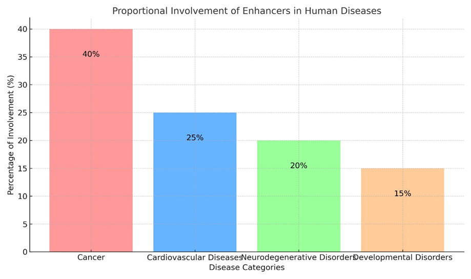
Fig : 3 Proportional involvements of enhancers in human diseases
Enhancers’ Potential for Therapeutic Use
Enhancers offer important therapeutic opportunities since they are important regulators of gene expression. Numerous illnesses, such as cancer, neurological disorders, autoimmune problems, and metabolic diseases, have been linked to enhancer dysregulation. Targeting enhancers’ regulatory activity, reestablishing normal function, or using their characteristics to promote therapeutic gene expression are all examples of using enhancers for therapeutic reasons. The therapeutic potential of enhancers and methods for using them in precision medicine are covered below.
Gene Regulation By adjusting enhancer activity, one can address the abnormal gene expression that frequently results from enhancer dysregulation. Influencers of Epigenetics:
Enhancer activity can be modulated by small compounds that target histone modifiers. For instance, histone acetylation at enhancers can be controlled by inhibitors of histone acetyltransferases (HATs) or histone deacetylases (HDACs), which can reactivate genes that have been silenced or repress genes that have been overexpressed. In malignancies caused by super-enhancer activation, bromodomain inhibitors (such as BET inhibitors) have demonstrated potential by preventing transcriptional coactivators from attaching to acetylated enhancers. Agents that Demethylate DNA: DNA methyltransferase inhibitors can be used to reactivate enhancers that have been silenced by DNA hypermethylation. This strategy is especially pertinent to malignancies when enhancer methylation silences tumor suppressor genes
Histone Methylation Modulators: To restore proper gene expression patterns, enhancer statuses can be modulated by targeting histone methyltransferases or demethylases.
In gene therapy, enhancers can be used to produce precise and regulated gene expression:
Enhancer-Based Gene Delivery: Therapeutic genes can be engineered to express themselves in a way that is particular to a given tissue or condition using synthetic enhancers. To reduce off-target effects, tissue-specific enhancers, for instance, can direct gene expression to the brain, liver, or heart.
Engineering Enhancer-Promoter: Promoters and enhancers can be used together to adjust the levels of therapeutic gene expression. This method is particularly useful for conditions like metabolic or enzyme-replacement illnesses that need for exact regulation of gene expression.
To treat disorders brought on by enhancer dysregulation, genome-editing technologies allow for the exact manipulation of enhancer sequences. Cas9/CRISPR Editing: removal of abnormal enhancers that promote the expression of oncogenes (for example, in tumors that have super-enhancer reprogramming). In genetic illnesses, enhancer mutations are corrected to return genes to normal control.
Activation of CRISPR (CRISPRa): CRISPRa systems, in which catalytically inactive Cas9 (dCas9) is linked to transcriptional activators such as VP64 or p300, can be utilized to activate enhancers of silenced genes. CRISPR Interference (CRISPRi): dCas9 coupled to repressor domains such as KRAB can target and repress overactive enhancers that drive disease-related genes.
Transcribed from active enhancers, enhancer RNAs (Erna’s) are becoming recognized as useful modulators of gene expression and possible targets for treatment: ernes Knockdown: Erna’s can be precisely broken down by antisense oligonucleotides (ASOs) or small interfering RNAs (siRNAs), which lowers the activity of related enhancers. This tactic is especially helpful in malignancies where oncogene activation is facilitated by Erna’s.
Treatments Based on RNA: An innovative method of regulating enhancer activity is to alter ernes interactions with transcriptional machinery or chromatin remodelers.
Super-enhancers are potential therapeutic targets since they control genes essential for cell identity and illness: BET Inhibitors: In malignancies, bromodomain inhibitors can interfere with super-enhancer-driven transcriptional processes, which lowers the expression of oncogenes. Preclinical models of neuroblastoma , breast cancer, and leukemia have demonstrated the effectiveness of this strategy.
Reprogramming the Super-Enhancer: In cancer immunotherapy, immune responses can be improved by altering immune cell super-enhancer activity. Alternatively, autoimmune illnesses can be treated by targeting super-enhancers that drive inflammatory genes.
Enhancers are essential for controlling the gene expression patterns needed for tissue regeneration and repair: Stem Cell Treatment: The effectiveness of stem cell differentiation into target cell types can be increased by modifying enhancers to activate lineage-specific genes. Induced pluripotent stem cells (iPSCs) can be differentiated for use in regenerative medicine under the guidance of enhancer engineering.
Reprogramming Enhancers for Regeneration: The heart, liver, or nervous system are among the damaged tissues where enhancers can be altered to activate restorative pathways.
Therapies Opportunities to alter immune responses in a variety of disorders are presented by enhancer targeting:
Autoimmune Conditions: IN autoimmune diseases, it is possible to lessen excessive immune activation by silencing enhancers that control genes that produce inflammatory cytokines. For instance, reducing inflammation in rheumatoid arthritis or Crohn’s disease can be achieved by blocking promoters of IL-6 or TNF-α.
Immunotherapy for cancer: Immune responses against malignancies can be enhanced by manipulating enhancers of immune checkpoint genes (e.g., PD-1 or CTLA-4). Immune cell activity can be increased through enhancer targeting, which facilitates efficient tumor removal.
Enhancers have potential as biomarkers due to their disease-specific and cell-type-specific activity:
Anticipatory Biomarkers: Genome-wide association studies (GWAS) have revealed disease-associated enhancer variations that can be used as indicators of disease risk and progression. For instance, autoimmune disease susceptibility can be predicted by SNPs in enhancers that control immune genes.
Diagnostic Biomarkers: Enhancer chromatin states or ernes levels can be utilized to track the effectiveness of treatments or make medical diagnoses. Enhancer profiling can assist in determining tumor subtypes and directing treatment choices for malignancies Obstacles and Prospects Even with their medicinal potential, there are still a number of obstacles to overcome:
Specificity of Enhancers: Enhancers frequently have context-specific functions, and environmental signals, transcription factors, and chromatin structure all affect how active they are. For therapeutic targeting to be effective, it is imperative to comprehend this specificity.
Enhancer Specificity: Enhancers frequently work in a context-specific way, and environmental signals, transcription factors, and chromatin structure all affect how active they are. For therapeutic targeting to be effective, it is imperative to comprehend this specificity.
Off-Target Effects: Targeting enhancers may unintentionally alter non-target gene expression, which could result in undesirable side effects. To reduce these hazards, better targeting technologies are required.
Methods of Delivery: Delivering enhancer-targeting treatments (such as RNA-based medications or CRISPR/Cas9 systems) effectively and tissue-specifically is still a significant hurdle.
Combining Other Therapies: To produce synergistic effects, enhancer-targeting techniques must be used with currently used treatments like immunotherapy or chemotherapy.
Conclusion
Enhancers have great potential as therapeutic targets since they provide chances to precisely and context-specifically alter gene expression. New opportunities to use enhancers in the treatment of autoimmune illnesses, cancer, genetic disorders, and more have been made possible by developments in RNA-based technologies, gene editing, and epigenetic modulators. Their potential to revolutionize precision medicine will only grow as our knowledge of enhancer biology advances.
Perspective and Future Directions
Viewpoint and Prospects Our knowledge of gene regulation and its function in development, health, and illness has been completely transformed by the research of enhancers. Enhancer research has made great strides, but there are still a number of obstacles and possibilities to overcome. By addressing these, we can improve their use in precision medicine and therapeutic approaches while also expanding our knowledge of enhancer biology. We highlight important viewpoints and potential avenues for enhancer research below.
Only a small percentage of the millions of possible enhancers that high-throughput technologies have found have been functionally described. Future initiatives should concentrate on: Enhancing Functional Validation: Applying techniques like as massively parallel reporter assays (MPRAs), CRISPR screens, and epigenomic editing to validate enhancer activity in a methodical manner. Studies by Cell Type and Context: Enhancers work very differently depending on the circumstance. Enhancer roles in particular cell types, tissues, and developmental stages will be revealed with the aid of single-cell technologies and organoid models. Cross-Species Comparisons: Research on enhancers from different species will shed light on both species-specific and conserved regulation mechanisms.
It is essential to understand the molecular processes that underlie enhancer activity: Interactions between Enhancer and Promoter: Better 3D genome mapping tools, such Hi-C and similar approaches, are required to clarify chromatin looping dynamics and enhancer-promoter interactions. Interactions between Enhancer and Enhancer: To understand the function of enhancer clusters, including super-enhancers, in controlling intricate gene networks, it is crucial to comprehend their cooperative behavior. Erna’s’ function: Enhancer RNAs’ (Erna’s’) role in transcriptional control and chromatin dynamics will become clearer with more research.
Given the large degree of variability and mutation susceptibility of enhancers, future studies should concentrate on: Comprehending Variants That Do Not Code:connecting the effects of enhancers’ non-coding genetic variations (such as SNPs, insertions, and deletions) on gene expression and disease characteristics. How to Interpret GWAS Data: combining functional genomics data with GWAS results to locate enhancers linked to disease and comprehend how they affect complex traits and illnesses. Applications of Rare Diseases: examining the part enhancer mutations play in uncommon genetic illnesses, which are still poorly understood in comparison to typical illnesses.
One promising class of medicinal targets is enhancers. Future paths consist of: Precision Epigenome editing involves developing methods to modify enhancer activity with high specificity and little off-target consequences, such as CRISPR/dCas9. Drug development is the process of creating biologics or small compounds that target proteins that are linked to enhancers, such as chromatin remodelers, coactivators, or transcription factors. Enhancer engineering is the process of creating artificial enhancers for regulated therapeutic gene expression in regenerative medicine and gene therapy
Enhancer research touches on a number of new fields in biology and medicine, including: Identifying active enhancers in uncommon cell groups and during dynamic processes like development and disease progression will be made possible by single-cell genomics. Machine learning and artificial intelligence (AI): AI-powered models are able to select disease-associated enhancers for functional research, predict enhancer activity, and interpret enhancer-promoter interactions. Organoids and In Vivo Models: Physiological contexts for researching enhancer function and evaluating treatment approaches will be provided by human organoids and sophisticated animal models.
The biological Enhancers frequently function inside intricate regulatory networks: GRNs, or gene regulatory networks: A systems-level knowledge of gene expression patterns in development and disease will be possible through the mapping of GRNs triggered by enhancers. Dynamic Regulation: Enhancer activity is not constant; examining how enhancer landscapes change over time will show how they orchestrate intricate biological processes.
Ethical and legal issues need to be resolved when enhancer-based treatments move closer to clinical use: Risks Off-Target: Because enhancer targeting affects several genes, it may have unforeseen consequences. To guarantee safety, extensive preclinical testing is required. Variability Particular to the Patient: Personalized techniques that account for individual genetic backgrounds and enhancer variants are critical for precision medicine. Ethical Considerations: The possibility of germline enhancer editing presents moral dilemmas that need to be resolved by public discussion and government regulation.
Prospects for the Future
Enhancer biology combined with state-of-the-art technologies will open up new avenues for basic science and translational medicine in the ensuing decades. Enhancers, which provide new therapeutic targets, biomarkers, and diagnostics, are positioned to play a key role in precision medicine. Their full potential will be realized through interdisciplinary cooperation, clinical validation initiatives, and the ongoing development of reliable technologies Our understanding and approach to treating complicated diseases are about to undergo a paradigm shift as a result of the close connection between enhancer research and therapeutic application. Enhancers are the keys to discovering the mysteries of human biology and illness, in addition to being controllers of gene expression.
CONCLUSION
The study of enhancers has significantly advanced in recent years, with new insights into their role in gene regulation, development, and disease. Enhancers, as non-coding DNA elements, play a crucial role in regulating gene expression by interacting with promoters, affecting cellular functions across various contexts. Their involvement in diseases such as cancer, neurological disorders, and metabolic conditions highlights their potential as therapeutic targets. The integration of high-throughput sequencing technologies, functional genomics, and epigenetic tools has greatly enhanced our understanding of enhancer biology, providing new avenues for disease diagnosis and treatment. Despite these advancements, the complexity of enhancer function and regulation remains an area of active research, with the need for further exploration into enhancer-promoter interactions, enhancer RNAs, and their implications in health and disease. Future studies focused on enhancer-targeted therapies and precision medicine hold promise for advancing treatments for various genetic disorders, cancers, and other diseases associated with enhancer dysregulation.
REFERENCES

Namrata Kujur, Active Enhancers : Recent Research Advances and Insights into Disease, Int. J. of Pharm. Sci., 2025, Vol 3, Issue 9, 3390-3406. https://doi.org/10.5281/zenodo.17226139
 10.5281/zenodo.17226139