KMCH College of Pharmacy, Coimbatore, Tamil Nadu, India
Nanotechnology has emerged as a powerful tool in modern pharmaceutical science, offering innovative solutions for targeted and controlled drug delivery. Traditional dosage forms often struggle with issues such as inadequate solubility, limited permeability, chemical degradation, and extensive first-pass metabolism, which ultimately compromise therapeutic performance. Novel nanocarriers—including phytosomes, ethosomes, and cubosomes—help address these problems effectively. Phytosomes are complexes of plant bioactives with phospholipids that enhance lipophilicity, membrane transport, and overall bioavailability. Ethosomes, which consist of phospholipids, ethanol, and water, are ultra-flexible vesicles capable of achieving deeper dermal and transdermal drug penetration. Cubosomes are lipid-based nanostructures with a cubic architecture that provide high drug-loading capacity, strong bioadhesion, and prolonged release suitable for multiple delivery routes. This review compares these nanocarrier systems in terms of formulation components, preparation methods, penetration mechanisms, evaluation parameters, benefits, drawbacks, and therapeutic applications, highlighting their ability to improve drug delivery efficiency, treatment effectiveness, and patient compliance.
In recent decades, nanotechnology has played a transformative role in the pharmaceutical and biomedical sciences by offering innovative solutions to overcome the inherent limitations of conventional drug delivery systems. Most bioactive compounds, particularly that of natural origin, have poor water solubility, low permeability, stability, and widespread first-pass metabolism leading to poor therapeutic activity (1). In order to enhance the solubility, stability, and bioavailability of synthetic and phytoconstituent-based therapies, nanocarrier-based drug delivery technologies have been developed. Among the wide range of nanocarriers, phytosomes, ethosomes, and cubosomes have gained considerable attention due to their unique structural and functional characteristics making them particularly effective for the delivery of plant-derived bioactives. Phytosomes are sophisticated herbal formulations that contain phytoconstituents complexed with phospholipids, usually phosphatidylcholine. This complexation increases the lipid compatibility and enhances the ability of the active compounds to pass through biological membranes. Consequently, phytosomes demonstrate significantly higher absorption and bioavailability compared to conventional herbal extracts (15). This system has been particularly beneficial for poorly soluble flavonoids, terpenoids, and polyphenolic compounds, thereby improving their therapeutic potential in treating chronic diseases such as inflammation, diabetes, and cancer. Ethosomes represent a novel class of flexible lipid vesicles composed of phospholipids, ethanol (20–45%), and water. The high ethanol acontent confers elasticity and enhances the penetration of the vesicles through the stratum corneum, making ethosomes highly effective for topical and transdermal delivery (9). Ethosomes not only improve the delivery of lipophilic drugs but also facilitate the transport of hydrophilic molecules, peptides, and even genetic materials through the skin. They provide a non-invasive method for systemic therapy and have been effectively utilized to administer medications including ketoconazole, acyclovir, and herbal substances. Cubosomes,in contrast, are amphiphilic molecules that self-assemble into a bicontinuous cubic phase, which are nanostructured lipid carriers. Both hydrophilic and hydrophobic medications can be encapsulated thanks to their interior three-dimensional honeycomb-like structure, which offers a vast surface area (4). Cubosomes exhibit remarkable physicochemical stability, bioadhesion, and controlled release characteristics, making them attractive for various routes of administration, including oral, ocular, transdermal, and intravenous. Collectively, these nanocarrier systems provide an efficient platform for enhancing the pharmacokinetic and pharmacodynamic properties of phytochemicals, reducing dosing frequency, and improving patient compliance. Furthermore, the ability to achieve targeted and controlled drug release positions these systems as key tools in modern phytopharmaceutical research. This review intends to provide an in-depth overview of phytosomes, ethosomes, and cubosomes (including their composition, method of preparation, modes of action, benefits, drawbacks, and the latest developments in phytosomes, ethosomes, and cubosomes usage and delivery of active compounds).
PHYTOSOMES
Phytosomes are vesicular drug delivery systems containing lipid and phytoconstituents on both surfaces (5). Lipid-based nanocarriers known phytosomes combine phospholipids with plant extracts or phytoconstituents (6).
When applied physically or taken orally, herbal extracts are more readily absorbed (5). The phospholipids that are used include phosphatidylcholine, phosphatidylserine, phosphatidylethanolamine, and phosphatidylinositol; however, phosphatidylcholine is the most often utilized because of its potential therapeutic benefits for liver illnesses include drug-induced liver damage, alcoholism, hepatitis, and alcoholic steatosis (5). Although the numerous treatments established to treat them, metabolic disorders (MDs) remain the primary cause of life-threatening diseases that affect individuals all over the world, including liver pathologies, cardiovascular diseases (CVDs), and diabetes mellitus (DM) (7). A naturally occurring flavonolignan molecule called Silibinin Phytosomes is extracted from milk thistle (Silybum marianum) and used in traditional medicine to treat liver conditions (6). Phytosomes loaded against sinigrin have a lot of promise as a skin cancer delivery mechanism. Combining the anticancer properties of Sinigrin-loaded Phytosomes with wound healing may result in more potent therapies with fewer adverse effects (6). The phospholipid bilayer shell that covers the (mitomycin c) Phytosomes and prevents their quick absorption into the systemic circulation is part of the Phytosome complex (6).
Table 1. Structural compounds and their chemical structure
|
S.NO |
CHEMICAL NAME |
CHEMICAL STRUCTURE |
|
|
Catechol |
|
|
|
Geraniol |
|
|
|
Linalool |
|
|
|
Menthol |
|
|
|
Isoquinoline alkaloid |
|
|
|
Pyrolidine |
|
Description
Based on their structural components, phytochemicals are divided into three main groups: polyphenolic compounds, alkaloids, and terpenoids. Many flavour and aromatic molecules, such as menthol, linalool, geraniol, and caryophyllene, are terpenoids, although phenolic substances include catechols, lignins, tannins, stilbenes, and flavonoids (15). Alkaloids are categorized into isoquinoline alkaloids, pyrrolidine, pyrrolidine-pyridine, and pyridine-piperidine based on their heterocyclic ring structures (9). Phytochemicals can affect enzyme processes in a number of ways, such as by functioning as substrates, cofactors, or inhibitors (15).
Structure:
Guggulosomes and Phytosomes are complexes of Phyto-phospholipids in their chemical
composition, are produced by the physical and chemical combination of the active
phytoconstituents and the polar head (choline moiety).
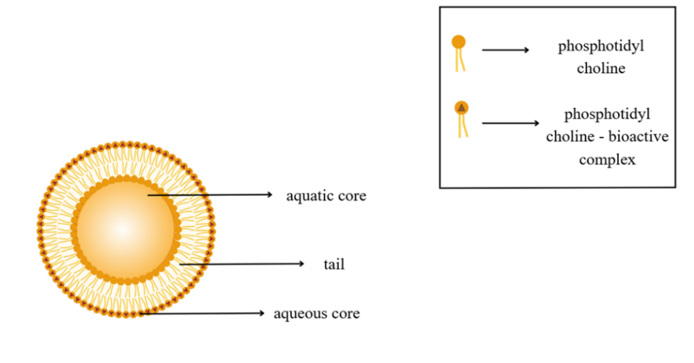
Fig. 1. Structure of phytosomes
Phospholipid head groups must be placed in these complexes. A fatty acid chain encapsulates the polar component in complexes that provide a lipophilic surface. The active component of a liposome can be detected inside a cavity between the many layers of the membrane (5). Particle size of phytosomes can range from 50nm to several hundred micrometers (>100μm).
Components of phytosomes
It has four important aspects of phyto-phospholipid complexes synthesis include solvents, phytoconstituents, phospholipid role, and stoichiometric ratio. The active component that can be incorporated in phytosomes other than polyphenols can include other compounds like siramasin, evodiamine, 20(R)-25-methoxyl-dammarane-3, and 12, 20-triol (25-OCH3-PPD). (15) According to theory, complexes derived from phytosomes might be combined with any active moiety because they are more than simply polyphenols (5).
Phytosome source
Several studies have reported the isolation and characterization of plant-derived phytosomal vesicles from a wide range of edible and medicinal sources, demonstrating their potential in anticancer, anti-inflammatory, and immunomodulatory applications.
Phytosomes obtained from fingerroot (Boesenbergia rotunda) have been shown to exert selective cytotoxic and apoptotic effects against cancer cells while sparing normal colon epithelial cells. These effects were primarily attributed to the induction of programmed cell death and disruption of intracellular redox balance, highlighting their potential as selective anticancer carriers.
Similarly, garlic (Allium sativum)-derived phytosomes have demonstrated significant cellular uptake by hepatocellular carcinoma (HepG2) cells, resulting in pronounced anti-inflammatory activity. The retention of these phytosomes within cancer cells has been associated with interactions involving the CD98 receptor, a mannose-rich glycoprotein known to facilitate vesicle–cell binding, although direct anticancer effects were limited.
Phytosomes isolated from lemon juice (Citrus limon) have been widely reported for their anticancer potential. In vitro studies revealed inhibition of cancer cell proliferation without damage to healthy cells, while in vivo investigations in tumor-bearing animal models demonstrated suppression of subcutaneous tumor growth. These effects were linked to the activation of angiogenesis inhibition and TRAIL-mediated apoptotic pathways, indicating a selective mechanism of tumor cell death.
Studies involving corn (Zea mays)-derived phytosomes have shown notable inhibition of colon cancer cell growth in vitro, along with effective suppression of tumor progression in animal models without observable systemic toxicity. These findings suggest a favorable safety profile combined with anticancer efficacy.
Phytosomes derived from cannabis (Cannabis sativa) have also been investigated for their biological activity. In vitro studies demonstrated dose- and time-dependent effects on hepatocellular carcinoma cell lines, while maintaining minimal impact on normal endothelial cells. The observed cellular responses were associated with mitochondrial-dependent apoptotic signaling pathways, including modulation of pro- and anti-apoptotic proteins, although data on their absorption and systemic disposition remain limited.
Tea leaf (Yongchuan Xiuya)-derived phytosomes have been reported to exhibit strong anticancer activity in both in vitro and in vivo models. These vesicles enhanced cytotoxicity in multiple breast cancer cell lines and significantly reduced tumor volume following oral and intravenous administration in animal studies. The anticancer mechanism was primarily attributed to elevated intracellular reactive oxygen species (ROS), leading to mitochondrial dysfunction, cell-cycle arrest, and apoptosis.
In addition, phytosomes obtained from apple fruits (Malus domestica, Golden Delicious variety) have demonstrated immunomodulatory and anti-inflammatory effects. In vitro studies showed reduced expression of pro-inflammatory cytokines such as IL-1β and IL-8 in activated macrophages, suggesting that these phytosomes can modulate immune responses and shift cells toward an anti-inflammatory phenotype.
Unique Characteristics of Phytosomes with Anticancer Activity
Safety, specificity, and efficacy are some of the most significant obstacles in the development of new medicines. Phytosome-based drug delivery circumvents the drawbacks of conventional therapies, which mainly rely on synthetic products or pure compounds obtained from natural sources, such as extracts from Rubia cordifolia, which are often boundup with adverse effects and can lead to tolerance if used over an extended period of time. Phytosomes offer a novel strategy to the plant-based treatment of cancer and chronic inflammation associated with cancer because research has generally demonstrated that some phytochemicals act on aberrant, changed cells, such as cancer cells, rather than adversely affecting healthy cells (11).
Phytosome characterization:
Study of morphology
Morphology was calculated of the phytosomes by use of transmission electron microscopy , potential, polydispersity index and particle size distribution study. DS was used to measure the phytosomes polydispersity index (PDI), zeta potential (ZP) and particle size (PS) distribution by using digital platform (12)
Estimation of Entrapment Efficiency
The entrapment efficiency is determined by centrifugation. Each formulation (phosphatidylcholine1, phosphatidylcholine2, and phosphatidylcholine3) was centrifuged in the 0.5 mL suspension of sample to a centrifuge tube (11). The excess liquid from formulations Phosphatidylcholine1, Phosphatidylcholine2, and Phosphatidylcholine3 was diluted with ethanol in 0.20, 0.30, and 0.25 mL amounts, respectively, causing vesicle disruption (12).
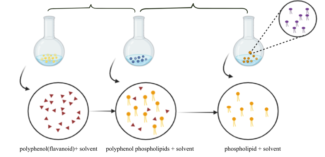
Mechanism of complexation
Standardized herbal extract and phospholipids react in a non-polar solvent in a stoichiometric ratio to form phytosomes. Phosphatidylcholine, an amphipathic molecule, is composed of the hydrophilic molecule choline and the lipophilic moiety phosphatidyl. The nonpolar phosphatidyl component envelops the choline-bound drug to form the body and tail, while the polar choline head interfaces and chemically links to the polar properties of the drug molecule. As the medication is incorporated in phospholipid layers, these are referred to as Phyto-phospholipid complexes, which form a small microsphere or cell (1).
Preparation of phytosomes
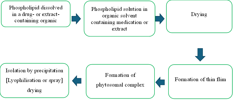
Fig. 2. General method of preparation
The complex can be separated by spraying, lyophilisation or precipitation mixed with non-solvents, including lithium-based hydrocarbons. During the complex formation of phytosomes, these two components form in a ratio of 0.5 to 2.0 moles. The ratio of flavonoids to phospholipids can be 1:1. shows how phytosomes are prepared methodically (5).
Methods:
Solvent evaporation method:
A marsupia-phospholipid complex is created using a mechanically dispersion-oriented precipitation of liquid antisolvent technique. When using the solvent evaporation process, the drug and the phospholipids (tetrahydrofuran and ethanol) are usually mixed in a flask with a suitable solvent or solvent system. Most studies have concluded that an ideal stoichiometric ratio for complex formation is 1:1. The oxymatrine-phospholipid complex was produced by mechanically agitating both solutions until all solvents had evaporated. The ratios of 1, 4, 2, 6, and 3 between the phospholipid and the medication were used. A composite design method was then used to improve the complex. At 60ºC for three hours, a 3:1 ratio was used to build the most productive complex. Developed an embelin-PC complex at molar ratios ranging from 1:0.5 to 1:3. The most effective formulation was made with a drug concentration of 80 g/l and a phospholipid to drug ratio of 0.9 (w/w). It was found that the complex's drug content was 45.78%, and the final formulation's combined proportion was 100%. They have mostly been replaced by aprotic solvents like methylene chloride, ethyl acetate, dioxane, etc. In their studies, researchers have utilized phospholipids from a variety of sources along with the solvent system (5).
Super critical fluid (SCF):
Supercritical liquids are an effective method to produce particles with diameters that range from 5 to 2000 nm. Three distinct conventional methods, involving lyophilization, solvent evaporation, and micronized puerarin, were used to produce purarin and phospholipid complexes. Supercritical fluids are utilized to improve the solubility characteristics of poorly soluble drug candidates (SEDS). Separate phospholipid and drug solutions were treated with a supercritical anti-solvent prior to the application of the ultimate pressure in the GAS procedure. The final result of the process achieved a 93% yield (5).
Lyophilization process:
A phospholipid-containing combination was added to another solution containing phytoconstituents after both natural and synthetic phospholipids and phytoconstituents were melted using different solvents. The mixture was then stirred until a complex formed. Lyophilization is a method to separate the formed complex. The acyl group present in the phospholipids used in phytosome formation can be phosphoryl choline, phosphatidylserine, and phosphatidylethanolamine, which are created from stearic, oleic, palmitic, and linoleic acids. Phytosomes' active principle develops into a structural element (5).
Salting out:
After dissolving in ethanol, the extract and PC are mixed and swirled. In order to produce a precipitate phytosome, n-hexane is added to the mixture throughout the precipitation formation process (5).
Thin layer hydration:
PC and fraction was dissolved in methanol, whereas cholesterol was dissolved in dichloromethane. The mixture is gradually evaporated at 45°C using a rotary evaporator once solvent has entirely evaporated and a thin, dry layer has developed at the container's bottom. After a night at room temperature, the resulting thin layer of lipid is treated using hydration and pumped with nitrogen gas The film layer was hydrated using Aquabidest on a rotating evaporator set at 45°C.Sonification and a homogeniser were also used to improve the process of determining particle size (5).
Applications
Table 2. Phytosomal preparations and their therapeutic use
|
Phytosome source |
Therapeutic uses |
|
Silibinin phytosome |
Traditional medicine for liver diseases Anticancer activities against cancer cells like Hepatocellular carcinoma, prostate cancer, breast cancer and lung cancer (6). |
|
Sinigrin phytosome |
Skin cancer (6). |
|
Mitomycin c phytosome |
Anti-tumor activity (6). |
|
Curcumin phytosome |
Anticancer, Curcumin and piperine is a natural alkaloid found in pepper used for Treating multiple myeloma (6). |
|
Gingo biloba |
Oxidative and inflammatory stress Hepatic disorder (7). |
|
Silymarin phytosome |
Dyslipidemia (7). |
|
Zingiber officinale |
Cardiovascular diseases (7). |
|
Rubia cordifolia |
Antibacterial, Rheumatoid arthritis, Uterine pain Joint pain(12). |
|
Europaeaoil |
Anti-inflammatory, Antioxidant, Cardiovascular protection, Anti-hyperlipidemia activities (10). |
|
Centellaasiatica |
Vein and skin disorders (10). |
|
Camelliasinensis |
Antioxidant activity (10) |
|
Rutagraveolens sophorajaponica |
Rheumatoid arthritis (10). |
|
Vitisvinifera |
Antioxidant activity (10). |
|
Echinaceaaugustifolia |
Immunomodulatory (10) |
|
Cartaegusmexicana |
Antioxidant activity (10) |
|
Carteagusspecies |
Antihypertensive activity (10) |
|
Ruscusaculeatus |
Anti-inflammatory activity (10) |
ETHOSOME
Introduction
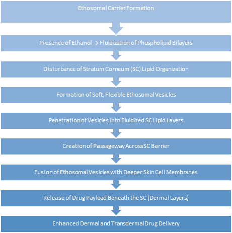
The skin is one of the body's largest and most accessible organs. Using the skin as a drug delivery method has a number of advantages over traditional methods, including lower fluctuations in plasma drug levels, avoidance of gastrointestinal problems and first-pass drug metabolism, and high patient compliance. One of the major disadvantages of transdermal drug delivery is the skin's low permeability, which restricts the number of drugs that may be given in this manner (14). The skin's barrier properties, which prevent the majority of medications from entering the body, present the biggest obstacle to transdermal drug administration. To increase transdermal permeability, a number of physical and chemical methods have been developed and evaluated to get beyond the stratum corneum barrier. Two new carriers have been introduced as a result of ongoing research with lipid-based systems, ethosomes and transfersomes. Phospholipids and an edge activator make up the deformable lipid vesicles known as transfersomes.
Often a surfactant molecule with a single chain. Touitou et al (1999). initially described ethosomes as an intriguing lipid-based carrier.
The non-invasive ethosomal drug delivery method transports the medication to the deep layers of the skin and into the bloodstream (15).
Ethosomes are flexible, soft vesicles that range in size from tens of nanometers to microns and are mostly made of phospholipids, ethanol, and water. The preparation process and use of methods such as sonication determine the size of ethosomes. These "soft vesicles" are an innovative vesicular carrier for improved skin delivery. The pliable, squishy vesicles are designed to distribute active substances more effectively. The formulation's high alcohol content (20–45%) gives the vesicles stability and soft, flexible qualities while also upsetting the skin's lipid bilayer structure, which increases membrane permeability (16).
Structure of ethosome
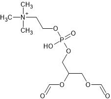
Ethosomes are soft, pliable lipid vesicles which consist of:
Phospholipids (2–10% w/w), including phosphatidylglycerol (PG), phosphatidylethanolamine (PE), and phosphatidylcholine (PC)
High levels of isopropyl alcohol or ethanol (20–45% v/v)
The aqueous phase is water.
Glycols (such as propylene glycol and transcutol) may act as penetration enhancers Cholesterol (0.1–1%) may be added for vesicular stability(14).
The ethosomal system encapsulates hydrophilic and lipophilic medications by forming multilamellar or unilamellar vesicles in which the lipid bilayer encloses both aqueous and hydroethanolic areas(18).
The vesicular bilayers are made flexible and malleable by the negative charge and fluidity that ethanol gives them, which is essential for deep skin penetration.
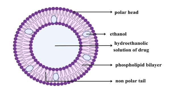
Fig.4. Structure of ethosomes
Composition
The ethosomal system is composed of phospholipids, ethanol, and water. Phospholipids with various chemical structures include phosphatidyl choline (PC), hydrogenated PC, phosphatidyl ethanolamine (PE), phosphatidyl glycerol (PPG), phosphatidyl inositol (PI), and hydrogenated PC.
Table 3. composition & its uses
|
Class |
Example(s) |
Function / Use |
|
Phospholipids Eg: phosphatidylcholine
|
Soya phosphatidylcholine (SPC) Egg phosphatidylcholine (EPC) Dipalmitoyl phosphatidylcholine (DPPC) Distearoyl phosphatidylcholine (DSPC) |
Act as vesicle-forming components that create the lipid bilayer structure of ethosomes, encapsulating both hydrophilic and lipophilic drugs. |
|
Polyols / Glycols Eg: propylene glycol
|
Propylene glycol Transcutol® RTM |
Function as skin penetration enhancers; improve vesicle deformability and enhance solubility of actives. |
|
Alcohols

Eg: Isopropyl alcohol
|
Ethanol & Isopropyl alcohol |
Provide softness and flexibility to vesicle membranes and serve as penetration enhancers by fluidizing stratum corneum lipids. |
|
Cholesterol
|
Cholesterol |
Acts as a membrane stabilizer, preventing vesicle leakage and improving rigidity when required. |
|
Dyes (for characterization) |
Rhodamine-123 Rhodamine red Fluorescein isothiocyanate (FITC) 6-Carboxyfluorescein |
Used in fluorescence microscopy and confocal studies to trace vesicle morphology, size, and penetration depth. |
|
Vehicle / Gel Base |
Carbopol 934 |
Used as a vehicle or gelling agent for topical application, improving spreadability and residence time on skin. |
The non-aqueous phase has a range of 22% to 70%.
The alcohol may be ethanol or isopropyl alcohol.
Ethosomes are often treated with dyes or amphiphilic fluorescent probes such as D-289, Rhodamine-123, fluorescence isothiocynate (FITC), and 6-carboxy fluorescence for characterisation investigations (14).
Mechanism of action
Method of preparation
Hot Method
Phospholipid + Water
↓
Colloidal solution formed
↓
Ethanol + Propylene glycol + Drug
↓
Organic phase added → Stir 5 min
↓
Sonication (reduce vesicle size)
↓
Final ethosomal formulation (stored)
(16)
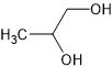
Cold Method:
Phospholipid + Drug + Lipid components + Ethanol
↓
Mixture heated to 30°C
↓
Preheated water added slowly with stirring
↓
Vesicle formation (after 5 min)
↓
Maintain vesicle stability (cold)
↓
Drug dissolved based on suitable solvent(7,23)
Evaluation
An essential first step in verifying the vesicular properties, stability, and effectiveness of ethosomal formulations in cutaneous or transdermal drug delivery is evaluation. Because of their high ethanol concentration, which provides flexibility, deformability, and improved permeability, ethosomes are different from traditional liposomes. For ethosomal systems, the following parameters are regularly evaluated.
Published studies consistently report that the performance of ethosomal drug-delivery systems is evaluated using a combination of physicochemical, biopharmaceutical, and stability-related parameters to confirm vesicle integrity, delivery efficiency, and transdermal performance.
Vesicle morphology and surface characteristics are commonly assessed to verify the formation of spherical, smooth, and multilamellar vesicles. Early investigations by Touitou et al. demonstrated the presence of multilamellar vesicular structures in phosphatidylcholine- and ethanol-based ethosomes, while subsequent studies confirmed homogeneous and smooth surface morphology in drug-loaded ethosomal systems, supporting their structural stability and suitability for dermal delivery (35,37).
Vesicle size and size distribution are critical determinants of skin penetration efficiency. Literature reports indicate that ethosomal vesicles generally fall within the nanometer range, with size strongly influenced by ethanol concentration and lipid composition. Increased ethanol content has been associated with reduced vesicle diameter and improved homogeneity due to enhanced membrane fluidity and electrostatic repulsion, as reported in studies where vesicle sizes ranged between 120 and 410 nm (18).
Surface charge (zeta potential) is widely used as an indicator of colloidal stability. Ethosomes typically exhibit a negative surface charge, which minimizes vesicle aggregation and enhances dispersion stability, thereby contributing to prolonged shelf life (16).
Drug entrapment efficiency (%EE) is regarded as a key parameter influencing ethosomal performance. Reported studies indicate that ethosomes are capable of achieving moderate to high drug encapsulation, with entrapment efficiencies generally ranging between 55 % and 90 %, depending on drug physicochemical properties, phospholipid concentration, and ethanol content (37,23).
Drug content uniformity and pH are evaluated to ensure formulation consistency and skin compatibility. Literature reports suggest that ethosomal formulations generally maintain a near-neutral pH suitable for topical application, while consistent drug content confirms reproducible drug loading and formulation stability (8,28).
In vitro drug-release behavior of ethosomal systems has been extensively investigated using diffusion-based models. Reported findings demonstrate that ethosomes typically exhibit sustained and controlled drug-release profiles, which are attributed to their flexible bilayer structure and ethanol-mediated enhancement of drug diffusion across membranes (18).
Ex vivo skin permeation studies described in the literature consistently demonstrate higher transdermal flux and deeper skin deposition for ethosomal formulations compared with conventional gels and liposomal systems. This enhanced permeation capability is primarily attributed to ethanol-induced disruption of stratum corneum lipids and the deformability of ethosomal vesicles (35,37).
Stability studies reported for ethosomal formulations indicate good physical and chemical stability during storage, with minimal changes in vesicle size, entrapment efficiency, and appearance over time, confirming their suitability for topical and transdermal applications (37,28).
Thermal behavior and membrane fluidity analyses have shown that ethanol lowers the lipid phase transition temperature, resulting in increased bilayer flexibility and deformability, which are essential attributes for effective skin penetration (21).
Finally, fluorescence-based tracking studies reported in the literature have confirmed enhanced penetration and localization of ethosomal vesicles within viable skin layers, providing visual evidence of improved dermal and transdermal drug delivery performance (37).
Applications
Ethosomes have been shown in numerous trials to be an effective treatment for viral and microbial skin infections. Animal models of deep skin infections were used to create and test the efficacy of the bacitracin and erythromycin ethosomal systems (11)
When it comes to transdermal drug administration, ethosomes have proven to be more effective than hydroalcoholic systems or traditional liposomes. Their ethanol-rich, flexible lipid bilayers facilitate deeper medication penetration and increase the skin lipids' fluidity (34,36).
Research has demonstrated that ethosomal formulations can deposit hydrophilic and lipophilic compounds in viable skin layers by transporting them across the stratum corneum. They have been successfully used to treat illnesses like arthritis, hormonal shortages, and cardiovascular disorders that call for continuous systemic delivery (23,28).
For corticosteroids and non-steroidal anti-inflammatory medications (NSAIDs), a number of ethosomal formulations have been created.
Ketoprofen Chourasia MK et al (2011), ibuprofen, and betamethasone-17-valerate are a few examples that showed improved penetration and extended anti-inflammatory activity following transdermal application. Furthermore, triptolide and ammonium glycyrrhizinate-containing ethosomal systems significantly reduced erythema and inflammation in in vivo models (23,28).
These investigations verify that ethosomes enhance skin deposition, patient tolerance, and anti-inflammatory effectiveness.
Ethosomal patches have been investigated for the treatment of menopausal symptoms in women and androgen deficiency in men. Compared to conventional transdermal techniques, these patches produced more systemic absorption and sustained plasma levels, according to in vivo investigations on rabbits (15).
This demonstrates how ethosomes can act as non-invasive transporters of hormones like estradiol and testosterone.
4. Uses in Dermatology and Anti-Pigmentation
When administered via ethosomes, formulations including linoleic acid for the treatment of melasma showed enhanced skin penetration and accumulation, providing targeted therapy of pigmentation problems (14). Additionally, by facilitating smoother penetration and longer-lasting action, ethosomes improve the skin distribution of active substances used in dermatological and cosmetic therapy.
Gold nanoparticles, distamycins, 5-aminolevulinic acid, metformin Magdy S et al (2022) have all been delivered via ethosomal carriers in cancer treatment. These formulations reduced systemic toxicity, improved photodynamic effects, and raised drug accumulation in tumor tissue. Deep tumor targeting via the skin was made possible by the flexible vesicular structure (14).
6. Uses in ocular therapy
Timolol-loaded ethosome Uner B et al (2023) have enhanced treatment of intraocular pressure called glaucoma
7.Applications for Hair Growth and Cosmetics
Minoxidil-containing commercial ethosomal formulations, including NanominoxTM, are effective hair tonics that encourage hair growth. For improved follicular distribution and extended effect, these medications make use of ethosomal vesicles. Other commercial examples are anti-cellulite and cosmetic formulations that are sold in different nations, such as Skin GenuityTM, LipoductionTM, and NoicelleXTM (23,7)
8. Delivery of Proteins and Peptides
Because ethosomes may pass through the epidermal barrier without breaking down, they can carry proteins and peptides. A promising method for non-invasive macromolecular medication delivery is made possible by the ethanol-phospholipid matrix, which stabilizes these biomolecules and promotes their transdermal transit (15).
CUBOSOMES
Introduction:
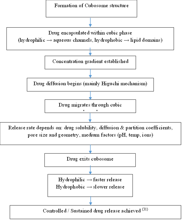
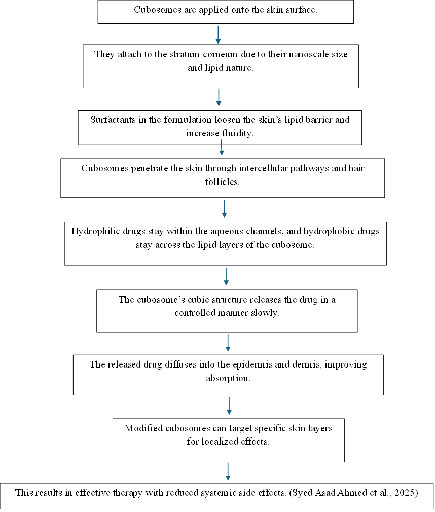
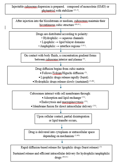
Cubosomes are tiny, nanoscale particles of bicontinuous cubic liquid crystals. The cubic crystalline phase, an appealing drug delivery mechanism, is a continuous, densely coiled layer of lipid with two equivalent cross-sectional aqueous channels. They can remain on the skin's surface for a longer period of time because of their larger surface area, that enables the medication to reach the intended location. The release of numerous pharmacological compounds, including antibiotics, anticancer medications, and local anaesthetics like mostly BCS class 2 and also other class drugs, can also be sustained by cubosomes (30).In order to accomplish targeted and controlled release, a vesicular drug delivery system encapsulates medications within vesicular structures. Depending on their purpose, these vesicles can serve as penetration enhancers to boost skin absorption or as carriers for high-molecular-weight medications. One type of vesicular system discovered in the 1980s is cubosomes, a lipid-based nanostructured colloidal carrier produced through the self-assembly of amphiphilic lipids in aqueous settings. These bicontinuous cubic liquid crystalline nanoparticles (10–500 nm) can encapsulate hydrophilic, hydrophobic, and amphiphilic medications due to their huge interior surface area and endurance. Cubosomes have great potential for advanced therapeutic applications, such as the treatment of melanoma, because of their biodegradability, controlled release capabilities, and excellent drug-loading effectiveness. (15)Different medicinal molecules with hydrophilic, hydrophobic, and amphiphilic properties can be encapsulated by cubosomes. The use of cubosomes as a medicine delivery mechanism has the following benefits:
Structure of cubosome:
Cubosomes are discrete, sub-micron or nanostructured particles that result from the steric stabilization and fragmentation of inverse bi-continuous cubic phases of lipids(12).
These structures consist of a curved bi-continuous lipid bilayer that extends in three dimensions and divides two congruent networks of water channels (6)
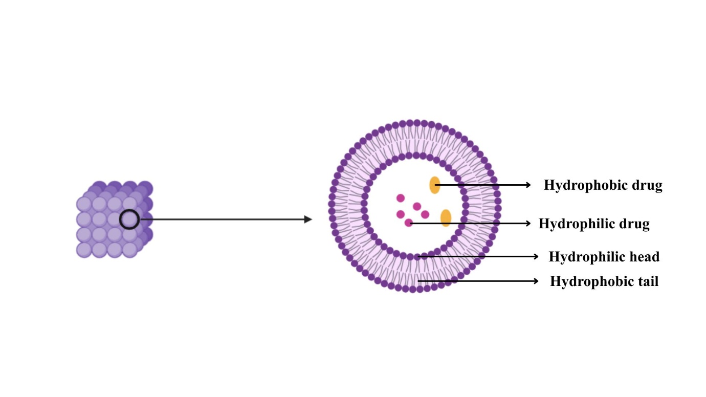
Fig.6. Structure of cubosomes
Because of their shape, cubosomes can simultaneously encapsulate hydrophilic, amphiphilic, and hydrophobic molecules(6).
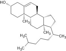
A lipid bilayer with cubic symmetry separates the two continuous but non-intersecting water channels that make up the internal nanostructure of cubosomes (26). The lipid bilayer is frequently composed of monoglycerides (monoolein) or phytantriol stabilized by a surfactant like poloxamer 407. (26)This stabilizer improves the colloidal stability of cubosomes by preventing aggregation (26)
Materials and Composition of Cubosomes:
Cubosomes are self-assembling nanostructured particles made mostly of aqueous components, stabilisers, and amphiphilic lipids. Their structural integrity, drug loading capacity, and release behaviour are all significantly influenced by their makeup. Below is a list of the essential components used in the creation of cubosomes.
Table 4. Materials and Composition of Cubosomes
|
Component Types |
Common Examples |
Function |
References |
|
Lipid Phase |
Glyceryl monooleate (GMO), Phytantriol, Oleic acid |
Structural matrix, drug encapsulation |
(6,30) |
|
Stabilizer |
Poloxamer 407, Poloxamer 188, PVA |
Steric stabilization, dispersion stability |
(6,15,30) |
|
Aqueous Phase |
Distilled/Milli-Q water, PBS |
Hydration medium, vesicle formation |
(4,26,30) |
|
Model Drugs |
Colchicine, Diclofenac sodium, 5-FU, Dexamethasone |
Therapeutic agents |
(15,30) |
|
Additives |
Carbopol 940, TEA, Propylene glycol |
Gelation, pH control, permeation |
(15,30) |
Because it lacks ester linkages and is less susceptible to hydrolysis, phytantriol (PYT) is an alternative lipid that provides higher chemical stability (26) When paired with GMO, oleic acid improves medication encapsulation effectiveness and membrane fluidity, particularly in topical applications (30). In order to adjust internal structures and release kinetics, other lipids including oleyl glycerate and monoglycerides have also been investigated (26)
Cubosomes' bicontinuous cubic liquid crystalline structure, which encloses both hydrophilic and hydrophobic molecules, is formed by the lipid phase. Because of its biocompatibility, biodegradability, and capacity to self-assemble in aqueous conditions, glyceryl monooleate (GMO) is the most widely utilised lipid (6,26,15).
In order to keep cubosomal dispersions colloidally stable and avoid aggregation, stabilisers are added.
The most used stabiliser is Poloxamer 407 (Pluronic F127), which improves dispersion stability and biocompatibility through its steric stabilisation effect (6,15,30). Another potential surfactant for improving dispersibility and lowering interfacial tension is poloxamer 188 (25,12). For improved stability and less particle coalescence, some formulations also use PEG-based surfactants and polyvinyl alcohol (PVA). (4)
Lipid hydration and vesicle formation depend on the aqueous phase, which is usually distilled or purified water. Phase behaviour, vesicle size, and interior nanostructure are all influenced by the water content. For physiological relevance, some investigations used phosphate-buffered saline (PBS) or Milli-Q water (4,30)Under certain hydration levels, the aqueous component helps lipids change from lamellar to cubic phases (26).
Cubosomes have been used to encapsulate a broad range of medications with varying solubility properties: Colchicine for transdermal anti-inflammatory use (25) Enhanced bactericidal activity through antimicrobial peptides (4)
Diclofenac sodium for topical anti-inflammatory treatment (15) 5-Fluorouracil (5-FU) as a targeted treatment for liver cancer (38) Dexamethasone for long-term topical vitiligo therapy (30).
The polarity, molecular weight, and therapeutic target of the medicine all influence the choice of lipid-drug combinations.
A number of auxiliary agents are employed to maximise formulation performance:
Triethanolamine (TEA) for pH correction in cubosomal gels with carbopol 940 as a gelling agent (15,30)
|
S.NO |
Name of material |
2D structure |
|
|
Glycerylmonooleate C21H40o4 |
|
|
|
Oleic acid C18H3402 |
|
|
|
Phytantriol C20H4203 |
|
|
|
Colchicine C22H25NO6 |
|
|
|
Diclofenac sodium C14H10CL2NNaO2 |
|
|
|
5-Flourouracil |
|
|
|
Dexamethasone C22H29FO5 |
|
|
|
Propylene glycol C3H8O2 |
|
Glycerol and propylene glycol as humectants and penetration boosters in topical systems (30).
Using ethanol as a precursor or co-solvent in bottom-up cubosome production methods (26)
Mechanism of action
Oral route
Transdermal route
Injectable route
Preparation Methods of Cubosomes: Top-down and bottom-up are the two key methods for creating cubosomes. To inhibit cubosome dispersion aggregation, these two approaches need the employment of a suitable stabiliser. Nonetheless, the most important factors to take into account when choosing the appropriate preparation method are stability, biocompatibility, and efficient drug release.
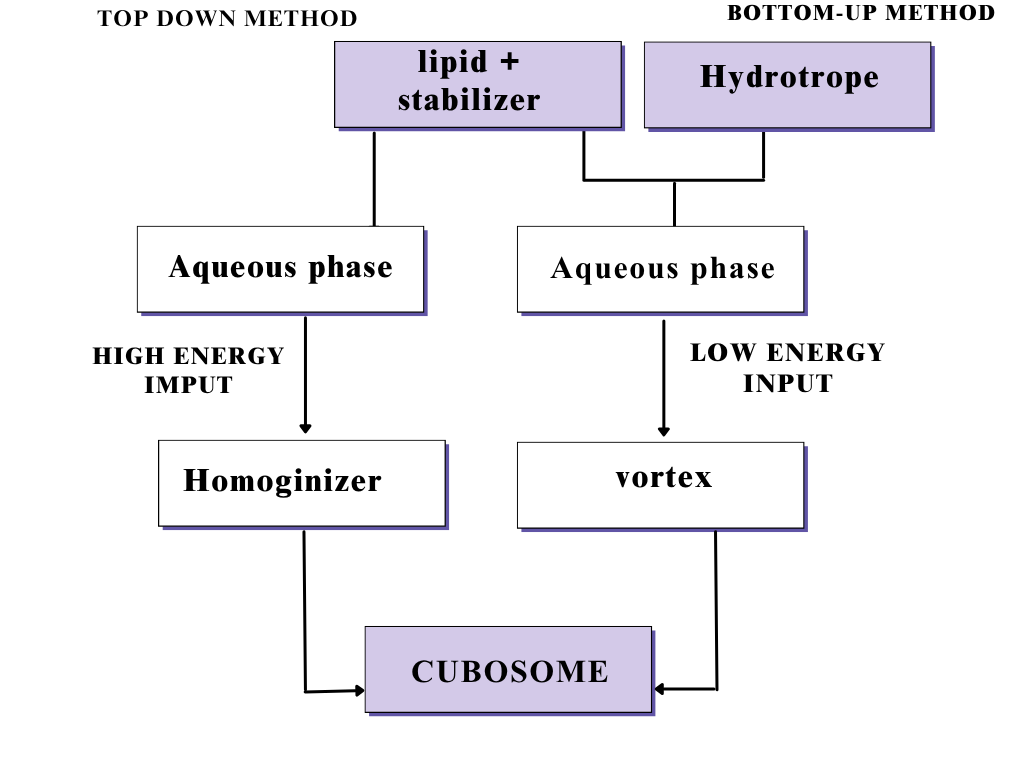
Fig.7.Preparation Methods of Cubosomes
Top-down method:
The widely used technique for making cubosomes is the top-down method, which consists of two major steps. To make the bulk viscous cubic aggregates, combine the lipid-forming cubosomes with an appropriate stabilizer. next, the cubosome is formed by dispersing the resulting viscous cubic aggregates in aqueous solutions using high energy, like sonication or a highpressure homogenizer. Fortunately, it has been discovered that cubosomes made by the top-down method are resilient against aggregation for as long as a year. In 36 Unfortunately, because the formation of viscous cubic aggregates requires a substantial energy input to be disseminated into cubosomes, this method has limitations in large-scale manufacturing when temperature-sensitive bioactive agents—particularly peptides and proteins—need to be incorporated.
Bottom-up method:
This method, also known as the solvent dilution method, utilizes very little energy to disseminate a combination that contains cubosomes that create lipid, a stabilizer, and a hydrotrope in excess of water. A crucial part of the bottom-up method is hydrotrope, which solubilizes water-insoluble lipids to create lipid precursors and stops liquid crystals from forming at high concentrations. A hydrotrope is a molecule may solubilize weakly soluble substances in aqueous solutions via using hydrotropic solubilization, which act of increasing the solubility of one solute by adding another. Among the most popular hydrotropes are urea, sodium alginate, and sodium benzoate. (12)
Evaluation and Characterization of Cubosomes:
Particle size and zeta potential
The cubosomes' zeta potential and particle size were assessed by dynamic light scattering. Samples were diluted in particle-free clean water and then analyzed at 250C. Particles are generally considered stable if their zeta potential is greater than either +30 mV or -30 mV. The polydispersity index (PDI) was calculated using cumulative analysis with the MALVERN program (15)
Morphological analysis
Scanning electron microscopy (SEM)
The SEM was utilized to evaluate the cubosomes' size and surface shape. The cubosomal dispersion was placed in a predetermined location on a copper grid. The size and surface characteristics of cubosmes were ascertained by taking photomicrographs using SEM (30).
Transmission electron microscopy (TEM) :
Cubosomal nanoparticle morphology is often examined using transmission electron microscopy (TEM). A small droplet of the cubosomal dispersion is placed on a carbon-coated copper grid, stained using a negative stain (such as 1% phosphotungstic acid or sodium phosphotungstate), and the excess solution is carefully removed with filter paper. The grid is then viewed under a microscope after being left to air dry at room temperature. (37) Smooth-surfaced, distinct, uniformly distributed nanoparticles with cubic or slightly spherical geometry are typically visible in TEM images. These findings confirm the bicontinuous cubic internal structure characteristic of cubosomes and show the successful creation of stable nanosystems (38).
Entrapment Efficiency (EE%):
Centrifugation or ultrafiltration techniques are used to measure entrapment efficiency. The reported EE% varies from 31 to 90% based on the drug's solubility and the ratio of lipid to surfactant. Because of their affinity for the lipid phase, hydrophobic medications show higher encapsulation. (25,24,30)
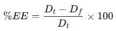
Where,
Dt – Total amount (entrapped)
Df – Amount offree (un entrapped) drug in the sample.
In vitro drug release studies:
Drugs are often released from cubosomes using phosphate buffer saline dialysis membranes. The release pattern usually exhibits a biphasic profile, with a preliminary burst release after a sustained phase, because of diffusion through the lipid bilayer. Their regulated release behavior validates their potential for prolonged pharmacological activity (24,30).
Drugs Formulated Using Cubosomes:
Table 5. Drugs Formulated Using Cubosomes
|
Drug |
Therapeutic Class / Use |
Route of Administration |
Key Outcome |
Reference |
|
Colchicine (rat) |
Anti-inflammatory |
Transdermal |
Enhanced skin permeation, sustained release |
(25) |
|
Antimicrobial peptides |
Antibacterial |
Topical |
Increased stability and bactericidal activity |
(4) |
|
Dexamethasone (rabbit eye ) |
Corticosteroid |
Ocular / Topical |
Improved bioavailability and sustained effect |
(12,30) |
|
Diclofenac sodium |
NSAID |
Percutaneous |
Prolonged anti-inflammatory action |
(15) |
|
Indomethacin |
NSAID |
Ocular |
Improved corneal permeation |
((12)) |
|
Erythromycin |
Antibacterial |
Topical |
Enhanced penetration, reduced irritation |
((12)) |
|
Dapsone |
Anti-inflammatory / antibacterial |
Topical |
Sustained release, better efficacy |
((12)) |
|
5-Fluorouracil |
Anticancer |
Parenteral (Liver) |
Liver targeting, prolonged circulation |
(38) |
|
Dacarbazine (melanoma animal) |
Anticancer |
Parenteral |
Reduced systemic toxicity |
((12)) |
|
Simvastatin (dogs) |
Lipid-lowering |
Oral |
Improved solubility and absorption |
((12)) |
|
Ibuprofen (beagle dogs) |
NSAID |
Oral |
Sustained release, enhanced bioavailability |
((12)) |
|
Resveratrol |
Neuroprotective |
Nasal |
Improved brain targeting |
((12)) |
Disadvantages of Cubosomes:
• They are difficult to create on a large scale due to the cubic phase's high viscosity (39).
• They have a low trapping efficiency for pharmacological compounds that are soluble in water because of the high water content of their structure.(6)
Comparative table
Table 6. Comparision between phytosome, ethosome, cubosome
|
S.NO |
PARAMETER |
PHYTOSOME |
ETHOSOME |
CUBOSOME |
|
|
Basic Definition |
Complex of phytoconstituents with phospholipids |
Soft, flexible lipid vesicles with high ethanol |
Nanostructured bicontinuous cubic lipid carriers |
|
|
Main Components |
Plant extract + phospholipids (mostly phosphatidylcholine) |
Phospholipids + ethanol (20–45%) + water |
Glyceryl monooleate / phytantriol + stabilizer (Poloxamer) + water |
|
|
Structure Type |
Phospholipid-bonded complex |
Unilamellar / multilamellar vesicles |
3D cubic liquid crystalline structure with aqueous channels |
|
|
Particle Size |
50 nm to 100 µm |
30 – 400 nm |
10 – 500 nm |
|
|
Drug Encapsulation Suitability |
Mainly plant-based hydrophobic phytochemicals |
Both hydrophilic & lipophilic drugs |
Hydrophilic, hydrophobic & amphiphilic drugs |
|
|
Mechanism of Action |
Improved membrane permeability via lipid compatibility |
Ethanol disrupts skin lipids → deep dermal penetration |
Sustained diffusion through interlinked aqueous channels |
|
|
Route of Administration |
Mostly oral, topical |
Dermal & transdermal |
Oral, transdermal, ocular, injectable |
|
|
Advantages |
High bioavailability of herbal compounds; better solubility |
Excellent skin penetration; non-invasive systemic delivery |
Sustained release; high drug loading; protects sensitive molecules |
|
|
Limitations |
Works mainly for phytochemicals; expensive phospholipids |
Skin irritation risk due to high ethanol |
Difficult large-scale production; low entrapment for water-soluble drugs |
|
|
Key Applications |
Liver diseases, cancer, inflammation, antioxidant therapy |
Anti-inflammatory, hormone therapy, antimicrobial, dermatology |
Cancer therapy, anti-inflammatory, ocular drugs, peptides, oral bioavailability enhancement |
|
|
Example Drugs / Extracts |
Silibinin, Curcumin, Ginkgo, Rubia cordifolia |
Ketoprofen, Acyclovir, Estradiol, Testosterone |
5-FU, Diclofenac, Dexamethasone, Colchicine, Ibuprofen |
|
|
Entrapment Efficiency |
Moderate |
55–90% |
31–90% (higher for lipophilic drugs) |
|
|
Release Pattern |
Faster release than liposomes |
Controlled + improved penetration |
Controlled / sustained (Higuchi diffusion) |
|
|
Commercial Use |
Herbal nutraceuticals & phytomedicines |
Cosmetics & transdermal patches |
Under development; limited marketed formulations |
DISCUSSION
The development of nanocarrier-based drug delivery systems has significantly advanced the bioavailability, therapeutic efficiency, and targeting capabilities of a wide range of bioactive compounds. Among the novel vesicular carriers explored in recent years, phytosomes, ethosomes, and cubosomes represent three distinct yet highly promising platforms with unique physicochemical characteristics and clinical relevance. Although all three systems aim to improve drug absorption and therapeutic outcome, the comparison reveals substantial differences in their composition, mechanism of drug permeation, release behavior, and applicability. Phytosomes are primarily designed to enhance the bioavailability of plant-derived phytoconstituents. The formation of a stoichiometric complex between phytochemicals and phospholipids significantly improves the lipophilicity and membrane permeation of herbal bioactives, thereby overcoming the poor solubility and low intestinal permeability associated with traditional herbal extracts. Their clinical efficiency has been extensively demonstrated in the treatment of chronic diseases, including liver disorders, cancer, inflammation, dyslipidemia, and cardiovascular complications. However, phytosome particle size ranges from nanometer to the micrometer scale, and the tendency of complexes to aggregate may limit their transport through barriers such as the skin and mucosa without additional size-reduction methods. Ethosomes, in contrast, have been engineered specifically for dermal and transdermal drug delivery. Their characteristic high ethanol content imparts membrane fluidity and deformability, facilitating deep penetration across the stratum corneum and enabling the systemic administration of drugs through a non-invasive route. A major advantage of ethosomes is to encapsulate both hydrophilic and lipophilic compounds while achieving significantly greater skin deposition compared to conventional liposomes. This makes ethosomes particularly valuable in managing inflammation, infections, hormonal disorders, dermatological conditions, and cosmetic applications. Nevertheless, the high ethanol concentration may cause irritation in sensitive skin and limit suitability for prolonged application in certain patients. Cubosomes represent a more structurally complex class of nanocarriers based on a bicontinuous cubic liquid crystalline architecture. Their three-dimensional lipid bilayer network creates extensive aqueous channels that enable the co-encapsulation of hydrophilic, lipophilic, and amphiphilic molecules with high loading capacity. This structural arrangement supports sustained and controlled drug release following Fickian or Higuchi diffusion, making cubosomes suitable for long-term delivery in transdermal, ocular, oral, and injectable applications. Furthermore, cubosomes demonstrate high bioadhesiveness, membrane affinity, and protection of fragile biomolecules such as peptides and nucleic acids. However, challenges remain in large-scale production owing to high viscosity of the bulk cubic phase and comparatively lower entrapment efficiency for highly water-soluble drugs. Overall, the comparison highlights that the choice of nanocarrier should be dictated by the therapeutic objective and the physicochemical profile of the drug. Phytosomes offer the greatest benefit for poorly soluble herbal molecules requiring improved oral bioavailability, whereas ethosomes are superior for non-invasive transdermal therapy with deep dermal penetration. Cubosomes, owing to their unique nanostructure and sustained-release kinetics, are best suited for controlled delivery and targeting applications across multiple administration routes. Collectively, these systems demonstrate complementary strengths rather than competition, emphasizing that the future of nanomedicine lies in application-specific design rather than universal drug-delivery platforms.
CONCLUSION
Nanocarrier-based drug delivery systems have appeared as a innovative strategy to overcome the inherent limitations of conventional dosage forms, including poor solubility, low permeability, metabolic instability, and short systemic retention of therapeutic molecules. Phytosomes, ethosomes, and cubosomes represent three innovative and highly promising lipid-based platforms, each offering distinct advantages based on their structural design, drug-loading profiles, and mechanisms of delivery. Phytosomes significantly enhance the lipophilicity and biological absorption of plant-derived phytoconstituents by creating a stable complex with phospholipids. This improvement in permeability and bioavailability has enabled phytosomes to address the long-standing challenge of delivering herbal actives for the treatment of chronic disorders such as cancer, metabolic diseases, and inflammatory conditions. Ethosomes, with their ethanol-rich flexible bilayer, introduce a breakthrough approach for dermal and transdermal delivery by achieving deep skin penetration while maintaining non-invasive systemic transport. Their effectiveness across anti-inflammatory, antimicrobial, hormonal, and dermatological therapies underscores their clinical versatility. Cubosomes, characterized by a bicontinuous cubic liquid crystalline phase with interconnected aqueous channels, offer exceptional drug-loading efficiency and controlled release kinetics. Their suitability for oral, ocular, transdermal, and injectable routes positions them at the forefront of long-acting and targeted drug delivery research.
Although each nanocarrier exhibits distinct limitations—such as aggregation tendencies in phytosomes, potential ethanol-induced irritation in ethosomes, and large-scale manufacturing challenges in cubosomes—their combined progress marks a significant leap forward in nanomedicine. Collectively, these systems demonstrate that no single platform is universally superior; rather, the optimal carrier is determined by the physiochemical properties of the drug and the intended therapeutic outcome. In conclusion, this review highlights phytosomes, ethosomes, and cubosomes collectively represent the next generation of drug-delivery technologies capable of enhancing therapeutic efficacy, reducing dosing frequency, improving patient compliance, and enabling targeted intervention. Continued research into formulation optimization, stability enhancement, and clinical translation will play a crucial role in unlocking their full potential and advancing them toward broader pharmaceutical and clinical applications.
REFERENCES

Dharani Priya B., Chokkalingam P. L., Sneha S., Subi Shankar K., Sudhan R., Varrun Sree Ram K., Advanced Nanocarriers in Drug Delivery: A Comparative Review of Phytosomes, Ethosomes, And Cubosomes, Int. J. of Pharm. Sci., 2026, Vol 4, Issue 2, 3388-3413. https://doi.org/10.5281/zenodo.18721980
 10.5281/zenodo.18721980
