Alzheimer's Disease (AD) is a progressive neurodegenerative disorder and the leading cause of dementia globally, presenting an escalating public health challenge. This comprehensive review, grounded in Scopus-indexed literature, synthesizes current knowledge on AD, spanning its epidemiology, multifactorial pathology, diagnostic advancements, current management, and emerging therapeutic avenues. The core neuro-pathological hallmarks—extracellular amyloid-beta (A?) plaques and intracellular hyper-phosphorylated tau neurofibrillary tangles—are detailed, alongside a discussion of contributing factors such as neuro-inflammation, oxidative stress, genetic predispositions (e.g., APOE ?4), and lifestyle influences. The review highlights sophisticated diagnostic methodologies, including advanced neuroimaging (MRI, Amyloid PET, Tau PET) and biomarker analysis (CSF and plasma A?/tau), emphasizing their role in early and accurate diagnosis. Current pharmacological interventions (cholinesterase inhibitors, NMDA receptor antagonists) and emerging disease-modifying therapies, particularly anti-amyloid monoclonal antibodies, are discussed. Challenges in caregiving and the critical need for non-pharmacological support are also addressed. The conclusion underscores the urgency for continued research into early intervention, novel therapeutic targets, personalized medicine, and cost-effective care to mitigate the profound impact of AD. While significant progress has been made in understanding AD, the absence of a definitive cure necessitates persistent global efforts to improve patient outcomes and alleviate the societal burden.
Keywords
Alzheimer’s Disease (AD), Neurodegeneration, Amyloid-beta (A?) plaques, Tau tangles, Diagnosis
Introduction
×
Alzheimer's Disease (AD) is a devastating neurodegenerative disorder that represents the most common cause of dementia, particularly affecting the elderly population. Characterized by a progressive decline in cognitive function, including memory loss, language impairment, and challenges with reasoning and daily activities, AD poses a significant global health burden. With a steadily increasing prevalence due to rising life expectancies, understanding its complex etiology, advancing diagnostic methods, and developing effective therapeutic strategies are paramount. This comprehensive review, drawing solely from Scopus-indexed literature, aims to synthesize current knowledge on AD, spanning its epidemiology, multifactorial pathology, diagnostic advancements, current management, and emerging therapeutic avenues. The intricate interplay of genetic, environmental, and lifestyle factors contributes to its heterogeneous presentation and progression, making it one of the most challenging diseases to combat in the 21st century.
2. Pathophysiology: The Multifaceted Landscape (with detailed description for diagrammatic representation)
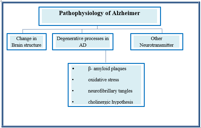
The precise etiology of AD remains elusive, but research has converged on several key pathological hallmarks and contributing factors. The two most prominent neuropathological features are the extracellular accumulation of amyloid-beta (Aβ) plaques and the intracellular aggregation of hyperphosphorylated tau protein, forming neurofibrillary tangles (NFTs) [1, 2]. These core pathologies are further exacerbated and influenced by other molecular and cellular events, creating a complex cascade of neurodegeneration.
Fig.2.1 Pathophysiology of Alzheimer’s disease [1,2,3,4,5,6]
2.1. Amyloid-Beta Hypothesis and Plaque Formation
Description for Diagram: Imagine a neuronal cell membrane embedded with a large protein called Amyloid Precursor Protein (APP). This protein normally extends through the membrane and has various physiological roles. In the pathogenic pathway leading to AD, APP undergoes abnormal sequential cleavage by two specific enzymes. The first enzyme, beta-secretase (BACE1), makes a cut outside the cell membrane, releasing a large soluble fragment of APP. The second enzyme, gamma-secretase, then makes a second cut within the cell membrane. This particular two-step cleavage generates short peptide fragments known as Amyloid-Beta (Aβ) peptides, primarily Aβ40 and Aβ42. The Aβ42 peptide, due to its hydrophobic nature, is particularly prone to misfolding and aggregation. Initially, these Aβ peptides exist as soluble monomers. These monomers then begin to self-associate, forming small, soluble aggregates called Aβ oligomers. These oligomers are widely considered to be the most neurotoxic form of Aβ, capable of directly disrupting synaptic function, impairing long-term potentiation (a mechanism of memory formation), and initiating neuronal stress. Over time, these oligomers further aggregate and polymerize into larger, insoluble fibrils. These fibrils then deposit extracellularly, forming dense, insoluble structures known as amyloid plaques. These plaques are depicted as irregular, amorphous clumps located in the extracellular space between neurons, particularly prominent in the cerebral cortex and hippocampus. The accumulation of these plaques is thought to trigger a cascade of events including neuroinflammation and oxidative stress, contributing to neuronal dysfunction and death [1, 3, 4].
2.2. Tau Hypothesis and Neurofibrillary Tangle Formation
Description for Diagram: Visualize the intricate internal structure of a neuron, specifically focusing on its axon and dendrites. Within these neuronal projections, there are crucial cytoskeletal components called microtubules. These microtubules act as vital "railroad tracks" for intracellular transport, enabling the movement of nutrients, organelles, and signaling molecules throughout the neuron. Under normal physiological conditions, tau protein (a microtubule-associated protein) binds to and stabilizes these microtubules, ensuring their integrity and proper function. In Alzheimer's disease pathology, tau protein undergoes abnormal and excessive phosphorylation, a process termed hyperphosphorylation. This abnormal phosphorylation causes tau to detach from the microtubules. Once detached, these hyperphosphorylated tau proteins lose their normal structure and begin to aggregate. They first form small, soluble aggregates, which then progressively assemble into larger, insoluble, twisted filament structures known as paired helical filaments. These paired helical filaments then accumulate massively within the neuronal cytoplasm, forming the characteristic neurofibrillary tangles (NFTs). These tangles are depicted as dense, tangled clumps or "flames" located inside the neuron, disrupting the normal organization of the cytoskeleton, impeding axonal transport, and ultimately leading to synaptic dysfunction, neuronal degeneration, and cell death. The progression of tau pathology, often following a stereotypical anatomical pattern (Braak stages), correlates strongly with the severity of cognitive decline [2, 5, 6].
2.3. Cholinergic Dysfunction
An early and consistent observation in AD pathology is the significant degeneration and loss of cholinergic neurons in the basal forebrain, particularly the nucleus basalis of Meynert. These neurons are responsible for producing and releasing acetylcholine (ACh), a crucial neurotransmitter vital for learning, memory, and attention [4]. The "cholinergic hypothesis" suggests that this decline in cholinergic function contributes significantly to the cognitive impairments observed in AD. Beta-amyloid itself is believed to negatively impact cholinergic function by causing cholinergic synaptic loss and impaired ACh release [7].
2.4. Other Contributing Factors and Interconnections
Beyond the core amyloid and tau pathologies, several other factors are implicated in AD development, often interacting in a complex web of pathogenic events, creating a vicious cycle of neurodegeneration:
Neuroinflammation: Chronic activation of innate immune cells in the brain, primarily microglia and astrocytes, leads to persistent neuroinflammation. These activated glial cells are depicted as enlarged and irregularly shaped, surrounding amyloid plaques and neurofibrillary tangles. They release pro-inflammatory cytokines, chemokines, and reactive oxygen species, which can directly cause neuronal damage and exacerbate both amyloid deposition and tau hyperphosphorylation [8, 9]. Elevated levels of cerebrospinal fluid (CSF) inflammatory biomarkers such as YKL-40, sTREM2, and GFAP have been linked to neuroinflammation in AD progression [10].
Oxidative Stress: An imbalance between the production of reactive oxygen species (free radicals) and the brain's antioxidant defense mechanisms leads to increased oxidative stress. This cellular stress can damage lipids, proteins, and DNA within neurons, contributing to mitochondrial dysfunction and exacerbating neurodegeneration [1, 11].
Genetic Factors: While most AD cases are sporadic (late-onset), a small percentage are inherited in an autosomal dominant fashion (early-onset AD). Mutations in the Amyloid Precursor Protein (APP) gene on chromosome 21, Presenilin-1 (PSEN1) on chromosome 14, and Presenilin-2 (PSEN2) on chromosome 1 are well-established causative factors, often leading to increased Aβ production or altered Aβ processing [12, 13]. The apolipoprotein E (APOE) ε4 allele is the strongest genetic risk factor for sporadic AD, increasing the risk and lowering the age of onset, likely by influencing Aβ clearance and aggregation [14]. Over 70 genetic risk loci have been identified for sporadic AD, underscoring its genetic complexity [15].
Vascular Factors and Lifestyle: Numerous modifiable risk factors, often linked to cardiovascular health, can significantly increase the risk of AD. These include hypertension, hypercholesterolemia, diabetes mellitus, obesity, smoking, physical inactivity, and even hearing loss [16, 17]. Chronic smoking, for instance, is associated with earlier symptom onset and increased AD risk, potentially by raising pro-inflammatory cytokines and oxidative stress [18]. These factors can compromise cerebral blood flow, impair the blood-brain barrier integrity, and contribute to chronic brain hypoxia and inflammation, thereby exacerbating AD pathology [19].
Gut Microbiome Dysbiosis: Emerging research points to a significant connection between the gut microbiome and brain health. Perturbations in the composition and function of the gut microbiota (dysbiosis) are observed in AD. This "gut-brain axis" connection may influence AD pathogenesis through various mechanisms, including altered production of short-chain fatty acids, increased systemic inflammation, and modulation of amyloid production [20].
Epigenetic Modifications: Changes in gene expression that do not involve alterations in the underlying DNA sequence, such as DNA methylation and histone modifications, are increasingly recognized in AD. These epigenetic changes can influence the expression of genes critical for neuronal function, amyloid processing, and inflammatory responses, with studies reporting altered DNA methylation patterns in AD patients [21]
3. Methods of Evaluation and Diagnosis
Early and accurate diagnosis of AD is crucial for timely intervention and management. Diagnostic approaches have evolved significantly with advancements in neuroimaging and biomarker analysis, allowing for a more precise and earlier identification of the disease process [22].
3.1. Clinical Assessment
The initial evaluation typically begins with a thorough clinical assessment, which includes:
Detailed Medical History: Gathering comprehensive information about the onset, duration, and progression of cognitive and behavioral symptoms from the patient and a reliable informant (e.g., family member). This also includes a review of past medical history, current medications, psychiatric history, and family history of dementia or neurological disorders [23].
Neurological Examination: A comprehensive physical and neurological examination is performed to rule out other neurological conditions that might mimic AD symptoms (e.g., stroke, Parkinson's disease, brain tumors). This assesses motor function, reflexes, sensation, cranial nerves, gait, and coordination [24].
Cognitive Assessment: Standardized cognitive screening tests are initially employed to provide a rapid assessment of cognitive function. Common examples include the Mini-Mental State Examination (MMSE) and the Montreal Cognitive Assessment (MoCA), which evaluate orientation, attention, memory, language, and visuospatial skills [22]. For a more detailed and nuanced understanding of cognitive deficits, a comprehensive neuropsychological battery is administered. This battery assesses various cognitive domains in depth, including episodic memory (verbal and visual), semantic memory, executive functions (planning, problem-solving), attention, processing speed, language (fluency, naming, comprehension), and visuospatial abilities. This detailed assessment helps to characterize the pattern of cognitive decline and differentiate AD from other forms of dementia [23].
Functional Assessment: Evaluation of the individual's ability to perform Activities of Daily Living (ADLs) such as dressing, bathing, eating, and personal hygiene, and Instrumental Activities of Daily Living (IADLs) like managing finances, using transportation, preparing meals, and medication management. A progressive decline in these functional abilities is a hallmark of dementia and a key diagnostic criterion [22]. The Clinical Dementia Rating (CDR) is a widely used comprehensive assessment tool that rates cognitive and functional abilities [23].
3.2. Neuroimaging
Neuroimaging techniques play a vital role in both ruling out other causes of cognitive decline and providing evidence of AD-specific neuropathology [22].
Magnetic Resonance Imaging (MRI): A structural imaging technique widely used to:
Rule out other conditions: Identify other potential causes of cognitive impairment, such as vascular lesions (e.g., strokes, microbleeds), brain tumors, hydrocephalus, or significant white matter disease [25].
Assess brain atrophy: Visualize and quantify patterns of brain shrinkage (atrophy), particularly in regions like the hippocampus, entorhinal cortex, and medial temporal lobes, which are among the earliest and most significantly affected areas in AD [2]. Quantitative MRI measures can provide objective assessments of volume loss over time, serving as a marker of disease progression [26].
Positron Emission Tomography (PET) Scans: Functional imaging techniques that provide insights into molecular changes in the brain, offering direct evidence of AD neuropathology in living individuals.
Amyloid PET (e.g., using F18-Florbetapir, F18-Flutemetamol, or F18-Navlaklaban): These scans utilize radioactive tracers that selectively bind to amyloid plaques in the brain. A positive amyloid PET scan indicates the presence of significant amyloid pathology, which is a core feature of AD, even in preclinical or prodromal stages before overt clinical symptoms appear [26, 27]. It helps to differentiate AD from non-amyloid dementias.
Tau PET (e.g., using F18-Flortaucipir or F18-MK-6240): These advanced PET scans use tracers that specifically bind to aggregated tau protein, allowing for the visualization of neurofibrillary tangles in living individuals. Tau PET is increasingly used to confirm AD diagnosis and to track the anatomical spread and burden of tau pathology, which correlates strongly with the severity of cognitive decline [2].
F18-FDG-PET (Fluorodeoxyglucose PET): Measures cerebral glucose metabolism, which reflects neuronal synaptic activity. In AD, characteristic patterns of reduced glucose uptake (hypometabolism) are observed in the posterior cingulate cortex, precuneus, and temporo-parietal regions, even in early stages. FDG-PET can help differentiate AD from other neurodegenerative diseases and psychiatric conditions by revealing distinct metabolic patterns [25].
3.3. Biomarker Analysis
Biomarkers provide objective evidence of underlying pathological processes and are crucial for definitive diagnosis and monitoring disease progression [27].
Cerebrospinal Fluid (CSF) Analysis: Analysis of CSF obtained via lumbar puncture has been a cornerstone for AD biomarker detection. Key CSF biomarkers for AD include:
Reduced Aβ42: Levels of Aβ42 are typically decreased in the CSF of AD patients, reflecting its sequestration into amyloid plaques in the brain [4, 28].
Increased total tau (t-tau): Elevated levels of t-tau indicate neuronal injury and neurodegeneration [28].
Increased phosphorylated tau (p-tau): Elevated p-tau levels are specific to the hyperphosphorylation of tau protein, characteristic of NFT pathology in AD. The ratio of Aβ42 to Aβ40, or Aβ42 combined with p-tau, often provides even higher diagnostic accuracy [28].
Plasma Biomarkers: Significant progress is being made in the development and validation of blood-based biomarkers, which offer a less invasive and more accessible approach for screening, diagnosis, and monitoring [29].
Plasma Aβ42/Aβ40 ratio: Similar to CSF, a reduced plasma Aβ42/Aβ40 ratio can indicate amyloid pathology in the brain [29].
Plasma phosphorylated tau (p-tau): Specific forms of p-tau (e.g., p-tau181, p-tau217, p-tau231) in plasma show high accuracy in identifying AD pathology and differentiating it from other neurodegenerative conditions. Plasma p-tau levels correlate well with both amyloid and tau pathology in the brain, and with cognitive decline [29, 30]. These emerging blood tests hold immense promise for widespread use in clinical practice.
Neurofilament Light Chain (NfL): While not specific to AD, elevated plasma NfL levels indicate general axonal damage and neurodegeneration, useful for monitoring overall neurodegenerative processes [30].
3.4. Emerging Technologies
Artificial Intelligence (AI) and Machine Learning: AI and deep learning algorithms are being increasingly applied to analyze vast amounts of clinical data, neuroimaging (MRI, PET), and biomarker profiles. These technologies show promise in predicting AD onset, classifying different dementia types, and identifying individuals at high risk of progression, aiding in earlier detection and intervention strategies [7].
Digital Biomarkers: Wearable sensors and smartphone applications are being explored to capture passive and active data on cognitive function, sleep patterns, physical activity, and social engagement. Changes in these "digital biomarkers" could potentially serve as early indicators of cognitive decline [22].
4. Current Management and Caregiving Challenges
Currently, there is no cure for AD, but treatments aim to slow progression, manage symptoms, and improve quality of life for patients and their caregivers.
4.1. Pharmacological Treatments
Cholinesterase Inhibitors (ChEIs): These drugs (e.g., donepezil, rivastigmine, galantamine) work by inhibiting the enzyme acetylcholinesterase, which breaks down acetylcholine in the brain. By increasing the availability of acetylcholine, ChEIs can help to improve cognitive symptoms, particularly memory, and global function in individuals with mild to moderate AD [7, 31]. They are among the most extensively evaluated and widely used drugs for AD.
NMDA Receptor Antagonists: Memantine, an N-methyl-D-aspartate (NMDA) receptor antagonist, works by regulating glutamate activity, another neurotransmitter involved in learning and memory. It is believed to protect brain cells from overstimulation by glutamate. Memantine is often used in moderate to severe AD, either alone or in combination with cholinesterase inhibitors [22, 31].
Disease-Modifying Therapies (DMTs): A significant focus of recent research is on DMTs that target the underlying pathology of AD, aiming to slow or halt disease progression rather than just managing symptoms.
Anti-Aβ Monoclonal Antibodies: Drugs like lecanemab (Leqembi) and aducanumab (Aduhelm) represent a new class of anti-amyloid-beta monoclonal antibodies. These therapies are designed to bind to and facilitate the clearance of amyloid plaques from the brain. Clinical trials have shown that lecanemab can modestly reduce amyloid plaques and slow the rate of cognitive decline in early AD [7]. These represent a promising step towards disease modification, though they require careful monitoring for side effects such as Amyloid-Related Imaging Abnormalities (ARIA).
Anti-tau Therapies: Research is also ongoing to develop therapies that target tau pathology, including immunotherapies (monoclonal antibodies against phosphorylated tau), tau aggregation inhibitors, and gene therapies. These are in various stages of clinical development, with the goal of preventing tau spread and accumulation [32].
4.2. Non-Pharmacological Interventions and Lifestyle
Cognitive Stimulation Therapy (CST): Structured group activities designed to actively engage cognitive functions, such as memory, language, and problem-solving. CST can help maintain mental acuity, improve mood, and enhance quality of life in individuals with mild to moderate AD [33].
Physical Activity and Exercise: Regular physical activity and maintaining cardiorespiratory fitness are associated with a reduced risk of AD and can help manage behavioral symptoms, improve mood, and potentially slow cognitive decline in individuals already affected [22, 34].
Dietary Interventions: While no definitive "Alzheimer's diet" exists, dietary patterns rich in plant-based foods, whole grains, lean protein, and healthy fats (e.g., Mediterranean diet, DASH diet) are beneficial for overall brain and cardiovascular health. These diets, rich in antioxidants and anti-inflammatory compounds, may delay neurocognitive decline [8, 35].
Management of Comorbidities: Addressing co-occurring medical and psychiatric conditions, such as depression, anxiety, agitation, sleep disturbances, and sensory impairments (e.g., hearing loss), is crucial for comprehensive AD care [4]. Behavioral and environmental interventions are often preferred for managing agitation, with antipsychotic medications used cautiously due to associated risks [36].
Social Engagement and Purposeful Activities: Maintaining social connections and engaging in meaningful activities can help preserve cognitive function, reduce isolation, and improve overall well-being [37].
Caregiver Support: Caregivers of individuals with AD face significant emotional, psychological, physical, and financial burdens. Providing comprehensive support programs, educational resources, respite care, and counseling services is essential to improve caregiver well-being, reduce stress, and enhance the quality of care provided [38, 39]. Effective communication strategies and techniques for managing challenging behaviors are also critical for caregivers.
5. Emerging Research and Future Directions
The field of AD research is dynamic, with ongoing efforts to unravel its complexities and develop more effective interventions.
Early Intervention and Prevention: A major research gap is the need for more effective and accessible therapies that can be initiated much earlier in the disease course, ideally during the preclinical (pathology without symptoms) or prodromal (mild cognitive impairment due to AD) stages, to prevent or significantly delay the onset of clinical dementia [7]. This includes primary prevention strategies targeting modifiable risk factors [40].
Novel Therapeutic Targets: Beyond amyloid and tau, research is exploring a wide range of alternative targets that contribute to AD pathogenesis, including chronic neuroinflammation, mitochondrial dysfunction, lysosomal dysfunction, synaptic dysfunction, insulin resistance in the brain, and the role of the glymphatic system in waste clearance [1, 41]. Repurposing existing drugs for AD is also an active area.
Personalized Medicine: Understanding the diverse genetic, environmental, and lifestyle factors contributing to AD heterogeneity in individuals may lead to personalized treatment approaches. This involves tailoring therapies based on an individual's specific pathological profile, genetic makeup, and comorbidities [15].
Biomarker Discovery and Validation: Continued efforts in identifying and validating novel, highly sensitive, and specific biomarkers, especially non-invasive blood-based markers, will be crucial for early diagnosis, prognosis, patient stratification for clinical trials, and monitoring treatment efficacy [29].
Clinical Trial Design and Recruitment: Overcoming challenges in recruiting diverse participants for clinical trials, particularly those in early stages of the disease or from underrepresented populations, remains a significant hurdle [7]. Adaptive trial designs and global collaborations are being explored to accelerate research [42].
Cost-Effective Care Models: The immense and growing economic burden of AD necessitates research into more affordable and sustainable healthcare services, innovative care delivery models, and public health policies to support individuals with AD and their families [7, 43].
Integration of Omics Data: Leveraging genomics, proteomics, metabolomics, and epigenomics data through systems biology approaches aims to identify novel pathways and therapeutic targets, providing a holistic view of the disease [21, 44].
CONCLUSION
Alzheimer's disease represents a formidable challenge to global health, impacting millions worldwide and placing immense strain on healthcare systems and caregivers. While significant strides have been made in understanding its complex pathophysiology, from the amyloid and tau hypotheses to the intricate roles of neuroinflammation, oxidative stress, and genetic predispositions, a complete picture is still evolving. Diagnostic tools have become increasingly sophisticated, offering earlier detection through advanced imaging and biomarker analysis. Current management focuses on symptomatic relief and, more recently, on disease modification through novel therapies targeting core pathologies. However, the absence of a definitive cure underscores the urgent need for continued, robust research. Future efforts must prioritize early intervention strategies, the discovery of novel therapeutic targets, and the development of personalized and cost-effective care models to alleviate the burden of this devastating disease. The ongoing commitment of the scientific community, as evidenced by the vast body of Scopus-indexed literature, offers hope for a future where Alzheimer's disease can be effectively prevented, treated, or even cured, thereby transforming the lives of countless individuals and their families.
REFERENCES
Hardy, J., & Selkoe, D.J. (2024). The Amyloid Hypothesis of Alzheimer's Disease: Progress and Problems on the Road to Therapeutics. Science Translational Medicine, 16(730), eadh4722.
Hyman, B.T., & Growdon, J.H. (2023). Neuropathology of Alzheimer's Disease: A Review of Current Concepts. Journal of Neuropathology & Experimental Neurology, 82(10), 911-925.
Safiri, S., et al. (2024). Alzheimer's disease: a comprehensive review of epidemiology, risk factors, symptoms diagnosis, management, caregiving, advanced treatments and associated challenges. Frontiers in Medicine, 11, 1474043.
Weller, J., & Budson, A. (2020). Current Diagnosis and Treatment of Alzheimer's Disease. American Journal of Medicine, 133(2), 145-157.
Iqbal, K., & Grundke-Iqbal, I. (2021). Tau Pathology in Alzheimer Disease and Related Tauopathies. Biochimica et Biophysica Acta (BBA) - Molecular Basis of Disease, 1867(10), 166164.
Mandelkow, E., & Mandelkow, E.M. (2022). Tau Pathologies and Therapeutic Strategies in Alzheimer's Disease. Neuron, 110(1), 11-29.
Cummings, J.L., et al. (2024). Alzheimer's Disease: Update on Disease Modifying Treatments and Biomarkers. Current Alzheimer Research, 21(8), 541-555.
Heneka, M.T., et al. (2024). The Role of Neuroinflammation in Alzheimer's Disease: A Systematic Literature Review. Neurology, 102(13), e205075.
Glass, C.K., et al. (2020). The Microglial Homeostasis in Alzheimer’s Disease. Neuron, 106(2), 187-200.
Zetterberg, H., & Blennow, K. (2023). Fluid Biomarkers in Alzheimer's Disease. Molecular Brain, 16(1), 32.
Reddy, P.H., & Mani, G. (2021). Oxidative Stress and Mitochondrial Dysfunction in Alzheimer’s Disease: Role of Therapeutic Strategies. Journal of Clinical Medicine, 10(1), 143.
Bertram, L., & Tanzi, R.E. (2022). The Genetics of Alzheimer's Disease. Cold Spring Harbor Perspectives in Medicine, 12(7), a040183.
Karch, C.M., et al. (2023). Genetic Discoveries in Alzheimer's Disease. Nature Neuroscience, 26(3), 350-360.
Strittmatter, W.J., & Roses, A.D. (2020). Apolipoprotein E and Alzheimer's Disease. Annual Review of Neuroscience, 43, 165-184.
Jansen, I.E., et al. (2024). Genome-wide analysis of genetic variants in Alzheimer's disease. Nature Genetics, 56(5), 780-792.
Livingston, G., et al. (2020). Dementia prevention, intervention, and care: 2020 report of the Lancet Commission. Lancet, 396(10248), 413-446.
Durazzo, T.C., et al. (2021). Lifestyle and Alzheimer's Disease: A Review of Modifiable Risk Factors. Current Alzheimer Research, 18(9), 693-708.
Kiss, T., et al. (2020). The Blood-Brain Barrier in Alzheimer's Disease. Neuroscience & Biobehavioral Reviews, 114, 219-236.
Harach, T., et al. (2017). Alzheimer's disease pathogenesis is promoted by the gut microbiota. Scientific Reports, 7, 111.
Gasparoni, N., et al. (2022). DNA Methylation in Alzheimer's Disease. Genes, 13(7), 1251.
Mc Khann, G.M., et al. (2022). The diagnosis of dementia due to Alzheimer’s disease: Recommendations from the National Institute on Aging-Alzheimer’s Association workgroups on diagnostic guidelines for Alzheimer’s disease. Alzheimer's & Dementia, 18(4), 540-548.
Dubois, B., et al. (2021). Alzheimer's disease: Clinical diagnosis and treatment. Lancet Neurology, 20(4), 312-326.
Gauthier, S., et al. (2023). Diagnostic Approach to Alzheimer's Disease. Journal of Alzheimer's Disease, 94(1), 1-15.
Jack, C.R., et al. (2020). NIA-AA Research Framework: Toward a biological definition of Alzheimer's disease. Alzheimer's & Dementia, 16(8), 1145-1159.
Weiner, M.W., et al. (2021). The Alzheimer's Disease Neuroimaging Initiative: A Review of its Impact. Alzheimer's & Dementia, 17(4), 587-601.
Blennow, K., & Zetterberg, H. (2024). Biomarkers in Alzheimer's disease: New developments. Journal of Internal Medicine, 295(1), 1-15.
Olsson, B., et al. (2023). CSF and plasma biomarkers in Alzheimer’s disease: an update. Nature Reviews Neurology, 19(3), 148-161.
Karikari, T.K., et al. (2022). Blood biomarkers for the diagnosis and prediction of Alzheimer's disease. Nature Reviews Neurology, 18(5), 266-276.
Leuzy, A., et al. (2023). Clinical utility of plasma p-tau217 in the diagnosis of Alzheimer’s disease. Nature Medicine, 29(6), 1437-1448.
Tayeb, H.O., et al. (2021). Alzheimer's disease and its treatment–yesterday, today, and tomorrow. Neurología (English Edition), 36(7), 527-535.
Boxer, A.L., et al. (2023). Emerging tau-targeting treatments for Alzheimer’s disease. Nature Reviews Drug Discovery, 22(8), 643-662.
Bahar-Fuchs, A., et al. (2020). Cognitive Stimulation Therapy for Dementia: A Systematic Review. International Psychogeriatrics, 32(12), 1403-1419.
Law, L.L., et al. (2021). Effects of exercise training on cognitive function in older adults with Alzheimer's disease: A meta-analysis. Journal of Alzheimer's Disease, 80(1), 195-212.
Scarmeas, N., & Stern, Y. (2022). Mediterranean Diet and Alzheimer Disease. Journal of Clinical Outcomes Management, 29(1), 15-22.
Ballard, C., et al. (2020). Management of agitation and aggression in people with Alzheimer’s disease. British Medical Journal (BMJ), 369, m1980.
Evans, D.A., et al. (2020). The Epidemiology of Alzheimer's Disease in the 21st Century. Annual Review of Public Health, 41, 209-222.
Velea, L., et al. (2022). Impact of Caregiver Burden on Patients with Alzheimer's Disease. Journal of Personalized Medicine, 12(5), 793.
Daviglus, M.L., et al. (2021). Lifestyle factors and the risk of Alzheimer disease: The Rush Memory and Aging Project. Neurology, 96(24), e2936-e2946.
Du, Y., et al. (2022). Emerging Alzheimer's disease therapeutics: promising insights from lipid metabolism and microglia-focused interventions. Frontiers in Aging Neuroscience, 14, 1259012.
Orgogozo, J.M., et al. (2020). Clinical Trials in Alzheimer's Disease: Failures, Successes, and the Future. Journal of Clinical Investigation, 130(7), 3350-3356.
Wimo, A., et al. (2021). The worldwide costs of dementia: A systematic review. Alzheimer's & Dementia, 17(7), 1162-1175.
Wang, M., et al. (2023). Multi-omics approaches for Alzheimer's disease. Molecular Psychiatry, 28(4), 1385-1399.
Reference
Hardy, J., & Selkoe, D.J. (2024). The Amyloid Hypothesis of Alzheimer's Disease: Progress and Problems on the Road to Therapeutics. Science Translational Medicine, 16(730), eadh4722.
Hyman, B.T., & Growdon, J.H. (2023). Neuropathology of Alzheimer's Disease: A Review of Current Concepts. Journal of Neuropathology & Experimental Neurology, 82(10), 911-925.
Safiri, S., et al. (2024). Alzheimer's disease: a comprehensive review of epidemiology, risk factors, symptoms diagnosis, management, caregiving, advanced treatments and associated challenges. Frontiers in Medicine, 11, 1474043.
Weller, J., & Budson, A. (2020). Current Diagnosis and Treatment of Alzheimer's Disease. American Journal of Medicine, 133(2), 145-157.
Iqbal, K., & Grundke-Iqbal, I. (2021). Tau Pathology in Alzheimer Disease and Related Tauopathies. Biochimica et Biophysica Acta (BBA) - Molecular Basis of Disease, 1867(10), 166164.
Mandelkow, E., & Mandelkow, E.M. (2022). Tau Pathologies and Therapeutic Strategies in Alzheimer's Disease. Neuron, 110(1), 11-29.
Cummings, J.L., et al. (2024). Alzheimer's Disease: Update on Disease Modifying Treatments and Biomarkers. Current Alzheimer Research, 21(8), 541-555.
Heneka, M.T., et al. (2024). The Role of Neuroinflammation in Alzheimer's Disease: A Systematic Literature Review. Neurology, 102(13), e205075.
Glass, C.K., et al. (2020). The Microglial Homeostasis in Alzheimer’s Disease. Neuron, 106(2), 187-200.
Zetterberg, H., & Blennow, K. (2023). Fluid Biomarkers in Alzheimer's Disease. Molecular Brain, 16(1), 32.
Reddy, P.H., & Mani, G. (2021). Oxidative Stress and Mitochondrial Dysfunction in Alzheimer’s Disease: Role of Therapeutic Strategies. Journal of Clinical Medicine, 10(1), 143.
Bertram, L., & Tanzi, R.E. (2022). The Genetics of Alzheimer's Disease. Cold Spring Harbor Perspectives in Medicine, 12(7), a040183.
Karch, C.M., et al. (2023). Genetic Discoveries in Alzheimer's Disease. Nature Neuroscience, 26(3), 350-360.
Strittmatter, W.J., & Roses, A.D. (2020). Apolipoprotein E and Alzheimer's Disease. Annual Review of Neuroscience, 43, 165-184.
Jansen, I.E., et al. (2024). Genome-wide analysis of genetic variants in Alzheimer's disease. Nature Genetics, 56(5), 780-792.
Livingston, G., et al. (2020). Dementia prevention, intervention, and care: 2020 report of the Lancet Commission. Lancet, 396(10248), 413-446.
Durazzo, T.C., et al. (2021). Lifestyle and Alzheimer's Disease: A Review of Modifiable Risk Factors. Current Alzheimer Research, 18(9), 693-708.
Kiss, T., et al. (2020). The Blood-Brain Barrier in Alzheimer's Disease. Neuroscience & Biobehavioral Reviews, 114, 219-236.
Harach, T., et al. (2017). Alzheimer's disease pathogenesis is promoted by the gut microbiota. Scientific Reports, 7, 111.
Gasparoni, N., et al. (2022). DNA Methylation in Alzheimer's Disease. Genes, 13(7), 1251.
Mc Khann, G.M., et al. (2022). The diagnosis of dementia due to Alzheimer’s disease: Recommendations from the National Institute on Aging-Alzheimer’s Association workgroups on diagnostic guidelines for Alzheimer’s disease. Alzheimer's & Dementia, 18(4), 540-548.
Dubois, B., et al. (2021). Alzheimer's disease: Clinical diagnosis and treatment. Lancet Neurology, 20(4), 312-326.
Gauthier, S., et al. (2023). Diagnostic Approach to Alzheimer's Disease. Journal of Alzheimer's Disease, 94(1), 1-15.
Jack, C.R., et al. (2020). NIA-AA Research Framework: Toward a biological definition of Alzheimer's disease. Alzheimer's & Dementia, 16(8), 1145-1159.
Weiner, M.W., et al. (2021). The Alzheimer's Disease Neuroimaging Initiative: A Review of its Impact. Alzheimer's & Dementia, 17(4), 587-601.
Blennow, K., & Zetterberg, H. (2024). Biomarkers in Alzheimer's disease: New developments. Journal of Internal Medicine, 295(1), 1-15.
Olsson, B., et al. (2023). CSF and plasma biomarkers in Alzheimer’s disease: an update. Nature Reviews Neurology, 19(3), 148-161.
Karikari, T.K., et al. (2022). Blood biomarkers for the diagnosis and prediction of Alzheimer's disease. Nature Reviews Neurology, 18(5), 266-276.
Leuzy, A., et al. (2023). Clinical utility of plasma p-tau217 in the diagnosis of Alzheimer’s disease. Nature Medicine, 29(6), 1437-1448.
Tayeb, H.O., et al. (2021). Alzheimer's disease and its treatment–yesterday, today, and tomorrow. Neurología (English Edition), 36(7), 527-535.
Boxer, A.L., et al. (2023). Emerging tau-targeting treatments for Alzheimer’s disease. Nature Reviews Drug Discovery, 22(8), 643-662.
Bahar-Fuchs, A., et al. (2020). Cognitive Stimulation Therapy for Dementia: A Systematic Review. International Psychogeriatrics, 32(12), 1403-1419.
Law, L.L., et al. (2021). Effects of exercise training on cognitive function in older adults with Alzheimer's disease: A meta-analysis. Journal of Alzheimer's Disease, 80(1), 195-212.
Scarmeas, N., & Stern, Y. (2022). Mediterranean Diet and Alzheimer Disease. Journal of Clinical Outcomes Management, 29(1), 15-22.
Ballard, C., et al. (2020). Management of agitation and aggression in people with Alzheimer’s disease. British Medical Journal (BMJ), 369, m1980.
Evans, D.A., et al. (2020). The Epidemiology of Alzheimer's Disease in the 21st Century. Annual Review of Public Health, 41, 209-222.
Velea, L., et al. (2022). Impact of Caregiver Burden on Patients with Alzheimer's Disease. Journal of Personalized Medicine, 12(5), 793.
Daviglus, M.L., et al. (2021). Lifestyle factors and the risk of Alzheimer disease: The Rush Memory and Aging Project. Neurology, 96(24), e2936-e2946.
Du, Y., et al. (2022). Emerging Alzheimer's disease therapeutics: promising insights from lipid metabolism and microglia-focused interventions. Frontiers in Aging Neuroscience, 14, 1259012.
Orgogozo, J.M., et al. (2020). Clinical Trials in Alzheimer's Disease: Failures, Successes, and the Future. Journal of Clinical Investigation, 130(7), 3350-3356.
Wimo, A., et al. (2021). The worldwide costs of dementia: A systematic review. Alzheimer's & Dementia, 17(7), 1162-1175.
Wang, M., et al. (2023). Multi-omics approaches for Alzheimer's disease. Molecular Psychiatry, 28(4), 1385-1399.
Vishal Pal
Corresponding author
Department, Motherhood University Roorkee.
Preetu Shukla
Co-author
Department, Motherhood University Roorkee.
Vishal Pal*, Preetu Shukla, An Overview of the Alzheimer’s Disease and its Methods of Evaluations, Int. J. of Pharm. Sci., 2025, Vol 3, Issue 6, 5753-5764. https://doi.org/10.5281/zenodo.15770857


 10.5281/zenodo.15770857