University College of Pharmaceutical Sciences, JNTUH, Sultanpur Telangana.
The study was aimed to formulate transferosomal transdermal patches and to compare the drug release with normal transdermal patch of lisnopril dehydrate. Various kinds of surfactants such as Tween 80, Labrosol, Span 80 as edge activators and soya lecithin were used the formulation. The transferosomal formulations were prepared by sonication method. The formulations were evaluated for morphology, size, Zeta potential, entrapment efficiency. The preparation having labrosol as were found to have optimum deformability and highest entrapment when compared with other formulations. The optimized transferosomal formulation was further made into transdermal patches with help of gums HPMC 15cps , HPMC 5cps and plasticizers propylene glycol and polyethylene glycol 400 by solvent evaporation method. The normal transdermal patches were prepared by directly adding drug in the gum solution. Both the patches were evaluated for cumulative drug release in vitro and exvivo for 24hrs. The cumulative drug release of transferosomal patches after 24hrs was 96% in vitro. The amount of lisnopril in normal transdermal patches was 62% in vitro. The transferosomal patches have shown better permeation studies, the flux was found to be 28± 2.68 (µg/cm2/hr) and cumulative amount of the drug permeated was found to be (Q24) 960 ±0.94 µg/cm2. . The formulation prepared with nanaocarriers incomparision with normal patches have shown better results. From this study it can be concluded that nanaocarrier mediated transdermal drug delivery is the best way to enhance transdermal permeation of drugs through the skin.
Background
Transdermal administration of drugs is generally limited by the barrier function of the skin. Vesicular systems are one of the most controversial methods for transdermal delivery of active substances. Permeability of the drug through the skin involves many limitations like the movement of the drug through stratum corneum and physical characters of the drug. These factors led to the incorporation of nanocarries in the preparation of transdermal formulation. These delivery systems were designed in an attempt to penetrate the drug through the skin into the blood. This helps in reducing the unwanted drug effects on the other organs of the body caused by oral drug delivery [1]. Permeability of the drug through the skin involves many limitations like the movement of the drug through stratum corneum and physical characters of the drug. These factors led to the incorporation of nanocarriers in the preparation of transdermal formulation. The nanocarriers have a prominent role in delivery drugs through various route of administration. The transdermal carriers made the drugs not only to move through the stratum corneum but made a wide verity of drugs to be suitable for the transdermal drug delivery. In the present research transdermal drug delivery patches were developed with incorporation of transferosomes as nanocarriers which are suitable for transdermal drug delivery. A comparative study is done among the normal transdermal patches with nanocarrier loaded transdermal patches using lisnopril. Lisinopril dehydrate is an effective blood pressure lowering drug [2], it is a correlates with lysine of enalaprilat. As lisnopril has poor oral oral bioavailability, this makes the drug suitable for transdermal drug delivery [3]. The present article aimed in the formulation of the nanocarriers suitable for the transdermal drug delivery and compared with normal transdermal patches the drug release studies and release kinetics were performed. A comparative illustration of both the results is made and the efficacy of nanocarriers was reported.
MATERIALS AND METHODS
Lisnopril dehydrate was obtained as sample from yarrow chem. Products Ghatkopar West, Mumbai, Maharashtra India. Cholesterol, Soya lecithin were purchased from Sigma Aldrich. HPMC E15 was purchased from CDH, New Delhi, India. Span 80, Tween 80 was obtained from Loba Chemie, Mumbai, India. All other chemicals used were of analytical grade.
Preparation of Nanovesicles:
Transferosomes were prepared by using film evaporation method. The surfactant and phospholipids were taken in round bottomed flask. The solvent mixture of methanol and chloroform were added and evaporated with the help of Rota evaporator. The film deposited after evaporation was hydrated with the help of buffer PBS (pH- 7.4). This results in the formation of a colloidal suspension [4]. The suspension formed is then ultrasonicated for the formation of uniform vesicles.
Table No. – 1: Formulation of Nanovesicles
|
Formulation Code |
T1 |
T2 |
T3 |
T4 |
T5 |
T6 |
|
Drug |
20 |
20 |
20 |
20 |
20 |
20 |
|
lecithin |
300 |
300 |
300 |
300 |
300 |
300 |
|
Tween 80 |
300 |
100 |
- |
- |
- |
- |
|
Labrosol |
- |
- |
300 |
100 |
- |
- |
|
Span 80 |
- |
- |
- |
- |
300 |
100 |
|
Chloroform: methanol |
3:1 |
3:1 |
3:1 |
3:1 |
3:1 |
3:1 |
|
buffer |
5ml |
5ml |
5ml |
5ml |
5ml |
5ml |
Characterization of transferosomes
Determination of the vesicle size, PDI and zeta potential of the prepared trasferosomes were determined based on laser diffraction using the Malvern Master seizer by diluting the sample using water as dispersant [5].
Determination of Vesicle size
Vesicle size of the nanoparticles was determined by using a Zetasizer 1000 HS (Malvern Instruments, UK). Light scattering was monitored at a 90º angle at 25ºC.
Entrapment efficiency: The amount of the drug entrapped was estimated separation of nanovesicles% Entrapment efficiency = Total amount of drug added – Nonbound drug
Total amount of drug
Transferosomes containing drug was separated from entrapped drug by centrifugation at 14,000 rpm for 30 min. The supernatant was filtered and assayed [6]
Preparation of transdermal patches with Nano formulation:
The transdermal patches along with nano suspension were prepared using solvent casting method. As mentioned in table no. 2 two different gums HPMC 15cps, HPMC 5cps with variable percentages were used and along with polyethylene glycol- 400 as plasticizer. All the ingredients were weighed accurately and taken in beaker along with nanosuspension [7]. Water is used as solvent and added to the mixture of ingredients with through mixing. The mixture is casted over a Teflon plate of 4cm diameter and was dried in hot air oven for at 300C for 24hrs. Keeping inverted funnel over the plate to control the rate of drying.
Table– 2: Formulation of Transferosomal Patches:
|
Formulation code |
HPMC 5cps (%) |
HPMC 15cps (%) |
PEG - 400 (ml) |
Nano suspension (ml) |
Water |
|
TL1 |
1 |
- |
0.5 |
0.5 |
5ml |
|
TL2 |
1.5 |
- |
0.5 |
0.5 |
5ml |
|
TL3 |
2 |
- |
0.5 |
0.5 |
5ml |
|
TL4 |
2.5 |
- |
0.5 |
0.5 |
5ml |
|
TL5 |
- |
1 |
0.5 |
0.5 |
5ml |
|
TL6 |
- |
1.5 |
0.5 |
0.5 |
5ml |
|
TL7 |
- |
2 |
0.5 |
0.5 |
5ml |
|
TL8 |
- |
2.5 |
0.5 |
0.5 |
5ml |
Preparation of transdermal patches without nanoparticles :
The transdermal patches without nanosuspension was prepared using solvent casting method. As mentioned in table no. 2 two different gums HPMC 15cps, HPMC 5cps with variable percentages were used and along with polyethylene glycol- 400 as plasticizer. All the ingredients were weighed accurately and taken in beaker. Water is used as solvent and added to the mixture of ingredients with through mixing. The mixture is casted over a Teflon plate of 4cm diameter and was dried in hot air oven for at 300C for 24hrs. Keeping inverted funnel over the plate to control the rate of drying [8].
Table 3 : Formulation of transdermal patches without nanovesicles:
|
Formulation code |
HPMC5cps (%w/v) |
HPMC15cps (%w/v) |
PEG-400 (ml) |
Water |
|
L1 |
1 |
|
0.5 |
5ml |
|
L2 |
1.5 |
|
0.5 |
5ml |
|
L3 |
2 |
|
0.5 |
5ml |
|
L4 |
2.5 |
|
0.5 |
5ml |
|
L5 |
|
1 |
0.5 |
5ml |
|
L6 |
|
1.5 |
0.5 |
5ml |
|
L7 |
|
2 |
0.5 |
5ml |
|
L8 |
|
2.5 |
0.5 |
5ml |
Evaluation of transdermal patch
Thickness Variation Test: Vernier calipers was used to measure the thickness of the films at Six points of the patch [9]
Weight Variation Test: The formulated films were prepared in triplicate. Three films from each batch were weighed one after the other and the average weight is noted.
Folding Endurance: Folding endurance of transdermal patches was estimated by folding a strip of film (2cmx2cm) at the one place continuously till it breaks. The number of times of folding at the same place without breaking was the value of folding endurance.
In-vitro diffusion studies: In vitro drug release studies were done by Franz diffusion cell. The cell was fabricated and receptor compartment volume was 25ml. A dialysis membrane was used to perform diffusion studies by placing patch uniformly on dialysis membrane.
Ex-vivo skin permeation studies
The exvivo studies were performed by taking pork skin
Flux (?g/cm2
The following equation:
Flux (Jss) = dMS
Where dM - Amount of drug permeated, S - Unit cross-section area, t -time(t).
From the equation, the slope of the steady state portion of the permeation curve is known by plotting the cumulative amount of drug permeated (µg) versus time in hours.
Lag Time (hrs): Lag time is the time required for the drug to get released from the nanovesicles. It is calculated by keeping cumulative amount of drug permeated vs time. The x-intercept value gives the lag time.
RESULTS AND DISCUSSION:
Characterization of Lisnopril dihydrate: lisnopril was identified and characterized as per official compendia. The drug purity was determined by IR spectra and melting point was found to be 162°C. The drug was found to be freely soluble in water. The partition coefficient of tolterodine tartrate in n-octanol: pH 7.4 phosphate buffer was found to be 1.62±0.16.
Evaluation of transferosomes:
The prepared transferosomes were formulated as mentioned in Table 1 and optimized by evaluating with different evaluation methods.
Table No. – 4: Evaluation of transferosomal nanoparticles
|
Code |
%Drug Entrapment |
Size |
Zeta |
PDI |
% Drug Release (24hrs) |
|
T1 |
80±0.4 |
143± 0.39 |
-39± 0.49 |
0.31± 0.39 |
93±0.4 |
|
T2 |
75±0.5 |
153± 0.43 |
-40± 0.64 |
0.45± 0.49 |
94±0.5 |
|
T3 |
89±0.6 |
140± 0.46 |
-40± 0.59 |
0.34± 0.17 |
98±0.6 |
|
T4 |
78± 0.4 |
146± 0.47 |
-46± 0.26 |
0.46± 0.25 |
92± 0.4 |
|
T5 |
44± 0.5 |
189± 0.47 |
-47± 0.35 |
0.42± 0.23 |
89± 0.5 |
|
T6 |
43± 0.3 |
158± 0.38 |
-49± 0.49 |
0.38± 0.27 |
83± 0.3 |
Note: Sample size (n=3)
From table no 4 it can be concluded that the prepared nanoparticles with labrosol as edge activator has better entrapment efficiency, optimum size and have shown better drug release when compared to other formulations.
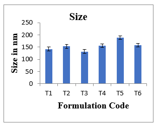
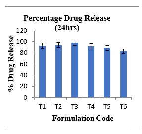
Figure No. -2: Bar diagram representation of percentage drug release for different formulations (n=3)
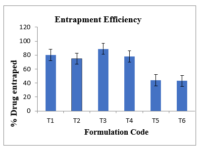
Fig. – 3: Comparative evaluation of entrapment Efficiency Of nanovesicles (n=3)
Figures 1,2,3 are showing a comparative evaluation of the formulation T1,T2,T3,T4,T5 and T6 of drug entrapment, percentage drug release and size. The results of the formulation with zetasizer are represented using figure 4,5. Figure 6 depicts the zeta value of optimized formulation.
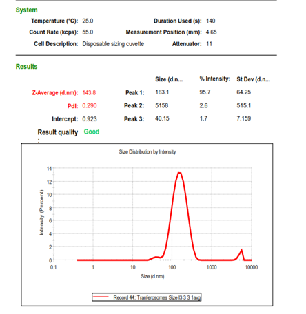
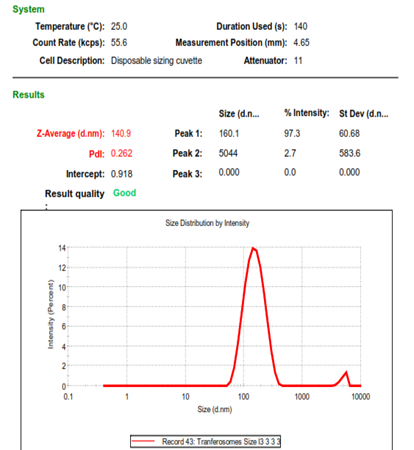
Fig. - 4: The zetasizer result for size Fig. -5 : The zetasizer results for particles with with 143nm 140nm
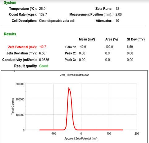
Fig. – 6 : The zeta potential results for nanoparticles
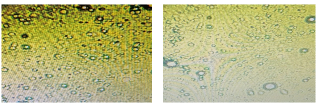
Fig -7 fluorescent microscopic images of nanovesicles formulation T3 with using Edge activator Labrosol, magnification at x 100
Fig -8 fluorescent microscopic images of nanovesicles formulation T3 Edge activator Labrosol, magnification at x 40
The figures 7&8 shows the morphology of the nanovesicles with two edge activators labrosol, from the images it is evident that the nanovesicles with T3 formulation has better morphology than the other.
Table No.-5: Optimization of transdermal patches with transferosomes (T3)
|
Formulation code |
Weight(mg) ± SD |
Thickness (mm) ± SD |
Folding endurance ± SD |
Diameter (cm) ± SD |
Area (cm2) ± SD |
Surface pH± SD |
%Drug Release For 24hrs± SD |
|
TL1 |
197.30 ± 2.68 |
0.052 ± 0.001 |
105.3 ± 2.52 |
4.03 ± 0.03 |
12.56±0.02 |
6.43±0.4 |
82 ±0.4 |
|
TL2 |
178.10 ± 3.64 |
0.051 ± 0.002 |
203.0 ± 1.73 |
4.06 ± 0.02 |
12.87±0.03 |
6.89±0.2 |
85 ±0.2 |
|
TL3 |
202.10 ± 2.48 |
0.046 ± 0.001 |
100.6 ± 2.51 |
4.10 ± 0.05 |
13.51±0.07 |
7.16±0.6 |
84 ±0.6 |
|
TL4 |
169.70 ± 2.32 |
0.048 ± 0.001 |
120.0 ± 1.73 |
4.03 ± 0.02 |
12.87±0.02 |
6.66±0.8 |
96 ±0.8 |
|
TL5 |
247.80 ± 2.74 |
0.053 ± 0.001 |
108.6 ± 2.08 |
4.06 ± 0.02 |
13.19±0.01 |
6.71±0.4 |
93 ±0.4 |
|
TL6 |
189.00 ± 2.16 |
0.049 ± 0.001 |
108.0 ± 2.64 |
4.06 ± 0.02 |
12.87±0.03 |
6.94±0.2 |
84±0.2 |
|
TL7 |
179.70 ± 1.34 |
0.051 ± 0.002 |
106.6 ± 2.08 |
4.08 ± 0.02 |
12.87±0.07 |
6.63±0.3 |
85±0.3 |
|
TL8 |
171.80 ± 2.21 |
0.049 ± 0.001 |
103.3 ± 1.53 |
4.05 ± 0.04 |
12.87±0.06 |
6.67±0.3 |
81±0.3 |
Note : Sample size (n=3)
The formulations with various percentages of HPMC 5cps (1%w/v,1.5% w/v, 2% w/v, 2.5% w/v) and HPMC 15cps (1%w/v,1.5% w/v, 2% w/v, 2.5% w/v) were prepared using PEG-400 as plasticizer with water as solvent [10]. The nanosuspension of 0.5ml was mixed with the above mixer. The patches prepare were evaluated for weight variation, folding endurance, thickness, diameter, area, surface pH and percentage drug release. The results were tabulated in table no 5.
From the table no.-5 it was inferred that all the formulations have optimum folding endurance and surface pH. Based on the percentage drug release studies for 24hrs the patches the prepared transdermal patches were optimized [11]
The transdermal patches with 2.5% w/v HPMC 15cps were found to have optimum drug release at 24th hr in comparison with other patches [12]. The patches with less percentage of HPMC 15Cps were found to deliver the drug prior to 24 hrs and stated decreasing the release at the end of 24 hrs.
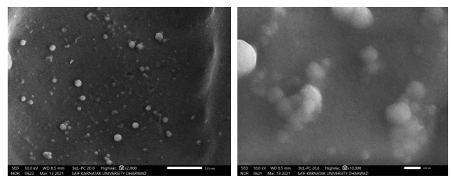
Fig. - 9: Scanning electron microscopic images of nanovesicles with labrosol as edge activator on transdermal patch
Fig.- 10: Scanning electron microscopic images of nanovesicles with labrosol activator on transdermal patch

Fig. 11 : Invitro drug release studies of transdermal patches
From figure 11 it is inferred that the formulations having HPMC 5cps 1%w/v, 1.5%w/v. 2%w/v are able to release the drug in 10hrs and there is a decrease observed in drug release at 24th hr. but HPMC 5CPS 2.5%w/v patches are able to release the drug at 24hrs in controlled manner. The patches prepared with HPMC 15cps 1%w/v was obseverd to release drug in controlled manner for 24hrs, the remaining concentration of HPMC 15cps (1.5%w/v. 2%w/v and 2.52%w/v) retarded the drug release of transferosomal formulation. Hence formulations T4 and T5 were selected as optimized formulations [13].
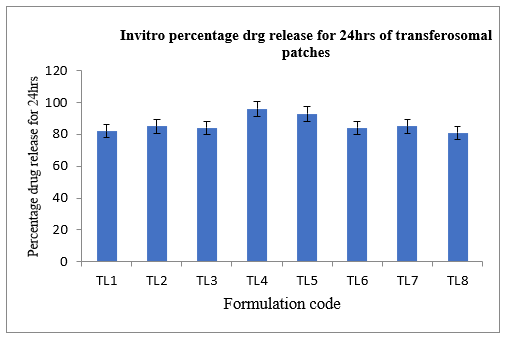
Fig.-12: Invitro percentage drug release studies of transferosomal transdermal patches for 24hrs
From figure No. 12 it can be noted that TL4 and TL5 formulations have optimum percentage of drug release for 24hrs.
Table No. 6: Evaluation of transdermal patches without transferosomes
|
Formulation code |
Weight(mg) ± SD |
Thickness (mm) ± SD |
Folding endurance ± SD |
Diameter (cm) ± SD |
Area (cm2) ± SD |
Surface pH) ± SD |
%Drug Release For 24hrs± SD |
|
L1 |
140.30 ± 2.68 |
0.052 ± 0.001 |
205.3 ± 2.52 |
4.03 ± 0.03 |
12.56±0.02 |
6.43±0.4 |
60 ±0.4 |
|
L2 |
139.10 ± 3.64 |
0.051 ± 0.002 |
203.0 ± 1.73 |
4.06 ± 0.02 |
12.87±0.03 |
6.89±0.2 |
62 ±0.2 |
|
L3 |
159.10 ± 2.48 |
0.046 ± 0.001 |
259.6 ± 2.51 |
4.10 ± 0.05 |
13.51±0.07 |
7.16±0.6 |
59 ±0.6 |
|
L4 |
149.70 ± 2.32 |
0.048 ± 0.001 |
290.0 ± 1.73 |
4.03 ± 0.02 |
12.87±0.02 |
6.66±0.8 |
58 ±0.8 |
|
L5 |
137.80 ± 2.74 |
0.053 ± 0.001 |
288.6 ± 2.08 |
4.06 ± 0.02 |
13.19±0.01 |
6.71±0.4 |
60 ±0.4 |
|
L6 |
149.00 ± 2.16 |
0.049 ± 0.001 |
278.0 ± 2.64 |
4.06 ± 0.02 |
12.87±0.03 |
6.94±0.2 |
58±0.2 |
|
L7 |
159.70 ± 1.34 |
0.051 ± 0.002 |
296.6 ± 2.08 |
4.08 ± 0.02 |
12.87±0.07 |
6.63±0.3 |
54±0.3 |
|
L8 |
151.80 ± 2.21 |
0.049 ± 0.001 |
253.3 ± 1.53 |
4.05 ± 0.04 |
12.87±0.06 |
6.67±0.3 |
52±0.3 |
Note: Sample size (n=3)
The evaluation of transdermal patches without nanovesicles are performed and tabulated in table no.6[14]. From the table it can be inferred that the drug release of the formulations without nanovesicles was found to have very less drug release.
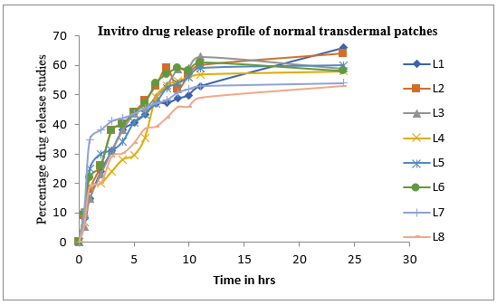
Fig. 13 : Drug release studies of the transdermal patches without nanovesicles
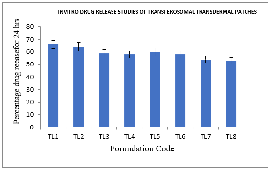
Fig. 13: A Comparitive invitro drug release profile of transdermal patches without transferosomes.
Table No-7: Exvivo Permeation Studies Using Pork Skin
|
Formulation Code |
Q24(µg/cm2) |
Flux (µg/cm2/hr) |
Lag time (hrs.) |
Skin content (?g/g) |
|
TL4 |
960± 0.68 |
28± 2.3 |
0.02± 1.4 |
42± 3.2 |
|
TL5 |
935± 0.8 |
26± 0.4 |
0.08± 1.8 |
31± 3.5 |
|
L2 |
624± 2.6 |
16± 1.1 |
0.5± 2.4 |
147± 2.3 |
|
L5 |
603± 1.3 |
14± 1.04 |
0.52± 2.8 |
156± 2.9 |
Note: Sample size (n=3), TL4, TL5- transferosomal patches; L2, L5- patches without
transferosomes.
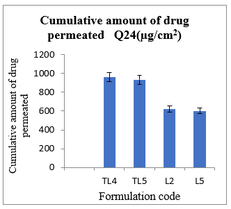
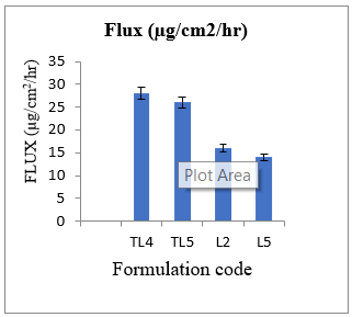
Fig. 14 : comparative evaluation of Q24(µg/cm2) Fig. 15 : comparative evaluation of Flux
The Table no.7 depicts the evaluation studies performed using transferosomal patches and and non transferosomal patches[15]. The cumulative amount of the drug permeated, flux and skin content were calculated and it was observed that the transferosomal patches have better Q24 , flux and less skin content compared to non transferosomal patches. The figures 14&15 represent the comparative evaluation of transdermal patches.
CONCLUSION:
In the present research the transferosomal patches with lisnoprildihydrate were prepared and compared with non transferosomal patches of the same drug. The nanaovesicles were evaluated for entrapment efficiency, size,and morphology from the results T3 formulation having edge activator(labrosol) and lecithin in 1:1 ratio was found to have better results. The prepared nanovesicles were fabricated into transdermal patches using HPMC 5cps, HPMC 15cps with various concentrations and evaluated. From the evaluation it was found that transferosomal patches having 1%w/v HPMC 15cps and 2.5% HPMC 5cps were found to have better mechanical properties and optimum drug release invitro drug release when compared with other transferosomal patches. The non transferosomal patches were prepared and compared with transferososmal patches. A comparative ex vivo studies were performed with two kinds of patches, based on the transdermal flux, Q24 and skin content it was found that the transferosomal patches have better transdermal permeability when compared to non transferosomal patches. This research helps in concluding that the nanocarrier based transdermal formulations can show efficient results. These formulations can overcome the disadvantages of conventional transdermal systems.
REFERENCES

Aparanjitha R.*, Sunitha Reddy M., Comparative Invitro and Exvivo Evaluation of Transferosomal Patches for Enhanced Drug Delivery of Lisinopril Dihydrate, Int. J. of Pharm. Sci., 2025, Vol 3, Issue 2, 727-738. https://doi.org/10.5281/zenodo.14848716
 10.5281/zenodo.14848716
