Department of pharmaceutics, St. Joseph’s College of Pharmacy, Cherthala, Alappuzha - 688524.
Silver nanoparticles have attracted considerable attention due to their wide range of applications in medicine, catalysis, and electronics. Silver nanoparticles exhibit strong interactions with other particles, enhancing their antibacterial properties, and have optical responses largely depend therefore gained significant attention due to their distinctive physical, chemical, and optical characteristics. Silver nanoparticles (AgNPs) have attained significant research interest owing to their distinct properties, including size- and shape-dependent optical, antimicrobial, and electrical characteristics. Numerous approaches have been explored for their synthesis, such as laser ablation, gamma and electron irradiation, chemical reduction, photochemical techniques, microwave-assisted processing, and biological methods. This review provides a comprehensive overview of these preparation strategies, broadly classified into physical, chemical, and biological routes. The article highlights the current progress in silver nanoparticle synthesis, while also discussing the advantages and limitations associated with each technique. In addition, the review emphasizes the future prospects and industrial relevance of these methods, aiming to outline their potential applications as well as the challenges that need to be addressed to advance large-scale and sustainable production laser. Silver nanoparticles exhibit strong interactions with other particles, and have therefore gained significant attention due to their distinctive physical, chemical and optical characteristics.
Silver nanoparticles are nanoparticles of silver having size range between 1 and 100 nm. Silver is one such metal which has been used for various biomedical applications. They penetrate the human body via different organs like liver, kidney, lungs, spleen and brain. AgNPs are extremely small, but their large surface area allows them to dissolve quickly. They exhibit several beneficial features, including chemical stability, strong conductivity, localized surface plasmon resonance, which make them effective against viruses. since they can act on multiple targets, the likelihood of causing drug resistance reduced. Nanotechnology, which focuses on the creation and manipulation of structures at the scale of 1–100 nm, has become a rapidly expanding area of modern research. Nanoparticles (NPs) are particularly valuable due to their broad range of applications in medicine, cosmetics, food, environmental monitoring, optics, catalysis, and electronics. Among the various branches of this field, nanobiotechnology has gained prominence, combining nanoscience with biological approaches to design functional nanomaterials with controlled size, shape, and dispersion. This interdisciplinary area involves contributions from chemistry, physics, biology, engineering, and medicine, and has enabled the development of innovative materials for healthcare and technology. Recently, increasing focus has been placed on green synthesis methods, which utilize non-toxic, environmentally friendly reagents and solvents. Such approaches offer sustainable alternatives to conventional chemical routes, reducing environmental risks while ensuring effective and reproducible nanoparticle production. These advancements highlight the potential of nanotechnology for both scientific innovation and industrial application.
Structure Of Silver Nanoparticles
Silver nanoparticles are defined as silver particles with a diameter ranging from 1 to 100 nanometers. While they are most commonly spherical, they can also be synthesized in various other forms, including diamond and octagonal shapes, as well as thin sheets. A key characteristic of silver quantum dots is their tunable bandgap, which can be adjusted by altering the dot's size. Furthermore, these nanoparticles exhibit Surface Plasmon Resonance (SPR), a property that makes them a highly desirable dopant material for use in photovoltaic cells to enhance their efficiency. Silver is a noble metal whose nanoparticles possess distinct and adjustable plasmonic characteristics. The frequency of their surface plasmon resonance (SPR) can be precisely controlled by modifying the particles' size and shape, as this determines how they interact with their surrounding environment. While nanoparticles made from pure, uncoated silver already demonstrate excellent physical and chemical properties, their functionality can be further tailored for specific applications. This is often achieved by coating them with a different metal, such as gold, to form a core-shell structure.
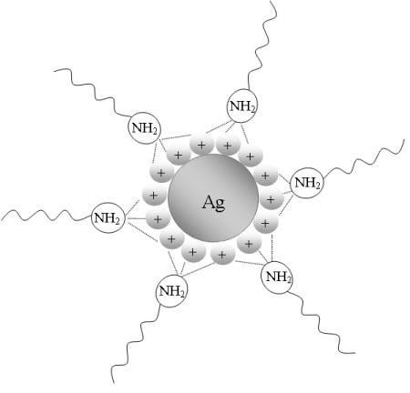
Figure 1 – Structure of Silver Nano Particles
Properties Of Nanoparticles
The physical and chemical characteristics of nanoparticles are used to describe their properties
Synthesis Of Silver Nanoparticles
There are two primary strategies for producing silver nanoparticles: the top -down and the bottom -up approaches. These approaches can be carried out using physical, chemical, or biological methods. Generally physical techniques follow the top – down route, while chemical and biological methods rely on bottom -up synthesis. In the top- down method, a bulk material is taken as the starting point and broken-down step by step into smaller particles until nanoparticles are obtained. On the other hand, bottom -up method builds nanoparticle by assembling atoms and molecules. This approach is generally less toxic, more cost effective and more efficient.
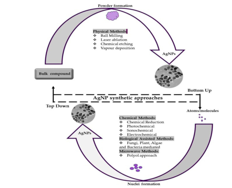
Figure 2 – Synthesis of Silver Nano Particles
Physical Methods
Physical approaches such as evaporation–condensation and laser ablation are among the most widely applied methods for producing silver nanoparticles. One advantage of these techniques is the absence of solvent contamination, which often occurs in chemical synthesis, thereby ensuring higher purity of the nanoparticles. Additionally, these methods allow greater uniformity in particle distribution. However, certain limitations exist. For example, synthesis using a tube furnace under atmospheric pressure requires a large amount of space, consumes significant energy, and generates high operating temperatures, which increases environmental concerns. Furthermore, achieving stable thermal conditions demands extended preheating times. A smaller ceramic heater has been proposed as an alternative, which can reduce energy requirements while maintaining adequate evaporation of silver to form nanoparticles. Rapid cooling of the vapor also promotes the formation of nanosized particles. Overall, physical methods are effective but less energy-efficient compared to chemical approaches, limiting their broader application. Among the physical techniques, evaporation–condensation and laser ablation are the most widely used methods. Their major advantages over chemical synthesis are the absence of solvent contamination in the resulting thin films and the uniform distribution of nanoparticles. However, producing silver nanoparticles with a tube furnace at atmospheric pressure has certain drawbacks. Tube furnaces require a large setup space, consume high amounts of energy, increase the surrounding temperature, and take considerable time to reach thermal stability. In addition, they typically demand power consumption of several kilowatts and need a long preheating period before achieving stable operating conditions. Studies have shown that silver nanoparticles can also be generated using a small ceramic heater with a localized heating zone. This approach allows efficient evaporation of the source material, and the vapor condenses quickly because the steep temperature gradient near the heater surface promotes rapid cooling. Compared to a tube furnace, this method is more efficient and facilitates the formation of smaller nanoparticles.
Chemical reduction is one of the most commonly employed strategies for preparing silver nanoparticles, as it enables large-scale production using relatively simple procedures. This approach involves reducing silver salts with organic or inorganic agents in the presence of stabilizers that prevent particle agglomeration. A wide range of reducing agents—including sodium borohydride, hydrazine, ascorbic acid, and glucose—have been reported. Stabilizers such as polyvinyl alcohol (PVA), polyvinylpyrrolidone (PVP), or natural polymers are often added to control particle size, shape, and dispersion. Despite its popularity, chemical synthesis faces challenges such as particle instability, aggregation, and the need for post-synthesis purification. Moreover, many reducing agents are toxic and raise environmental concerns. To address these issues, researchers are increasingly exploring “green” chemical methods that utilize eco-friendly solvents and biological stabilizers while maintaining efficiency and reproducibility. Such modifications aim to overcome the drawbacks of conventional chemical routes and improve their suitability for biomedical and industrial purpose. The synthesis of silver nanoparticles (AgNPs) is most commonly achieved through chemical reduction using either organic or inorganic reducing agents. Typically, compounds like sodium citrate, ascorbate, sodium borohydride (NaBH?), elemental hydrogen, polyol methods, Tollens’ reagent, N, N-dimethylformamide (DMF), and poly (ethylene glycol)-based copolymers are employed to reduce silver ions (Ag?) in aqueous or non-aqueous systems. These agents convert Ag? into metallic silver (Ag?), which subsequently aggregates into small oligomeric clusters. Over time, these clusters evolve into metallic colloidal silver nanoparticles. To ensure stable dispersions during nanoparticle synthesis, protective agents are necessary. They safeguard the nanoparticles by preventing aggregation and enabling them to bind or adsorb onto surfaces. Surfactants containing functional groups such as thiols, amines, acids, and alcohols play a key role in particle stabilization. They promote controlled particle growth, reduce sedimentation, and preserve the surface properties Surfactants containing functional groups such as thiols, amines, acids, and alcohols play a key role in particle stabilization. They promote controlled particle growth, reduce sedimentation, and preserve the surface properties of nanoparticles. Additionally, polymers like poly (vinyl alcohol), poly(vinylpyrrolidone), and poly (ethylene glycol) are often incorporated as stabilizing agents during synthesis.
Table 1. Different reducing agents and stabilizing agents for AgNPs Synthesis.
|
Silver salt |
Reducing Agent |
Capping Agent/ Stabilizer |
Particle size (nm) |
|
|
Silver nitrate |
Hydrazine hydrate and sodium citrate |
Sodium dodecyl sulphate |
10 -20 |
|
|
Silver nitrate |
Sodium borohydride |
- |
35 -6 |
|
|
Silver nitrate |
Glucose |
Poly vinyl pyrrolidine |
20-80 |
|
|
Silver nitrate |
Gallic acid |
Gallic acid |
7-89 |
|
|
Silver nitrate |
Poly ethylene glycol |
Poly ethylene glycol |
15-30 |
|
|
Silver nitrate |
Sodium hydroxide |
Alkali lignin |
5-100 |
|
|
Silver nitrate |
Glucose and sodium hydroxide |
- |
8-24 |
|
|
Silver nitrate |
PVP and gelatin |
Glucose, Fructose |
35 |
|
|
Silver nitrate |
D glucose |
Carboxymethylcellulose |
5-15 |
|
Biological Method
Various eco-friendly synthesis techniques such as the use of polyoxometalates, pollens, polysaccharides, and irradiation methods are employed for producing silver nanoparticles. Green synthesis refers to the application of biologically compatible materials like plants, bacteria, and fungi for nanoparticle preparation. These methods offer significant advantages over conventional chemical synthesis, as they avoid many of its limitations. One widely used approach involves plant extracts, which serve as both reducing and capping agents in the formation of silver nanoparticles. This method is environmentally safe and non-toxic compared to traditional chemical reduction techniques. The bioactive molecules present in plant extracts—such as amino acids, vitamins enzymes, and proteins—help in reducing silver ions (Ag?) and stabilize the nanoparticles, ensuring uniform size and shape. A wide range of plants, including moringa, bitter apple, fenugreek, neem, and others, have been utilized for this purpose.
Other Methods
Microwave assisted synthesis is a promising technique for producing silver nano particle. Compared to traditional oil bath heating, microwave heating consistently yields nanostructures with smaller sizes, narrower size distributions, and higher crystallization degrees. This method offers shorter reaction times, lower energy consumption, and better product yields, reducing particle agglomeration. Beyond eliminating the need for an oil bath, microwave assisted synthesis with environmentally friendly reaction media can significantly reduce chemical waste and reaction times in various organic syntheses and chemical transformations. It has been reported that silver nanoparticles can be synthesized using microwave assistance with carboxy methyl cellulose sodium as both a reducing and stabilizing agent. The size of the nanoparticles depends on the concentrations of carboxy methyl cellulose and silver nitrate. The resulting nanoparticles were uniform, stable, and showed specific characteristics.
Laser irradiation can be used to synthesize silver nanoparticles in an aqueous solution containing silver salt and surfactant. This method produces nanoparticles with a well-defined shape and size distribution. In a photo sensitization method using benzophenone, laser irradiation at low powers created silver nanoparticles of about 20 nm, while higher powers resulted in nanoparticles of about 5 nm. Both lasers and mercury lamps can serve as light sources for producing silver nanoparticles.
The electrochemical synthetic method is used to synthesize silver nanoparticles. By adjusting electrolysis parameters and changing the composition of electrolytic solutions we can control particle size and improve the homogeneity of silver nanoparticles
Silver nanoparticles can be produced through several photoinduced or photocatalytic reduction techniques. This photochemical approach is a clean method known for its high precision, ease of use, and adaptability. A key advantage is the ability to create nanoparticles in diverse environments such as cells, emulsions, polymer films, surfactant micelles, and glasses. In one application , polyelectrolyte capsules served as microreactors for the photoinduced synthesis of silver nanoparticles with an average size of 8 nm .The same photoinduced process has also been used to transform spherical silver nanoparticles into triangular nanocrystals with controlled edge lengths between 30 and 120 nm , this morphological change was managed using a dual beam illumination technique with citrate acting as stabilizers .
UV-initiated photoreduction is a straightforward and efficient technique for synthesizing silver nanoparticles (NPs). This process employs compounds like citrate, polyvinylpyrrolidone, poly (acrylic acid), and collagen to facilitate the reaction. For example, one study used laponite clay suspensions as a stabilizer to prevent aggregation during the UV photoreduction of silver nitrate. The characteristics of the resulting nanoparticles were found to depend on the duration of UV exposure. After three hours of irradiation, the NPs had a bimodal size distribution and were relatively large. Continued UV exposure broke these particles down into smaller sizes with a single, stable distribution mode. In another application of this technique, silver nanostructures—including nanospheres, nanowires, and dendrites—were produced at room temperature using poly (vinyl alcohol) as a protective stabilizing agent. The final morphology of the nanostructures was significantly influenced by the concentrations of both the poly (vinyl alcohol) and the silver nitrate.
Microemulsion methods offer a way to create uniform silver nanoparticles (NPs) with controllable sizes. This process uses a two-phase (aqueous-organic) system where the reactants—a metal precursor and a reducing agent—are initially separated into two immiscible liquids. A quaternary alkyl-ammonium salt acts as a mediator, controlling the transport and interaction rate between these phases at their interface. Metal clusters form at this liquid interface. They are stabilized by coating with surfactant molecules present in the non-polar organic medium and are then shuttled into the organic phase by the same mediator. A significant drawback of this technique is the requirement for large quantities of harmful organic solvents and surfactants. These must later be separated and removed from the final nanoparticle product, complicating the process. Additionally, a separate green chemistry approach has been documented. This method enables the one-step synthesis and stabilization of silver nanostructures in water at room temperature.
Silver nanoparticles (NPs) can be produced through laser ablation, a process that involves using a laser to vaporize material from a solid silver target immersed in a solution. The properties of the resulting nanoparticles are influenced by several key parameters:
A significant benefit of laser ablation over chemical synthesis methods is that it does not require chemical reagents. This allows for the creation of pure, contaminant-free silver colloids ideal for sensitive applications. For instance, one study generated silver nano spheroids (20-50 nm) in water using an 800 nm femtosecond laser. When compared to ablation with nanosecond pulses, the femtosecond method resulted in a lower production efficiency and generally yielded smaller colloidal particles.
Types Of Silver Nanoparticles
Nanoparticles (NPs) are categorized into three primary groups: inorganic, carbon-based, and organic nanoparticles.
Inorganic Based NPs
This classification encompasses nanoparticles that do not contain carbon atoms. This group primarily includes nano-sized particles composed of metals or metal oxides.
1. Metal NPs
This category includes nanoparticles made from metals such as cadmium (Cd), aluminum (Al), copper (Cu), cobalt (Co), gold (Au), iron (Fe), silver (Ag), zinc (Zn), and lead (Pb). Their distinctive properties—which arise from their size and inherent characteristics—include a high surface area, specific pore size, surface charge density, and varied forms like cylindrical and spherical shapes. They also exhibit diverse colors and can have either amorphous or crystalline structures. Furthermore, external environmental conditions like air, heat, sunlight, and moisture can influence these properties.
Since certain metals readily form oxides, their oxide nanoparticles are synthesized to improve and enhance their material characteristics. Due to the propensity of certain metals to form oxides, their oxide nanoparticles are synthesized to achieve enhanced material properties. For instance, iron nanoparticles readily oxidize in the presence of oxygen (O?) at room temperature, forming iron oxide (Fe?O?), which exhibits greater reactivity than its metallic counterpart. Commonly synthesized metal oxide nanoparticles—including titanium oxide (TiO?), silicon oxide (SiO?), zinc oxide (ZnO), magnetite (Fe?O?), iron oxide (Fe?O?), aluminum oxide (Al?O?), and cerium oxide (CeO?)—are known for their improved efficiency, reactivity, and overall performance. Literature indicates that these metal oxide NPs often possess superior properties compared to pure metal nanoparticles.
Organic-Based NPs
Organic-based nanoparticles, also known as nano capsules, are generally non-toxic and environmentally friendly. This category includes ferritin, liposomes, micelles, and dendrimers—all of which are composed of polymeric or organic materials. These nanoparticles demonstrate high responsiveness to external stimuli such as light and heat. Owing to their distinct and advantageous characteristics, they are increasingly preferred by researchers as a promising alternative in various applications. These distinctive properties make organic NPs a superior and preferred choice for applications like drug delivery. Their stability, high drug-loading capacity, and ability to specifically adsorb or encapsulate therapeutic compounds position them as highly efficient carriers for active pharmaceutical ingredients. Their effectiveness is further defined by their unique surface morphology, size, shape, and chemical composition. In targeted drug delivery systems, organic NPs are specifically designed to transport and release active agents at precise sites of action within the body.
Carbon-Based NPs
Nanoparticles that are composed entirely of a carbon framework are classified as carbon-based nanoparticles. This category includes several distinct types such as graphene, fullerenes, carbon nanofibers, carbon nanotubes, black carbon, and activated carbon.
Fullerenes are spherical carbon nanoparticles formed by carbon atoms connected via sp² hybridization. Their structure can consist of a single layer (monolayer) or multiple layers (poly-layered). Monolayer fullerenes, composed of roughly 28 to 1500 carbon atoms, can reach diameters up to 8.3 nm, while poly-layered versions typically range from 4 to 36 nm in diameter.
Graphene is a two-dimensional, planar allotrope of carbon where the atoms are arranged in a hexagonal lattice. A single layer of this material has an extremely small thickness of approximately 1 nm.
3. Carbon Nanotubes
Carbon nanotubes are produced by rolling a graphene nano-foil into a hollow, cylindrical tube. Single-layer (monolayer) nanotubes have a very small diameter, typically less than0.7 nm. Multi-layer nanotubes can vary significantly in length, from micrometers to several millimeters, and their ends may be either sealed or open.
4. Nanofibers of Carbon
Carbon nanofibers are also fabricated from graphene nano-foils. However, unlike the uniform cylinders of nanotubes, they are coiled into shapes such as cups or cones.
5. Black Carbon
Black carbon nanoparticles are amorphous, spherical particles with diameters between 20 and 70 nm. They exhibit strong particle interactions and tend to combine into larger aggregates, forming agglomerates around 500 nm in size.

Figure 3 - showing Different types of silver nanoparticles
Characterization Of Silver Nanoparticles
Analyzing the physicochemical properties of nanoparticles is essential for understanding their behavior, distribution within biological systems, safety, and effectiveness. A variety of analytical techniques are employed to characterize silver nanoparticles (AgNPs) and evaluate their functional performance. Key methods include:
The following sections detail the fundamental principles of these primary techniques.
1. UV-Visible Spectroscopy
This technique is a fundamental and widely used tool for the initial characterization of nanoparticles, particularly for monitoring their synthesis and stability. AgNPs possess distinct optical properties, notably a surface plasmon resonance (SPR) absorption band. This band arises from the collective oscillation of free electrons in the nanoparticles when they interact with specific wavelengths of light. UV-vis spectroscopy is favored because it is rapid, straightforward, sensitive, and can be used on colloidal suspensions without the need for calibration.
2. X-Ray Diffraction (XRD)
XRD is a versatile analytical method used to determine the crystalline structure of materials at the atomic level. It is applied for phase identification, quantitative analysis, and measuring properties like crystallinity and particle size. The technique operates on Bragg's law, analyzing the diffraction patterns produced when X-rays interact with a crystalline sample. It is non-destructive and can be used to characterize a wide array of organic, inorganic, and nanoscale materials.
3. Dynamic Light Scattering (DLS)
DLS is a primary technique for determining the size distribution of nanoparticles in a solution or suspension, typically in the 2–500 nm range. It works by measuring the fluctuations in the intensity of laser light scattered by particles undergoing Brownian motion. This data is used to calculate the hydrodynamic diameter of the particles, providing crucial information about their size in a liquid medium.
4. Fourier Transform Infrared (FTIR) Spectroscopy.
FTIR spectroscopy is used to identify molecular fingerprints and functional groups present on the surface of nanoparticles. It is highly accurate and can detect very small absorbance changes. This makes it invaluable for confirming the involvement of biomolecules in green synthesis processes and for studying functional molecules attached to nanomaterials like silver, carbon nanotubes, and graphene.
5. Scanning Electron Microscopy (SEM).
SEM is a high-resolution imaging technique that uses a focused beam of electrons to provide detailed information on the surface morphology, size, shape, and distribution of nanoparticles. When combined with Energy-Dispersive X-ray spectroscopy (EDX), SEM can also provide the elemental composition of the sample, allowing researchers to correlate structure with chemistry.
Applications Of Silver Nanoparticles
Silver has been utilized for its antibacterial properties for over 5,000 years. As nanoparticles (AgNPs), silver is particularly valued because it combines potent antibacterial effects with low toxicity to human cells. This antibacterial activity is defined as the ability to kill or inhibit the growth of bacteria while leaving surrounding cells unharmed. Beyond this, AgNPs have a wide range of applications, including antiviral, antifungal, and anti-inflammatory uses, as well as in cosmetics, water treatment, healthcare, biosensing, drug delivery, imaging, and textiles.
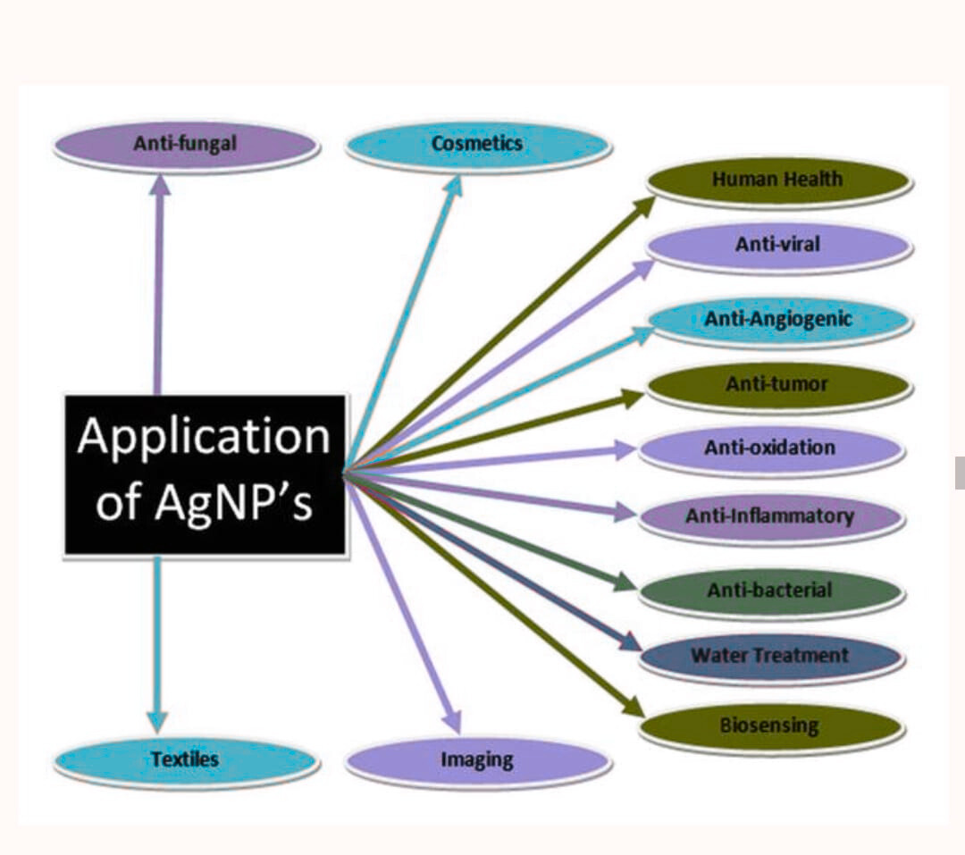
Figure 4 – Application Of Silver Nano particles
A significant advantage of AgNPs is their ability to combat bacteria that have developed resistance to conventional antibiotics. Their effectiveness is attributed to their high surface-area-to-volume ratio and unique surface crystallographic structure. Studies have shown that AgNPs can destroy multiple drug-resistant bacterial strains. Gram-negative bacteria are generally more susceptible to AgNPs than Gram-positive bacteria. This difference is due to their distinct cell wall structures:
Furthermore, the antibacterial mechanism is enhanced by electrostatic attraction. Bacterial cell membranes are typically negatively charged due to carboxyl, phosphate, and amino groups. The positive charge often found on AgNPs causes them to be attracted to and attach to the cell membrane, disrupting its function and leading to cell death. Immunocompromised individuals are highly susceptible to fungal infections, which are notoriously difficult to treat due to the limited number of effective and biocompatible antifungal drugs available. Silver nanoparticles (AgNPs) have demonstrated significant potential in this area.
Inflammation is a biological response to injury or infection, characterized by swelling, redness, heat, and pain. While AgNPs are primarily recognized for their antibacterial and antimicrobial properties, their role as anti-inflammatory agents, though less documented, is also important. They contribute to this field by modulating the body's immunological response to foreign particles.
Cancer involves the uncontrolled proliferation of cells and is a prevalent global health issue. Conventional treatments like chemotherapy and radiation therapy are often associated with severe side effects, significant patient discomfort, and high costs. Consequently, there is a pressing need for more effective, affordable, and lower-molecular-weight treatments. AgNPs present a promising alternative. Their suitability stems from their ability to serve as targeted drug delivery systems; therapeutic agents can be encapsulated within the nanoparticles and directed specifically to tumor cells, minimizing damage to healthy tissues.
Other Applications
In recent years, peptide-capped silver nanoparticles have been extensively researched for colorimetric sensing applications. These studies primarily investigate the interaction between the peptide and silver, and how the peptide influences nanoparticle formation. Furthermore, fluorescent sensors based on silver nanoparticles demonstrate high efficiency and can surpass conventional detection limits.
2. Optical Probes
Silver nanoparticles are commonly employed as probes in surface-enhanced Raman scattering (SERS) and metal-enhanced fluorescence (MEF). They offer superior performance compared to other noble metal nanoparticles due to their higher extinction coefficients, sharper extinction bands, and significantly enhanced field effects.
3. Catalyst-
Silver nanoparticles exhibit catalytic redox properties for both biological agents (e.g., dyes) and chemical agents (e.g., benzene). Their catalytic performance is strongly influenced by their chemical environment. Catalysis typically occurs through the adsorption of reactant species onto the nanoparticle surface. However, when stabilizers such as polymers, complex ligands, or surfactants are used to prevent nanoparticle coalescence, the catalytic efficiency often decreases due to reduced adsorption capacity. Silver nanoparticles are frequently combined with titanium dioxide to serve as catalysts in various chemical reactions.
Advantages Of Silver Nanoparticles
Silver nanoparticles are promising candidates for antimicrobial applications because of their minute size, significant surface-area-to-volume ratio, and unique physicochemical characteristics. These properties enable them to interact with microbes by entering cells and clumping together, which causes toxicity and cell death. A drug-delivery system using ligands to control the release of silver can create a powerful synergistic antimicrobial effect. This synergy benefits both the drug and the silver nanoparticles, particularly when the ligands have short carbon chains and oxygen atoms that form weak bonds. Consequently, tailoring the nanoparticles' surface ligands—by carefully selecting their coordinating atoms, carbon chain lengths, and terminal groups—is essential for developing commercially viable treatments for infectious diseases. Studies indicate that engineered silver nanoparticles can be effective in drug delivery and may help reduce a drug's inherent toxicity. This research also notes that higher concentrations of these silver nanoparticle conjugates resulted in only a minimal decrease in their cytotoxic effects relative to the cells' own cytotoxicity. After exposure, AgNPs can trigger localized inflammation and oxidative stress. They also possess the ability to cross biological barriers and enter systemic circulation. Once in the bloodstream—either directly from injection or after translocation from other entry points—AgNPs are distributed to various organs, where they can induce distinct pathophysiological effects.
Limitations Of Silver Nanoparticles
Cytotoxicity of Silver Nanoparticles Humans can be exposed to silver nanoparticles (AgNPs) through various routes, including dermal contact, inhalation, ingestion, and entry into the bloodstream. Numerous in vivo and in vitro studies have been published on nanoparticle toxicity, demonstrating that the cytotoxic effects of AgNPs are influenced by their concentration, environmental conditions, physical dimensions, and duration of exposure. Smaller AgNPs generally exhibit greater cytotoxicity than their larger counterparts due to their higher surface area-to-mass ratio. Production of Reactive Oxygen Species (ROS) A key mechanism behind AgNPs toxicity is the excessive generation of reactive oxygen species (ROS). While ROS are essential for normal cellular functions, their overproduction can lead to oxidative stress, resulting in damage to cellular components like DNA. Research on the harmful effects of AgNPs in liver cells and fibroblasts indicates that these particles can trigger antioxidant defense mechanisms within mitochondria. Interaction with Proteins The interaction between proteins and silver nanoparticles is a critical factor in their toxicity. This phenomenon has been widely investigated. For instance, studies using spectroscopic methods have examined the binding of AgNPs to human hemoglobin, a blood protein. These interactions are both time- and concentration-dependent, with nanoparticles binding to specific sites on the hemoglobin molecule, such as the heme group and amide linkages. Similar interactions have been observed with cytoskeletal proteins.
Future Perspectives
Silver nanoparticles (AgNPs) hold significant promise for the healthcare sector, particularly in treating infectious diseases and combating the growing problem of antibiotic-resistant bacteria, in addition to their recognized anti-inflammatory capabilities. Their utility extends beyond medicine to various biological and research fields, including electrochemistry, biochemistry, textiles, detergents, soaps, water purification systems, and surgical instruments. A notable advancement is the integration of AgNPs into artificial implants, which reduces reliance on traditional antibiotics. Research also indicates their potential in developing innovative pharmaceutical formulations and treating conditions such as bladder inflammation. Furthermore, AgNPs are being utilized in animal models for biosensor detection. A critical focus for future research is to establish a reliable mechanism explaining the notable biological activity of AgNPs. There is also considerable potential in achieving controlled release of silver and enhancing the stability of AgNPs for use in medical and mechanical device.
CONCLUSION
This comprehensive review details the classification, synthesis strategies, and diverse applications of silver nanoparticles (AgNPs). It serves as a consolidated reference on their characteristics, types, biological properties, medicinal uses, role in water treatment, and associated potential hazards. The article examines the primary synthesis approaches—physical, chemical, and biological—and identifies green synthesis as the most advantageous method. This preference is due to its environmental friendliness, cost-effectiveness, operational simplicity, regenerative nature, and low energy requirements. A key focus in nano-chemistry is particle size, and evidence shows that different plants can produce varying sizes of AgNPs from the same metal ions, highlighting their unique reducing capacities. A noted limitation of green synthesis, however, is its typically lower nanoparticle yield. The review also emphasizes the critical need for thorough assessments of side effects, an area requiring further intellectual investigation. Owing to their significant antimicrobial properties, AgNPs are increasingly applied in water purification and disinfection strategies.
REFERENCES


Akhila K. A.*, Praveen Raj R., Milu M. R., Detailed Review on Synthesis of Silver Nanoparticles, Int. J. of Pharm. Sci., 2025, Vol 3, Issue 9, 416-431 https://doi.org/10.5281/zenodo.17051609
 10.5281/zenodo.17051609
