Anuradha College of Pharmacy, Chikhli, Buldhana, Maharashtra.
Chemically induced hepatotoxicity in rats, also known as drug-induced liver injury (DILI), is a widely used model for studying liver damage caused by various chemicals and drugs. Researchers use this model to understand the mechanisms of hepatotoxicity, develop new therapies, and assess the safety of new drugs before they are tested in humans. Commonly used hepatotoxins in rats include Carbon Tetrachloride (CCl4), Thioacetamide (TAA), Paracetamol (Acetaminophen), D-Galactosamine, and Dimethyl nitrosamine (DMN). Histopathological and biochemical parameters are assessed through examination of liver tissue under a microscope and measurement of liver enzymes and markers in blood samples. The study aims to assess the in-vivo hepato-protective activity of a polyherbal extract against chemically induced-hepatotoxic rats. The objectives include conducting a literature survey, selecting plant material, procuring and authenticating it, preparing a poly-herbal combination, evaluating liver injury caused by hepatotoxic substances, investigating the test compound's ability to protect the liver against hepatotoxicity, comparing its efficacy with known hepato-protectants, and evaluating the safety profile of the test compound or herbal extract in hepatotoxic rats. In Present study, we can conclude that, This formulation preserves the activity of serum marker enzymes, which aids in the healing of hepatic tissue. At the oral dosages examined, the formulation was well tolerated and showed no symptoms of hepato-, reno-, or hematotoxicity, nor any indication of clinical toxicity. These results are confirmed by the findings of our study which is further supported by the histopathological findings. By acute and general toxicities following oral ingestion, we were able to prove that, it is a non-toxic hepatoprotective medication. This Polyherbal extract containing different types of the extract can be a best alternative for treatment of hepatotoxicity.
Chemically induced hepatotoxicity in rats, also known as drug-induced liver injury (DILI), is a widely used model for studying liver damage caused by various chemicals and drugs. Researchers use this model to understand the mechanisms of hepatotoxicity, develop new therapies, and assess the safety of new drugs before they are tested in humans. Commonly used hepatotoxins in rats include Carbon Tetrachloride (CCl4), Thioacetamide (TAA), Paracetamol (Acetaminophen), D-Galactosamine, and Dimethyl nitrosamine (DMN). Hepatotoxicity can be induced through intraperitoneal injection, oral gavage, or subcutaneous injection. Dose-dependent administration is used to control the dose and duration of exposure to the hepatotoxin. Rats are used in hepatotoxicity research to understand the mechanisms of DILI, develop new therapies, and assess drug safety. Hepatotoxicity in rats can be a warning sign that a drug may also be hepatotoxic in humans, leading to changes in drug development or use. Histopathological and biochemical parameters are assessed through examination of liver tissue under a microscope and measurement of liver enzymes and markers in blood samples (1-5). The study aims to assess the in-vivo hepato-protective activity of a polyherbal extract against chemically induced-hepatotoxic rats. The objectives include conducting a literature survey, selecting plant material, procuring and authenticating it, preparing a poly-herbal combination, evaluating liver injury caused by hepatotoxic substances, investigating the test compound's ability to protect the liver against hepatotoxicity, comparing its efficacy with known hepato-protectants, and evaluating the safety profile of the test compound or herbal extract in hepatotoxic rats.
2. MATERIALS AND METHODS
2.1 Collection, authentication and processing of the Crude Drugs
Seeds of Moringa oleifera, Barks of Cinnamomum zeylanicumand Rhizomes of Curcuma longa were identified and collected from Taxonomist, Department of Botany, Shri Shivaji Science and Art college, Chikhli, Maharashtra in the month of July-August when the plants bear flower. The collected plant materials were shade dried. The dried materials were coarsely powdered individually by means of mechanical grinder. Then they were passed though sieve no.40 (aperture size-425mm) to get moderately coarse powder. The resulting powdered materials were used for further studies.
2.2 Preparation of Polyherbal Formulation
We selected 2.5:2.5:2.5 ratio of methanolic extracts of Seeds of Moringa oleifera, bark of Cinnamomum zeylanicum and Rhizomes of Curcuma longa for polyherbal syrup. For preparation of polyherbal syrup, sugar base was prepared by mixing of sucrose and 50 ml of water, heated to boiling point. The liquid was strained and volume made up to 100 ml with distilled water. The preservatives were dissolved in few milliliter of boiled and cooled water and added to a sugar base. Extracts were dissolved in propylene glycol at 45–50°C and this glycerin and sorbitol were added. The remaining sweetening agents were added and mixed thoroughly. Adjust the pH between 5.5 and 7.0 with, if necessary (6).
2.3 Acute Toxicity Study
Total 6 rats of 10-12 weeks age were selected and randomly divided into 2 groups. Group I was vehicle control group which received vehicle (gum acacia 1% w/v in distilled water) while group II was test group that received MEPE. Each group consisted of 3 animals (females). Females were nulliparous and non-pregnant. Dose selected for the present study is limit test dose as mentioned in the guidelines. The starting dose for limit test 2000 mg/kg was selected on the basis of dose suggested in OECD guideline 423 (7).
2.4 Evaluation of in-vivo hepato-protective activity of Poly Herbal Extract (PHE) against chemically induced-hepatotoxicity in rats
A study involving rats was conducted to determine the effects of different treatments on their health. The animals were divided into five groups, each with six rats. The first group was given 10 mL of distilled water daily for 28 days while fasting. The second group received 10 mL of distilled water daily for 27 days, followed by a single intraperitoneal injection of 1 mL/kg CCl4 on day 28. The third group received 200 mg of Liv 52 daily per kilogram of body weight while fasting. The fourth group received 400 mg of PHF daily for 27 days while fasting, followed by a single intraperitoneal injection of 1 mL/kg CCl4 on day 28 (8, 9).
2.5 Parameters to be determined
Blood was collected from the retro orbital plexus of anaesthesized animals and collected in a centrifuge at 4000 rpm to separate the plasma. Four milliliters of the sample were put into heparin for biochemical analysis, and two milliliters into EDTA for hematological analysis. Biochemical parameters were estimated for levels of bilirubin and transaminase enzymes in the plasma using ELISA kits. Lipid profile parameters were determined using ELISA kits. The liver homogenate was prepared by weighing liver tissue samples, preserving tissue morphology, flash-freezing it using liquid nitrogen, and storing it at -80°C to create liver homogenate. The liver was then crushed using dry ice and mixed in a buffer with protease inhibitors using a low-speed homogenizer. The homogenate was centrifuged and removed to save some of the supernatant for future use. Parameters were determined using ELISA kits for cytokine levels (IL-6 and TNF-α) and oxidative stress parameter (GSH and Catalase) assay in the liver homogenate. Liver cross sections were investigated by isolating animal livers from different groups, cutting them into 1cm pieces, and keeping them in 10% phosphate buffered formalin for at least 24 hours. After ethanol and xylene washing and dehydration, the slices were stained and seen under a light microscope to check for changes in histology (10, 11).
3. RESULTS AND DISCUSSION
3.1 Acute Toxicity Study
It was observed that the Poly Herbal Extract (PHF) LD50 was more than 2000 mg/kg body weight. So, it can be concluded that HAPE were safe up to a dose of 2000 mg/kg body weight based on the observation made during the toxicity studies that an oral dose of 2000 mg/kg body weight did not cause drug-related toxicity and mortality, abnormal clinical signs, remarkable body weight, or gross pathological changes in the animals. According to the Globally Harmonized method, the test substance is classified as "unclassified" or "category - 5" since its LD50 was found to be more than 2000 mg/kg body weight.
3.2 Evaluation of in-vivo hepato-protective activity of Poly Herbal Extract (PHE) against chemically induced-hepatotoxicity in rats
3.2.1 Effect on Biochemical Parameter levels of the transaminase enzymes (AST, ALT, and ALP) in the plasma
Carbon tetrachloride produces a significant (p<0.001)increase in the levels of transaminase enzymes (AST, ALT, and ALP)as compared to normal control group. Treatment with Standard drug (Liv. 52) shows a significant decrease in the levels of transaminase enzymes (AST, ALT, and ALP) alongwith the treatment with PHE at a dose level of 200 and 400 mg/kg, BW but this effect was concentration dependant effect (Table 1; Figure 1-4).
Table 1: Effect on Biochemical Parameter levels of the Total Bilirubin and transaminase enzymes (AST, ALT, and ALP) in the plasma
|
Groups |
Treatment |
AST (SGOT) (IU/L) |
ALT (SGPT) (IU/L) |
ALP (IU/L) |
Total bilirubin (mg/dL) |
|
G-I |
Normal Control |
35.56 ± 0.873 |
45.74 ± 0.620 |
77.62 ± 1.114 |
1.55 ± 0.147 |
|
G-II |
Experimental Control |
161.52 ± 0.735 |
169.66 ± 1.025 |
191.28 ± 0.216 |
8.57 ± 0.193 |
|
G-III |
Standard Control (Himalaya Liv 52) |
37.66 ± 0.245 |
49.54 ± 0.566 |
80.66 ± 0.445 |
1.93 ± 0.168 |
|
G-IV |
PHE-I |
70.54 ± 0.004 |
78.69 ± 0.364 |
115.54 ± 0.915 |
2.28 ± 0.145 |
|
G-V |
PHE-II |
40.45 ± 0.498 |
51.56 ± 0.954 |
83.45 ± 0.166 |
2.01 ± 0.925 |
|
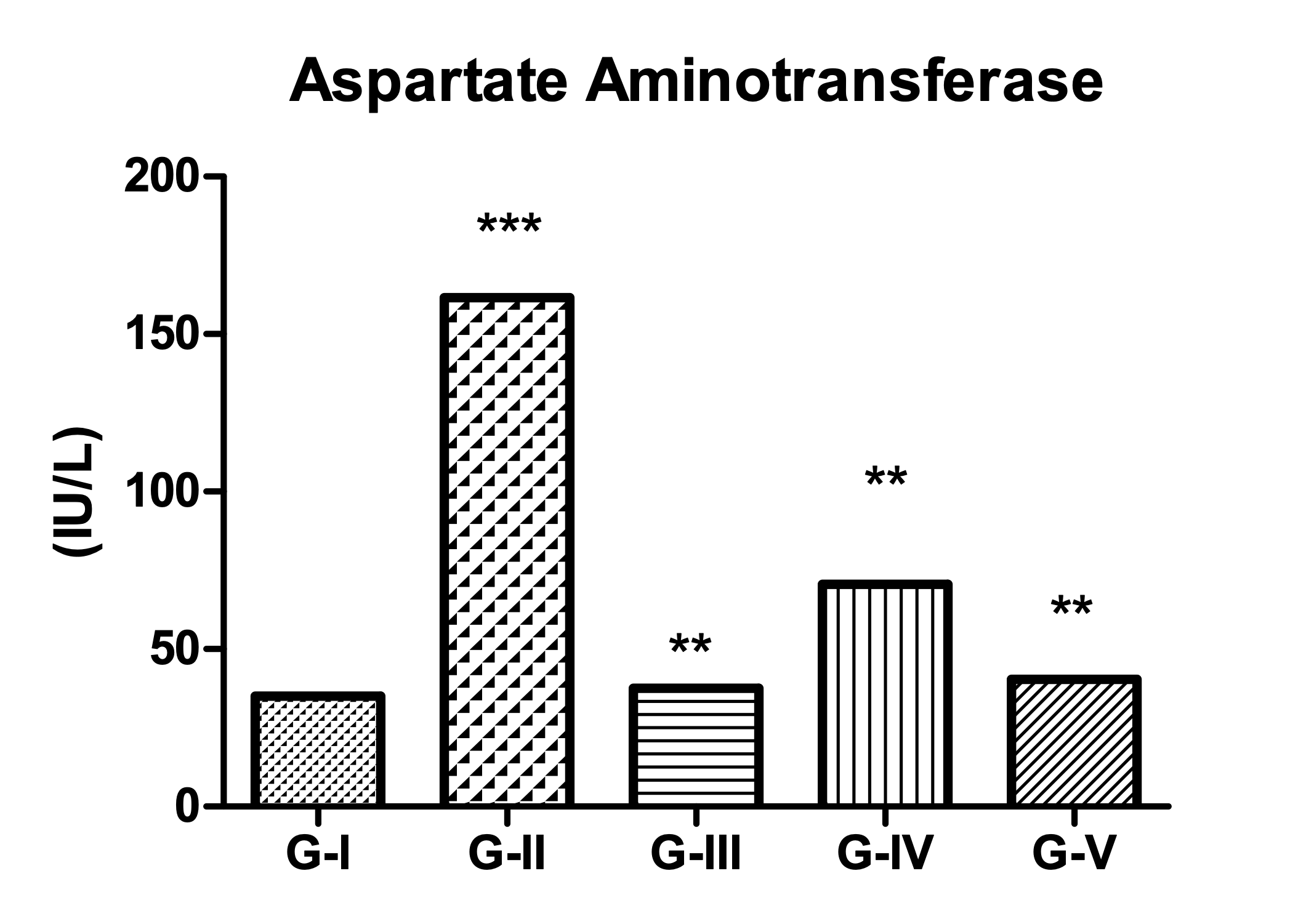
Figure 1: Effect of Treatment on AST (SGOT) levels (All values are mean ± SEM, n=6, ***p<0.001 compared to control group.) |
|||||
|
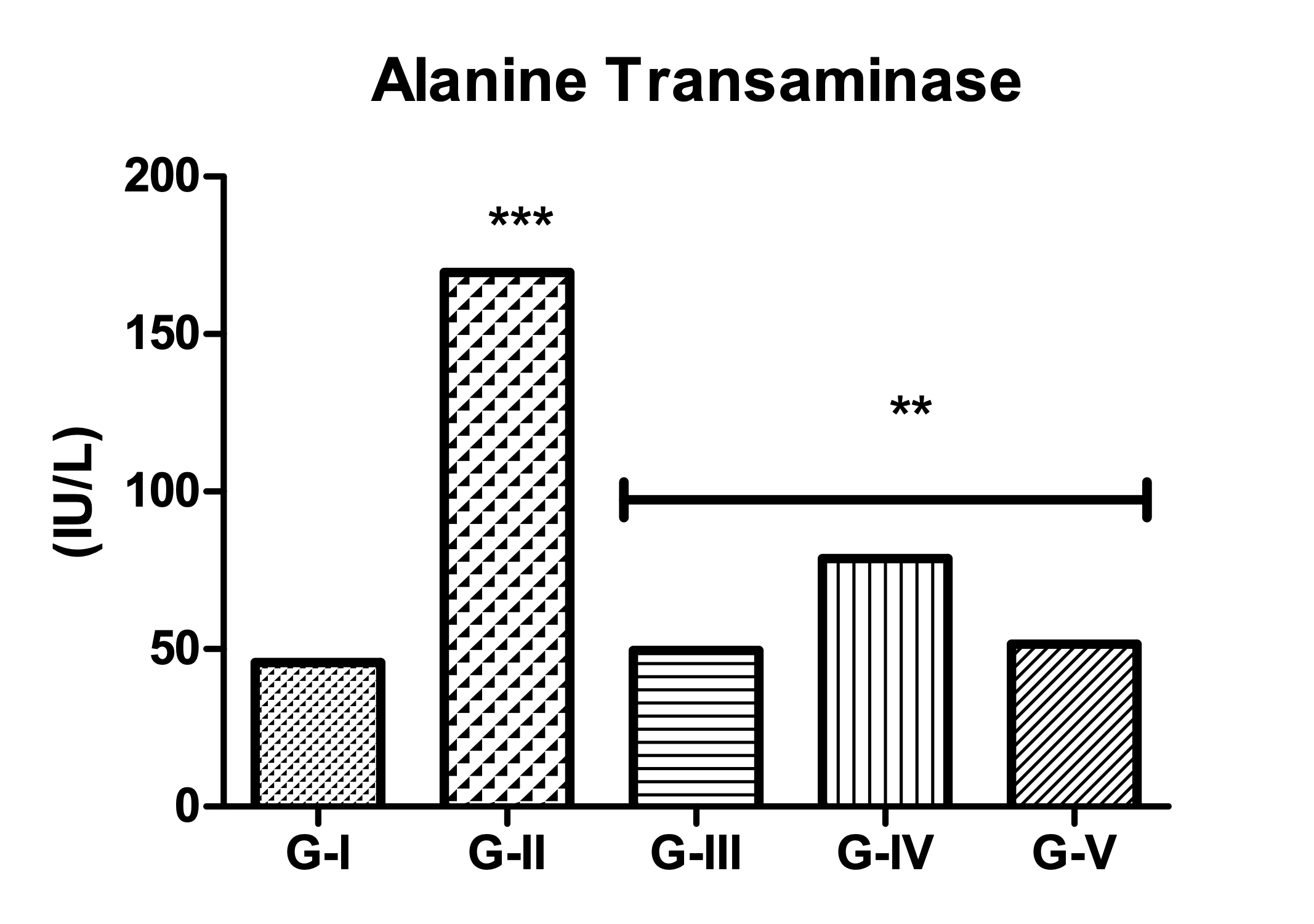
Figure 2: Effect of Treatment on ALT (SGPT) levels (All values are mean ± SEM, n=6, ***p<0.001 compared to control group.) |
|||||
|
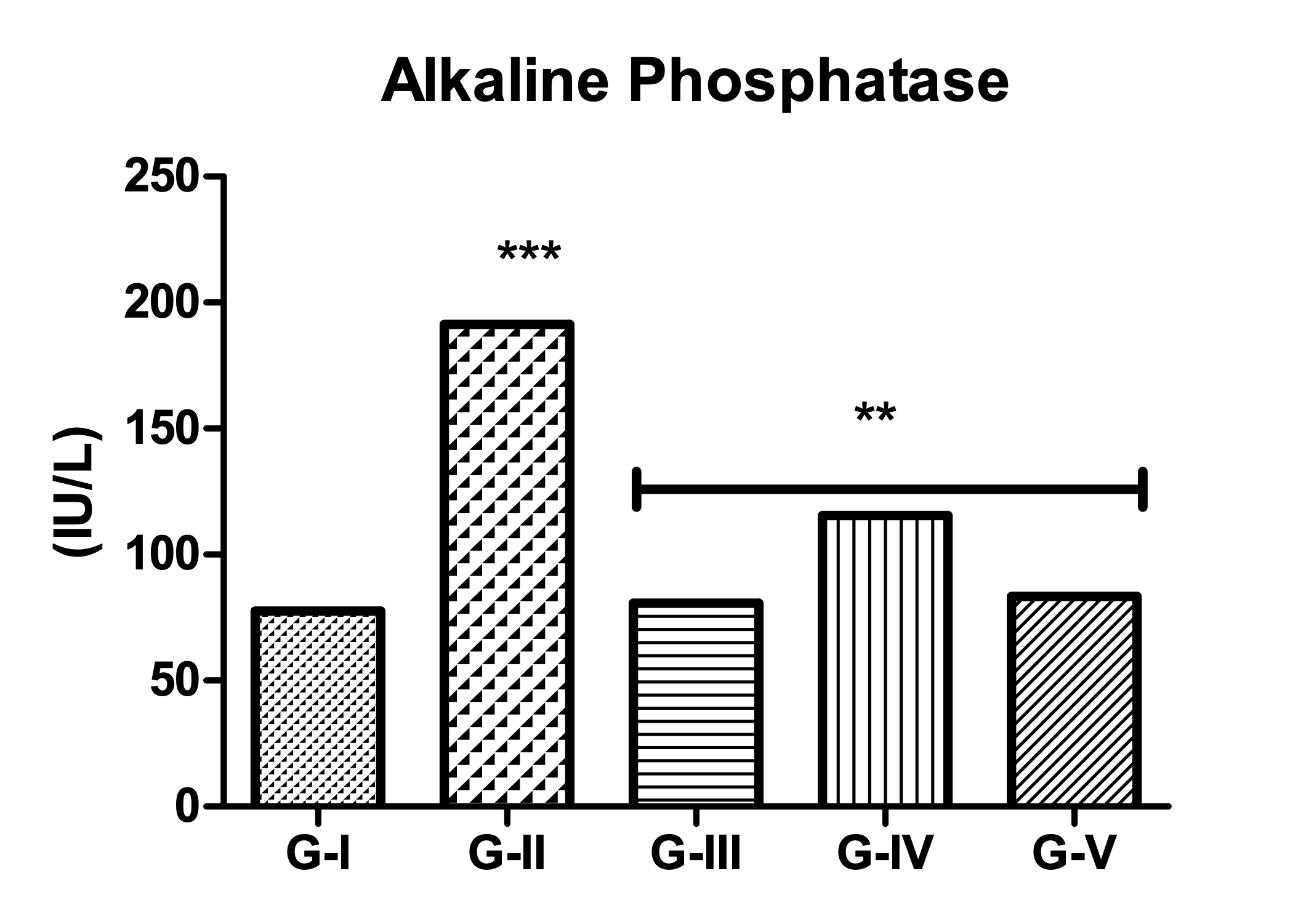
Figure 3: Effect of Treatment on ALP levels (All values are mean ± SEM, n=6, ***p<0.001 compared to control group.) |
|||||
|

Figure 4: Effect of Treatment on Total Bilirubin levels (All values are mean ± SEM, n=6, ***p<0.001 compared to control group.) |
|||||
3.2.2 Effect on Lipid Profile parameters in the plasma
Carbon tetrachloride producessignificant alterations in the various parameters of lipid profile assay as compared to the normal control animals. Treatment with Standard drug (Liv. 52) shows a significant reversal of toxicity in the levels of lipid profile parameters alongwith the treatment with PHE at a dose level of 200 and 400 mg/kg, BW but this effect was concentration dependant effect (Table 2; Figure 5-8).
Table 2: Effect on Lipid profile parameters in the plasma
|
Groups |
Treatment |
Triglyceride (mg/dL) |
Total cholesterol (mg/dL) |
HDL (mg/dL) |
LDL (mg/dL) |
|
G-I |
Normal Control |
72.65 ± 1.085 |
95.40 ± 1.327 |
36.62 ± 0.877 |
48.67 ± 0.462 |
|
G-II |
Experimental Control |
106.45 ± 2.101 |
60.75 ± 0.833 |
24.50 ± 0.993 |
25.25 ± 1.172 |
|
G-III |
Standard Control (Himalaya Liv 52) |
76.64 ± 0.515 |
93.96 ± 0.486 |
33.45 ± 0.159 |
46.26 ± 0.666 |
|
G-IV |
PHE-I |
88.83 ± 1.650 |
78.64 ± 1.120 |
26.78 ± 0.571 |
40.64 ± 0.540 |
|
G-V |
PHE-II |
67.51 ± 1.272 |
85.60 ± 1.039 |
29.64 ± 0.643 |
44.56 ± 0.647 |
|
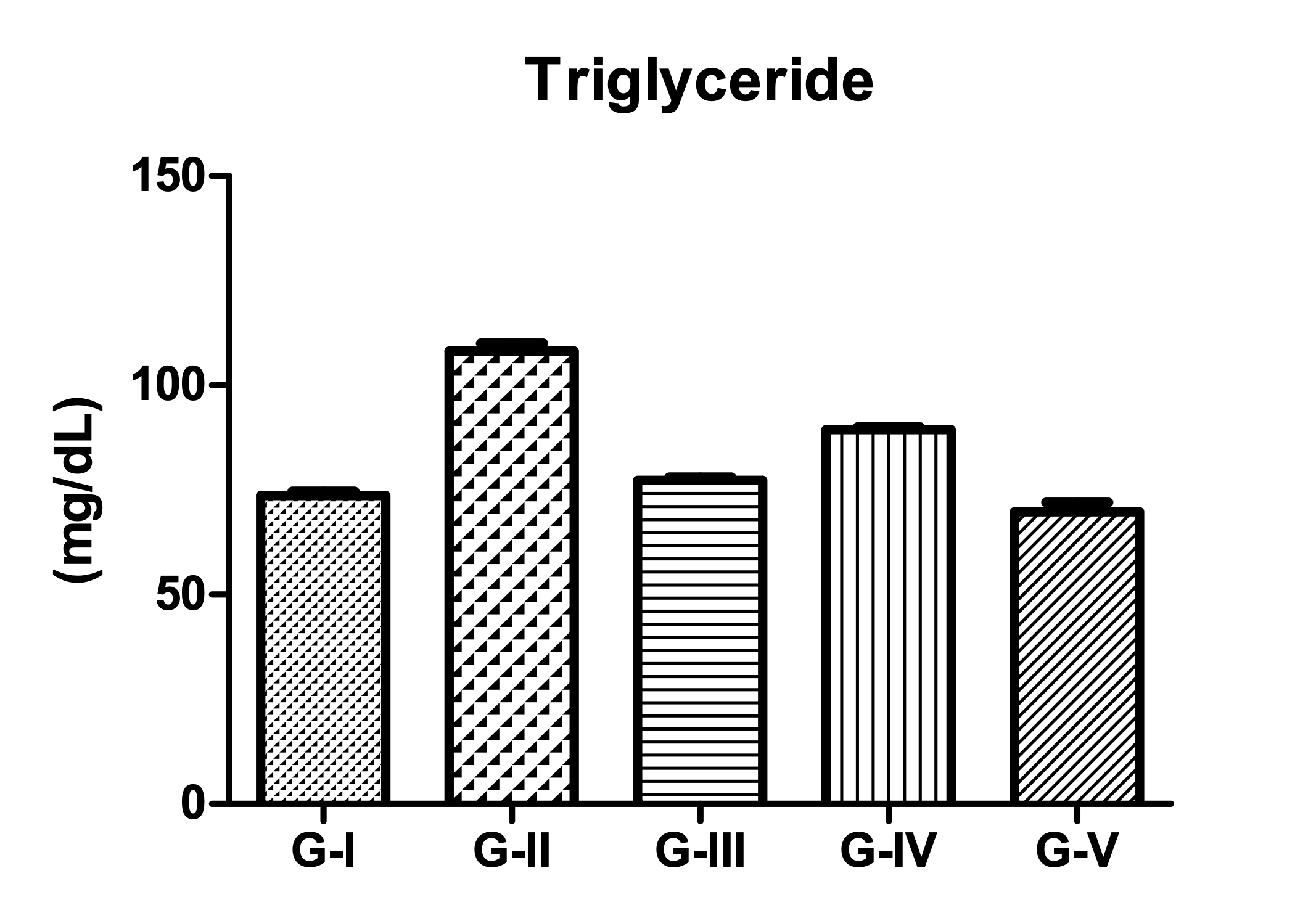
Figure 5: Effect on Triglyceride (mg/dL) (All values are mean ± SEM, n=6, ***p<0.001 compared to control group.) |
|||||
|
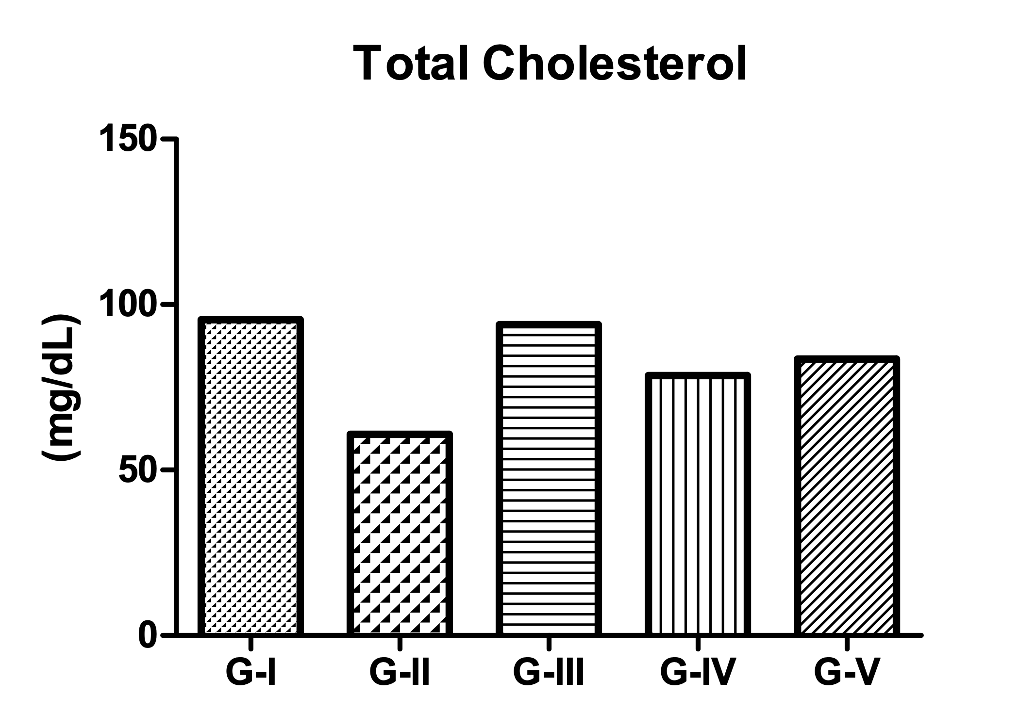
Figure 6: Effect on Total Cholesterol (mg/dL) (All values are mean ± SEM, n=6, ***p<0.001 compared to control group.) |
|||||
|
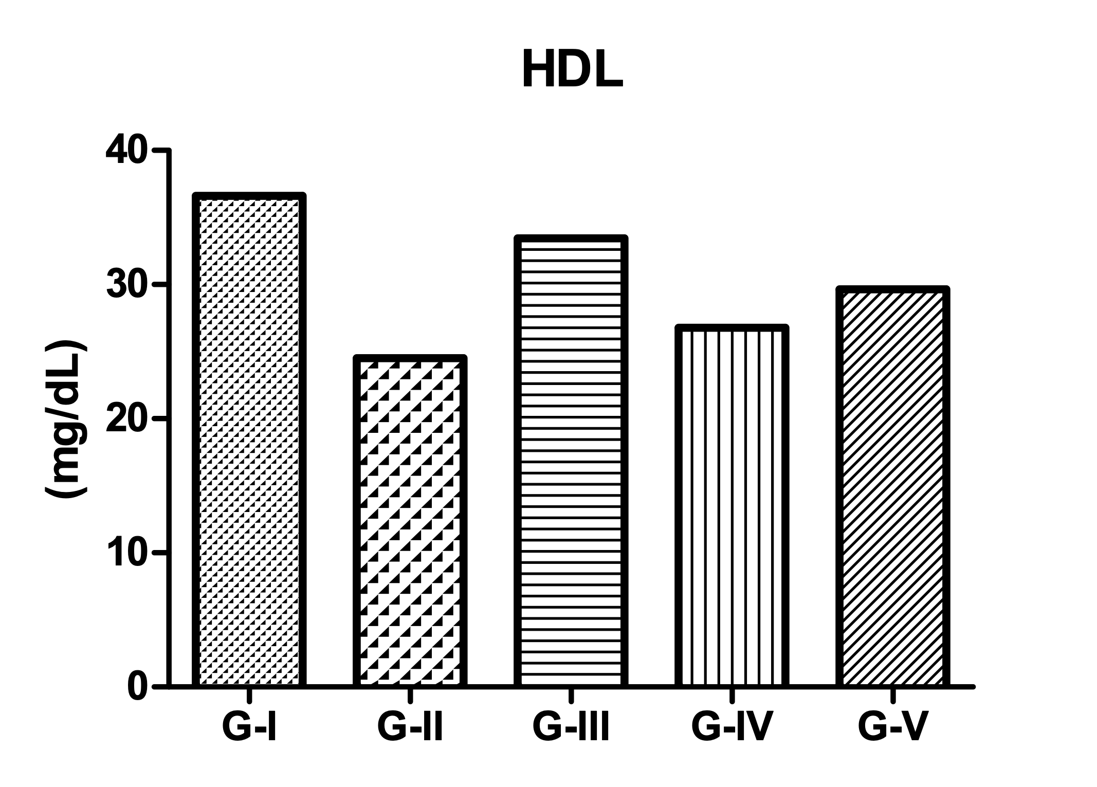
Figure 7: Effect on HDL (mg/dL) (All values are mean ± SEM, n=6, ***p<0.001 compared to control group.) |
|||||
|
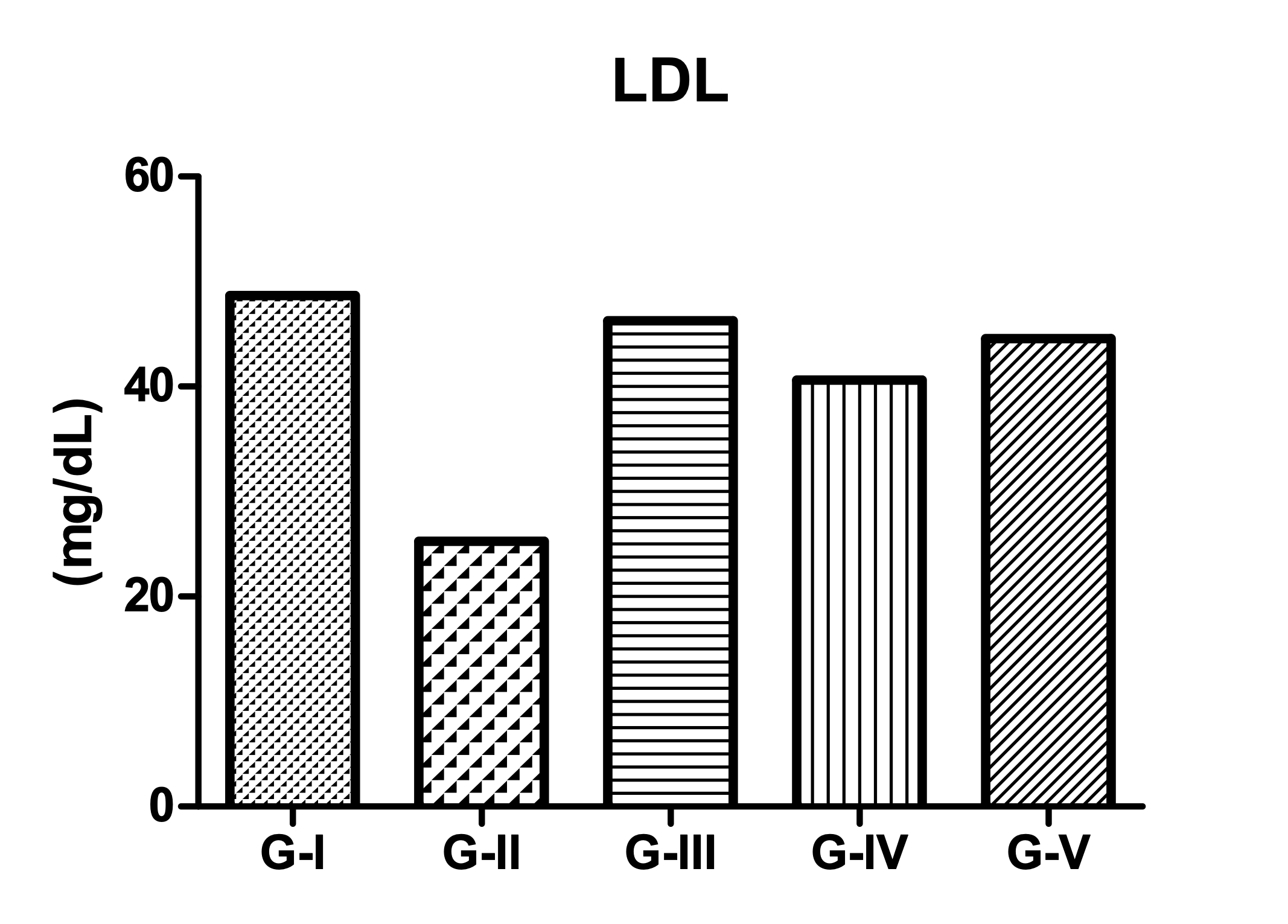
Figure 8: Effect on LDL (mg/dL) (All values are mean ± SEM, n=6, ***p<0.001 compared to control group.) |
|||||
3.2.3 Determination of Cytokines Levels (IL-6 and TNF-α) and Oxidative Stress parameter (GSH and Catalase) assay in Liver Homogenate
Carbon tetrachloride produces significant alterations in the Cytokines Levels (IL-6 and TNF-α) and Oxidative Stress parameter (GSH and Catalase) as compared to the normal control animals. Treatment with Standard drug (Liv. 52) shows a significant reversal of levels in the Cytokines Levels (IL-6 and TNF-α) and Oxidative Stress parameter (GSH and Catalase) alongwith the treatment with PHE at a dose level of 200 and 400 mg/kg, BW but this effect was concentration dependant effect (Table 3; Figure 9-12).
Table 3: Effect on Cytokines Levels (IL-6 and TNF-α) and Oxidative Stress parameter (GSH and Catalase) in Liver Homogenate
|
Groups |
Treatment |
IL-6 (pg/mg of protein) |
TNF-α (pg/mg of protein) |
GSH (µg/mg of protein) |
Catalase (µg/mg of protein) |
|
G-I |
Normal Control |
42.56 ± 0.546 |
43.59 ± 0.489 |
50.86 ± 0.587 |
66.69 ± 0.933 |
|
G-II |
Experimental Control |
73.56 ± 0.489 |
77.98 ± 0.695 |
29.10 ± 0.604 |
42.99 ± 0.920 |
|
G-III |
Standard Control (Himalaya Liv 52) |
40.26 ± 0.694 |
41.56 ± 0.695 |
48.98 ± 0.680 |
64.84 ± 0.250 |
|
G-IV |
PHE-I |
69.45 ± 0.964 |
63.65 ± 0.895 |
35.45 ± 0.454 |
58.36 ± 0.359 |
|
G-V |
PHE-II |
50.54 ± 0.556 |
55.56 ± 0.695 |
43.54 ± 0.694 |
68.04 ± 0.959 |
|
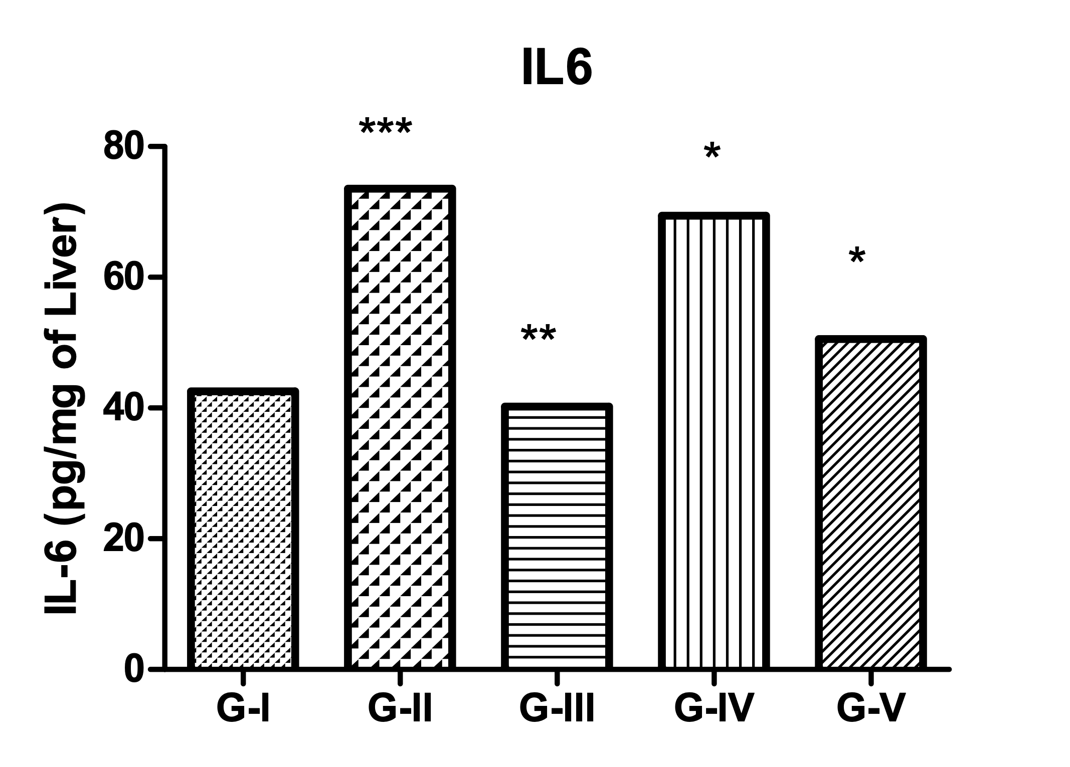
Figure 9: Effect on IL-6 (pg/mg of protein) (All values are mean ± SEM, n=6, ***p<0.001 compared to control group.) |
|||||
|
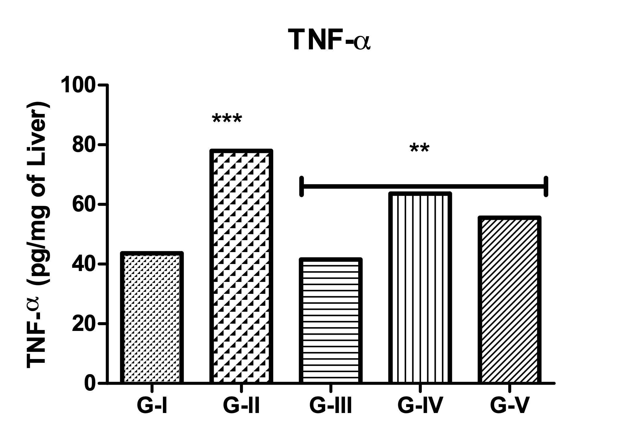
Figure 10: Effect on TNF-α (pg/mg of protein) (All values are mean ± SEM, n=6, ***p<0.001 compared to control group.) |
|||||
|
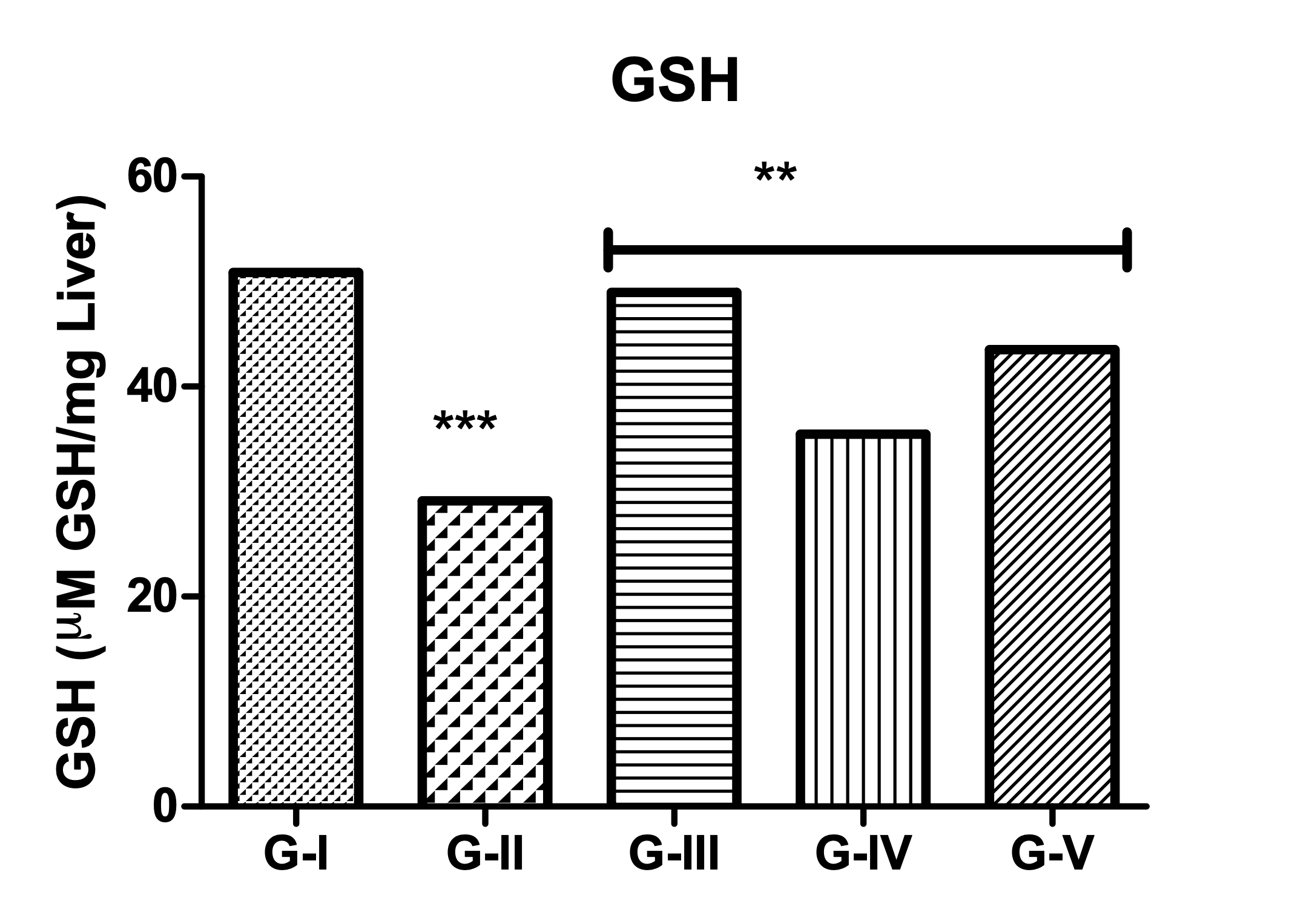
Figure 11: Effect on GSH (µg/mg of protein) (All values are mean ± SEM, n=6, ***p<0.001 compared to control group.) |
|||||
|
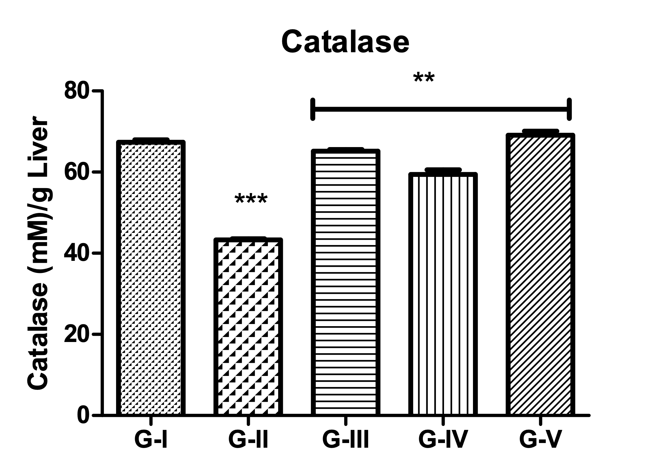
Figure 12: Effect on Catalase (µg/mg of protein) (All values are mean ± SEM, n=6, ***p<0.001 compared to control group.) |
|||||
3.3 Histopathological Examination
The control and the Standard group liver samples revealed normal ultrastructure with evident normal hepatocyte, blood sinusoid, and central vein. In the CCl4 group, histopathological abrasions like congested central vein and inflammatory cell infiltration; the hepatocytes appeared degenerated with vacuolated cytoplasm. However, in the PHF + CCl4 group, we observed amelioration regarding the degeneration, congestion and infiltration of inflammatory or fatty cells (Figure: 13).
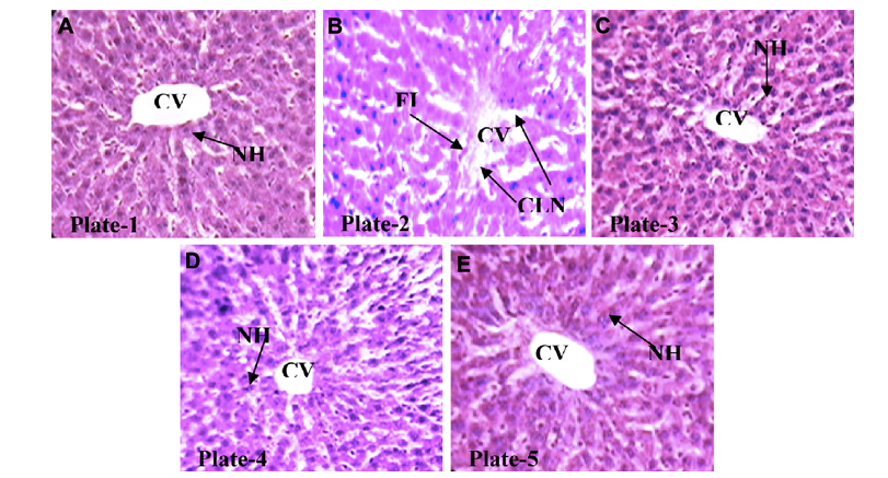
Figure 13: Histopathological Examination of Liver
[PHF impact on histopathological alterations in the liver of male albino rats is shown in (A) Healthy control, (B) Toxic control (C) Standard control (D) PHF I pretreatment and CCL4, and (E) PHF II pretreatment and CCL4 with eosin stain, original magnification 400. CLN: central lobular necrosis, CV: central vein, FI: fatty infiltration, and NH: normal hepatocyte].
4. CONCLUSIONS
In Present study, we can conclude that, This formulation preserves the activity of serum marker enzymes, which aids in the healing of hepatic tissue. At the oral dosages examined, the formulation was well tolerated and showed no symptoms of hepato-, reno-, or hematotoxicity, nor any indication of clinical toxicity.These results are confirmed by the findings of our study which is further supported by the histopathological findings. By acute and general toxicities following oral ingestion, we were able to prove that, it is a non-toxic hepatoprotective medication. This Polyherbal extract containing different types of the extract can be a best alternative for treatment of hepatotoxicity.
REFERENCES

Mrunali Dhakare*, Dr. Gopal Bihani, Dr. Pavan N. Folane, Dr. Kailash Biyani, Evaluation of In-Vivo Hepato-Protective Activity of Polyherbal Extract Against Chemically Induced-Hepatotoxic Rats, Int. J. of Pharm. Sci., 2025, Vol 3, Issue 5, 3823-3835. https://doi.org/10.5281/zenodo.15490469
 10.5281/zenodo.15490469
