Department of Pharmaceutics PES’s Modern College of Pharmacy Nigdi.
The current research focuses on the formulation and evaluation of a herbal transdermal patch containing Ocimum sanctum (Tulsi) extract, utilizing the film-forming technique for enhanced wound healing application. Tulsi is renowned for its broad-spectrum antimicrobial, antioxidant, and anti-inflammatory properties, which are essential in managing wound infections and promoting skin regeneration. Ethanolic extract of Tulsi leaves was incorporated into a polymeric film matrix composed of hydroxypropyl methylcellulose (HPMC) and polyvinyl alcohol (PVA), with glycerin serving as a plasticizer to impart flexibility and uniformity. The film-forming technique was employed to prepare transdermal patches with consistent thickness and smooth surface characteristics. The prepared patches were evaluated for various physicochemical parameters such as film thickness, folding endurance, tensile strength, surface pH, moisture content, , and drug content uniformity. In vitro drug release studies demonstrated sustained release of active constituents over a 24-hour period, indicating the potential for prolonged therapeutic action. The study concludes that the transdermal patch formulated using the film-forming technique offers a promising herbal delivery system for wound management, ensuring controlled release, antimicrobial protection, and potential for future clinical application.
Transdermal drug delivery is one of the most prevalent and widely utilized drug delivery methods. When compared to other routes of distribution, the transdermal route has attracted more attention in medication delivery due to its flexibility in palatability and convenience [1]. The transdermal route is one of the most appropriate, older, easy, safe, and cost-effective medication delivery methods. The main objectives of a transdermal medication delivery system are to target a specific region of action and to manage the rate of delivery. Transdermal drug delivery devices are self-contained, discrete dosage forms that are applied to undamaged skin. release medications into the systemic circulation at a controlled rate[2]. Wound healing is a complex physiological process involving multiple overlapping phases: hemostasis, inflammation, proliferation, and tissue remodeling. This intricate process can be hindered by factors such as microbial infection, oxidative stress, and chronic diseases, which delay healing and increase the risk of complications like chronic ulcers and secondary infections [17]. Thus, there is a pressing need for wound care strategies that accelerate healing, prevent infection, and promote tissue regeneration. Herbal medicines have gained significant interest in wound management due to their broad pharmacological activities, low toxicity, and biocompatibility. Ocimum sanctum (Tulsi), a sacred and widely used medicinal herb in Ayurveda, is reported to have a wide range of therapeutic effects including antimicrobial, anti-inflammatory, antioxidant, and wound-healing properties [18]. Phytoconstituents such as eugenol, ursolic acid, flavonoids, and tannins contribute to its bioactivity, promoting epithelialization, reducing inflammation, and protecting against microbial invasion [19]. These properties make Tulsi an ideal candidate for topical wound-healing formulations. The drug (Ocimum sanctum) was chosen because they demonstrate wound healing properties and have been tested and shown to be safe. (6,7). Transdermal drug delivery systems (TDDS) offer distinct advantages in wound care, such as sustained and localized delivery of active ingredients, bypassing first-pass metabolism, enhancing patient compliance, and providing a protective barrier over the wound surface [20]. Among these, film-forming patches are particularly promising due to their ease of application, flexibility, and capacity to create an occlusive environment favorable for wound healing [21]. In this study, a transdermal patch containing Ocimum sanctum extract was formulated using the film-forming technique. The aim was to deliver the active phytoconstituents directly to the wound site in a controlled and sustained manner, while evaluating the patch for its physicochemical properties, in vitro drug release, and antimicrobial activity. This herbal formulation seeks to combine traditional healing practices with modern pharmaceutical delivery systems for effective wound management [22].
2. MATERIALS AND METHODS
Herbal extract powder of Ocimum sanctum was obtained from the process of extracting dry leaves and roots. Hydroxypropyl Methylcellulose (HPMC), propylene glycol, DMSO
METHODOLOGY:
2.1.1 Preparation of Ocimum sanctum Extract
The leaves of plants were washed three times with tap water and then once with deionized water to wash away dirt. The washed leaves were dried in the shade at room temperature. Using a blending machine Coarse powder from the dried leaves was prepared for solvent extraction. The coarse leaf powder (50g) was soaked for 3 days in 500ml of ethanol using the cold maceration extraction method. Evaporation was used to concentrate the extracts, which were then stored in an airtight container at a cool temperature for future use. (5).
2.1.1.2 Phytochemical Analysis of Extract:
a) Dragendroff’s Test: A A volume of 2 mL of the extract solution was mixed with a few drops of Dragendroff’s reagent, which is a potassium bismuth iodide solution. The formation of an orange-red precipitate indicated the presence of alkaloids. (8).
b) Alkaline reagent test: 3 mL of the extract solution was mixed with 10 mL of distilled water and shaken. Then, 1 mL of 10% sodium hydroxide was added. The appearance of a yellow color confirmed the presence of flavonoids. (9)
c) Foam Test: The extract solution 3mL was diluted with 2 mL of distilled water in a test tube and vigorously shaken for 5 minutes. A stable foam layer at the surface signaled the presence of saponins (10).
d)Test for essential oils: Sudan III is a fat-soluble dye that selectively stains lipophilic substances like essential oils. When added to a sample containing lipids or essential oils, the dye dissolves into the oil phase, giving a distinct red or orange-red coloration, indicating the presence of oil.
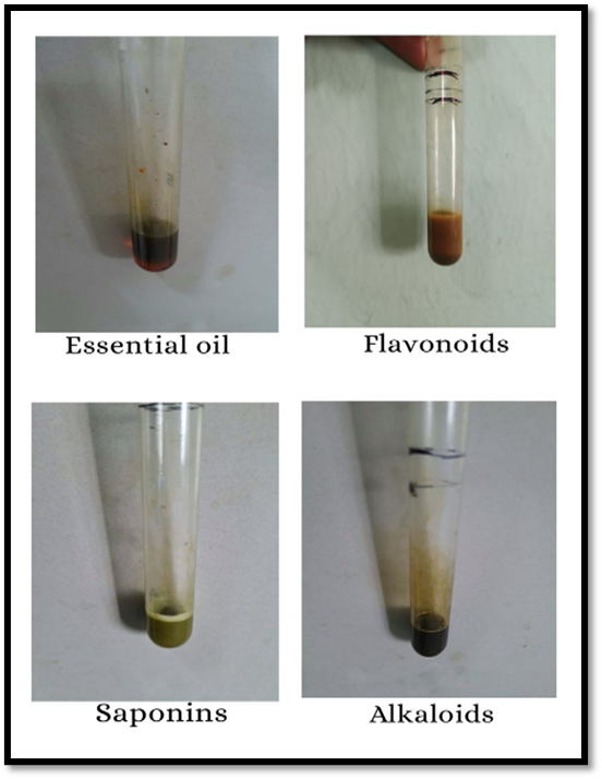
Fig 1: Phytochemical Test
2.1.2 Formulation of a transdermal patch:
Table 1: Formulation trial batches
|
Ingredient |
F1 |
F2 |
F3 |
F4 (Optimized) |
|
Ocimum sanctum |
1gm |
1gm |
1gm |
1gm |
|
HPMC |
4gm |
5gm |
6gm |
6gm |
|
Distilled water |
35ml |
35ml |
35ml |
35ml |
|
Propylene glycol |
5ml |
5ml |
5ml |
5ml |
|
DMSO |
5ml |
5ml |
5ml |
5ml |
Patch formulation
Solvent Evaporation method
Preparation of casting solution
HPMC was measured accurately and dissolved in a solvent mixture containing distilled water and ethanol in a 1:1 ratio, with continuous stirring over a hot water bath. Once the mixture reached a temperature of 25 degrees Celsius, Ocimum sanctum was incorporated into the solution. Subsequently, glycerol and propylene glycol were added as plasticizers with gentle stirring. The resulting mixture was transferred to a film-former machine and kept undisturbed for 1 hr to allow film formation.
Collection of patches
After film formation, The team removed the transdermal patches from the machine without breaking them. They cut the patches into small pieces measuring 2 × 4 cm. The prepared pieces were collected and stored in a desiccator for future use.
Fig 2: Transdermal Patch
2.1.2.1 Evaluation of Transdermal Patch:
i). Organoleptic Characteristics:
The physical appearance of the patch was examined based on its look, color, clarity, flexibility, and smoothness.
ii). Physico-Chemical Evaluation:
Fig 3: Thickness
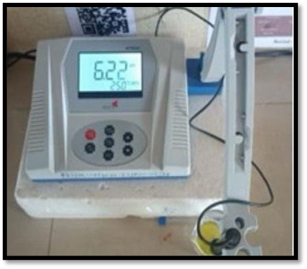
Fig 4: Ph measurement
e. Moisture Content: The films were weighed and placed in a desiccator with calcium chloride for 24 hours (see Fig. 3). They were then reweighed to calculate the percentage of moisture content using the formula below.
%Moisture content = (Initial weight - Final weight) / Initial weight × 100
f. Drug Content: A 2×4 cm area of the patch was dissolved in a phosphate buffer solution. The solution was stirred to dissolve the film, then transferred to a volumetric flask. The absorbance of the solution was measured at a wavelength of 247 nm to find the drug content.
Drug content (%) = Absorbance / Total amount of drug × 100 In-vitro Study. (11-14)
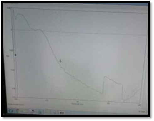
Fig 5: Drug content
In vitro drug permeation was studied using a Franz diffusion cell. This device has a donor chamber and a receptor chamber that are clamped together. The transdermal patch was placed between these two chambers. The donor chamber was filled with phosphate buffer solution, while the receptor chamber held the medium for sampling. During the experiment, the drug moved from the donor chamber through the transdermal patch into the receptor chamber. Samples were taken from the receptor chamber every 30 minutes through a sampling port. A UV spectrophotometer was used to analyze the samples and determine the total amount of drug that permeated across the membrane (15,16).
RESULT & DISCUSSION:
1.Characteristics The prepared transdermal patch of batch B1 was found to be flexible and smooth, indicating good film-forming ability and uniform surface texture. The opaque appearance suggests that the polymer and incorporated drug/extract were well dispersed but not in a crystalline transparent form. The patch was non-sticky, which is desirable for better handling and patient compliance. The light yellow colour may be attributed to the presence of plant extract and polymer combination. These properties collectively indicate that the patch possesses the necessary physical attributes for effective transdermal delivery.
Table 2: Characteristics
|
Batch |
Flexibility |
Smoothness |
Transparency |
Stickiness |
Colour |
|
B1 |
Flexible |
Smooth |
Opaque |
Non-sticky |
Light yellow |
2. Phytochemical Analysis of Extract
The ethanolic extract showed the presence of essential oils, flavonoids, saponins, and alkaloids, as confirmed by their respective qualitative tests. Essential oils (Sudan Red III test) and flavonoids (alkaline reagent test) were positive, indicating the potential for antimicrobial and anti-inflammatory activity. Saponins (foam test) were present, which may contribute to permeability enhancement and bioactivity. Alkaloids (Dragendorff’s test) were also detected, suggesting possible analgesic and therapeutic effects. The presence of these bioactive compounds supports the extract’s potential role in enhancing the pharmacological efficacy of the transdermal patch.
Table 3: Phytochemical Analysis of Extract
|
S. No. |
Constitute |
Test |
Ethanolic extract |
|
1 |
Essential oils |
Sudan Red III test |
+ve |
|
2 |
Flavonoids |
Alkaline Reagent test |
+ve |
|
3 |
Saponins |
Foam test |
+ve |
|
4 |
Alkaloids |
Dragendroff’s test |
+ve |
3. Evaluation of Transdermal Patch
The prepared transdermal patch was evaluated for its physicochemical parameters, and the findings are summarized in Table 4. The thickness of the patch was found to be 0.22 mm, indicating uniform fabrication and proper distribution of the polymer matrix. The surface pH was 6.22, which is within the skin’s physiological pH range (4.5–6.8), thereby minimizing the risk of skin irritation upon application. The percent moisture content was 4.98%, suggesting adequate moisture retention that helps maintain flexibility without promoting microbial growth. The drug content of 2.55% confirmed uniform drug incorporation within the patch matrix, ensuring dose consistency. Folding endurance was recorded at 35, indicating good flexibility and resistance to breaking upon repeated folding, which is essential for patient comfort during use. The uniformity of weight (0.0863 g) demonstrated consistent patch preparation without significant variation between samples. Overall, all parameters complied with the standard requirements, confirming the suitability of the patch for transdermal delivery.
Table 4: Evaluation of Transdermal Patch
|
Sr no |
Physicochemical Evaluation |
RESULT |
Inference |
|
1 |
Thickness of Patch |
0.22mm |
Complies |
|
2 |
Determination of Surface pH |
6.22 |
Complies |
|
3 |
Percent moisture content |
4.98% |
Complies |
|
4 |
% drug content |
2.55% |
Complies |
|
5 |
Folding endurance |
35 |
Complies |
|
6 |
Uniformity of weight |
0.0863gm |
Complies |
4: Invitro Drug Release
The cumulative drug release from the formulation increased progressively over the sampling period. At 10 min the drug release was 34.39%, which rose to 40.74% at 20 min, 42.22% at 30 min and 44.00% at 40 min. Continued sampling showed further release up to 47.90% (50 min), 54.32% (60 min), 58.27% (70 min), 62.22% (80 min) and reached 66.17% at 90 min. The profile demonstrates a steady, time-dependent increase in percent drug released reaching ~66% by 90 minutes.
Table 5: Invitro Drug Release
|
Time |
Drug Release % |
|
10 |
34.39 |
|
20 |
40.74 |
|
30 |
42.22 |
|
40 |
44 |
|
50 |
47.90 |
|
60 |
54.32 |
|
70 |
58.27 |
|
80 |
62.22 |
|
90 |
66.17 |
Conflict of Interest: The authors declared no conflict of interest.
Author Contributions: All authors have equally contributed.
Source of Support: Nill
Funding: The author declared that this study has received no financial support.
Informed Consent Statement: Not applicable.
Ethics Approval: Not Applicable
REFERENCES

Dr. Sangita A. Kale*, Mahesh D. Raut, Mahesh R. Reddy, Jaie R. Zore, Girish. P. Funde, Formulation And Evaluation of a Transdermal Patch of Ocimum Tenuiflorum for Wound Healing, Int. J. of Pharm. Sci., 2025, Vol 3, Issue 9, 1144-1151 https://doi.org/10.5281/zenodo.17090395
 10.5281/zenodo.17090395
