View Article

  • Galangin as a Therapeutic Agent for Wound Healing: Pharmacology, Challenges, and Advanced Delivery Systems

  • Matoshri College of Pharmacy, Ekhlahare, Nashik, Maharashtra, India 422105

Abstract

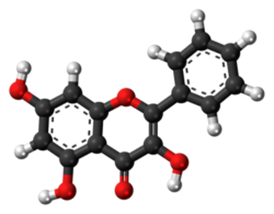
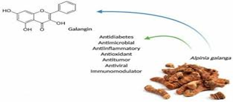
Galangin (3,5,7-trihydroxyflavone) is a naturally occurring flavonoid predominant in plants such as Alpinia officinarum (lesser galangal), galangal rhizomes and propolis. Its broad pharmacological profile includes antioxidant, anti-inflammatory, antimicrobial, anti-fibrotic, and cell-modulatory effects. Although most research has focused on cancer, cardiovascular, and fibrotic diseases, emerging evidence suggests potential roles for galangin in wound healing and dermal repair processes. However, critical challenges such as poor solubility, low bioavailability, rapid metabolism, and limited clinical translation have impeded its therapeutic development. Advanced delivery systems — including hydrogels, nanoparticles, and biopolymer carriers — provide promising strategies to overcome these limitations. This review synthesizes the current evidence on galangin’s pharmacology in wound healing, discusses mechanistic pathways, highlights challenges for clinical application, and explores advanced delivery platforms designed to enhance its therapeutic potential.Wound healing is a highly coordinated biological process involving inflammation, cell migration, angiogenesis, extracellular matrix deposition, and tissue remodeling. Impairment in any of these phases can lead to chronic, non-healing wounds, representing a significant clinical challenge. Galangin (3,5,7-trihydroxyflavone), a naturally occurring flavonoid found in Alpinia officinarum, propolis, and related plant sources, has attracted increasing attention due to its diverse pharmacological activities. Galangin exhibits potent anti-inflammatory, antioxidant, antimicrobial, and anti-fibrotic properties, which are highly relevant to key stages of wound repair. Experimental studies suggest that galangin can modulate inflammatory mediators, regulate fibroblast migration and proliferation, suppress excessive collagen deposition through the TGF-?/Smad signaling pathway, and reduce oxidative stress, thereby promoting more effective wound healing. Despite these promising effects, the therapeutic application of galangin is limited by poor aqueous solubility, low bioavailability, rapid metabolism, and insufficient in vivo and clinical evidence.

Keywords

Galangin; Wound healing; Flavonoids; Anti-inflammatory activity; Antioxidant activity; Fibroblast migration; TGF-?/Smad signaling; Chronic wounds; Drug delivery systems; Hydrogels; Nanoparticles; Controlled drug delivery

Introduction

Wound healing is a complex, multi-step physiological process involving inflammation, cell migration, angiogenesis, collagen deposition, and remodeling. Chronic wounds — including diabetic foot ulcers, venous leg ulcers, and pressure sores — present a major clinical burden due to impaired healing and infection. Natural products, particularly flavonoids such as quercetin, naringenin, and galangin, have attracted interest due to their multifaceted bioactivities that may positively influence key pathways in wound repair. Flavonoids can modulate inflammation, oxidative stress, fibroblast proliferation, extracellular matrix remodeling, and microbial growth — all of which are crucial determinants in healing outcomes.

TYPES OF WOUNDS

Wounds can be categorized into various types based on their cause, depth, and characteristics. Wounds can be categorized in following types:

  1. Incisional Wounds: These wounds are caused by a sharp object, such as a knife or scalpel, resulting in a clean, straight cut through the skin and underlying tissues.
  2. Lacerations: Lacerations are irregular, jagged wounds caused by blunt trauma or a tearing force, resulting in tissue damage with rough, uneven edges.
  3. Abrasion: Abrasions, also known as scrapes, occur when the skin rubs against a rough surface, causing the superficial layers of the skin to be scraped away.
  4. Puncture Wounds: Puncture wounds are caused by a sharp, pointed object piercing the skin, creating a small hole or puncture. These wounds are prone to infection due to their depth and narrow opening.
  5. Avulsion: Avulsion wounds occur when a portion of the skin and underlying tissues is torn away from the body, often as a result of a traumatic injury or accident.
  6. Penetrating Wounds: Penetrating wounds involve the penetration of foreign objects into the body, such as bullets, knives, or glass shards, causing damage to internal tissues and organs.
  7. Burns: Burns are injuries to the skin and underlying tissues caused by exposure to heat, chemicals, electricity, or radiation. They can range from mild superficial burns to severe deep tissue damage.
  8. Ulcers: Ulcers are open sores or lesions that develop on the skin or mucous membranes, often as a result of poor circulation, pressure, or underlying medical conditions such as diabetes or venous insufficiency.
  9. Pressure Injuries: Pressure injuries, also known as pressure ulcers or bedsores, develop when prolonged pressure or friction on the skin leads to tissue damage, particularly over bony prominences.

WOUND HEALING PROCESS:-

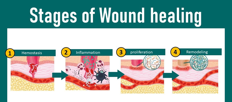
Wound healing is a complex biological process that involves several distinct but overlapping stages. These stages include hemostasis, inflammation, proliferation, and remodeling. There are the steps of wound healing:

  1. Hemostasis: Hemostasis is the initial response to injury aimed at stopping bleeding. Blood vessels constrict to reduce blood flow, and platelets aggregate at the site to form a temporary clot. This process is crucial for preventing excessive blood loss and initiating the wound healing cascade.
  2. Inflammation: Inflammation is the body's natural response to injury or infection. In this stage, immune cells such as neutrophils and macrophages migrate to the wound site to remove debris, pathogens, and damaged tissue. Inflammatory cytokines and growth factors are released to orchestrate the healing process and promote tissue repair.
  3. Proliferation: During the proliferation phase, new tissue is generated to fill the wound gap. Fibroblasts produce collagen, a key structural protein that provides strength to the healing tissue. Endothelial cells form new blood vessels (angiogenesis) to restore blood supply to the injured area. Epithelial cells at the wound edges migrate and proliferate to cover the wound surface (re-epithelialization).
  4. Remodeling:- The remodeling phase involves the maturation and remodeling of the newly formed tissue. Collagen fibers are rearranged and cross-linked to increase tensile strength, and excess scar tissue is gradually remodeled and refined. This phase can last for months to years and results in the restoration of tissue strength and function.

ADVANCE MEDICATION THERAPY INVOLVED IN WOUND HEALING

Growth Factors:- Platelet-derived growth factor (PDGF), transforming growth factor-beta (TGF-β), and epidermal growth factor (EGF) are commonly used growth factors in wound healing(4). Antimicrobial Agents: Silver-based dressings, antimicrobial peptides, and nanoparticles are among the advanced antimicrobial agents used in wound care.

Anti-inflammatory Drugs:- Corticosteroids, nonsteroidal anti-inflammatory drugs (NSAIDs), and cytokine inhibitors are advanced antiinflammatory drugs used to modulate the inflammatory response in wound healing.

Extracellular Matrix (ECM) Modulators:- Collagen-based dressings, hyaluronic acid derivatives, and extracellular matrix scaffolds are advanced ECM modulators used to facilitate tissue remodeling and regeneration.

Stem Cell Therapies: Mesenchymal stem cells (MSCs), adipose-derived stem cells (ASCs), and induced pluripotent stem cells (iPSCs) are being investigated for their potential to promote tissue regeneration in wound healing.

Gene Therapy: Gene-based approaches, such as the delivery of growth factors and cytokines, hold promise for enhancing wound healing by promoting angiogenesis and tissue regeneration.

Bioactive Dressings: Bioactive dressings containing growth factors, antimicrobial agents, and extracellular matrix components provide sustained release and localized delivery of therapeutic molecules to the wound site.

ADVANCE DRUG DELIVERY SYSTEM

Advanced drug delivery systems for wound healing have emerged as a promising approach to improve the efficacy of wound care treatments. These systems aim to optimize the delivery of therapeutic agents to the wound site, enhancing their bioavailability, and promoting faster and more efficient healing. There are some notable advanced drug delivery systems for wound healing:

Hydrogels: Hydrogels are highly hydrated polymeric networks that can retain large amounts of water. They are attractive for wound healing applications due to their ability to provide a moist environment conducive to wound healing. Hydrogels can be loaded with therapeutic agents such as growth factors, antimicrobial agents, and anti-inflammatory drugs, which are released slowly over time to the wound site.

Nanoparticles:- Nanoparticles offer unique advantages for drug delivery, including high drug loading capacity, sustained release profiles, and the ability to target specific cells or tissues. Various types of nanoparticles, such as liposomes, polymeric nanoparticles, and dendrimers, have been investigated for wound healing applications. These nanoparticles can encapsulate therapeutic agents and protect them from degradation, enabling controlled release at the wound site.

Microneedle patches:- Microneedle patches consist of arrays of micron-scale needles that penetrate the skin's outer layer, facilitating the delivery of drugs into the underlying tissue. These patches can be loaded with various therapeutic agents, including growth factors, antibiotics, and anti-inflammatory drugs, and applied directly to the wound site for targeted delivery and enhanced efficacy.

Electrospun nanofibers:- Electrospun nanofibers are produced by electrospinning polymer solutions into nanoscale fibers, which can mimic the extracellular matrix structure of natural tissues. These nanofibers can be loaded with therapeutic agents and fabricated into wound dressings, providing sustained release of drugs and promoting cell proliferation and tissue regeneration.

3D printed scaffolds: 3D printing technology enables the fabrication of customized scaffolds with precise control over their architecture and porosity. These scaffolds can be loaded with therapeutic agents and implanted at the wound site to provide structural support, promote cell attachment and proliferation, and facilitate controlled drug release for enhanced wound healing.

MARKETED FORMULATION USED AS ADVANCED DRUG DELIVERY SYSTEM FOR THE TREATMENT OF WOUND HEALING

Advanced drug delivery systems hold great promise for improving the treatment outcomes of various types of wounds, including acute and chronic wounds, burns, and ulcers, by enhancing the delivery and efficacy of therapeutic agents at the wound site. Several advanced drug delivery systems and marketed products are utilized in the treatment of wound healing:

Ag Hydro fiber Dressing:- Ag is a sterile, nonwoven dressing composed of sodium carboxymethylcellulose and ionic silver. It forms a soft gel when in contact with wound exudate, providing a moist wound healing environment. while delivering antimicrobial benefits due to the presence of silver ions. This dressing is effective for managing wounds with high levels of exudate and controlling infection.

Hydro-Alginate Antimicrobial Dressing:- Ag is a sterile, non-woven dressing composed of alginate fibers and silver ions. It absorbs exudate from the wound while providing sustained antimicrobial activity against a broad spectrum of pathogens. The combination of alginate and silver facilitates autolytic debridement and promotes granulation tissue formation.

Antimicrobial Activated Charcoal Dressing:- Silver 220 is an activated charcoal dressing containing silver ions. It absorbs Odor and exudate from the wound while providing broad-spectrum antimicrobial activity. The activated charcoal component helps manage malodor associated with infected wounds.

Dermal Regeneration Template:- It is a bilayer membrane system consisting of a porous matrix of bovine collagen and glycosaminoglycan (GAG) and a semipermeable silicone outer layer. It serves as a dermal substitute for full-thickness wounds, promoting vascularization and cellular infiltration. It is commonly used in the treatment of burn injuries and complex wounds requiring soft tissue reconstruction.

SOME HERBAL APPROACHESUSED AS ADVANCED DRUG DELIVERY SYSTEM FOR THE TREATMENT OF WOUND HEALING

Turmeric (Curcuma longa) Loaded Nanoparticles:- Turmeric, containing curcumin as its active ingredient, has been traditionally used for wound healing due to its anti-inflammatory and antioxidant properties. Incorporating curcumin into nanoparticles enhances its stability and bioavailability, enabling controlled release at the wound site.

Aloe vera Loaded Hydrogels:- Aloe vera is known for its wound healing properties attributed to its anti-inflammatory and antimicrobial effects. Incorporating Aloe vera extract into hydrogel formulations provides a moist wound environment and sustained release of bioactive compounds, promoting tissue regeneration.

Honey-based Nanofiber Scaffolds:- Honey has been used for wound healing for centuries due to its antimicrobial and anti-inflammatory properties. Fabricating honey into nanofiber scaffolds enhances its application to wounds, providing sustained release and improved efficacy.

Neem (Azadirachta indica) Oil-loaded Liposomes:- Neem oil possesses antimicrobial and anti-inflammatory properties beneficial for wound healing. Encapsulating neem oil in liposomes improves its stability and enhances its penetration into the skin, facilitating wound closure and tissue regeneration.

Pharmacology of Galangin in Wound Healing

Galangin accelerates the wound healing process by modulating several biochemical pathways across the phases of tissue repair (hemostasis, inflammation, proliferation, and remodeling).

  • Anti-inflammatory Activity: Galangin suppresses the production of pro-inflammatory cytokines such as TNF-$\alpha$, IL-6, and IL-1$\beta$. It inhibits the NF-$\kappa$B and MAPK signaling pathways, which are often overactive in chronic wounds, preventing excessive tissue damage.
  • Antioxidant Effects: It acts as a powerful scavenger of reactive oxygen species (ROS). By upregulating antioxidant enzymes (like SOD and CAT) and activating the Nrf2/HO-1 pathway, it protects fibroblasts and keratinocytes from oxidative stress-induced apoptosis.
  • Antimicrobial Potential: Galangin exhibits broad-spectrum activity against wound pathogens, including Staphylococcus aureus. It disrupts bacterial cell membranes and inhibits the expression of genes related to biofilm formation, which is a major barrier to healing in infected wounds.
  • Tissue Remodeling: It modulates Matrix Metalloproteinases (MMP-2 and MMP-9), ensuring that the extracellular matrix (ECM) is properly reorganized rather than excessively degraded, which helps in reducing scar formation.

This review explores the therapeutic potential of Galangin ($3,5,7$-trihydroxyflavone), a natural flavonol primarily derived from Alpinia galanga (galangal) and propolis, in the context of cutaneous wound healing. While galangin exhibits potent pharmacological properties, its clinical application is hindered by poor solubility and bioavailability, necessitating the development of advanced delivery systems.

1. Pharmacology of Galangin in Wound Healing

Galangin accelerates the wound healing process by modulating several biochemical pathways across the phases of tissue repair (hemostasis, inflammation, proliferation, and remodeling).

  • Anti-inflammatory Activity: Galangin suppresses the production of pro-inflammatory cytokines such as TNF-2$\alpha$, IL-6, and IL-1$\beta$.3 It inhibits the NF-4$\kappa$B and MAPK signaling pathways, which are often overactive in chronic wounds, preventing excessive tissue damage.
  • Antioxidant Effects: It acts as a powerful scavenger of reactive oxygen species (ROS).6 By upregulating antioxidant enzymes (like SOD and CAT) and activating the Nrf2/HO-1 pathway, it protects fibroblasts and keratinocytes from oxidative stress-induced apoptosis.
  • Antimicrobial Potential: Galangin exhibits broad-spectrum activity against wound pathogens, including Staphylococcus aureus. It disrupts bacterial cell membranes and inhibits the expression of genes related to biofilm formation, which is a major barrier to healing in infected wounds.
  • Tissue Remodeling: It modulates Matrix Metalloproteinases (MMP-2 and MMP-9), ensuring that the extracellular matrix (ECM) is properly reorganized rather than excessively degraded, which helps in reducing scar formation.

 Challenges in Therapeutic Application

Despite its efficacy in in vitro models, galangin faces several hurdles for human clinical use:

Challenge

Description

Low Solubility

Galangin is highly hydrophobic, making it difficult to incorporate into traditional water-based topical gels or creams.

Poor Bioavailability

Rapid metabolism and low skin penetration limit the amount of active compound that reaches the deeper dermal layers.

Stability Issues

It is sensitive to environmental factors such as light, temperature, and pH, which can degrade the molecule before it exerts its effect.

Rapid Washout

When applied as a simple solution or powder, it is easily removed by wound exudate, requiring frequent reapplication.

Advanced Delivery Systems

To overcome these challenges, researchers are utilizing nanotechnology and biomaterials to enhance galangin's delivery to the wound site.

Nano-Carrier Systems

  • Liposomes & Niosomes: Encapsulating galangin in lipid bilayers improves its solubility and allows for a controlled, sustained release. These vesicles can fuse with skin cell membranes to enhance deep-tissue penetration.
  • Polymeric Nanoparticles (PLGA/ Chitosan): Biodegradable polymers protect galangin from enzymatic degradation in the wound bed. Chitosan-based particles also provide inherent antimicrobial synergy.
  • Gold Nanoparticles (AuNPs): Galangin-conjugated AuNPs have shown promise in modulating angiogenesis and cell migration, potentially speeding up the proliferative phase of healing.

Scaffolds and Hydrogels

  • Electrospun Nanofibers: Mimicking the natural ECM, these fibers can be loaded with galangin to provide a high surface-area-to-volume ratio for drug release while providing a physical scaffold for cell growth.
  • Smart Hydrogels: These "intelligent" dressings can respond to the wound environment (e.g., pH or temperature changes) to trigger the release of galangin exactly when inflammation or infection levels rise.

Pharmacology: How Galangin Heals

Galangin acts as a "biological orchestrator" that accelerates the body's natural repair mechanisms through several distinct pathways:

  • Anti-inflammatory Control: It suppresses the NF-$\kappa$B and MAPK signaling pathways. By doing so, it reduces the release of pro-inflammatory cytokines such as TNF-$\alpha$ and IL-6, preventing wounds from becoming "stuck" in a chronic inflammatory state.
  • Oxidative Stress Reduction: It activates the Nrf2/ARE pathway, which triggers the production of internal antioxidants like Superoxide Dismutase (SOD) and Catalase. This protects new cells from being destroyed by Reactive Oxygen Species (ROS).
  • Promotion of Angiogenesis: Galangin has been shown to upregulate VEGF (Vascular Endothelial Growth Factor), which stimulates the formation of new blood vessels, ensuring the healing tissue receives oxygen and nutrients.
  • Extracellular Matrix (ECM) Stability: It regulates Matrix Metalloproteinases (MMPs), ensuring that collagen is deposited in an organized fashion rather than being prematurely broken down.

Clinical Challenges: The Solubility Barrier

Despite its high efficacy in laboratory tests, Galangin faces three primary "bottlenecks" that hinder its use as a standard medical treatment:

  1. Poor Solubility: It is highly hydrophobic (water-hating), meaning it doesn't dissolve well in standard medical gels or blood, leading to poor absorption.
  2. Low Bioavailability: When applied to the skin, very little of the drug actually reaches the deep dermal layers where the most critical healing occurs.
  3. Environmental Sensitivity: It can degrade quickly when exposed to light or the varying pH levels of a wound environment.

3. Advanced Delivery Systems

To solve these problems, researchers are using Nanotechnology to "wrap" Galangin in protective carriers:

Delivery System

Mechanism of Action

Benefit

Nanofibrous Scaffolds

Mimics the skin's physical structure.

Provides a "home" for cells to grow while releasing drug slowly.

Liposomes/ Niosomes

Fatty "bubbles" that carry the drug.

Penetrates the skin's lipid barrier more effectively.

Hydrogel Nanocomposites

Smart gels that respond to pH or heat.

Releases Galangin only when the wound is inflamed or infected.

Gold Nanoparticles

Metallic carriers conjugated with Galangin.

Enhances antimicrobial action and improves targeted delivery.

Pharmacological Mechanisms in Wound Healing

Galangin accelerates tissue repair by orchestrating cellular responses across the four stages of wound healing (Hemostasis, Inflammation, Proliferation, and Remodeling).

A. Anti-inflammatory & Immunomodulatory Activity1

Chronic wounds often stall in the inflammatory phase.2 Galangin effectively "unlocks" this stage by:

  • NF-$\kappa$B Inhibition: It prevents the phosphorylation of I$\kappa$B$\alpha$, thereby blocking the NF-$\kappa$B pathway and reducing the production of pro-inflammatory cytokines like TNF-$\alpha$, IL-6, and IL-1$\beta$.
  • MAPK Pathway Regulation: By modulating Mitogen-Activated Protein Kinase (MAPK) signaling, it controls the recruitment of neutrophils and macrophages to the wound site.

B. Oxidative Stress Mitigation

Excessive ROS (Reactive Oxygen Species) destroys healthy tissue. Galangin acts as a "bio-shield" through:

  • Nrf2/HO-1 Activation: It stimulates the Nrf2 pathway, leading to increased production of antioxidant enzymes like Superoxide Dismutase (SOD) and Catalase (CAT).
  • Lipid Peroxidation Reduction: It decreases levels of Malondialdehyde (MDA), preserving the integrity of cell membranes in fibroblasts and keratinocytes.

C. Angiogenesis and Tissue Regeneration

  • Growth Factor Modulation: Galangin has been shown to upregulate VEGF (Vascular Endothelial Growth Factor) and TGF-$\beta$1, promoting the formation of new capillary networks essential for oxygenating the wound bed.
  • Stem Cell Activation: Recent 2024–2025 studies suggest Galangin can activate Tendon-Derived Stem Cells (TDSCs) and mesenchymal stem cells via JAK2/STAT3 signaling, facilitating deeper tissue repair.

D. Remodeling & Anti-Scarring

  • MMP Regulation: It inhibits Matrix Metalloproteinases (MMP-2 and MMP-9), ensuring that the extracellular matrix (ECM) is built effectively without excessive degradation.
  • Hypertrophic Scar Prevention: By modulating the TGF-6$\beta$/Smad pathway, it balances collagen types I and III, reducing the risk of abnormal, raised scarring.

Key Challenges in Therapeutic Application

Despite its benefits, Galangin’s "natural" form presents significant hurdles for clinical use:

Barrier

Impact on Wound Healing

Hydrophobicity

Virtually insoluble in water; difficult to formulate into standard gels.

Poor Bioavailability

Rapidly metabolized and poorly absorbed through the thick skin barrier.

Stability Issues

Highly sensitive to light and pH; degrades quickly in the alkaline environment of chronic wounds.

Washout Effect

Free Galangin is quickly diluted or removed by wound exudate (fluid).

3. Advanced Delivery Systems (The "Solution")

To bridge the gap between lab and clinic, researchers are using Nano-Medicine to deliver Galangin effectively:

  • Nanofibrous Scaffolds: Electrospun fibers (often made of Chitosan or PLGA) mimic the skin's natural structure. They act as a "second skin" that releases Galangin slowly while allowing the wound to "breathe."
  • Nanostructured Lipid Carriers (NLCs): These lipid-based "bubbles" encapsulate Galangin, allowing it to penetrate the fatty layers of the skin and reach the deep dermis where regeneration occurs.
  • Stimuli-Responsive Hydrogels: "Smart" hydrogels that release Galangin only when they sense a specific trigger, such as a change in wound temperature or the presence of bacterial enzymes (pH-triggered release).
  • Metallic Nanoparticles: Conjugating Galangin with Gold Nanoparticles (AuNPs) has been shown to enhance its antimicrobial and anti-tumor properties, providing a dual-action dressing for infected wounds or post-surgical recovery.

CONCLUSION

In summary, the transition of Galangin ($3,5,7$-trihydroxyflavone) from a traditional herbal constituent to a sophisticated therapeutic agent represents a significant advancement in the field of regenerative medicine. This review has delineated the multifaceted role of Galangin in wound healing, underscoring its ability to modulate the complex biochemical environment of injured tissue through a synergy of pharmacological actions, while also addressing the technological hurdles that have historically limited its clinical utility.

Pharmacological Integration

The pharmacological profile of Galangin is remarkably comprehensive, addressing the primary barriers to successful tissue repair. Its ability to act as a potent anti-inflammatory agent via the inhibition of the NF-1$\kappa$B and MAPK pathways is critical in preventing the chronicity of wounds.2 Furthermore, its role in oxidative stress management through the Nrf2/HO-1 axis provides a "bio-shield" for migrating fibroblasts and keratinocytes. Perhaps most importantly, Galangin’s influence on angiogenesis and the regulation of Matrix Metalloproteinases (MMPs) ensures that the remodeling phase results in functional tissue rather than pathological fibrosis. This multi-target approach makes it superior to single-target synthetic drugs that often fail to address the holistic complexity of the wound bed.

Addressing the Bioavailability Gap

Despite these benefits, the "Galangin Paradox"—the gap between its high in vitro efficacy and poor in vivo performance—remains the central challenge. The molecule’s inherent hydrophobicity, low aqueous solubility, and susceptibility to rapid metabolic degradation necessitate a departure from conventional topical formulations. Simple ointments and creams are insufficient to maintain the sustained therapeutic concentrations required for deep-tissue repair, particularly in the presence of wound exudates that can easily wash away free active compounds.

The Future of Advanced Delivery

The emergence of advanced delivery systems marks the "enabling" phase of Galangin research. The integration of nanotechnology, including liposomes, polymeric nanoparticles, and electrospun scaffolds, has successfully transformed Galangin into a viable clinical candidate. These systems do more than just deliver the drug; they protect the molecule from environmental degradation, enhance its penetration through the skin barrier, and provide a sustained-release profile that reduces the need for frequent dressing changes. The development of "smart" hydrogels, which can release Galangin in response to specific wound triggers such as pH shifts or bacterial enzymes, represents the next frontier in personalized wound management.

Final Verdict

In conclusion, Galangin stands as a promising "all-in-one" natural therapeutic for the treatment of acute and chronic wounds, including diabetic ulcers and severe burns. While the laboratory evidence is robust, the future of Galangin-based therapy depends on the successful translation of these advanced delivery platforms into human clinical trials. By bridging the gap between ancient pharmacology and modern material science, Galangin-loaded biomaterials are poised to become a cornerstone in the next generation of wound-care products, offering a cost-effective, natural, and highly efficient solution for global healthcare challenges.

REFERENCES

  1. Lynch SE, Trippel SB, Finkelman RD, Hernandez RA, Kiritsy CP, Antoniades HNJWR, et al. The combination of platelet? derived growth factor? BB and insulin? like growth factor? I stimulates bone repair in adult Yucatan miniature pigs. 1994;2(3):182-90.
  2. Cowan JB, Bedi A, Carpenter JE, Robbins CB, Gagnier JJ, Miller BSJJoS, et al. Evaluation of American Academy of Orthopaedic Surgeons Appropriate Use Criteria for the management of fullthickness rotator cuff tears. 2016;25(7):1100-6.
  3. Gurtner GC, Werner S, Barrandon Y, Longaker MTJN. Wound repair and regeneration. 2008;453(7193):314-21.
  4. Barrientos S, Brem H, Stojadinovic O, Tomic? Canic MJWR, Regeneration. Clinical application of growth factors and cytokines in wound healing. 2014;22(5):569-78.
  5. Bowler P, Duerden B, Armstrong DGJCmr. Wound microbiology and associated approaches to wound management. 2001;14(2):244-69.
  6. Eming SA, Krieg T, Davidson JMJJoID. Inflammation in wound repair: molecular and cellular mechanisms. 2007;127(3):514-25.
  7. Veves AJDC. Apligraf Diabetic Foot Ulcer Study. Graftskin, a human skin equivalent, is effective in the management of noninfected neuropathic diabetic foot ulcers: a prospective randomized multicenter clinical trial. 2001;24:290-5
  8. Rees PA, Greaves NS, Baguneid M, Bayat AJAiwc. Chemokines in wound healing and as potential therapeutic targets for reducing cutaneous scarring. 2015;4(11):687-703.
  9. Zhang F, Xu C-L, Liu C-MJDD, Development, Therapy. Drug delivery strategies to enhance the permeability of the blood–brain barrier for treatment of glioma. 2015:2089-100.
  10. Schultz GS, Davidson JM, Kirsner RS, Bornstein P, Herman IMJWR, Regeneration. Dynamic reciprocity in the wound microenvironment. 2011;19(2):134-48.
  11. Li J, Mooney DJJNRM. Designing hydrogels for controlled drug delivery. 2016;1(12):1-17.
  12. Giovagnoli S, Mancuso F, Vannini S, Calvitti M, Piroddi M, Pietrella D, et al. Microparticle-loaded neonatal porcine Sertoli cells for cell-based therapeutic and drug delivery system. 2014;192:249-61.
  13. Larrañeta E, Lutton RE, Woolfson AD, Donnelly RFJMS, Reports ER Microneedle arrays as transdermal and intradermal drug delivery systems: Materials science, manufacture and commercial development. 2016;104:1-32.
  14. Sill TJ, Von Recum HAJB. Electrospinning: applications in drug delivery and tissue engineering. 2008;29(13):1989-2006.
  15. Murphy SV, Atala AJNb. 3D bioprinting of tissues and organs. 2014;32(8):773-85.
  16. Bowler P, Jones S, Walker M, Parsons DJJoBC, Rehabilitation. Microbicidal properties of a silver-containing Hydrofiber® dressing against a variety of burn wound pathogens. 2004;25(2):192-6.
  17. Thomas S, McCubbin PJJowc. A comparison of the antimicrobial effects of four silver-containing dressings on three organisms. 2003;12(3):101-7.
  18. Swanson T, Angel D, Sussman G, Cooper R, Haesler E, Ousey K, et al. Wound infection in clinical practice: principles of best practice. 2016.
  19. Burke JF, Yannas IV, Quinby Jr WC, Bondoc CC, Jung WKJAos. Successful use of a physiologically acceptable artificial skin in the treatment of extensive burn injury. 1981;194(4):413.
  20. Refaat H, Mady FM, Sarhan HA, Rateb HS, Alaaeldin EJIjop. Optimization and evaluation of propolis liposomes as a promising therapeutic approach for COVID-19. 2021;592:120028.
  21. Glover RL, Nyanganyura D, Mufamadi MS, Mulaudzi RB. Green Synthesis in Nanomedicine and Human Health: CRC Press; 2021.
  22. Almal SH, Padh HJIJoPS. Variation in Copy Number of MTUS1 Gene among Healthy Individuals and Cancer Patients from Gujarat. 2017;79(2).   

Reference

  1. Lynch SE, Trippel SB, Finkelman RD, Hernandez RA, Kiritsy CP, Antoniades HNJWR, et al. The combination of platelet? derived growth factor? BB and insulin? like growth factor? I stimulates bone repair in adult Yucatan miniature pigs. 1994;2(3):182-90.
  2. Cowan JB, Bedi A, Carpenter JE, Robbins CB, Gagnier JJ, Miller BSJJoS, et al. Evaluation of American Academy of Orthopaedic Surgeons Appropriate Use Criteria for the management of fullthickness rotator cuff tears. 2016;25(7):1100-6.
  3. Gurtner GC, Werner S, Barrandon Y, Longaker MTJN. Wound repair and regeneration. 2008;453(7193):314-21.
  4. Barrientos S, Brem H, Stojadinovic O, Tomic? Canic MJWR, Regeneration. Clinical application of growth factors and cytokines in wound healing. 2014;22(5):569-78.
  5. Bowler P, Duerden B, Armstrong DGJCmr. Wound microbiology and associated approaches to wound management. 2001;14(2):244-69.
  6. Eming SA, Krieg T, Davidson JMJJoID. Inflammation in wound repair: molecular and cellular mechanisms. 2007;127(3):514-25.
  7. Veves AJDC. Apligraf Diabetic Foot Ulcer Study. Graftskin, a human skin equivalent, is effective in the management of noninfected neuropathic diabetic foot ulcers: a prospective randomized multicenter clinical trial. 2001;24:290-5
  8. Rees PA, Greaves NS, Baguneid M, Bayat AJAiwc. Chemokines in wound healing and as potential therapeutic targets for reducing cutaneous scarring. 2015;4(11):687-703.
  9. Zhang F, Xu C-L, Liu C-MJDD, Development, Therapy. Drug delivery strategies to enhance the permeability of the blood–brain barrier for treatment of glioma. 2015:2089-100.
  10. Schultz GS, Davidson JM, Kirsner RS, Bornstein P, Herman IMJWR, Regeneration. Dynamic reciprocity in the wound microenvironment. 2011;19(2):134-48.
  11. Li J, Mooney DJJNRM. Designing hydrogels for controlled drug delivery. 2016;1(12):1-17.
  12. Giovagnoli S, Mancuso F, Vannini S, Calvitti M, Piroddi M, Pietrella D, et al. Microparticle-loaded neonatal porcine Sertoli cells for cell-based therapeutic and drug delivery system. 2014;192:249-61.
  13. Larrañeta E, Lutton RE, Woolfson AD, Donnelly RFJMS, Reports ER Microneedle arrays as transdermal and intradermal drug delivery systems: Materials science, manufacture and commercial development. 2016;104:1-32.
  14. Sill TJ, Von Recum HAJB. Electrospinning: applications in drug delivery and tissue engineering. 2008;29(13):1989-2006.
  15. Murphy SV, Atala AJNb. 3D bioprinting of tissues and organs. 2014;32(8):773-85.
  16. Bowler P, Jones S, Walker M, Parsons DJJoBC, Rehabilitation. Microbicidal properties of a silver-containing Hydrofiber® dressing against a variety of burn wound pathogens. 2004;25(2):192-6.
  17. Thomas S, McCubbin PJJowc. A comparison of the antimicrobial effects of four silver-containing dressings on three organisms. 2003;12(3):101-7.
  18. Swanson T, Angel D, Sussman G, Cooper R, Haesler E, Ousey K, et al. Wound infection in clinical practice: principles of best practice. 2016.
  19. Burke JF, Yannas IV, Quinby Jr WC, Bondoc CC, Jung WKJAos. Successful use of a physiologically acceptable artificial skin in the treatment of extensive burn injury. 1981;194(4):413.
  20. Refaat H, Mady FM, Sarhan HA, Rateb HS, Alaaeldin EJIjop. Optimization and evaluation of propolis liposomes as a promising therapeutic approach for COVID-19. 2021;592:120028.
  21. Glover RL, Nyanganyura D, Mufamadi MS, Mulaudzi RB. Green Synthesis in Nanomedicine and Human Health: CRC Press; 2021.
  22. Almal SH, Padh HJIJoPS. Variation in Copy Number of MTUS1 Gene among Healthy Individuals and Cancer Patients from Gujarat. 2017;79(2).   

Photo
Pushkar Zambre
Corresponding author

Matoshri College of Pharmacy, Ekhlahare, Nashik, Maharashtra, India 422105

Photo
Nishigandha Dhokale
Co-author

Matoshri College of Pharmacy, Ekhlahare, Nashik, Maharashtra, India 422105

Pushkar Zambre, Nishigandha Dhokale, Galangin as a Therapeutic Agent for Wound Healing: Pharmacology, Challenges, and Advanced Delivery Systems, Int. J. of Pharm. Sci., 2026, Vol 4, Issue 1, 1717-1728. https://doi.org/10.5281/zenodo.18280445

More related articles
Assessment Of Incidence & Pattern of Drug Induced ...
Binu KM, H. Doddayya, Praise Marin Sabbu, Sagnik Ghosh, Nimesh Go...
Artificial Intelligence in Pharmaceutical Research...
Shifa Siddiqui, Aamir Patel, Vanshika Arvind Gujral, Tushar , Kir...
Formulation And Evaluation of Punarnava Syrup for ...
Maya Jadhav, Komal Chavan, Shalini Kawade, Rupal Kachkure, Madhav...
Recent Advances in Antiviral Drug Discovery ...
Sakshi Jadhav, Sameer Pawar , Dr. Amol Shirode, Dr. Vinod Bairagi , ...
Mpox - Rapidly implementing Disease ...
Borphale Pallavi Rajendra , Patel Muskan Rajjak, Dharashive Pradnya Shivsamb, Giram Tushar Bankat , ...
Related Articles
Formulation Development and Analytical Method Development of Dapsone and Ethoxzo...
Babu Anmulwad , Asha Chopde , Dr. Pawan Avhad , Kiran Thorat, Pritam Ghadge , ...
A Systemic Review on Neuroprotective Activity of Various Herbal Plants...
Pallavi N, Iyswarya G M, Kavya P Reddy, Karthik S, Kiran G M, ...
Review Article On Uterine Cancer ...
Maya Jadhav , Prof. Komal Chavan , Dr.kavita kulkarni , ...
Assessment Of Incidence & Pattern of Drug Induced Gastrointestinal Disorder in a...
Binu KM, H. Doddayya, Praise Marin Sabbu, Sagnik Ghosh, Nimesh Godwin Johny, Ahmed Bin Md. Batis, Mu...
More related articles
Assessment Of Incidence & Pattern of Drug Induced Gastrointestinal Disorder in a...
Binu KM, H. Doddayya, Praise Marin Sabbu, Sagnik Ghosh, Nimesh Godwin Johny, Ahmed Bin Md. Batis, Mu...
Artificial Intelligence in Pharmaceutical Research: Current Applications, Limita...
Shifa Siddiqui, Aamir Patel, Vanshika Arvind Gujral, Tushar , Kiran Grewal, Deeksha D Ghatge, Ojasw...
Formulation And Evaluation of Punarnava Syrup for Uti...
Maya Jadhav, Komal Chavan, Shalini Kawade, Rupal Kachkure, Madhavi Kakphale , ...