View Article

Abstract

Wound healing is a complex biological process often delayed by infection, inflammation, and dehydration. Traditional gauze dressings offer minimal therapeutic action. Recently, medicated gauze pads integrated with controlled drug delivery systems have emerged as promising platforms for active wound management. This review highlights advancements in natural and synthetic materials, nanotechnology-enabled drug carriers, incorporation techniques, and release mechanisms that enhance the performance of medicated gauze pads. Clinical examples, evaluation methodologies, and the potential for smart dressing applications are discussed. The integration of therapeutic and diagnostic functions may define the next generation of wound care products.

Keywords

Controlled drug delivery, medicated gauze pad, Nano carriers, hydrogels, wound dressing, sustained release, wound healing, natural fibers

Introduction

The skin is the largest organ of the human body, with an extensive surface area of approximately 1.8 m2, playing a vital role in the protection and function of biological systems, such as hydration, vitamin D synthesis initialization, and thermal regulation. It protects internal tissues and organs from damaging external factors, acts as a physical– chemical barrier against pathogens, and maintains the body’s homeostasis. Through its site and roles, the skin is susceptible to damage, and it can be affected by a plethora of physical, chemical, and biological factors that can produce severe acute and chronic injuries, such as bruises, burns, ulcers (e.g., diabetic ulcers, venous ulcers), and deep cuts (e.g., surgical wounds). Also, systemic factors such as age, metabolic, vascular, autoimmune diseases, or various treatments may affect the healing process. Acute injuries tend to be repaired using a well-organized and effective healing process, which results in long-term skin recovery. Chronic wounds, on the other hand, are described as wounds that cause superficial, partial, or full-thickness skin loss, heal through secondary intention, and do not maintain maximum anatomical and functional integrity. Wound dressings are materials applied to wounds to promote healing, protect them from infection, and prevent further injury. They come in different forms and types; each with its own purposes and benefits. That said, the primary function of wound dressings is to provide a moist environment for wound healing, which promotes the growth of healthy cells and facilitates the process of healing. Wounds, whether acute or chronic, present significant clinical challenges. Conventional gauze dressings primarily serve as protective barriers without offering therapeutic value. Bacteria are known to form a protective polysaccharide coating, especially in chronic wounds, called a ‘‘biofilm.’’ This coating goes unrecognized by host defenses and is impermeable to most systemic and topical antimicrobials. It is for this reason that the primary intervention of wound debridement is essential, disrupting this film and allowing permeation of beneficial host cells and interactive materials from dressings.

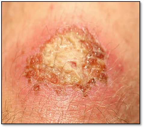
Fig no. 1: Wound marks

The evolution of wound dressings into medicated systems, particularly those capable of controlled drug release, has created a paradigm shift in wound care. Controlled-release gauze pads aim to deliver bioactive agents such as antimicrobials, analgesics, anti-inflammatories, and growth factors in a sustained and localized manner, improving healing outcomes and reducing systemic side effects. There are two categories of dressings: Adhesive dressing and Gauze dressing. An Adhesive dressing is a piece of medical plaster that is used in case of injuries that do not require a full-fledged dressing. They are meant for small injuries or wounds. The Gauge dressing are thick cotton pads that are used to cover large wounds. They are held in place by wrapping with a gauze strip (bandage) or a tape.

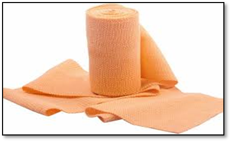
Fig no.2: Crepe Bandage

There are three main categories of “Bandages” that are Roller bandage, Tubular bandage, and Triangular bandage. They are all used in different ways for one common intention and that is to either cover wounds or to apply pressure in controlling bleeding and support the strain or the sprain. Roller bandage are long strips of bandages that can further be subdivided into two types: An Elastic roller bandage that is applied to support a strain or a sprain and is wrapped around the joint or limbs many times. It should be applied firmly. Cotton or Linen roller bandages are used to cover gauze dressings. These  bandage can come in any number of widths and lengths and can be used for almost any bandage application, including holding a dressing in place

Materials Used in Controlled-Release Gauze Pads

Substrate Materials

Cotton gauze:

Widely used due to affordability and high porosity. Requires surface modification for advanced functions. However, untreated cotton lacks antimicrobial properties and can adhere to wounds, causing pain upon removal. Surface functionalization (e.g., with chitosan or silver nanoparticles) improves antimicrobial performance and bioactivity.

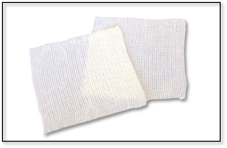
Fig. no.3: Cotton fiber bandage

Cotton gauze is primarily composed of natural cotton fibers, which consist mainly of cellulose along with small amounts of other organic and inorganic compounds. The table below outlines the typical chemical composition of processed medical-grade cotton gauze.

Table No. 1: Chemical Composition Cotton Gauze

Component

Approximate Percentage

Function/Role

Cellulose

88–96%

Main structural polymer; provides strength and absorbency.

Hemicellulose

2–6%

Supports cellulose structure; less crystalline.

Lignin

0.5–1%

Binds fibers in raw cotton (mostly removed during processing).

Waxes and Fats

0.4–1%

Natural protective coating on fibers (removed by scouring).

Pectin

0.4–1%

Help bind cell walls; also removed during processing.

Proteins

0.3–0.5%

Minor content; removed during bleaching.

Ash (minerals)

0.5–1.2%

Includes calcium, magnesium, potassium salts.

Moisture content

~7–8%

Natural water content in cotton fibers (varies with humidity).

Banana pseudo stem fiber: A biodegradable, lignocellulosic natural fiber rich in cellulose. Exhibits high moisture retention, flexibility, and tensile strength. When woven into gauze and coated with bioactive agents (e.g., honey, curcumin), it forms a sustainable substrate for medicated dressings. Eco-friendly and abundantly available in tropical regions.  Table 2 shows the chemical composition of banana plant fibers, and their physical properties. It is noted that cellulose is the main constituent of plant fibers followed by hemi-celluloses and lignin interchangeably and pectin respectively. Cellulose is also the reinforcement for lignin, hemi cellulose and Pectin.

Table no.2: Chemical composition of banana fibers

Component

Percentage

Function/Role

Cellulose (%)

60-65

Main structural polymer; provides strength and absorbency.

Hemi cellulose (%)

6-19

Supports cellulose structure; less crystalline.

Lignin (%)

5-10

Binds fibers (mostly removed during processing)

Pectin (%)

3-5

Natural protective coating on fibers (removed by scouring).

Ash (%)

1-3

Help bind cell walls; also removed during processing.

Extractives (%)

3-6

……

Chitosan-coated fibers: Biopolymer offering intrinsic antimicrobial properties and film-forming ability.

Drug-Delivery Platforms

Since ancient times, many different materials have been used to treat wounds in an attempt to stop bleeding, absorb exudates, and promote healing. Some of these materials consisted of honey, animal oils or fat, cobwebs, mud, leaves, sphagnum moss, or animal dung. Although most of these readily available natural substances would later prove to provide little benefit, others such as honey have been studied and shown to provide some value.

Hydrogels: PVA, PEG, and other crosslinked systems enable high drug loading and moisture retention. Hydrogels are complex hydrophilic organic cross-linked polymers, consisting of an 80%–90% water base. These gels are available in a free-flowing amorphous or fixed flexible sheet form. They can absorb a minimum amount of fluid by swelling, but they also can donate moisture to a dry wound, thereby facilitating autolytic debridement and maintaining a moist wound environment that is thermally insulated. They have also been shown to promote granulation and epithelialization and reduce the temperature of a wound bed by up to 5C. They are permeable to gas and water and have proven to be a less effective bacterial barrier than occlusive dressings. The main application of these dressings is hydrating dry wound beds and softening and loosening slough and necrotic wound debris. They are unable to absorb heavy drainage due to their high-water concentration; they absorb very slowly and therefore are not useful on bleeding wounds, and they generally require a secondary dressing. They can be used on a variety of wounds including pressure ulcers, partial and full thickness wounds, and vascular ulcers. Maceration can be of concern, as peri wound skin areas need to be protected from excess hydration. Among its benefits, hydrogels can be used in conjunction with topical medications or antibacterial agents. The fixed form of hydrogels should not be used in infected wounds. Hydrogels need to be covered with secondary dressings while remaining in place for up to 3 days.

Nanoparticles: Polymeric (PLGA), silver, or lipid-based for enhanced drug stability and targeted release. Promotes cell migration , mimic extracellular matrix and enables controlled drug release.

Liposomes & niosomes: Effective for encapsulating both hydrophilic and lipophilic drugs. Delivery of antibiotics such as gentamicin, ciprofloxacin. Delivery of herbal extract such as curcumin and aloe vera. Minimizing scar formation and infection.

Microspheres: Offer prolonged release and protection of encapsulated actives.

Types of Drugs Used in Gauze Pad Systems

Table no.3: Drugs Used in Gauze Pad

Sr.no.

Drug Type

Examples

Purpose

1.

Antimicrobials/ Antibiotics

Silver nitrate, gentamicin, curcumin, Neomycin, bacitracin,

Prevent infection

2.

Antiseptics

Povidone iodine (eg. Betadine), chlorhexidine, hydrogen peroxide, Alcohol based solutions.

Prevent and reduce infection by killing or inhibiting the growth of microorganism.

3.

Anti-inflammatories

Diclofenac, ibuprofen, hydrocortisone

Reduce inflammation and swelling

4.

Analgesics/ Anesthetic

Lidocaine, ketorolac, Benzocaine

Enhance tissue regeneration

5.

Growth factors

Platelet derived growth factor (PDGF), epidermal growth factor (EGF)

Enhance tissue regeneration

6.

Hemostatic agent

Oxidized cellulose, gelatin sponge, chitosan, calcium alginate

Stop bleeding by promoting clot formation

7.

Debriding agent

Collagenase, papain – urea ointments

Help to remove dead or damaged tissue from wounds

8.

Antifungal agents

Clotrimazole, miconazole , nystatin

Used when there is a risk of fungal infection.

9.

Herbal extracts

Neem, turmeric, aloe Vera

Natural healing support

Drug Incorporation Techniques

  • Soaking and drying: Ideal for water-soluble drugs but less precise.
  • Electrospinning: Produces nanofiber layers for high surface area and precise control.
  • Layer-by-layer assembly: Facilitates sequential drug release.
  • Supercritical fluid impregnation: Suitable for thermolabile or poorly soluble drugs.
  • 3D printing: Emerging tool for customized drug distribution and patterning.

Sterilized gauze (cotton or banana fiber-based)

Drug substance (e.g., antiseptic or antibiotic) ; Solvent/base (e.g., ethanol, water, gel base) ; Beakers, measuring cylinders ; Magnetic stirrer or shaker Drying oven or desiccator Sterile packaging materials (e.g., aluminum foil, pouches);Tongs, gloves, laminar air flow cabinet.

Procedure:

  1. Preparation of Drug Solution/Dispersion: Accurately weigh the required quantity of the drug. Dissolve or disperse it in a suitable solvent or base to form a uniform solution. Mix using a magnetic stirrer for 15–30 minutes for complete dissolution.
  2. Selection and Sterilization of Gauze: Use sterile gauze pads (cotton or banana pseudo stem fibres-based). If not pre-sterilized, autoclave at 121°C for 15 minutes and cool.
  3. Drug Loading
  • Method A (Soaking): Immerse the gauze in the drug solution for a specific time (e.g., 5–10 min).
  • Method B (Spraying): Uniformly spray the drug solution onto the gauze surface.
  • Method C (Coating): Dip the gauze in a coating solution or apply using a casting method.
  1. Drying: Place the drug-loaded gauze in a hot air oven at 40–50°C until completely dry. Avoid high temperatures that may degrade the drug.
  2. Packaging: Pack the medicated gauze in sterile pouches under aseptic conditions. Seal immediately to avoid contamination.
  3. Storage: Store in a cool, dry place away from light and moisture until use.

Mechanisms of Controlled Drug Release

1. Diffusion-based: Gradual release through matrix or reservoir systems.

Application:

  1. most common mechanism in polymer based dressings.
  2. Used for antibiotics, antiseptic.

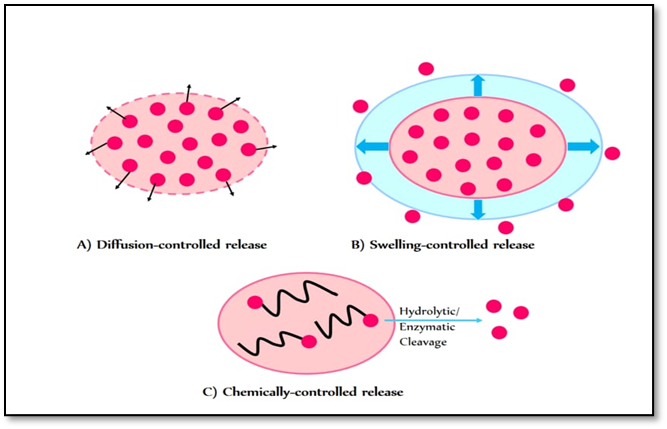
Fig no. 4: Diffusion mechanism

2. Swelling-triggered: Hydrophilic matrices expand in presence of exudate, allowing drug diffusion.

Application:

Hydrogels or cellulose based dressings that absorb moisture before releasing the drug.

3. Degradation-controlled: Release dependent on breakdown of polymeric carrier.

Application:

Useful for sustained release antibiotics, enzymes or anti-inflammatory drugs.

4. Stimuli-responsive: Environmental triggers (pH, temperature, enzymes) regulate release, especially useful in infected or inflamed wounds.

Application:

Advanced gauze pads that release antimicrobials when infection is detected (low pH or high enzyme activity)

Evaluation Techniques to be used

Table no.4: Evaluation Parameter

Sr. no.

Parameter

Method

1.

Drug release

Franz diffusion cell, dialysis membrane

2.

Antimicrobial activity

Zone of inhibition, broth dilution

3.

Tensile strength

Universal testing machine

4.

Fluid absorption

Gravimetric or centrifuge retention test

5.

Biocompatibility

MTT assay, haemolysis test

Commercial and Clinical Insights

1. Acticoat™: Nanocrystalline silver gauze with sustained antimicrobial effect.

Advantage:

  1. low cost
  2. Minimize bacterial colonization.

Disadvantage:

  1. not for wound with high exudatea
  2. Cytotoxicity may cause wound healing delay.
  3. Constant removal may be traumatic, disrupting the granulation tissue.

2. Aquacel Ag™: Combines hydrofiber technology with silver for exudate absorption and microbial control.

Advantages:

  1. moist microenvironment promotes healing.

Disadvantage:

  1. High cost.
  2. Destruction of dressing results in unpleasant color and odor often confused with infection.

3. Medihoney™: Medical-grade honey in gauze format for infection control and healing promotion

Challenges and Future Perspectives

  • Sterilization issues: Many drugs or nanocarriers degrade under heat or radiation.
  • Regulatory complexity: Classification as drug-device combinations requires dual compliance.
  • Cost-effectiveness: Advanced systems must balance efficacy and affordability.
  • Smart dressing potential: Future gauze pads may include sensors for real-time monitoring and feedback-controlled release.

CONCLUSION

Controlled-release medicated gauze pads represent a significant advancement in wound care by integrating therapeutic delivery with protective functions. Innovations in materials, drug-carrier systems, and release mechanisms offer opportunities to tailor treatments for various wound types. While challenges remain in terms of scalability and regulatory approval, the future of wound dressings lies in smart, multifunctional platforms that merge therapy with diagnostics.

REFERENCES

  1. Boateng JS, Matthews KH, Stevens HN, Eccleston GM. Wound healing dressings and drug delivery systems: a review. J Pharm Sci. 2008;97(8):2892–2923.
  2. Pereira RF, Bartolo PJ. Traditional therapies for skin wound healing. Adv Wound Care. 2016;5(5):208–229.
  3. Dhivya S, Padma VV, Santhini E. Wound dressings – a review. Biomedicine (Taipei). 2015;5(4):22.
  4. Yadav MK, Chae SW, Im GJ. Applications of chitosan-based nanomaterials in the treatment of infections caused by drug-resistant bacteria. Nanomedicine. 2021;16(2):121–133.
  5. Thomas S, Loveless P. A comparative study of the properties of twelve hydrocolloid dressings. World Wide Wounds. 2000.
  6. Liu Y, Zhang X, Yu D, et al. Smart bandages for wound healing. Nat Rev Bioeng. 2023;1:214–228.
  7. Wang T, Zhu X, Wang P, et al. Antibacterial and anti-inflammatory properties of silver nanocomposite wound dressings. Mater Sci Eng C. 2020;117:111273.
  8. D.R. de Araújo, D.C. da Silva, R.M. Barbosa, M. Franz-Montan, C.M. Cereda, C. Padula, P. Santi, E. de Paula Strategies for delivering local anesthetics to the skin: Focus on liposomes, solid lipid nanoparticles, hydrogels and patches Expert Opin. Drug Deliv., 10 (2013), pp. 1551-1563
  9. L.G. Ovington Bacterial toxins and wound healing Ostomy Wound Manage, 49 (2003), pp. 8-12
  10. T.A. Mustoe, K. O'Shaughnessy, O. Kloeters Chronic wound pathogenesis and current treatment strategies: a unifying hypothesis Plast. Reconstr. Surg., 117 (2006), pp. 35-41
  11. A. Sood, M.S. Granick, N.L. Tomaselli  Wound dressings and comparative effectiveness data Adv. Wound Care, 3 (2012), pp. 511-529
  12. Sisco M and Mustoe TA: Animal models of ischemic wound healing: toward an approximation of human chronic cutaneous ulcers in rabbit and rat. Methods Mol Med 2003; 78: 55.
  13. Brigham PA and McLoughlin E: Burn incidence and medical care use in the United States: estimate, trends, and data sources. J Burn Care Rehabil 1996; 17: 95.
  14. Medical Data International, Inc.: U.S. markets for wound management products. Irvine, CA: Medical Data International, August 1997.
  15. Morin RJ and Tomaselli NL: Interactive dressings and topical agents. Clin Plast Surg 2007; 34: 643.
  16. Chambers, E.S.; Vukmanovic-Stejic, M. Skin barrier immunity and ageing. Immunology 2020, 160, 116–125. 
  17. Lima, T.d.P.d.L.; Passos, M.F. Skin wounds, the healing process, and hydrogel-based wound dressings: A short review. J. Biomater. Sci. Polym. Ed. 2021, 32, 1910–1925.
  18. Luo, R.; Dai, J.; Zhang, J.; Li, Z. Accelerated Skin Wound Healing by Electrical Stimulation. Adv. Healthc. Mater. 2021, 10, 2100557.
  19. Tottoli, E.M.; Dorati, R.; Genta, I.; Chiesa, E.; Pisani, S.; Conti, B. Skin Wound Healing Process and New Emerging Technologies for Skin Wound Care and Regeneration. Pharmaceutics 2020, 12, 735.
  20. Falanga, V.; Isseroff, R.R.; Soulika, A.M.; Romanelli, M.; Margolis, D.; Kapp, S.; Granick, M.; Harding, K. Chronic wounds. Nat. Rev. Dis. Primers 2022, 8, 50.
  21. WHO. Burns. Available online: https://who.int/news-room/fact-sheets/detail/burns (accessed on 9 January 2025).
  22. Jeschke, M.G.; van Baar, M.E.; Choudhry, M.A.; Chung, K.K.; Gibran, N.S.; Logsetty, S. Burn injury. Nat. Rev. Dis. Primers 2020, 6, 11.
  23. Shi, C.; Wang, C.; Liu, H.; Li, Q.; Li, R.; Zhang, Y.; Liu, Y.; Shao, Y.; Wang, J. Selection of Appropriate Wound Dressing for Various Wounds. Front. Bioeng. Biotechnol. 2020, 8, 182.
  24. Rezvani Ghomi, E.; Khalili, S.; Nouri Khorasani, S.; Esmaeely Neisiany, R.; Ramakrishna, S. Wound dressings: Current advances and future directions. J. Appl. Polym. Sci. 2019, 136, 47738.
  25. Verdolino, D.V.; Thomason, H.A.; Fotticchia, A.; Cartmell, S. Wound dressings: Curbing inflammation in chronic wound healing. Emerg. Top. Life Sci. 2021, 5, 523–537.
  26. Nguyen, H.M.; Ngoc Le, T.T.; Nguyen, A.T.; Thien Le, H.N.; Pham, T.T. Biomedical materials for wound dressing: Recent advances and applications. RSC Adv. 2023, 13, 5509–5528.
  27. Weir, D. Wound Dressings. In Local Wound Care for Dermatologists; Alavi, A., Maibach, H.I., Eds.; Springer International Publishing: Cham, Switzerland, 2020; pp. 25–34.
  28. Kaya, S.; Acar, S. Properties of ideal wound dressingideal yara örtüsünün özellikleri. Ank. Univ. Eczaci. Fak. Derg. 2023, 47, 5.
  29. Nazarko, L. Wound healing and moisture balance: Selecting dressings. Nurs. Resid. Care 2009, 11, 286–291
  30. Obagi, Z.; Damiani, G.; Grada, A.; Falanga, V. Principles of Wound Dressings: A Review. Surg. Technol. Int. 2019, 35, 1–57.
  31. Niculescu, A.-G.; Grumezescu, A.M. An up-to-date review of biomaterials application in wound management. Polymers 2022, 14, 421.
  32. Ahmadi, S.A.; Rastegarpanah, H.; Ansari, N.; Dortaj, H.; Shafaghi, S.; Ghorbani, F.; Shafaghi, M. Preparation and in vivo Evaluation of an Astragalus Gummifer Hydrogel-Based Dressing for Excisional Wound. Biointerface Res. Appl. Chem. 2024, 14, 95.
  33. Brumberg, V.; Astrelina, T.; Malivanova, T.; Samoilov, A. Modern Wound Dressings: Hydrogel Dressings. Biomedicines 2021, 9, 1235.
  34. Gravelle, A. J., & Choi, S. H. (2014). "Biocompatible fibers in wound care: Their structure, properties, and use in medicated gauze." Journal of Biomedical Materials Research, 15(3), 259-265. DOI: 10.1002/jbm.32068
  35. Vishwakarma, A., & Bhatt, P. (2019). "Natural fibers and their applications in wound care: A comprehensive review." Journal of Natural Fibers, 16(4), 634-643.DOI: 10.1080/15440478.2018.1476981
  36. Shukla, A., & Ghosh, S. K. (2020). "Polymeric and natural fibers in wound care: A review on their applications in medicated gauze and dressing." International Journal of Polymeric Materials and Polymer Composites, 69(6), 329-336.DOI: 10.1080/00914037.2019.1694990
  37. Chowdhury, A., & Shukla, R. (2018). "Fibers and their role in drug delivery: Medicated gauze and dressing materials." Journal of Controlled Release, 297, 55-66.DOI: 10.1016/j.jconrel.2018.03.023
  38. Gouda, A., & Pradhan, S. (2013). "Advances in fiber-based wound dressings and drug delivery systems." International Journal of Pharmaceutics, 457(1-2), 67-79.DOI: 10.1016/j.ijpharm.2013.07.025
  39. Devi, P., & Rajendran, A. (2015). "Effectiveness of natural fibers in medicated wound dressings: A comparative review." Journal of Applied Polymer Science, 132(21), 42491.DOI: 10.1002/app.42491
  40. Sivaraman, K., & Kumar, R. (2020). "Functionalization of natural fibers for controlled drug release in wound dressings." Journal of Industrial Textiles, 49(1), 94-107.DOI: 10.1177/1528083719882599
  41. Gokce, M., & Arslan, G. (2019). "Cellulose-based fibers in the development of medicated gauze for wound healing applications." Carbohydrate Polymers, 206, 201-210.DOI: 10.1016/j.carbpol.2018.10.078
  42. Ahmed, A., & El-Sayed, M. (2016). "A review on fiber-reinforced polymer composites in wound healing applications." Journal of Wound Care, 25(11), 675-682. DOI: 10.12968/jowc.2016.25.11.675
  43. Zhang, Y., & Wei, Y. (2017). "Hydroxyapatite and fiber-based wound dressings for enhanced drug delivery." Biomaterials, 134, 65-75.DOI: 10.1016/j.biomaterials.2017.03.024
  44. Müller, M., & Pires, A. (2015). "Biodegradable fiber-reinforced wound dressings: Innovations in drug delivery." Journal of Biomaterials Science, Polymer Edition, 26(8), 521-532.DOI: 10.1080/09205063.2015.1067404
  45. Zhou, X., & Zhao, H. (2018). "Natural fiber composites for medical textiles: A new approach for wound care." Composites Part B: Engineering, 146, 143-153.DOI: 10.1016/j.compositesb.2018.04.032
  46. Soman, P., & Dinesh, S. (2017). "Natural fibers as novel drug delivery vehicles in wound healing." International Journal of Biological Macromolecules, 103, 745-753.DOI: 10.1016/j.ijbiomac.2017.04.015
  47. Lee, H. L., & Park, S. (2016). "Applications of natural fibers in wound healing: A review of their material properties." Textile Research Journal, 86(6), 630-640.DOI: 10.1177/0040517515591361
  48. Pradeep, S., & Karthik, G. (2014). "Fibers in wound healing: New opportunities for drug incorporation and controlled release." Journal of Drug Delivery Science and Technology, 24(2), 212-221 DOI: 10.1016/j.jddst.2014.01.005
  49. Gonzalez, E., & Gomez, M. (2012). "Modified fibers for medicated gauze and wound care applications." Biomaterials Science, 3(4), 517-529.DOI: 10.1039/C2BM20147J
  50. Narayana, K., & Sundararajan, V. (2020). "Polymer and fiber-based composites for enhanced drug delivery in wound dressings." Polymer Bulletin, 67(7), 949-960.DOI: 10.1007/s00289-020-03357-0
  51. Basak, S., & Singh, S. (2017). "Smart fiber-based wound dressings for controlled drug release: A review." Journal of Biomaterials and Nanobiotechnology, 8(2), 136-150.DOI: 10.4236/jbnb.2017.82014
  52. Kang, I. K., & Han, D. W. (2014). "Smart wound dressings: Emerging trends in the use of fibers for wound care applications." Journal of Biomaterials Science, Polymer Edition, 25(9), 993-1012.DOI: 10.1080/09205063.2014.924484
  53. Chakraborty, S., & Maitra, S. (2017). "The role of biodegradable fibers in advanced wound care and drug delivery systems." Materials Science and Engineering: C, 74, 83-95.DOI: 10.1016/j.msec.2017.02.036
  54. Bennett, N. E., & Hudson, C. (2005). "Silver dressings in the management of burns and chronic wounds." Journal of Wound Care, 14(3), 105-112. DOI: 10.12968/jowc.2005.14.3.26679
  55. Bowers, S. L. (2005). "The role of Acticoat in the management of burns." Journal of Burn Care & Research, 26(6), 522-528.DOI: 10.1097/01.BCR.0000188230.12283.7A
  56. Davis, M. S., & Darlene, T. J. (2012). "Antimicrobial properties of silver in wound care." Burns, 38(8), 1079-1085.DOI: 10.1016/j.burns.2012.02.015
  57. Cullen, B., & Finlay, M. (2012). "Silver dressings in the management of burn wounds: Evidence and cost analysis." Burns, 38(5), 655-660.DOI: 10.1016/j.burns.2012.02.010
  58. Nussbaum, S. R., & Sibbald, R. G. (2016). "Silver in wound care: A review of its antimicrobial efficacy and its role in wound healing." Journal of Wound Care, 25(5), 265-271. DOI: 10.12968/jowc.2016.25.5.265
  59. Salama, M., & Mounir, M. (2006). "Use of Acticoat in burn care: Review of the evidence." Wounds, 18(6), 184-191.DOI: 10.1016/j.wound.2005.12.001
  60. West, D. P., & Phillips, T. J. (2009). "Silver-based wound dressings: The current status of clinical use." Journal of Clinical Dermatology, 30(2), 109-116.DOI: 10.1016/j.jcd.2009.02.009
  61. Harvey, L., & Miller, K. (2014). "Effectiveness of silver dressings in treating chronic wounds: A systematic review." Wound Repair and Regeneration, 22(4), 506-510.DOI: 10.1111/wrr.12155
  62. Chowdary, M., & Chandrasekaran, S. (2015). "A review on the mechanisms of silver ion action on microbial cells." Antimicrobial Agents, 39(9), 342-350. DOI: 10.1016/j.ijantimicag.2015.04.002
  63. Aly, A., & Maibach, H. (2017). "Clinical uses of silver dressings in wound management: Review." Journal of Dermatological Treatment, 28(3), 240-247.DOI: 10.1080/09546634.2017.1292767
  64. Lemaitre, L. M., & Lefebvre, J. (2018). "Review of antimicrobial dressings: Efficacy and use in clinical practice." International Wound Journal, 15(5), 873-880.DOI: 10.1111/iwj.12934
  65. Jull, A. B., & Walker, N. (2013). "Silver-containing dressings for the treatment of infected wounds: A systematic review." Cochrane Database of Systematic Reviews, 6(CD006478).DOI: 10.1002/14651858.CD006478.pub2
  66. Martins, M. M., & Ribeiro, A. (2017). "The use of silver-based wound dressings in wound management: A review." Infection Control & Hospital Epidemiology, 38(2), 160-166.DOI: 10.1017/ice.2016.282
  67. Rigo, G., & Shulze, A. (2010). "Efficacy of silver-containing dressings on wound healing: A comparative review." Clinical Therapeutics, 32(10), 1781-1792.DOI: 10.1016/j.clinthera.2010.09.015
  68. Shan, S. S., & Zhen, P. (2011). "Clinical performance of silver dressings in burn and chronic wound care." Journal of Burn Care & Research, 32(4), 563-568.DOI: 10.1097/BCR.0b013e31822cc9c1
  69. Parks, J. T., & Fischer, S. A. (2014). "Silver dressings in the treatment of chronic wounds: An evidence-based review." Journal of Wound Care, 23(8), 398-403.DOI: 10.12968/jowc.2014.23.8.398
  70. Wong, E., & Goh, T. (2015). "Silver-based antimicrobial wound dressings and their role in clinical management." Wound Medicine, 10(2), 15-21. DOI: 10.1016/j.wndm.2015.04.001
  71.  Aslam, S. K., & Sood, A. (2007). "The antimicrobial properties of silver and its role in wound care." Indian Journal of Dermatology, 52(2), 41-44.DOI: 10.4103/0019-5154.29172
  72. Bordes, D. A., & Doran, L. (2012). "Silver nanoparticles in the treatment of chronic wounds: A comprehensive review." Journal of Clinical Aesthetic Dermatology, 5(8), 47-51.
  73. Mazzola, L., & Lippi, D. (2011). "The role of Acticoat in modern wound care." Burns, 37(5), 726-732.DOI: 10.1016/j.burns.2010.12.004

Reference

  1. Boateng JS, Matthews KH, Stevens HN, Eccleston GM. Wound healing dressings and drug delivery systems: a review. J Pharm Sci. 2008;97(8):2892–2923.
  2. Pereira RF, Bartolo PJ. Traditional therapies for skin wound healing. Adv Wound Care. 2016;5(5):208–229.
  3. Dhivya S, Padma VV, Santhini E. Wound dressings – a review. Biomedicine (Taipei). 2015;5(4):22.
  4. Yadav MK, Chae SW, Im GJ. Applications of chitosan-based nanomaterials in the treatment of infections caused by drug-resistant bacteria. Nanomedicine. 2021;16(2):121–133.
  5. Thomas S, Loveless P. A comparative study of the properties of twelve hydrocolloid dressings. World Wide Wounds. 2000.
  6. Liu Y, Zhang X, Yu D, et al. Smart bandages for wound healing. Nat Rev Bioeng. 2023;1:214–228.
  7. Wang T, Zhu X, Wang P, et al. Antibacterial and anti-inflammatory properties of silver nanocomposite wound dressings. Mater Sci Eng C. 2020;117:111273.
  8. D.R. de Araújo, D.C. da Silva, R.M. Barbosa, M. Franz-Montan, C.M. Cereda, C. Padula, P. Santi, E. de Paula Strategies for delivering local anesthetics to the skin: Focus on liposomes, solid lipid nanoparticles, hydrogels and patches Expert Opin. Drug Deliv., 10 (2013), pp. 1551-1563
  9. L.G. Ovington Bacterial toxins and wound healing Ostomy Wound Manage, 49 (2003), pp. 8-12
  10. T.A. Mustoe, K. O'Shaughnessy, O. Kloeters Chronic wound pathogenesis and current treatment strategies: a unifying hypothesis Plast. Reconstr. Surg., 117 (2006), pp. 35-41
  11. A. Sood, M.S. Granick, N.L. Tomaselli  Wound dressings and comparative effectiveness data Adv. Wound Care, 3 (2012), pp. 511-529
  12. Sisco M and Mustoe TA: Animal models of ischemic wound healing: toward an approximation of human chronic cutaneous ulcers in rabbit and rat. Methods Mol Med 2003; 78: 55.
  13. Brigham PA and McLoughlin E: Burn incidence and medical care use in the United States: estimate, trends, and data sources. J Burn Care Rehabil 1996; 17: 95.
  14. Medical Data International, Inc.: U.S. markets for wound management products. Irvine, CA: Medical Data International, August 1997.
  15. Morin RJ and Tomaselli NL: Interactive dressings and topical agents. Clin Plast Surg 2007; 34: 643.
  16. Chambers, E.S.; Vukmanovic-Stejic, M. Skin barrier immunity and ageing. Immunology 2020, 160, 116–125. 
  17. Lima, T.d.P.d.L.; Passos, M.F. Skin wounds, the healing process, and hydrogel-based wound dressings: A short review. J. Biomater. Sci. Polym. Ed. 2021, 32, 1910–1925.
  18. Luo, R.; Dai, J.; Zhang, J.; Li, Z. Accelerated Skin Wound Healing by Electrical Stimulation. Adv. Healthc. Mater. 2021, 10, 2100557.
  19. Tottoli, E.M.; Dorati, R.; Genta, I.; Chiesa, E.; Pisani, S.; Conti, B. Skin Wound Healing Process and New Emerging Technologies for Skin Wound Care and Regeneration. Pharmaceutics 2020, 12, 735.
  20. Falanga, V.; Isseroff, R.R.; Soulika, A.M.; Romanelli, M.; Margolis, D.; Kapp, S.; Granick, M.; Harding, K. Chronic wounds. Nat. Rev. Dis. Primers 2022, 8, 50.
  21. WHO. Burns. Available online: https://who.int/news-room/fact-sheets/detail/burns (accessed on 9 January 2025).
  22. Jeschke, M.G.; van Baar, M.E.; Choudhry, M.A.; Chung, K.K.; Gibran, N.S.; Logsetty, S. Burn injury. Nat. Rev. Dis. Primers 2020, 6, 11.
  23. Shi, C.; Wang, C.; Liu, H.; Li, Q.; Li, R.; Zhang, Y.; Liu, Y.; Shao, Y.; Wang, J. Selection of Appropriate Wound Dressing for Various Wounds. Front. Bioeng. Biotechnol. 2020, 8, 182.
  24. Rezvani Ghomi, E.; Khalili, S.; Nouri Khorasani, S.; Esmaeely Neisiany, R.; Ramakrishna, S. Wound dressings: Current advances and future directions. J. Appl. Polym. Sci. 2019, 136, 47738.
  25. Verdolino, D.V.; Thomason, H.A.; Fotticchia, A.; Cartmell, S. Wound dressings: Curbing inflammation in chronic wound healing. Emerg. Top. Life Sci. 2021, 5, 523–537.
  26. Nguyen, H.M.; Ngoc Le, T.T.; Nguyen, A.T.; Thien Le, H.N.; Pham, T.T. Biomedical materials for wound dressing: Recent advances and applications. RSC Adv. 2023, 13, 5509–5528.
  27. Weir, D. Wound Dressings. In Local Wound Care for Dermatologists; Alavi, A., Maibach, H.I., Eds.; Springer International Publishing: Cham, Switzerland, 2020; pp. 25–34.
  28. Kaya, S.; Acar, S. Properties of ideal wound dressingideal yara örtüsünün özellikleri. Ank. Univ. Eczaci. Fak. Derg. 2023, 47, 5.
  29. Nazarko, L. Wound healing and moisture balance: Selecting dressings. Nurs. Resid. Care 2009, 11, 286–291
  30. Obagi, Z.; Damiani, G.; Grada, A.; Falanga, V. Principles of Wound Dressings: A Review. Surg. Technol. Int. 2019, 35, 1–57.
  31. Niculescu, A.-G.; Grumezescu, A.M. An up-to-date review of biomaterials application in wound management. Polymers 2022, 14, 421.
  32. Ahmadi, S.A.; Rastegarpanah, H.; Ansari, N.; Dortaj, H.; Shafaghi, S.; Ghorbani, F.; Shafaghi, M. Preparation and in vivo Evaluation of an Astragalus Gummifer Hydrogel-Based Dressing for Excisional Wound. Biointerface Res. Appl. Chem. 2024, 14, 95.
  33. Brumberg, V.; Astrelina, T.; Malivanova, T.; Samoilov, A. Modern Wound Dressings: Hydrogel Dressings. Biomedicines 2021, 9, 1235.
  34. Gravelle, A. J., & Choi, S. H. (2014). "Biocompatible fibers in wound care: Their structure, properties, and use in medicated gauze." Journal of Biomedical Materials Research, 15(3), 259-265. DOI: 10.1002/jbm.32068
  35. Vishwakarma, A., & Bhatt, P. (2019). "Natural fibers and their applications in wound care: A comprehensive review." Journal of Natural Fibers, 16(4), 634-643.DOI: 10.1080/15440478.2018.1476981
  36. Shukla, A., & Ghosh, S. K. (2020). "Polymeric and natural fibers in wound care: A review on their applications in medicated gauze and dressing." International Journal of Polymeric Materials and Polymer Composites, 69(6), 329-336.DOI: 10.1080/00914037.2019.1694990
  37. Chowdhury, A., & Shukla, R. (2018). "Fibers and their role in drug delivery: Medicated gauze and dressing materials." Journal of Controlled Release, 297, 55-66.DOI: 10.1016/j.jconrel.2018.03.023
  38. Gouda, A., & Pradhan, S. (2013). "Advances in fiber-based wound dressings and drug delivery systems." International Journal of Pharmaceutics, 457(1-2), 67-79.DOI: 10.1016/j.ijpharm.2013.07.025
  39. Devi, P., & Rajendran, A. (2015). "Effectiveness of natural fibers in medicated wound dressings: A comparative review." Journal of Applied Polymer Science, 132(21), 42491.DOI: 10.1002/app.42491
  40. Sivaraman, K., & Kumar, R. (2020). "Functionalization of natural fibers for controlled drug release in wound dressings." Journal of Industrial Textiles, 49(1), 94-107.DOI: 10.1177/1528083719882599
  41. Gokce, M., & Arslan, G. (2019). "Cellulose-based fibers in the development of medicated gauze for wound healing applications." Carbohydrate Polymers, 206, 201-210.DOI: 10.1016/j.carbpol.2018.10.078
  42. Ahmed, A., & El-Sayed, M. (2016). "A review on fiber-reinforced polymer composites in wound healing applications." Journal of Wound Care, 25(11), 675-682. DOI: 10.12968/jowc.2016.25.11.675
  43. Zhang, Y., & Wei, Y. (2017). "Hydroxyapatite and fiber-based wound dressings for enhanced drug delivery." Biomaterials, 134, 65-75.DOI: 10.1016/j.biomaterials.2017.03.024
  44. Müller, M., & Pires, A. (2015). "Biodegradable fiber-reinforced wound dressings: Innovations in drug delivery." Journal of Biomaterials Science, Polymer Edition, 26(8), 521-532.DOI: 10.1080/09205063.2015.1067404
  45. Zhou, X., & Zhao, H. (2018). "Natural fiber composites for medical textiles: A new approach for wound care." Composites Part B: Engineering, 146, 143-153.DOI: 10.1016/j.compositesb.2018.04.032
  46. Soman, P., & Dinesh, S. (2017). "Natural fibers as novel drug delivery vehicles in wound healing." International Journal of Biological Macromolecules, 103, 745-753.DOI: 10.1016/j.ijbiomac.2017.04.015
  47. Lee, H. L., & Park, S. (2016). "Applications of natural fibers in wound healing: A review of their material properties." Textile Research Journal, 86(6), 630-640.DOI: 10.1177/0040517515591361
  48. Pradeep, S., & Karthik, G. (2014). "Fibers in wound healing: New opportunities for drug incorporation and controlled release." Journal of Drug Delivery Science and Technology, 24(2), 212-221 DOI: 10.1016/j.jddst.2014.01.005
  49. Gonzalez, E., & Gomez, M. (2012). "Modified fibers for medicated gauze and wound care applications." Biomaterials Science, 3(4), 517-529.DOI: 10.1039/C2BM20147J
  50. Narayana, K., & Sundararajan, V. (2020). "Polymer and fiber-based composites for enhanced drug delivery in wound dressings." Polymer Bulletin, 67(7), 949-960.DOI: 10.1007/s00289-020-03357-0
  51. Basak, S., & Singh, S. (2017). "Smart fiber-based wound dressings for controlled drug release: A review." Journal of Biomaterials and Nanobiotechnology, 8(2), 136-150.DOI: 10.4236/jbnb.2017.82014
  52. Kang, I. K., & Han, D. W. (2014). "Smart wound dressings: Emerging trends in the use of fibers for wound care applications." Journal of Biomaterials Science, Polymer Edition, 25(9), 993-1012.DOI: 10.1080/09205063.2014.924484
  53. Chakraborty, S., & Maitra, S. (2017). "The role of biodegradable fibers in advanced wound care and drug delivery systems." Materials Science and Engineering: C, 74, 83-95.DOI: 10.1016/j.msec.2017.02.036
  54. Bennett, N. E., & Hudson, C. (2005). "Silver dressings in the management of burns and chronic wounds." Journal of Wound Care, 14(3), 105-112. DOI: 10.12968/jowc.2005.14.3.26679
  55. Bowers, S. L. (2005). "The role of Acticoat in the management of burns." Journal of Burn Care & Research, 26(6), 522-528.DOI: 10.1097/01.BCR.0000188230.12283.7A
  56. Davis, M. S., & Darlene, T. J. (2012). "Antimicrobial properties of silver in wound care." Burns, 38(8), 1079-1085.DOI: 10.1016/j.burns.2012.02.015
  57. Cullen, B., & Finlay, M. (2012). "Silver dressings in the management of burn wounds: Evidence and cost analysis." Burns, 38(5), 655-660.DOI: 10.1016/j.burns.2012.02.010
  58. Nussbaum, S. R., & Sibbald, R. G. (2016). "Silver in wound care: A review of its antimicrobial efficacy and its role in wound healing." Journal of Wound Care, 25(5), 265-271. DOI: 10.12968/jowc.2016.25.5.265
  59. Salama, M., & Mounir, M. (2006). "Use of Acticoat in burn care: Review of the evidence." Wounds, 18(6), 184-191.DOI: 10.1016/j.wound.2005.12.001
  60. West, D. P., & Phillips, T. J. (2009). "Silver-based wound dressings: The current status of clinical use." Journal of Clinical Dermatology, 30(2), 109-116.DOI: 10.1016/j.jcd.2009.02.009
  61. Harvey, L., & Miller, K. (2014). "Effectiveness of silver dressings in treating chronic wounds: A systematic review." Wound Repair and Regeneration, 22(4), 506-510.DOI: 10.1111/wrr.12155
  62. Chowdary, M., & Chandrasekaran, S. (2015). "A review on the mechanisms of silver ion action on microbial cells." Antimicrobial Agents, 39(9), 342-350. DOI: 10.1016/j.ijantimicag.2015.04.002
  63. Aly, A., & Maibach, H. (2017). "Clinical uses of silver dressings in wound management: Review." Journal of Dermatological Treatment, 28(3), 240-247.DOI: 10.1080/09546634.2017.1292767
  64. Lemaitre, L. M., & Lefebvre, J. (2018). "Review of antimicrobial dressings: Efficacy and use in clinical practice." International Wound Journal, 15(5), 873-880.DOI: 10.1111/iwj.12934
  65. Jull, A. B., & Walker, N. (2013). "Silver-containing dressings for the treatment of infected wounds: A systematic review." Cochrane Database of Systematic Reviews, 6(CD006478).DOI: 10.1002/14651858.CD006478.pub2
  66. Martins, M. M., & Ribeiro, A. (2017). "The use of silver-based wound dressings in wound management: A review." Infection Control & Hospital Epidemiology, 38(2), 160-166.DOI: 10.1017/ice.2016.282
  67. Rigo, G., & Shulze, A. (2010). "Efficacy of silver-containing dressings on wound healing: A comparative review." Clinical Therapeutics, 32(10), 1781-1792.DOI: 10.1016/j.clinthera.2010.09.015
  68. Shan, S. S., & Zhen, P. (2011). "Clinical performance of silver dressings in burn and chronic wound care." Journal of Burn Care & Research, 32(4), 563-568.DOI: 10.1097/BCR.0b013e31822cc9c1
  69. Parks, J. T., & Fischer, S. A. (2014). "Silver dressings in the treatment of chronic wounds: An evidence-based review." Journal of Wound Care, 23(8), 398-403.DOI: 10.12968/jowc.2014.23.8.398
  70. Wong, E., & Goh, T. (2015). "Silver-based antimicrobial wound dressings and their role in clinical management." Wound Medicine, 10(2), 15-21. DOI: 10.1016/j.wndm.2015.04.001
  71.  Aslam, S. K., & Sood, A. (2007). "The antimicrobial properties of silver and its role in wound care." Indian Journal of Dermatology, 52(2), 41-44.DOI: 10.4103/0019-5154.29172
  72. Bordes, D. A., & Doran, L. (2012). "Silver nanoparticles in the treatment of chronic wounds: A comprehensive review." Journal of Clinical Aesthetic Dermatology, 5(8), 47-51.
  73. Mazzola, L., & Lippi, D. (2011). "The role of Acticoat in modern wound care." Burns, 37(5), 726-732.DOI: 10.1016/j.burns.2010.12.004

Photo
Nutan Wakale
Corresponding author

Samarth Institute of Pharmacy, Belhe

Photo
Dr. Sachin Bhalekar
Co-author

Samarth Institute of Pharmacy, Belhe

Photo
Ganesh Lamkhade
Co-author

Samarth Institute of Pharmacy, Belhe

Photo
Shraddha Khaladkar
Co-author

Samarth Institute of Pharmacy, Belhe

Nutan Wakale, Sachin Bhalekar, Ganesh Lamkhade, Shraddha Khaladkar, Innovations in Controlled Drug Delivery Systems: A Review on Advanced Medicated Gauze Pad Designs for Wound Management, Int. J. of Pharm. Sci., 2025, Vol 3, Issue 7, 790-800. https://doi.org/10.5281/zenodo.15826835

More related articles
Ethnopharmacological Approaches to Epilepsy: Mecha...
Nethravathi N, Dr. Rupesh Kumar M, Manjunath C, ...
A Review Article on: Drug Discovery and Developmen...
Shruti Shinde , Aaditee Gore, Dr. Megha Salve, ...
Comprehensive Review Status: Tablets Coating Compo...
Hemant Kumar Singh, Kumar Sanbhav Singh, ...
Development And Evaluation of Stomach-Specific Drug Delivery System...
Pooja bodade, Dr. Yogesh Gurav, Nutan Kamble, Gouri Ithape, Prajakta Kamble, ...
A Review Plumeria alba as an anti-ulcer agent ...
Shipra Mishra, Dr. Suprabha Devi, ...
A Review On Preparation Of Herbal Shampoo, Uses And Products Available In Market...
Shital Rohidas kamble , Sameera M. Shaikh, Rauf Shaikh, Pradnya S. Shingare, Vaishnavi V. Shisodiya,...
More related articles
A Review Article on: Drug Discovery and Development...
Shruti Shinde , Aaditee Gore, Dr. Megha Salve, ...