1Dept. of Fish Nutrition, WBUAFS, Kolkata West Bengal.
2Dept. of Bioscience and biomedical engineering, IIT Indore, Indore.
3Dept. of Business Analytic, ISMS, Pune, Maharashtra.
4Dept of Zoology, Ananda Mohan College, Kolkata.
5Dept. of Physiology, Calcutta University, Kolkata.
This study investigates the presence and antibiotic resistance of Escherichia coli isolated from sewage water. Sewage water, a habitat for diverse microorganisms, often harbors contaminants from industrial and hospital sources, including antibiotics and toxic materials that can promote antibiotic resistance. The study aimed to isolate, characterize, and assess antibiotic sensitivity of E. coli from sewage water samples. Following sample collection, serial dilution techniques and selective media (EMB agar) were used to isolate and purify E. coli. Morphological and biochemical tests, including Gram staining and biochemical tests were performed to confirm bacterial identity. Antibiotic susceptibility testing was conducted by molecular docking also using the Kirby-Bauer disk diffusion method with a variety of antibiotics to evaluate the resistance profile of the isolated strains. Results showed significant resistance to Erythromycin and Galtifloxacin and varying degrees of resistance to other antibiotics, highlighting concerns about the potential spread of resistant E. coli strains through sewage water. The study emphasizes the need for regular monitoring and effective wastewater treatment to mitigate the spread of antibiotic-resistant bacteria in the environment.
Water plays the key role in supportive all forms of life on earth. But, in case of water contaminated with a variety of contaminants as in sewage water, for suitable conditions of this place to become growing different types of microorganisms which may have probable for distribution a verity of diseases. Sewage water is untreated water collected form of many sources like hospital and industries. By utilizing the organic and inorganic waste, the microorganisms will grow under sewage water. In sewage water very significant to know that which types of bacteria are present and studies their isolation and characterizations of their enzymes produced by the isolated bacteria. Sewage water contain a mixture of contaminants such as inorganic or organic particles, many salts, some toxic materials, drugs and antibiotics may lead to the development of antibiotic resistance in microorganisms (Mahes et al., 2017). There are several bacteria are present in sewage water mainly members of coliforms group bacteria, enterococcus, Pseudomonas, proteus, Streptococcus, Staphylococcus etc. According to Reinthaler, in his experiment observed that 80.5% of feces samples of healthy persons contained with antibiotics resistant bacteria and these bacteria are 98% of E. coli. After isolating this bacterium used in treatment plants, even in well-functioning biological plants and also treated sewage by entering the environment of these bacteria. In the effluent of a large treatment plant as many as 103 CFU/ml resistant coliform bacteria were found, 17% of which had a six-fold resistance to antibiotics (Reinthaler et al., 2002).
In this study is pointed to isolate, characterize, observe biochemical activities and then check for antibiotics sensitivity of E. coli bacteria.
MATERIALS AND METHODOLOGY: -
There is two steps of our experiment. One is molecular docking assay for identification of antibiotic sensitivity of E.coli and another one is isolation and identification of E coli. for the first experiment we perform the following steps:
Samples collection - The sewage water sample were collected aseptically in a sterile glass bottle by directly dipping the bottle into the surface of the water. Sample was collected in a sterile bottle and transported to laboratory. Physical properties like pH, temperature, colour was recorded at the site of sample collections. The pH was determined using pH meter. Then the sewage water sample is transferred to the laboratory instantly and stored at 4°C to avoid any physical-chemical changes in the sewage-water (S. Garcha et al., 2016).
Isolation of bacteria in mix culture– After isolation of sewage water sample were transported to the laboratory in cool conditions and processed within two hours of collection. In this serial-dilutions process, sewage water sample are diluted in 10-1 to 10-6 are prepared in sterile distilled water. The sample is taken in a test tube and six test tubes, each with 9 ml of distilled water are taken. 1 ml of appropriately mixed sample is taken and added to the first tube to make the total volume of 10 ml. This provides an initial dilution of 10-1. Next 1 ml of mixture sample taken from the 10-1 dilutions and is collapsed into the second tubes. Now, the second tube is providing total dilutions factor of 10-2. For the remaining tubes, same process is then repeated, taking 1 ml from the previous tube and adding it to the next 9 ml diluents. After, six tubes are used, the final dilutions for the bacterial/cells will be 10-6 (Sapkota et al., 2020).
Then 100 μL of sample is collected from 10-5 and 10-6 dilution and are spread on two nutrient agar plate. Brain heart infusion agar plate technique used for a general-purpose medium supporting growth of a wide range of non-fastidious organisms. The plates were incubated at 37° C for 24-48 hours. After proper incubation different bacterial colonies were grown in mix culture (Mahesh et al., 2017).
Isolation of E. coli in pure culture- After successful growth of microorganisms in BHIA agar plate, the colonies of bacteria are sub-cultured on selective and differential medium. After incubation, based on colony morphology representative colonies are picked and sub-cultured on EMB agar (Eosin methylene blue) plate. EMB is a differential media medium, which slightly inhibits the growth of Gram-negative bacteria and provides a colour indicator distinguishing between organisms that ferment lactose and those that do not. EMB agar contain peptone, lactose, sucrose, dipotassium & PO4, agar, eosin Y, methylene blue, distilled water and adjust pH to 7.1 (Tankeswar et al., 2013). Then the EMB agar plate and incubate 35-37° C for 18-24-hour and protect from light. After proper incubation pink colonies was shown.
Morphological Test- Gram Staining
Gram staining is a common technique used to differentiating bacterial species whether it is Gram-positive or Gram-negative based on the physical and chemical properties of their cell walls. According to Mahesh et al, make a thin smear of culture on glass slides, dried the smear and heat fix, cover the smear one by one with crystal violet (60 seconds), gram’s iodine (60 seconds), 95% C2H5OH (20 seconds) and safranin (40 seconds). Air dried the slides after washing with distilled water and observed under microscope (Mahesh et al., 2017).
Biochemical Test-
Biochemical tests are one of the traditional methods for the identifications of microorganisms usually performed with phenotypic identifications basis of their biochemical activities towards different biochemical compounds.
Before biochemical test a lactose broth tube is prepared. Take colonies from pure culture plate then inoculate into the lactose broth tube and incubation for 24 hours at 37°C.
Antibiotics susceptibility test – Antibiotics susceptibility test of E. coli determine using Kirby-Bauer standard disk diffusion method describes by Monges et al 2014. Once the bacterial isolation and identifications are completed, then next step is antibiotics susceptibility testing. In this test, bacterial inoculum is prepared by suspending the freshly grown bacteria in sterile saline. Then the turbidity is adjusted to that of a 0.5 McFarland standard. After that newly bacterial inoculum is spread using sterile cotton swab on a sterile Petri dish Mueller Hinton agar medium. The antibiotics disks we used as - chloramphenicol (30 μg), and tetracycline (30 μg), galtifloxacin (5 µg), and erythromycin (15 µg) are placed on the top of the previously inoculated Mueller Hinton agar medium surface with the help of sterile forceps. To ensure that each disk must be press down of complete interaction with the agar surface. Then plates are incubated at 37°C for 18-24 hours. The inhibitions zone around each cup is observed against each antibiotic disk (Monges et al., 2014).
For the second stage we perform following steps:
The study employed Molecular Docking of four antibiotics- Erythromycin, Gatifloxacin, Tetracycline and Chloramphenicol with two different proteins found in E. coli to rule out the best drug of choice for treatment of E. coli mediated infections. The docking is done against two protein targets namely- papG and OmpT whose functions are discussed earlier.
The protein structures are downloaded from RCSB PDB1 database and then the water molecules, non-standard amino acids, bound ligands are deleted from the structures. The PDB ID for papG protein is 1J8S and for OmpT, it is 1IJ8. The 2D structures of antibiotics are downloaded using Pub Chem, followed by their conversion into PDB format from SDF with the help of Open Babel software. The protein structures are shown in Figure 2.
The docking is done using Vina Wizard of PyRx software2. It is an easy to use, reliable software used for Virtual Screening and Molecular docking that uses RF-Score V2 scoring function. The scoring function is based on Machine Learning approach that has better prediction ability when compared to Autodock Vina3. First, the protein is loaded on to the software and it is made as “macromolecule.” The ligands/antibiotics are loaded in PDB format and minimized using ‘uff’ (umbrella force field) to remove steric clashes and unwanted interactions. The macromolecule and ligands are put inside a grid box having dimensions (X=36.15, Y=50.59, Z=50.18) and docked with an exhaustivess of 8. The results are portrayed showing the RMSD of the ligands and binding affinity in kcal/mol. The binding poses of the ligands are saved as PDB and the complex (protein-ligand) is visualized in PyMol4 and Chimera5.
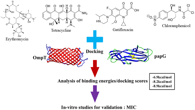
Figure1: Schematic diagram of the methods performed in this study.
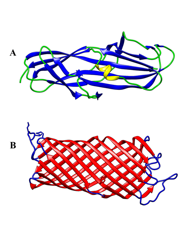
Figure 2: A. Structure of papG protein (PDB ID:1J8S), beta sheet is coloured in blue, alpha helix in yellow and coils in green. B. Structure of OmpT (PDB ID:1IJ8), beta barrel coloured in red and coils in blue.
RESULT AND DISCUSSION
Bacterial isolation and identification

Fig. 3 E. coli colony growth by striking method on BHIA plate
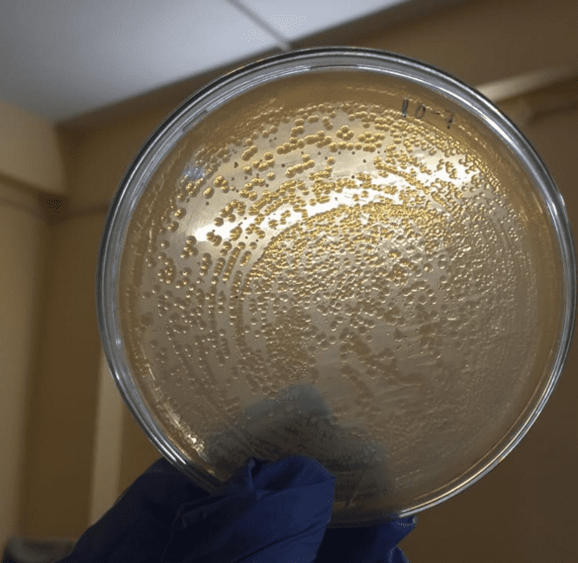
Fig. 4 E. coli colony growth by spread plate technique
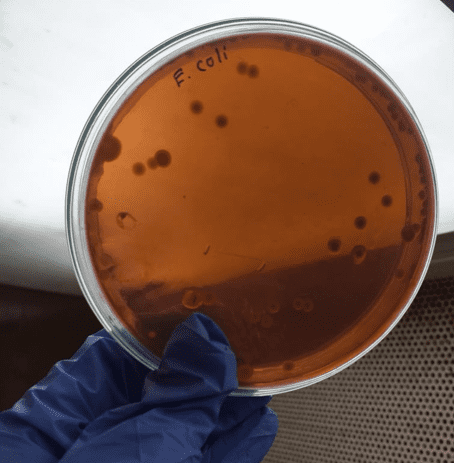
Fig. 5 E. coli colony growth in Eosin Methylene Blue (EMB) agar media plate
Bacteria was isolated from sewage water. Firstly, it was grown on brain heart infusion agar media plate by both spread plate and striking plate technique. Again the E. coli strain was confirmed from selective media colony growth.
Morphological identification
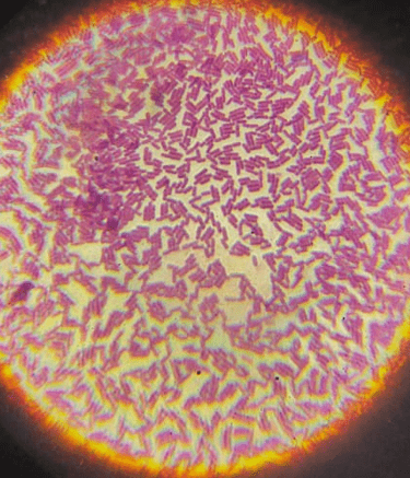
Fig. 6-gram staining of E. coli
In Gram stain E. coli strain was showing rod shape purple colour morphology. It was confirmed gram negative strain of E. coli.
Biochemical identification
Table 1 Biochemical test for E. Coli test confirmation
|
Characteristics |
E. Coli |
|
Motility |
Motile |
|
Catalase |
Positive (+) |
|
Oxidase |
Negative (-) |
|
Oxidative/Fermentative |
+/+ |
|
Indole |
Positive (+) |
|
Citrate |
Negative (-) |
|
Urease |
Negative (-) |
|
Nitrate Reduction |
Positive (+) |
|
H2S |
Negative (-) |
|
Gas |
Positive (+) |
|
Arabinose |
Positive (+) |
|
Glucose |
Positive (+) |
|
Lactose |
Positive (+) |
|
Sorbitol |
Positive (+) |
E. coli is tested with kit and other biochemical process. it was confirmed with those tests.
Antibiotic susceptibility test
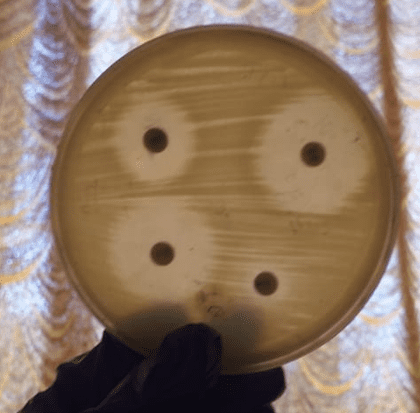
Fig. 7 antibiotic susceptibility test by disc diffusion method
Table 2 Zone of inhibition
|
Strain |
Antibiotics |
Zone of inhibition (in cm) |
|
E. Coli |
Chloramphenical |
2.9 |
|
Galtifloxacin |
1.1 |
|
|
Erthromycin |
0.5 |
|
|
Tetracycline |
2.6 |
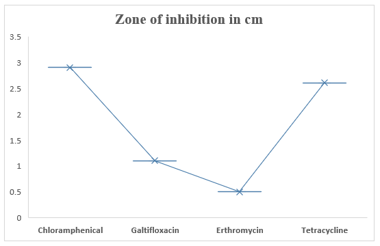
Fig.8 Graphical representation of antibiotic susceptibility assay with different antibiotics
Table 3. Descriptive Statistics
|
Statistic |
Value |
|
Mean |
1.775 |
|
Standard Deviation |
1.159 |
|
Minimum |
0.5 |
|
Median |
1.85 |
|
Maximum |
2.9 |
|
IQR |
7.725 |
|
Range |
2.4 |
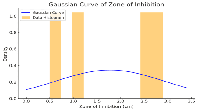
Fig.9 Normal distribution of Zone inhibition (cm)
We are using different antibiotic disc for checking antibiotic susceptibility. We get highest zone of inhibition in chloramphenical, then tetracycline, galtifloxacin and least zone observed in erythromycin.
Table 4 depicts the binding affinity of papG with various antibiotics and the affinity with which antibiotics bind to the OmpT protein. Docking score is a numerical value that is given to a protein-ligand complex to understand their binding affinity. It gives an estimated score of how strongly a ligand binds to the protein by taking the intermolecular forces into consideration. Using search algorithms, the best fit ligand (RMSD 0.0) is found out and then the binding poses are given a score based on scoring functions. FF-based Scoring function is widely used to calculate the docking score of a complex. The docking score has several applications- like prediction of a drug that binds well with this protein, biomolecular interactions, drug designing and lead optimization. The more negative the docking score, higher is the binding affinity. This concept can be explained based on Gibb’s free energy change.
Table 4: Docking scores of two different proteins with four different ligands.
|
Protein |
Ligand |
Docking score (in kcal/mol) |
|
papG |
Gatifloxacin |
-6.1 |
|
Chloramphenicol |
-5.7 |
|
|
Erythromycin |
-6.2 |
|
|
Tetracycline |
-6.9 |
|
|
OmpT |
Gatifloxacin |
-8.2 |
|
Chloramphenicol |
-7.0 |
|
|
Erythromycin |
-7.9 |
|
|
Tetracycline |
-8.9 |
In this study, we have docked papG and OmpT separately with four different antibiotics. The results vividly portray that the antibiotics are binding strongly with the later as compared to the former, as the docking scores are more negative in case of OmpT when compared with papG. Hence, it can be inferred that targeting OmpT would be a better option since there would be enhanced drug binding and the disease must be cured faster. Both the proteins serve for bacterial diseases like UTI (Urinary Tract Infection). OmpT’s ability to degrade antimicrobial peptides indirectly enrich papG’s activity to adhere strongly to the host cells. Thus, it is aiding in bacterial defense mechanism and hence if we inhibit it then there would be loss of bacterial ability to protect itself against antibacterials. Consequently, papG’s function would be hindered and the person would be cured effectively. The various docked structures are shown in Figure 10 and Figure 11.

Figure 10: Docked structures of papG protein with various antibiotics- Gatifloxacin (A), Erythromycin (B), Chloramphenicol (C) and Tetracycline (D). The protein is coloured in pink and the ligand in orange. Visualization is made using PyMol and ChimeraX.
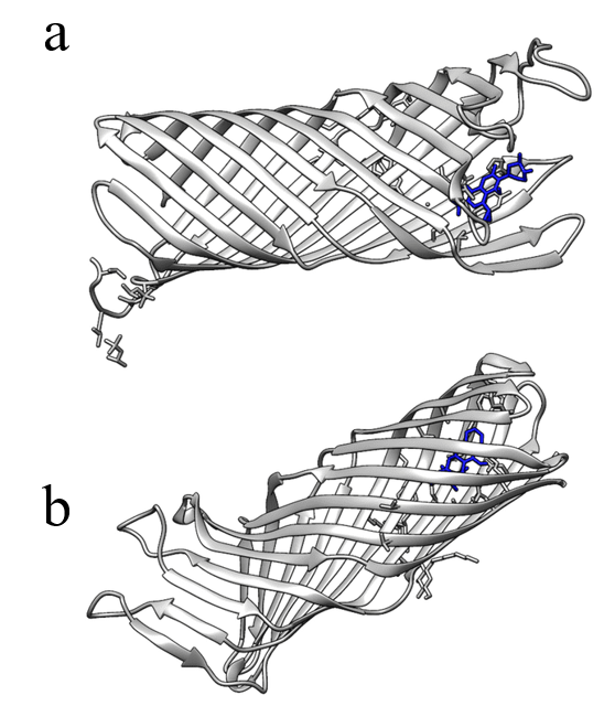
Figure 11: Docked structures of OmpT protein with various antibiotics- Gatifloxacin (a), and Tetracycline (b). The protein is coloured in gray and the ligand in blue. Visualization is made using PyMol and Chimera.
From the results, it can be inferred that Tetracycline binds very strongly with OmpT protein (-8.9kcal/mol) as compared to the other three antibiotics. Gatifloxacin is the second-best binding drug (-8.2kcal/mol). Hence, Tetracycline in combination with Gatifloxacin would be best drug of choice to treat patients suffering from E. coli mediated infections like UTI and diarrhea6. Tetracycline would block the action of 30S ribosomal subunit, thereby hindering protein synthesis and Gatifloxacin will have a role in blocking the DNA replication. We have also calculated the Inhibition constant value (Ki) of the drug as descripted in Table 2. Lower the Ki value, better is the drug binding and vice versa. Thus, Tetracycline is having a lower value suggesting good binding as compared to Gatifloxacin. Therefore, apart from these antibiotics we must design a better drug using Virtual screening and Lead Optimization techniques as over usage of these might lead to Anti-microbial Resistance which is very challenging to treat.
There are five Hydrogen bonds observed when Gatifloxacin is docked with OmpT. The amino acids which are participating in hydrogen bonding are shown in Table 2. Histidine 212 of OmpT is bonding to Fluorine atom of Gatifloxacin as seen in Figure 12. Two unfavorable interactions are observed in case of Tetracycline, but there are six H-bonds, that contribute to a better binding with the target protein.
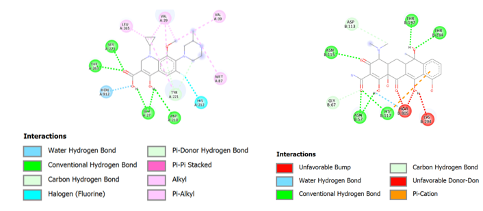
Figure 12: Interactions between gatifloxacin and Omp T shown on the left and on the right hand site, the molecular interactions between tetracycline and Omp T is shown. These are visualized in BIOVIA Discovery Studio7.
Table 5
|
Protein |
Ligand |
RMSD |
Binding affinity (in kcal/mol) |
Number of H-bonds (Protein-Ligand) |
Inhibition constant (in μM) |
Amino acids involved in H-bonding |
|
OmpT |
Gatifloxacin |
0.0 |
-8.2 |
5 |
1.06 |
Glu27, Asp210, Ser223, Thr263 |
|
Tetracycline |
0.0 |
-8.9 |
6 |
0.32 |
Asn75, Asn115, Lys117, Thr184 and Thr197 |

Figure 13: Effects of Antibiotics (Tetracycline and Gatifloxacin) on OmpT as a target protein8.
CONCLUSION
In our present work we can identified E. coli strains in sewage water samples and assessed their antibiotic resistance profiles. The findings confirm that sewage water is a reservoir for antibiotic-resistant E. coli, likely due to contamination from industrial and medical sources. The molecular docking results of the antibiotic susceptibility tests demonstrated high resistance to several commonly used antibiotics, including complete resistance to ampicillin and varying levels of resistance to other antibiotics. These findings highlight the potential risk of disseminating antibiotic-resistant bacteria into natural water bodies and the broader environment. The presence of antibiotic-resistant E. coli in sewage underscores the importance of implementing effective wastewater treatment strategies to minimize the release of resistant bacteria into ecosystems. Continued monitoring and control of antibiotic contamination in sewage are crucial to reducing the spread of resistance, protecting public health, and maintaining the efficacy of antibiotics.
No conflict of interest between all authors.
No funding is applicable for this work.
REFERENCES

Soma Garani*, Ahana Chakraborty, Subhrodipto Basu Choudhury, Sanjay Dey, Tufan Dewan Badshah, Investigation of Antibiotic Resistance in E. Coli Isolated from Sewage Water, Int. J. of Pharm. Sci., 2025, Vol 3, Issue 12, 1929-1941 https://doi.org/10.5281/zenodo.17892291
 10.5281/zenodo.17892291
