
We use cookies to ensure our website works properly and to personalise your experience. Cookies policy
The Oxford College of Pharmacy, Hongasandra, Bommanahalli, Bangalore.
Liposomes are the spherical vesicles that comprises of phospholipid bilayers which is most important in the area of drug delivery, cosmetics as well as nanomedicine. They have capacity to incorporate both amphiphilic or amphipathic substances which makes them a versatile carrier for targeted therapies liposomes play a crucial role in increasing drug solubility, to improve bioavailability, along with reducing toxicity. Their capacity is to deliver therapeutic agents to specific sites and make them an essential component in modern medicine. The field of liposomal research has expanded widely, with its applications in cancer therapy, gene therapy, immunotherapy and vaccine delivery. The applications of liposomes protrude over medicine, reaching the food, cosmetic, and agricultural industries, where they improve the stability of bioactive compounds. This article describes the fundamentals of liposomes with their structural composition, classification, and various preparation methods. It also describes the applications of liposomes in pharmaceuticals, cosmetics etc as well as the challenges that are faced in large – scale production and stability, with this the review highlights ongoing research and the future trends in liposome technology, such as artificial intelligence – assisted liposomes design and hybrid lipid – polymer vesicles with the continuous advancements, liposomes are set to revolutionize targeted drug delivery and precision medicine, ensuring improved therapeutic outcomes for patients worldwide.
Liposomes are small, globular vesicles comprises of one or more concentric lipid bilayers enclosing an internal aqueous compartment. Their membranes are made from natural or synthetic lipids, known for being biocompatible, biodegradable and non-immunogenic. Due to their unique bilayer structure, liposomes are widely used as delivery systems for both hydrophilic and lipophilic substances. Hydrophilic substances are encapsulated within the aqueous core, whereas lipophilic drugs are predominantly incorporated into the lipid bilayers.1. Liposomal research is becoming significant in natural, pharmaceutical and medical research as a result of these liposomes that are found towards having most productive carriers for the introduction of every single factor within the cells.2.
Liposomes can also be loaded with nanoparticles of different types and sizes, significantly expanding their applications. This combination influences the advantages of both liposomes and nanoparticles, which has played a central role in scientific research over the past several decades.3. Since the 1990’s, the use of liposomes as carriers for anticancer drugs has resulted in the approval pf several lipid-based formulations. Recently, interest in liposomes has grown even further driven by the growing demand for effective delivery systems associated with emerging therapeutic approaches such as oligonucleotides, DNA and m-RNA based vaccines, and CRISPR technologies.4.
These liposomes were initially developed in year 1961 by Alec D. Bangham. There remains an ongoing challenge across multiple industries and research fields including biochemistry, pharmacy, biophysics and colloid science, which is used to develop innovative methods for fabricating liposomes and expand their range of applications. This is especially important for advancing their use as advanced drug delivery systems, particularly for targeting specific tissues or organs.5. The name “liposome” was taken from the Greek words “lipo” that denotea fat where as “soma” that indicates body, meaning lipid-based body or structure.6.
In clinical studies, liposomes have demonstrated the ability to improve pharmacokinetics and biodistribution of therapeutic agents. These properties help to minimize systemic toxicity by enhancing drug accumulation at the desired site of action.7. Liposomes are colloidal structures that typically ranges from 5 to 200 nm in diameter and consist of membranes that has 4nm of thickness and arranged in concentric or non-concentric layers.8.
Recent breakthrough in liposome vaccines, especially mRNA technology, have made them even more important in immunology-they help vaccines stay stable, show antigens better, and boost our immune response. Liposome-based COVID -19 vaccines proved how powerful they can be-they’ll likely play a huge role in future vaccine developments. Even outside healthcare, liposomes are being explored in agriculture and food science to deliver nutrients, good compounds, and pesticides safely. As research continuous, liposomes will probably become even more crucial in medicine industry and biotech.
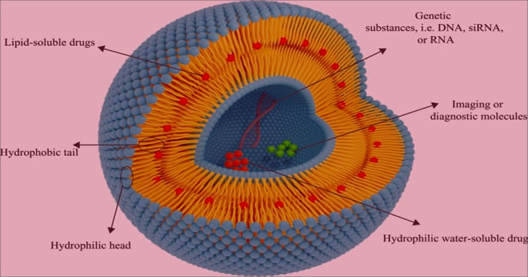
Figure 1: Structure of Liposomes
CLASSIFICATION OF LIPOSOMES
Liposomes are categorized according to their preparation process, number of lamellae, and vesicle size. The degree of encapsulation of drug in the liposomes is determined by the size and number of bilayers, and the vesicle size plays an essential role in determining the liposomes half-life. Liposomes fall into a number of categories-
I. Based on structure or number of lipid layers:
1. Multilayered liposomes:
These are made up of multiple phospholipid bilayers. These liposomes have sizes ranging from 0.1 to 0.5 µm. MLVs have a structure that looks like an onion, and their primary benefit is that they are stable and easy to manufacture. But the main drawback of liposomes is their small loading compound space due to the presence of more layers. So, it is also limiting their injectable use.9.
2. Unilamellar liposomes:
These liposomes consist of a single phospholipid bilayer with an aqueous solution enclosed by a sphere. There are three types of unilamellar vesicles-
3. Multivesicular liposomes:
These vesicles are made up of multiple non-concentric vesicles that are enclosed in a single bilayer. These multipurpose liposomes are between 2 and 40µm in size.11
4. Oligolamellar liposome:
Compared to multilamellar liposomes, oligolamellar liposomes have fewer lamellar layers. The size of these liposomes ranges from 0.1 to 10µm.11
5. Giant liposome:
These are the biggest liposomes, with sizes ranging from 10 to 1000µm. There are several medical and diagnostic uses for these liposomes. They function as an LUV as well as an SUV. 11
II. Based on method of preparation:1, 25
1. Reverse Phase Evaporated Vesicles (REV): Reverse phase evaporated vesicles (REV) are a kind of vesicular drug delivery system made by dissolving lipids in an organic solvent to make a water-in-oil emulsion, then removing the solvent to form a gel that, when agitated and evaporated, creates large unilamellar vesicles (LUVs). Drugs that are hydrophilic or lipophilic can be encapsulated using this technique because of its high entrapment efficiency.
2. Multilamellar vesicle made by reverse phase evaporation method (MLV-REV): The reverse-phase evaporation (REV) method can be used to create multilamellar vesicles (MLVs), although it is more frequently linked to large unilamellar vesicles (LUVs) or other vesicular structures.
3. Stable Pluri Lamellar Vesicles (SPLV): These are a type of multilamellar liposome with enhanced stability, characterized by their multiple bilayers and a lack of osmotic compression. They are prepared through lipid dispersion in an organic solvent, emulsification with an aqueous phase, and subsequent solvent evaporation. These have unique physical and biological properties.
4. Frozen and thawed multilamellar vesicles (FATMLV): It is a technique which significantly enhances solute trapping efficiency and it can alter their structure, further it leads to larger vesicle sizes or more unilamellar nature due to osmotic stress and bilayer changes. Multilamellar vesicles prepared by repetitive freeze-thaw cycles.
5. Vesicle prepared by extrusion technique (VET): These are prepared by forcing a lipid suspension through a series of filters with defined pore sizes under pressure, which breaks down larger, multilamellar vesicles (MLVs) into smaller, more uniform, and typically unilamellar vesicles (LUVs). Vesicle size is determined by the pore size of membrane used.
6. Dehydration-Rehydration Vesicles (DRV): This method involves encapsulating therapeutic molecules, such as medicine or vaccines, within a liposome. Pre-formed empty liposome is freeze-dried with a sugar solution, rehydrated with the material to be encapsulated, and then the resulting vesicles are formed. This technique uses mild conditions and is easy to use. High entrapment efficiency can be seen by this technique for a variety of compounds, including large sized and particulate substances.
7. Vesicles prepared by fusion (FUV): In this technique, vesicle is created by fusion of small, pre-existing vesicle (SUV) to form a single, continuous structure.
8. Vesicle prepared by French press (FPV): A French press can be used to create unilamellar vesicles by applying high pressure to lipid dispersions and pressing them through a tiny opening. Although this technique is restricted to smaller sample amounts and may involve pre-processing to prevent clogging, it produces comparatively big, stable vesicle with excellent entrapment efficiency. The vesicle obtained by this method mainly dependent on the pressure.
III. Based on composition and application:1, 25
1. Conventional liposome: The first generation of lipid-based drug delivery systems are conventional liposomes, which have an aqueous core enveloped in a phospholipid bilayer that also includes cholesterol. They are made to transport drugs that are hydrophilic and hydrophobic nature in the lipid bilayer.
2. Fusogenic liposome: These are specialized lipid-based nanoparticles have the ability to fuse with cell membranes directly, allowing them to transfer therapeutic materials such as proteins, peptides or DNA into the cytoplasm of the cell without being destroyed in the lysosome.
3. pH sensitive liposome: These are type of drug delivery system which is made to release its components in reaction to pH variations. They are usually made up of lipids, when exposed to acidic environments, change structurally, such as shifting from a membrane-like structure to a hexagonal phase. They are helpful in delivering the medications to specialized areas of the body with lower pH values than usual, including acidic tumour cells during intracellular drug delivery.
4. Cationic liposome (CL): These are positively charged lipid-based vesicles which are mostly employed as non-viral vectors to introduce therapeutic medicines and genetic materials (DNA, siRNA, and mRNA) into cells. They combine with negatively charged nucleic acids to create complexes known as lipoplexes. They have been utilized in vaccines and are great for delivering genetic material.
5. Long circulating liposome (LCL): It is a drug delivery vesicle which is made to prolong its half-life in the bloodstream by resisting the reticuloendothelial system (RES) fast clearance. This is usually accomplished by applying hydrophilic polymers, such as polyethylene glycol (PEG), to the liposome surface. This makes the liposome invisible to immune cell. This liposome extends circulation and makes it possible for them to more efficiently aggregate in target tissues, such as cancers improving therapeutic efficacy.
6. Immuno liposome: In this type, antibodies or their fragments are conjugated to the surface of liposomes. These changes allow the liposome to target specific antigens on the surface of cell and leading to more efficient delivery of therapeutic agents to the target tissue and reducing side effects on healthy tissues.
METHOD OF PREPARATION
Initially a mixture of phospholipid and cholesterol is taken in the round-bottomed flask and is dissolved in organic solvent, and these solvent contents are removed using a rotary evaporator. The thin layer of phospholipid and cholesterol is present in the RBF, and then the aqueous buffer solution was added to the RBF with shaking, and it led to the formation of vesicles. This type of preparation is used in the formation multilamellar vesicles (MLVs). Further, if required, this size of a vesicle is reduced using the sonication and extrusion method.12
Multilamellar vesicle (MLV) liposomes, which are prepared by thin film hydration or by other preparation methods, is placed in a chamber of a French pressure cell, also it is extruded at the range of 20,000 psi within a small opening. So, it produces liposome of unilamellar vesicles (SUV). While carrying out the preparation, extreme care and handling should be there. This preparation technique is considered a simpler and more reliable method.12.
Multilamellar vesicle (MLV) liposomes are converted into unilamellar vesicles using the sonicator.
There are two types of sonicators:
1. Probe sonication:
It works by the principle of ultrasonic cavitation. The sonicator is placed in the liposome (MLV). When it produces high-frequency vibration, it forms the bubble, which expands and collapses, which disrupts the particle size and leads to the formation of unilamellar vesicles. The container should be placed in the ice bath as it produces intense force and heat.13
2. Bath sonication:
In this type, liposomes (MLV) are placed in the cylinder, and then it is kept in the bath sonicator. It utilizes the ultrasonic waves to break the liposome into smaller vesicle sizes. When compared to a probe sonicator, maintaining the temperature is easier. The temperature is maintained at 4-6°C in order to prevent the degradation of the liposome.13.
3. Freeze-thawed liposome:
This method of preparation is commonly done to improve the entrapment efficiency and reduce the layers of lipid. Liposomes are frozen using liquid nitrogen, and it leads to the formation of crystals; then it is slowly thawed, which helps in the disruption of lipid layers. Multiple times it has been done to improve the drug loading.13.
4. Membrane extrusion method:
In this type, prepared liposome (MLV) is converted to SUV and LUV, and it is done by passing the liposome through the polycarbonate membrane filter, and it forms the liposome of particle size 120-140 nm.14.
5. Freeze drying:
This method is also known as lyophilization. In this method of preparation, liposome suspension is frozen at -40°C. Then the water content is removed by placing it under a vacuum by sublimation. Then secondary drying is done to remove remaining water content, and then they are stored as dry powders. These dry powders show stability, an increase in shelf life, and a reduction in degradation. Powder form can be reconstituted by the addition of water or buffer.15.
6. Non-shaking vesicle:
In this type of preparation, the drug, lipoid, and cholestrin are diffused in the natural solvent, and then it is transferred to round-bottom flask. Solvent content is separated by utilizing a rotary vacuum evaporator, and it forms a narrow lipid layer over surface of the flask. Then this layer is hydrated by using an aqueous buffer. This process of preparation does not require vigorous shaking.16.
7. Nano emulsification:
A nano-emulsifier is utilized to prepare a liposome. Lipids are taken into a fluidizer, and pumps the fluid at high pressure. It leads to the breakdown of large liposomes into smaller sizes. It forms the thermodynamically stable liposomes of smaller size.17.
8. Ethanol injection:
In this preparation, phospholipids are dissolved in the ethanol, and then it is injected into the stirred aqueous buffer. Dilution of ethanol in the aqueous solution leads to precipitation of lipids and formation of liposomes. Then the excess ethanol is removed using a rotary evaporator or by some other method. It leads to the evolution of concentrated nanoparticles.18
CHALLENGES IN LIPOSOME FORMULATION19
Liposomes are great but forming them isn't easy. There are stability problems and large-scale production issues.
Liposomes degrade easily due to their phospholipid composition. This makes them prone to hydrolysis and oxidation. Researchers are looking into antioxidants, lyophilization, and cholesterol to fix this.
Another issue is liposomes getting cleared quickly from the body. Stealth liposomes that are with PEG coating help, but even those get cleared eventually. Modifying lipid content and charge can improve circulation time.
Producing liposomes in bulk is tough. Lab methods like thin-film hydration and extrusion don’t scale up well. Microfluidic technology and high-pressure homogenization are being investigated instead.
Liposomes as to hold onto drugs well. Hydrophilic drugs leak out over time, while lipophilic drugs clump together. Optimizing lipid blend, vesicle size, and prep methods helps.
Liposomes are sensitive to temperature and pH. Refrigeration helps, but freeze-thaw cycles cause issues. Lyophilization with cryoprotectants improves shelf life, but rehydration can be tricky.
Most liposomes are biocompatible, but some cause immune reactions or toxicity. Cationic lipids are cytotoxic, and are limiting their use in gene therapy. Careful lipid selection and surface modification are crucial.
Liposomal drugs face strict regulations. Manufacturers must prove uniform drug release, safety, and efficacy through trials. Regulatory bodies like FDA and EMA require detailed data on stability, biocompatibility.
Liposome production is expensive due to complex processes and specialized equipment. High-purity lipids, sterilization, and quality control add costs. Researchers seek improvements in lipid synthesis, large-scale techniques, and automation.
EVALUATION PARAMETERS20
1. Shape and Lamellarity of vesicle
An electron microscope is used to analyse the vesicle size. It is evaluated by using transmission electron microscopy (TEM) and scanning electron microscopy (SEM) which provide direct visualization of their spherical structure.
2. Granule Size
Particle size of liposome is measured by using zeta sizer instrument.
3. Pdi
It is also known as “particle size distribution” it is obtained by photon correlation spectroscopic analysis.
4. Zeta Potential
Zeta potential is the charges which are present on the superficial layers of liposome. The charge is due to ingredients that are used in manufacturing of liposome. Presence of charge produce repulsion which prevents the coagulation of particles. Zeta potential is measured by using zeta sizer instrument having Malvern software. It is conducted at 25°C with a 90° detection angle.
5. Entrapment Efficiency
This quantifies the quantity and rate at which liposomes water-soluble compounds are trapped. For measurement of entrapment of drug present in supernatants it was measured by after centrifugation by UV spectrometer at 254 nm. The entire amount of drug used in the preparation was then reduced from total amount of drug used in the supernatant. It is measured by the given below formula,
% Entrapment efficiency=W-wW ×100
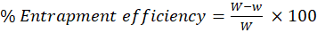
Where,
W – Total quantity of drug used during the formulation.
w – Quantity of drug in supernatant after centrifugation.
6. Efficiency of Loading
0.1M HCL is used to remove the medication from the liposomes, then drug content was measured. Here liposome (150gm) was mixed with 50ml of HCL. Then it was filtered using proper dilution at 254nm using UV spectroscopy.
L%= QnWn ×100
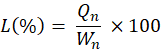
Where,
Qn–the drug’s concentration in the liposome
Wn –liposome weight
7. Invitro Release of Drug
Liposome present in aqueous suspension was separated using ultracentrifugation. Liposome (2mg) was mixed with 10ml of 7.4 phosphate buffer. Then the buffer was placed in dialysis membrane page. Make 900ml of phosphate buffer and transfer to dissolution apparatus beaker. USP paddle type was used for dissolution. Temperature was maintained at 37°C. at different time interval, 1ml of buffer in dissolution apparatus was removed and it is replaced with 1ml of fresh phosphate buffer. The content of drug released was measured using UV visible spectrometer at 275nm.20
8. Dynamic Light Scattering
Dynamic light scattering (DLS) can be used to calculate the intensity of light scattered as a function of time by suspended particles undergoing Brownian motion. This method offers a number of benefits, including precise, repeatable, and accurate size analysis in its natural surroundings, direct measurement of high concentration and turbid samples, fully automated measurement, ease of setup, measurement of small sizes <1 nm and molecular weight <1000 Da, and low sample volume.26
FUTURE PERSPECTIVES AND INNOVATIONS
Liposome research advances with attempts to overcome challenges and expand applications. Nanotechnology improvements enable enhanced stability, targeted delivery, and large-scale manufacturing.
1. Smart and Stimuli-Responsive Liposomes
Stimuli-responsive liposomes release drugs upon activation by pH, temperature, ultrasound, or enzymes. pH-sensitive liposomes target cancer cells, while temperature-sensitive one’s release drugs at elevated temperatures.
2. Lipid Nanoparticles (LNPs) in RNA Therapeutics
LNP technology delivers mRNA vaccines like COVID-19 vaccines. LNPs secure genetic compounds like mRNA and siRNA, enabling innovative treatments for genetic disorders, infectious diseases, and targeted medicine.
3. Hybrid and Multifunctional Liposomes
Hybrid liposomes combine liposomes with polymers, nanoparticles, or dendrimers. They enhance drug encapsulation, circulation times, and targeting capabilities. Multifunctional liposomes deliver multiple therapeutic agents.
4. Artificial Intelligence in Liposome Design
AI and machine learning optimize liposome formulation parameters, predict drug release behaviour, and ensure stability.21.
APPLICATION
Many liposomal based preparations were successfully applied in the clinical field as antiviral, anticancer, antifungal or anti-infective, analgesics for pain management and also in photodynamic therapy.
Liposomal therapy shows significant therapeutic benefits in the prevention of cancer and fungal diseases.22.
1. In Cancer Therapy
2. Fungal Treatment
3. Pain Management
4. Photodynamic therapy
5. Antiviral agents
6. Liposomes in Vaccination
Liposomes act as a immunological adjuvant.
Therefore, mRNA coding used for the protein spike of the Coronavirus, that will be encapsulated within the liposomes which are developed to make stable for circulating the blood until they are taken up by the phagocytic cells in the body through endocytosis.
Hence mRNA is indicated as the spike Protein results in promoting the immune response to it and kill the invading virus.
7. Liposomes in clinical applications:
Many liposomal formulations have been extensively implemented in the clinical field as anticancer, antifungal, or anti-infective analgesics for pain management and also in photodynamic therapy.
Liposome therapy shows significant therapeutic benefits in the treatment of cancer and fungal infections.
CONCLUSION
Liposomes are becoming a game changer in medicine, science and industry. Their capability to incorporate both amphiphilic or amphipathic molecules improves their strength and also sustained release thus leading them effective in treatments such as cancer therapies and RNA-based vaccines.
Despite of existing challenges like limited stability, high production rates etc, research and innovations continue to drive improvements in liposomal technologies. Innovations in nanotechnology, artificial intelligence are expanding the potential of liposomes for their use in medicines, agriculture, and environmental sustainability.
As the technology improves the possibilities of the liposomes continues to grow. These are set to play an important role in transforming healthcare and biotechnology. With continued investment in research and development, liposomes are assured to become foundation of next generation therapies and industrial solutions that delivers broad benefits to patients and global industries.
REFERENCES


Uma Prabha R, Nandhini R, Nanditha. R, Navya R. Reddy, Neelakanta, Nisarga S T, Liposome: Structure, Preparation and Future Perspectives, Int. J. of Pharm. Sci., 2025, Vol 3, Issue 9, 2932-2943. https://doi.org/10.5281/zenodo.17197885
 10.5281/zenodo.17197885