Department of Pharmaceutics, College of Pharmaceutical Sciences, Govt. Medical College, Kozhikode, India
Nanoparticles (NPs) have gained considerable interest owing to their distinctive physicochemical properties and enhanced functional performance compared with their bulk counterparts, enabling broad technological and biomedical applications. Conventional physical and chemical synthesis approaches commonly employ toxic reducing agents, generate hazardous by-products, and require high energy input, thereby limiting their environmental sustainability. In response, green synthesis has emerged as a viable and eco-friendly alternative that utilizes biological systems for nanoparticle fabrication. Biological entities such as bacteria, actinomycetes, fungi, algae, yeast, and plants act as natural reducing and stabilizing agents, offering a clean, safe, cost-effective, and scalable route for NP synthesis. This review provides a comprehensive overview of nanoparticles, focusing on their classification, intrinsic properties, synthesis strategies with particular emphasis on green synthesis characterization techniques, and key applications across various fields. Furthermore, current challenges, limitations, and future perspectives associated with biologically synthesized nanoparticles are critically discussed, highlighting their potential contribution to the advancement of sustainable nanotechnology.
Overview of Nanoparticles
Materials with diameters typically between 1 and 100 nm are known as nanoparticles (NPs), and they have distinct physicochemical characteristics from their bulk counterparts. Nanoparticles have unique optical, electrical, catalytic, and biological properties because of their incredibly small size, high surface-area-to-volume ratio, and increased surface reactivity. Because of these characteristics, nanoparticles may successfully interact with biological systems, which has led to their widespread use in industries including biomedical science, electronics, environmental remediation, and catalysis[1,2].
Importance of Nanotechnology in Medicine
Because it makes it possible to create novel diagnostic and treatment approaches, nanotechnology has become a revolutionary field in contemporary medicine. While reducing systemic toxicity, nanoparticles can enhance therapeutic solubility, stability, and targeted delivery. Because of their antibacterial qualities and capacity to interact with microbial cells and biological membranes, metal and metal oxide nanoparticles—such as silver, gold, zinc oxide, copper, and iron nanoparticles—have drawn special attention. Applications of these nanoparticles in medication delivery, biosensing, imaging, wound healing, and antimicrobial therapies[3,4].
Antimicrobial Resistance Problem
Antimicrobial resistance (AMR) has emerged as one of the most major global public health issues despite tremendous advancements in antimicrobial therapy. Through processes such enzymatic drug degradation, alteration of antibiotic targets, and activation of efflux pumps, pathogenic microbes have acquired resistance to several antibiotics. In addition to decreasing the efficacy of traditional antibiotic therapies, the rise of multidrug-resistant bacteria has raised morbidity, death, and healthcare costs globally[5,6].
Role of Biofilms in Infections
Microorganisms ‘capacity to build biofilms is a significant factor in antibiotic resistance. Biofilms are organized colonies of microorganisms embedded in an extracellular polymeric substance (EPS) matrix that is mostly made up of proteins, lipids, polysaccharides, and extracellular DNA. Both biological tissues and abiotic surfaces, like medical equipment, catheters, implants, and prosthetic materials, can support the growth of these microbial communities. Biofilm-associated infections are difficult to treat and frequently chronic because microorganisms within biofilms are far more resistant to medicines and host immune responses than planktonic cells[7,8].
2. BACTERIAL BIOFILMS
2.1 Biofilm formation
Gram-negative bacteria have developed a defence mechanism called bacterial biofilms to keep things out of their cells. Extracellular polymeric substances (EPS), which are mostly made of proteins, nucleic acids, and polysaccharides linked to microbial colonies known as bacterial biofilms, make up the biofilm. Biofilms, which can grow on a range of living and non-living surfaces, including industrial machinery and medical equipment, are thought to be responsible for over 80% of chronic infections. One of the most prevalent and stable microbial lives on Earth, biofilms are frequently found in soil, groundwater, seawater, and ocean sediments, where they are crucial to biogeochemical cycles.[8]
Biofilms are extremely resistant to chemical and physical stressors due to the biofilm matrix and modifications in bacterial metabolism, such as the development of dormant cells. Although biofilms are helpful in many industrial operations, there are significant health hazards associated with them. In addition to contaminating drinking water, biofilms can infect medical equipment, including catheters, implants, and prosthetic materials, resulting in chronic illnesses that are challenging to cure.[7]
Biofilm communities have unique architectural characteristics in natural settings, such as the existence of macro- and micro-colonies divided by interstitial spaces. Nutrients, gases, and antibacterial chemicals can more easily diffuse throughout the biofilm structure because of these voids. Biofilms, on the other hand, are extremely dynamic and can alter their structure in response to shifts in both internal physiological processes and external environmental factors. Significant phenotypic and functional variability within the biofilm community is caused by the close proximity of cells within biofilms, which facilitates the interchange of quorum-sensing molecules and extrachromosomal genetic material, such as plasmids. [9]
2.2 Biofilm Formation on Medical Devices
One of the main causes of nosocomial and healthcare-associated infections is biofilm formation on medical devices. Urinary and intravascular catheters, endotracheal tubes, orthopaedic implants, cardiac valves, and prosthetic devices are examples of indwelling and implantable medical devices that offer perfect surfaces for microbial adherence and the subsequent formation of biofilms. Both Gram-positive and Gram-negative bacteria that cause illnesses linked to medical devices have been found to use biofilm formation as their main pathogenicity mechanism[10].
The initial attachment of planktonic microbes is facilitated by the adsorption of host-derived conditioning films made of proteins, fibrin, and other organic molecules onto the device surface, which starts the biofilm formation process. Bacterial aggregation, microcolony development, and biofilm maturation follow this attachment's transition from a reversible to an irreversible state. Extracellular polymeric substances (EPS), which include polysaccharides, proteins, lipids, and extracellular DNA, are secreted by microbes during this process, creating a highly organized and protective biofilm matrix.
Biofilms formed on medical devices exhibit enhanced resistance to antimicrobial agents and host immune defences. The EPS matrix limits antibiotic penetration, while altered metabolic states and the presence of persister cells further contribute to antimicrobial recalcitrance. In addition, biofilm communities facilitate horizontal gene transfer, promoting the spread of antimicrobial resistance genes among microbial populations. These characteristics often lead to persistent and recurrent infections that are difficult to eradicate using conventional antimicrobial therapy and frequently necessitate device removal or replacement. [10]
Biofilm-associated medical device infections have a significant clinical impact, leading to longer hospital stays, higher medical expenses, and higher morbidity and mortality, especially in critically ill and immunocompromised patients. Therefore, developing effective preventive and therapeutic measures, such as antifouling surface modifications, antimicrobial coatings, and early biofilm detection technologies, requires an understanding of the principles driving biofilm formation on medical devices.
2.3 Stages of Biofilm Formation
Planktonic bacteria change into organized, surface-associated populations embedded in a matrix of self-produced extracellular polymeric substance (EPS) through a highly controlled, multi-step biological process known as biofilm development. Over 60% of chronic infections are linked to biofilms, which are the most common mode of microbial growth in natural, clinical, and industrial settings [11,12]. Initial attachment, irreversible adhesion, microcolony formation, maturity, and dispersion are among the successive and overlapping phases of the biofilm developmental cycle. [8,13]
2.3.1. Initial Attachment
When free-floating planktonic cells come into contact with a biotic or abiotic surface in a hydrated environment, biofilm formation starts. Van der Waals interactions, electrostatic forces, and hydrophobic interactions between microbial cell surfaces and the substratum are examples of weak physicochemical forces that facilitate initial attachment, which is usually reversible [12,13]. This early stage is heavily influenced by environmental factors such as ionic strength, shear stress, nutrition availability, and surface roughness [14]. Attached cells can still readily separate at this time and revert to their planktonic form.
2.3.2. Irreversible Attachment
Microorganisms reinforce their connection after reversible adhesion by expressing surface adhesins, pili, fimbriae, and flagella, which secure cells to the surface [14]. Alongside the start of EPS formation, this change signifies the beginning of irreversible attachment. Extracellular DNA (eDNA), proteins, lipids, and polysaccharides are the main components of EPS that function as binding agents to maintain cell-surface and cell-cell connections [11,15]. A crucial transition from individual cell survival to community-based growth is represented by the release of EPS.
2.3.3. Microcolony Formation
Once irreversibly attached, microbial cells proliferate and aggregate, forming microcolonies that serve as the foundational units of the biofilm. During this stage, EPS synthesis increases significantly, accounting for up to 90% of the biofilm biomass [16]. eDNA released from lysed cells plays a vital structural role by reinforcing the EPS matrix and facilitating horizontal gene transfer, thereby enhancing genetic diversity and adaptability within the biofilm [17]. Cell-to-cell communication becomes increasingly prominent as population density rises.
2.3.4. Biofilm Maturation
During biofilm maturity, a complex three-dimensional architecture with a heterogeneous cell distribution and internal water pathways that allow the transfer of waste, nutrients, and oxygen is formed[18]. A signalling mechanism that is dependent on cell density, quorum sensing controls the regulation of genes linked to metabolic adaptability, virulence, stress response, and EPS generation[19]. Because of gradients in nutrients and oxygen, mature biofilms show significant physiological variability, with bacterial cells living in various metabolic states[20]. Together, these characteristics give biofilms their exceptional resistance to host immunological responses and antimicrobial drugs.
2.3.5. Dispersion
The last phase of the biofilm life cycle, dispersion, allows microbial cells to spread out and occupy new habitats. Environmental stress, quorum-sensing-regulated signalling pathways, or nutritional deficiency may all initiate this process [21]. In order to release planktonic cells from the biofilm matrix, dispersion entails the enzymatic breakdown of EPS components, decreased adhesin expression, and enhanced motility. The development and recurrence of infections linked to biofilms are significantly influenced by dispersed cells, which frequently display increased virulence.
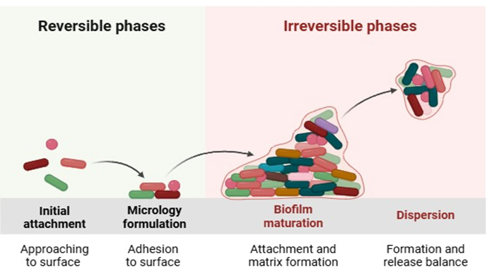
Fig 1: Mechanism of biofilm formation (created in https://www.biorender.com)
3. ANTIBIOTIC RESISTANCE IN BIOFILMS
Mechanisms of Antimicrobial Resistance in Biofilms
Biofilms possess inherent properties that confer intrinsic tolerance to antimicrobial agents, including metabolic dormancy and protection provided by the extracellular polymeric substance (EPS). Beyond this intrinsic tolerance[22], multiple biological and ecological factors within biofilms facilitate the evolution, persistence, and dissemination of antibiotic resistance both within and between bacterial species[23].
3.1. Role of the Biofilm Matrix in Antimicrobial Resistance
The biofilm matrix is a structurally robust and defining feature of biofilm architecture?. Composed primarily of polysaccharides, proteins, lipids, and extracellular DNA (eDNA), the matrix acts as both a physical and chemical barrier protecting embedded cells from antibiotics and host immune defences[16,24].
Limited antibiotic penetration is one of the primary resistance mechanisms in biofilms. The dense polymeric network reduces diffusion rates and lowers the effective concentration of antimicrobial agents reaching bacterial cells[20]. Certain antibiotics interact directly with matrix components; for example, positively charged aminoglycosides bind to negatively charged eDNA, further slowing drug diffusion[25]. During chronic infections, host-derived DNA released from lysed immune cells integrates into the matrix, reinforcing its density and protective function[26].
High bacterial cell density within biofilms also contributes to reduced drug susceptibility. This phenomenon, known as the inoculum effect, requires higher antimicrobial concentrations to achieve bactericidal activity[26].
3.2. Structural and Functional Roles of Extracellular DNA
Extracellular DNA is a critical structural and functional component of biofilms. Initially regarded as a byproduct of cell lysis, eDNA is now recognized as an actively produced and regulated component essential for biofilm stability and development.
Structurally, eDNA stabilizes biofilm architecture by forming networks that link cells and matrix components. DNase-mediated degradation of eDNA has been shown to prevent biofilm formation and disrupt established biofilms across multiple species. Functionally, eDNA contributes to antimicrobial resistance by binding positively charged antibiotics and reducing their mobility within the matrix[26–29].
3.3. Biofilm Matrix as a Reservoir for Biomolecules
The biofilm matrix functions as a dynamic reservoir for enzymes, nutrients, proteins, and genetic material. Matrix-associated enzymes degrade substrates into utilizable nutrients and modify matrix composition to adapt biofilm properties. Structural proteins such as amyloid fibres contribute to mechanical stability and organization[30,31].
Cell lysis within biofilms releases intracellular DNA and plasmids, which may serve as genetic material for horizontal gene transfer¹?. The immobilization and close proximity of cells facilitate frequent cell-to-cell interactions, creating optimal conditions for antimicrobial resistance gene exchange[32,33].
3.4. Horizontal Gene Transfer in Biofilms
Horizontal gene transfer (HGT) occurs at significantly higher frequencies in biofilms compared to planktonic populations. Genetic material can be exchanged through transformation, transduction, and conjugation.
Conjugation is particularly important in multidrug-resistant Gram-negative bacteria, where resistance genes are commonly located on conjugative plasmids. Biofilms promote plasmid stability and persistence even in the absence of antibiotic selection pressure[31,34,35].
Emerging mechanisms such as lateral transduction and outer membrane vesicle–mediated gene transfer further facilitate resistance dissemination.
3.5. Evolution of Resistance Within Biofilms
Biofilm-associated bacteria frequently survive high antibiotic concentrations. Exposure to sub-inhibitory antimicrobial levels can select for resistant mutants through point mutations, acquisition of resistance genes, or adaptive phenotypic changes.
Environmental stressors beyond antibiotics, including heavy metals and disinfectants, also contribute to resistance evolution within biofilms by promoting genetic exchange and selection of tolerant phenotypes[36,37].
3.6. Tolerance and Persistence in Biofilms
Antimicrobial survival in biofilms is not solely dependent on genetic resistance. Physiological heterogeneity results in populations of slow-growing or dormant cells that exhibit antibiotic tolerance.
Tolerance refers to survival without resistance-conferring mutations. Within this population, persister cells enter a reversible dormant state characterized by metabolic inactivity²³. Because many antibiotics target active cellular processes, persisters survive treatment and can repopulate the biofilm once antibiotic pressure is removed[38].
Persister formation is associated with stress responses and toxin–antitoxin systems that suppress essential cellular functions[39,40].
3.7. Interplay Between Resistance, Tolerance, and Persistence
Resistance, tolerance, and persistence are distinct but interconnected phenotypes. Persister-mediated survival during prolonged therapy increases the likelihood of resistance mutations emerging. Tolerance has also been shown to accelerate resistance evolution[41,42].
3.8. Polymicrobial Interactions and Antimicrobial Resistance
Most clinical biofilms are polymicrobial. Cooperative interactions can enhance collective resistance, while competitive interactions induce adaptive responses such as increased EPS production and efflux pump upregulation.
Some species degrade antibiotics or modify microenvironments, indirectly protecting neighbouring organisms[43].
3.9. Quorum Sensing and Resistance Regulation
Quorum sensing (QS) is a cell-density–dependent communication system regulating biofilm formation, virulence, stress responses, and antimicrobial tolerance.
QS coordinates protective responses across biofilm communities. Disruption of QS pathways impairs biofilm development and increases antibiotic susceptibility[44,45].
3.10. Competition and Cooperation in Biofilms
Bacterial species within biofilms engage in competitive and cooperative interactions. Community-level resistance mechanisms, including enzymatic antibiotic degradation and shared EPS protection, benefit all members[43].
4. NANOTECHNOLOGY
The use of nanoparticles can be traced back to ancient civilizations, where nanoscale materials were unintentionally employed to obtain unique optical and mechanical properties. A notable early example is the Roman Lycurgus Cup (4th century AD), whose dichroic behaviour arises from the presence of gold and silver nanoparticles dispersed within a glass matrix[46]. Similar nanoparticle-induced coloration was later observed in medieval stained-glass windows and ceramic glazes, attributed to metallic nanoparticles embedded in glassy materials[47].
The scientific understanding of nanoparticles began to emerge in the nineteenth century through the pioneering work of Michael Faraday, who investigated the optical properties of colloidal gold and demonstrated that particle size significantly influences light absorption and scattering[48]. Faraday’s work is considered a foundational contribution to colloid chemistry and nanoparticle science.
A major conceptual breakthrough in nanoscience occurred in 1959, when Richard Feynman introduced the idea of manipulating matter at the atomic scale in his seminal lecture “There’s Plenty of Room at the Bottom,” which laid the theoretical foundation for modern nanotechnology?. The term nanotechnology was later formally introduced in 1974 by Norio Taniguchi, referring to the precision processing of materials at the nanometre level[49].
Progress in nanoparticle research accelerated with the development of advanced nanoscale characterization techniques and the discovery of new nanomaterials such as fullerenes, carbon nanotubes, quantum dots, and graphene, which significantly expanded the field of nanoscience and established nanoparticles as key components of modern nanotechnology with applications in medicine, electronics, catalysis, energy systems, agriculture, and environmental remediation[50].
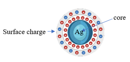
Fig1: structure of nanoparticle surrounded by charge
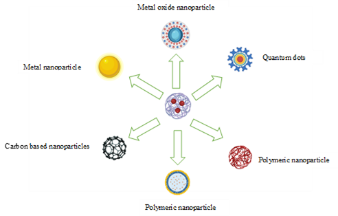
Fig 2: Types of nanoparticles
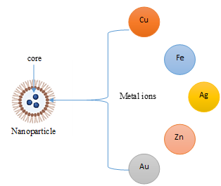
Fig 3: types of metal nanoparticles
4.2 Types of Nanoparticles
Table 1: Classification of Nanoparticles.
|
Category of NPs |
Description / Key Characteristics |
Typical Size Range |
Major Applications |
Reference |
|
Carbon-based NPs |
Add carbon nanotubes (CNTs) and fullerenes (spherical, hollow cages). CNTs can be single-, double-, or multi-walled structures made of rolled graphene sheets. Show excellent mechanical strength, flexibility, electron affinity, and electrical conductivity. |
~1–2 nm (CNT diameter) |
Electronics, sensors, nanocomposites, energy storage |
[2] |
|
Metal NPs |
exhibit localized surface plasmon resonance (LSPR), which produces unique electrical and optical properties, and are composed of pure metals like as Au, Ag, and Cu. absorb a lot in the region that is visible. |
1–100 nm |
Catalysis, sensing, antimicrobial agents, biomedical applications |
[51] |
|
Ceramic NPs |
inorganic, non-metallic nanoparticles that may be amorphous or crystalline. recognized for its hardness, thermal stability, and resistance to chemicals. |
1–100 nm |
Catalysts, batteries, coatings, structural materials |
[52] |
|
Lipid-based NPs |
spherical lipid-containing nanoparticles with a solid lipid core and a lipophilic matrix. suitable and biocompatible with biological systems. |
10–1,000 nm |
Drug delivery, gene delivery, pharmaceutical formulations |
[51] |
|
Semiconductor NPs |
possess characteristics halfway between those of metals and non-metals. able to emit and absorb light with band gaps that vary in size. |
1–100 nm |
LEDs, solar cells, bioimaging, cancer therapy, electronics |
[53] |
|
Polymeric NPs |
have properties that are in between those of metals and non-metals. capable of producing and absorbing light with different-sized band gaps.1–100 nm |
1–1,000 nm |
Controlled drug delivery, biomedical applications |
[54]
|
|
Zinc Oxide NPs (ZnONPs) |
broad band-gap semiconductor (3.37 eV). exhibit catalytic, optoelectronic, and photochemical properties. Different physical and chemical techniques are employed to produce nanoparticles. |
1–100 nm |
Catalysis, sensors, UV-blocking materials, biomedical uses |
[31] |
5. TYPES OF METAL-BASED NANOPARTICLES
Metal-based nanoparticles are composed of metallic precursors and exhibit unique optoelectrical, catalytic, and biological properties primarily due to localized surface plasmon resonance (LSPR). Their physicochemical characteristics are strongly influenced by particle size, shape, and surface structure.
5.2 Silver Nanoparticles (AgNPs)
Silver nanoparticles are typically 1–100 nm in size and possess high surface area–to–volume ratios, resulting in enhanced optical and antimicrobial properties compared to bulk silver. AgNPs are widely synthesized by chemical reduction methods and are extensively used in antimicrobial coatings, medical devices, and sensing applications[55].
5.3 Zinc Oxide Nanoparticles (ZnONPs)
ZnO nanoparticles are wide band gap semiconductor materials (3.37 eV) known for their catalytic, optoelectronic, and photochemical properties. They are produced using various physical and chemical methods and are commonly applied in catalysis, sensors, UV-blocking materials, and biomedical fields[56].
5.4 Copper Nanoparticles (CuNPs)
Copper nanoparticles generally range from 1–100 nm and exhibit unique optical and fluorescence properties. Due to their cost-effectiveness and electrical conductivity, CuNPs are employed in electronics, catalysis, and antimicrobial applications. Physical synthesis approaches such as electrical explosion of wire are commonly used for their preparation[57].
5.5 Gold Nanoparticles (AuNPs)
Gold nanoparticles exhibit size- and shape-dependent optical properties and strong absorption in the visible and near-infrared regions. They are extensively studied due to their chemical stability, biocompatibility, and surface functionalization capabilities, making them valuable in biomedical imaging, drug delivery, diagnostics, and nanoelectronics[58].
5.6 Aluminium Nanoparticles (AlNPs)
Aluminium nanoparticles are highly reactive materials with applications in energetic formulations, hydrogen generation, and the synthesis of alumina-based nanostructures. Their high energy density makes them suitable for advanced propulsion and energy-related applications[59].
5.7 Iron Nanoparticles (FeNPs)
Iron nanoparticles, typically 1–100 nm in size, possess magnetic properties that enable applications in catalysis, environmental remediation, energy storage, and biomedicine. They are widely used as contrast agents in magnetic resonance imaging and as carriers for targeted drug delivery[51].
6. TYPES OF NANOPARTICLES AND THEIR INFLUENCE ON BIOFILM
6.1 Silver Nanoparticles (AgNPs)
The broad-spectrum antibacterial activity, physicochemical stability, and multifunctional biological features of silver nanoparticles (AgNPs) make them one of the metallic nanomaterials that have been investigated the most. AgNPs have drawn a lot of attention recently as potential substitutes or supplements to traditional antibiotics, especially in light of the rising rates of antimicrobial resistance (AMR) and diseases linked to biofilms.[60]
6.1.1 Physicochemical Properties of AgNPs
AgNPs have distinct optical, electrical, and biological characteristics that set them apart from bulk silver. They usually range in size from 1 to 100 nm. Effective interactions with microbial cells are made possible by their high surface-area-to-volume ratio, which increases surface reactivity. Particle size, shape, surface charge, dispersity, and surface functionalization all have a significant impact on AgNPs' antibacterial activity. Due to improved cellular contact and decreased aggregation, smaller, spherical AgNPs with high negative zeta potential are typically more stable and work better against microorganisms.[61]
AgNPs exhibit characteristic surface plasmon resonance (SPR) in UV-vis profiles around 400–450 nm, indicating nanoscale formation and size distribution. Factors like particle shape and surface chemistry play crucial roles in determining antimicrobial potency and biocompatibility.[62]
Characterization typically employs UV–Vis spectroscopy, TEM, SEM, XRD, DLS, FTIR, and zeta potential analysis to confirm size, morphology, crystalline structure, and stability. [63]
6.1.2 Mechanism of Action of Silver Nanoparticles
1. Initial Interaction and Membrane Disruption
Silver nanoparticles (AgNPs) initiate their antimicrobial action through electrostatic interactions with the negatively charged bacterial cell surface. The presence of lipopolysaccharides in Gram-negative bacteria and teichoic acids in Gram-positive bacteria facilitates nanoparticle adhesion. Upon attachment, AgNPs accumulate on the membrane surface, causing structural perturbations such as membrane thinning, pore formation, and increased permeability. These alterations result in leakage of intracellular constituents, including potassium ions, ATP, and proteins, ultimately leading to loss of membrane integrity and cell death. Membrane damage is considered one of the primary bactericidal mechanisms of AgNPs and has been consistently reported in ultrastructural studies [64,65].
2. Silver Ion (Ag?) Release and Enzyme Inhibition
AgNPs function as reservoirs for silver ions (Ag?), which are gradually released through oxidative dissolution in aqueous environments. The liberated Ag? ions exhibit high affinity for thiol (-SH) groups present in proteins and enzymes, leading to enzyme inactivation and disruption of essential metabolic pathways such as cellular respiration and ATP synthesis. Additionally, Ag? ions can bind to phosphorus-containing biomolecules, including DNA, thereby interfering with replication and transcription processes. This dual nanoparticle–ion effect significantly enhances antimicrobial potency[66,67].
3. Reactive Oxygen Species (ROS) Generation
A critical aspect of AgNP-mediated toxicity is the induction of oxidative stress through the generation of reactive oxygen species (ROS), including superoxide radicals, hydroxyl radicals, and hydrogen peroxide. ROS production results from surface redox reactions and disruption of electron transport chains. Excessive ROS accumulation overwhelms bacterial antioxidant defense systems, causing lipid peroxidation, protein oxidation, and DNA strand breaks. Oxidative damage amplifies cellular dysfunction and accelerates microbial cell death [68,69].
4. Intracellular Penetration and DNA Interaction
Smaller AgNPs, particularly those below 20 nm, can penetrate the bacterial cell wall and localize within the cytoplasm. Once internalized, they interact with ribosomal subunits, impairing protein synthesis. Furthermore, silver ions released intracellularly can bind directly to nucleic acids, leading to chromosomal condensation and inhibition of DNA replication. These intracellular interactions contribute to irreversible cellular damage and inhibition of bacterial proliferation [64,69].
5. Quorum Sensing and Biofilm Inhibition
Beyond planktonic bacteria, AgNPs demonstrate significant antibiofilm activity. They can diffuse through the extracellular polymeric substance (EPS) matrix due to their nanoscale size and disrupt its structural components, including polysaccharides and extracellular DNA. AgNPs also interfere with quorum sensing systems by downregulating signalling molecules responsible for coordinated biofilm formation and virulence expression. This leads to inhibition of initial adhesion, suppression of biofilm maturation, and destabilization of established biofilms [70,71].
6. Multifactorial and Synergistic Effects
The antimicrobial activity of AgNPs is multifactorial, combining membrane damage, silver ion release, oxidative stress induction, enzyme inhibition, DNA interaction, and biofilm disruption. This simultaneous targeting of multiple cellular pathways reduces the likelihood of resistance development compared to conventional antibiotics. Moreover, AgNPs often exhibit synergistic effects when combined with antimicrobial agents, enhancing therapeutic efficacy against multidrug-resistant strains [68,72].
6.1.3 Antibiofilm Activity of AgNPs
Strong antibiofilm activity is demonstrated by silver nanoparticles (AgNPs), which target the initial adhesion, maturation, and persistence stages of biofilm growth. Complex microbial colonies known as biofilms are encased in an extracellular polymeric substance (EPS) matrix made up of proteins, lipids, polysaccharides, and extracellular DNA. This matrix confers increased resistance to host immune responses and antibiotics. By changing the characteristics of the bacterial cell surface and interfering with membrane-associated adhesion proteins, AgNPs prevent the early attachment stage. AgNPs can enter the EPS matrix and interact directly with its structural elements because of their small size and strong surface reactivity. This causes matrix destabilization and a decrease in biofilm biomass. Moreover, AgNPs produce reactive oxygen species (ROS), which oxidatively damage proteins, lipids, and nucleic acids in cells trapped in biofilms. The continuous discharge of silver ions (Ag?) enhances this effect by binding to thiol groups in enzymes, interfering with metabolic pathways, and inhibiting DNA replication. In addition, AgNPs have been shown to suppress quorum sensing signalling pathways, thereby reducing coordinated gene expression responsible for biofilm maturation and virulence factor production. Through this multifactorial mechanism combining membrane disruption, oxidative stress induction, ion-mediated toxicity, EPS degradation, and quorum sensing inhibition AgNPs effectively prevent biofilm formation and promote the eradication of established biofilms, including those formed by multidrug-resistant pathogens[66,67,71,73].
6.1.4 Biomedical Applications of AgNPs
Because of their broad-spectrum antibacterial activity, anti-inflammatory qualities, and potential therapeutic flexibility, silver nanoparticles (AgNPs) have attracted a lot of interest in the biomedical area. Their special nanoscale properties, including as regulated release of silver ions (Ag?) and a high surface area-to-volume ratio, increase biological interactions and boost efficacy in medicinal applications [74,75].
Wound healing and dressings are among the most well-known uses of AgNPs. AgNP-based dressings speed up tissue regeneration, lower inflammation, and stop microbial colonization. By inhibiting harmful microorganisms like Pseudomonas aeruginosa and Staphylococcus aureus that are frequently linked to wound infections, the prolonged release of Ag? ions lower the probability of biofilm development and chronic infection[76,77]. Wound care techniques have been greatly enhanced by their inclusion into hydrogels, nanofibers, and polymeric films[78].
AgNPs are also frequently utilized in the coatings of medical devices, including dental materials, orthopaedic implants, cardiovascular stents, and catheters. Bacterial adherence and biofilm formation on implant surfaces can lead to device-associated illnesses. By limiting microbial attachment and inhibiting biofilm formation, coating these devices with AgNPs offers long-term antimicrobial protection, improving device longevity and safety[79,80].
AgNPs operate as carriers in drug delivery systems because of their tiny size and capacity for surface functionalization. To accomplish controlled and site-specific drug release, they might be coupled with medicinal drugs, antibodies, or targeting ligands. This method lowers systemic toxicity and increases medication absorption[81,82]. AgNPs also show promise in anticancer therapy, where they use apoptosis, mitochondrial malfunction, and oxidative stress to cause lethal effects in tumour cells[83,84]. They are intriguing options for cancer treatment based on nanomedicine because of their capacity to specifically impact rapidly dividing cells.
AgNPs are useful in managing viral infections and fungal diseases because they also exhibit antiviral and antifungal action. Research has demonstrated that AgNPs can interact with viral surface proteins and genetic material to prevent viral entrance and replication[85]. Additionally, their anti-inflammatory properties can lessen tissue damage during infection[86].
Concerns about cytotoxicity, biodistribution, and long-term safety continue to be important factors despite their potential applications[87]. In order to improve biocompatibility and reduce side effects, surface modification, green production techniques, and controlled release tactics are being used more frequently.
6.2 Zinc nanoparticles (ZnNPs)
The unique physicochemical, optical, and biological characteristics of zinc nanoparticles, especially zinc oxide nanoparticles (ZnO NPs), have garnered significant interest. Strong UV absorbance, photocatalytic activity, and chemical stability are all provided by ZnO, a broad band gap semiconductor (~3.37 eV) with a high exciton binding energy (60 meV). When compared to bulk materials, zinc nanoparticles' high surface area-to-volume ratio and improved surface reactivity at the nanoscale greatly enhance their interaction with microbial and human cells. The comparatively good biocompatibility of ZnO NPs at regulated concentrations is partly due to zinc, an essential trace element involved in many enzymatic and metabolic activities. Zinc nanoparticles have been extensively investigated for use in antimicrobial coatings due to their affordability and adaptable production by physical, chemical, and environmentally friendly methods. Drug delivery, wound healing, environmental remediation, food packaging, and agriculture. Nevertheless, their dose-dependent cytotoxicity and potential environmental impact require careful evaluation to ensure safe and sustainable use[88–91].
6.2.1 Physicochemical Properties of Zinc Nanoparticles
Zinc nanoparticles (ZnNPs), particularly zinc oxide nanoparticles (ZnO NPs), exhibit unique physicochemical properties that make them highly attractive for biomedical, environmental, and industrial applications. These properties are primarily governed by their nanoscale size, surface chemistry, and crystal structure[92,93].
ZnNPs generally range in size from 10 to 100 nm, and their small dimensions result in a high surface-area-to-volume ratio, which enhances surface reactivity and interaction with biological and chemical systems[94]. Morphologically, ZnNPs can appear as spherical, hexagonal, rod-shaped, or flower-like structures depending on the synthesis method and experimental conditions[94,95].
Structurally, ZnNPs—especially ZnO nanoparticles—exhibit a crystalline nature, commonly adopting a hexagonal wurtzite crystal structure[89]. This crystallinity contributes to their thermal stability, mechanical strength, and functional durability. ZnNPs also possess notable optical properties, including strong ultraviolet absorption and a wide band gap, which are advantageous for photocatalytic and antimicrobial applications[51,96].
6.2.2 Mechanism of Action of Zinc Nanoparticles (ZnNPs)
Zinc nanoparticles (ZnNPs), particularly zinc oxide (ZnO) and zinc sulphide (ZnS) nanoparticles, exhibit potent antimicrobial and antibiofilm activity through multiple synergistic mechanisms that target bacterial structure, metabolism, and signalling pathways.
1. Interaction with Bacterial Cell Surface
ZnNPs possess a high surface area and surface charge that enables strong electrostatic attraction toward negatively charged bacterial cell walls. This interaction destabilizes the membrane structure, increases permeability, and causes leakage of intracellular components such as proteins and nucleic acid[97].
2. Generation of Reactive Oxygen Species (ROS)
ZnNPs induce the formation of reactive oxygen species (ROS), including hydroxyl radicals, superoxide anions, and hydrogen peroxide. ROS cause oxidative stress, leading to lipid peroxidation, protein oxidation, and DNA damage, ultimately resulting in bacterial cell death[98,99]
3. Release of Zn²? Ions
ZnNPs release Zn²? ions in aqueous and physiological environments. These ions penetrate microbial cells and disrupt enzymatic systems by binding to sulfhydryl groups, inhibiting respiratory enzymes, and interfering with protein synthesis, leading to metabolic dysfunction[95,100].
4. Disruption of Cell Membrane Integrity
Direct contact between ZnNPs and bacterial membranes causes structural deformation, pore formation, and loss of membrane integrity. Electron microscopy studies have shown membrane rupture and cytoplasmic leakage following ZnNP exposure[51].
5. Inhibition of Biofilm Formation
ZnNPs effectively inhibit biofilm formation by preventing initial bacterial adhesion, reducing extracellular polymeric substance (EPS) production, and disrupting mature biofilm architecture. This results in reduced biofilm biomass and metabolic activity[96,101].
6. Anti-Quorum Sensing Activity
ZnNPs interfere with quorum sensing (QS) systems by inhibiting autoinducer signaling pathways. This downregulates the expression of virulence genes, EPS synthesis, and motility factors, thereby reducing bacterial pathogenicity without directly inducing resistance[51,102].
6.2.3 Antibiofilm Activity of ZnNP
Zinc nanoparticles (ZnNPs) have garnered significant interest due to their strong antibiofilm activity against a variety of pathogenic bacteria, especially zinc oxide (ZnO) and zinc sulfide (ZnS) nanoparticles. ZnNPs prevent the formation of biofilms at several phases, such as early bacterial adhesion, the growth of microcolonies, and the maturation of already-formed biofilms. Strong interactions with bacterial cell envelopes are made possible by their large surface area and surface charge, which change the hydrophobicity of the surface and decrease adhesion to both biotic and abiotic surfaces. Furthermore, ZnNPs decrease the biofilm matrix's structural integrity by inhibiting the production of extracellular polymeric substances (EPS). By causing oxidative stress, upsetting metabolic pathways, and affecting enzymatic processes crucial for antibiofilm efficacy, the production of reactive oxygen species (ROS) and the prolonged release of Zn2+ ions biofilm maintenance. Importantly, ZnNPs have been shown to interfere with quorum sensing signalling systems, resulting in downregulation of genes associated with virulence, motility, and EPS production. These combined mechanisms lead to reduced biofilm biomass, compromised biofilm architecture, and enhanced susceptibility of bacterial cells to antimicrobial agents, highlighting zinc nanoparticles as promising candidates for controlling biofilm-associated infections, particularly on medical devices and implanted surfaces[97,103,104]
6.2.4 Biomedical Applications of Zinc Nanoparticles
1. Antimicrobial and Antibiofilm Applications
ZnNPs exhibit broad-spectrum antibacterial, antifungal, and antiviral activity. Their antimicrobial efficacy is primarily attributed to ROS generation, membrane disruption, Zn²? ion release, and interference with microbial metabolic pathways[93,105]. In addition, ZnNPs inhibit biofilm formation by reducing extracellular polymeric substance (EPS) production and disrupting quorum sensing signalling systems[93,96]. These properties have led to their incorporation into wound dressings, surgical sutures, implant coatings, and catheter materials to prevent biofilm-associated infections.
2. Wound Healing and Tissue Repair
ZnNPs play a significant role in accelerating wound healing by promoting fibroblast proliferation, collagen synthesis, angiogenesis, and re-epithelialization[106]. Their anti-inflammatory and antimicrobial effects reduce infection risk while enhancing tissue regeneration. Consequently, ZnNP-based hydrogels, nanofibers, and ointments are being explored for chronic wounds, diabetic ulcers, and burn injuries.
3. Drug Delivery Systems
Due to their high surface area, ease of functionalization, and pH-responsive dissolution behaviour, ZnNPs serve as efficient nanocarriers for drug delivery. They enable controlled and targeted release of therapeutic agents, particularly in acidic tumour microenvironments, thereby improving drug bioavailability and minimizing systemic toxicity. ZnNPs have been investigated for delivering anticancer drugs, antibiotics, and genetic materials[107].
4. Anticancer Therapy
ZnNPs demonstrate selective cytotoxicity toward cancer cells through ROS-mediated apoptosis, mitochondrial dysfunction, and DNA damage[94]. Their preferential toxicity to rapidly proliferating tumour cells, compared to normal cells, makes them promising candidates for nanomedicine-based cancer therapy. Additionally, ZnNPs are being studied in combination therapies to overcome multidrug resistance.
5. Biosensing and Bioimaging
ZnNPs possess unique optical and semiconducting properties that support their application in biosensors and diagnostic imaging. Their fluorescence and surface reactivity allow detection of biomolecules, pathogens, and disease biomarkers. These characteristics make ZnNPs useful in developing sensitive and rapid diagnostic platforms[108].
6. Tissue Engineering and Regenerative Medicine
ZnNPs contribute to tissue engineering by enhancing cell adhesion, proliferation, and differentiation. When incorporated into scaffolds and biomaterials, they improve mechanical strength and provide antimicrobial protection while supporting bone and soft tissue regeneration. Zinc-based nanomaterials are particularly promising in bone tissue engineering due to zinc’s role in osteogenesis[92].
6.3 Copper nanoparticles CuNPs
Because of their high surface-to-volume ratio and quantum size effects, copper nanoparticles (CuNPs), which are metallic nanomaterials with sizes typically between 1 and 100 nm, have special physicochemical characteristics that set them apart from bulk copper[109]. In the visible spectrum, these nanoparticles exhibit exceptional thermal stability, electrical conductivity, catalytic activity, and surface plasmon resonance. CuNPs have drawn a lot of interest in industrial, biological, and environmental applications because they are more affordable and readily available than noble metals like gold and silver[107]. Interestingly, they have broad-spectrum antibacterial activity against both Gram-positive and Gram-negative bacteria, fungi, and some viruses, mainly by means of membrane rupture, copper ion release, and the production of reactive oxygen species. However, issues like oxidation susceptibility and possible toxicity need for careful synthesis and suitable surface stabilizing techniques for secure and effective applications[110].
6.3.1 Physiochemical properties
Copper nanoparticles (CuNPs) exhibit unique physicochemical properties compared to bulk copper due to their nanoscale size, high surface-to-volume ratio, and quantum confinement effects[109]. Typically ranging from 1–100 nm, the reduction in particle size significantly increases surface area, enhancing surface reactivity, catalytic efficiency, and interaction with biological systems. One of the most characteristic optical properties of CuNPs is surface plasmon resonance (SPR), observed in the visible region (approximately 560–590 nm), resulting from collective oscillation of conduction band electrons upon light excitation². CuNPs also demonstrate excellent electrical and thermal conductivity, making them suitable for applications in electronics, conductive inks, sensors, and thermal management systems[107]. Their high surface energy contributes to strong catalytic activity in redox reactions and environmental remediation processes. However, a major limitation of CuNPs is their susceptibility to oxidation, leading to the formation of copper oxide species (Cu?O and CuO), which can alter their optical and electrical properties[110]. Additionally, surface charge and zeta potential play critical roles in determining colloidal stability.
6.3.2 Antibiofilm Activity of Copper Nanoparticles
Copper nanoparticles (CuNPs) exhibit significant antibiofilm activity against a broad spectrum of Gram-positive and Gram-negative multidrug-resistant pathogens. Biofilms are structured microbial communities embedded in an extracellular polymeric substance (EPS) matrix composed of polysaccharides, proteins, and extracellular DNA, which restricts antibiotic penetration and enhances microbial resistance. Biogenic CuNPs have been shown to effectively inhibit biofilm formation and disrupt pre-formed biofilms at relatively low concentrations compared to conventional antibiotics[111] .In microtiter plate assays and scanning electron microscopy (SEM) analysis, CuNPs caused structural damage to biofilm architecture and induced morphological alterations in Staphylococcus aureus, indicating their ability to interfere with bacterial adhesion and matrix integrity[112].
Mechanistically, copper nanoparticles exert antibiofilm effects through multiple pathways. CuNPs generate reactive oxygen species (ROS) via redox cycling between Cu? and Cu²? states, leading to oxidative stress, lipid peroxidation, protein oxidation, and DNA damage[113]. Additionally, CuNPs disrupt bacterial cell membranes through electrostatic interactions, increase membrane permeability, and promote intracellular copper ion release (often described as the “Trojan horse” mechanism), which enhances cytotoxicity and impairs biofilm stability[113].
Recent studies also demonstrated that biosynthesized CuNPs significantly reduced bacterial attachment and decreased viable cells within mature biofilm matrices. Importantly, CuNPs downregulated biofilm-associated genes such as carO, bssS, and pelA in Acinetobacter baumannii, Klebsiella pneumoniae, and Pseudomonas aeruginosa, confirming their molecular-level interference with biofilm regulatory mechanisms[113] Moreover, CuNPs have been reported to inhibit efflux pump activity and reduce bacterial motility, further preventing biofilm establishment and persistence [114]
6.3.3 Biomedical Applications of Copper Nanoparticles
1. Antibacterial and Antibiofilm Activity
Copper nanoparticles (CuNPs) exhibit potent antimicrobial activity against multidrug-resistant Gram-positive and Gram-negative bacteria. They inhibit biofilm formation and eradicate mature biofilms through reactive oxygen species (ROS) generation, membrane disruption, and suppression of biofilm-associated genes[111,115]. These properties make CuNPs promising agents for controlling persistent biofilm-related infections.
2. Dental Applications
Copper-containing nanoparticles have been incorporated into dental restorative materials and implant coatings to prevent colonization by oral pathogens. Their antimicrobial mechanism involves ROS production and intracellular copper ion release, making them suitable for preventing dental caries and peri-implant infections[116].
3. Synergistic Use with Antibiotics
CuNPs enhance the activity of conventional antibiotics by increasing membrane permeability and inhibiting efflux pump activity, thereby restoring antibiotic sensitivity in resistant strains[117]. This synergistic interaction supports their potential role as adjunctive therapeutic agents.
4. Emerging Therapeutic Applications
Green-synthesized copper oxide nanoparticles have demonstrated additional biomedical benefits, including antioxidant, antiviral, and antidiabetic activities, broadening their therapeutic applications beyond antimicrobial use[116].
6.4 Gold nanoparticles (AuNPs)
Gold nanoparticles (AuNPs) have emerged as one of the most extensively studied nanomaterials due to their unique size-dependent physicochemical, optical, and surface properties[118]. At the nanoscale (1–100 nm), gold exhibits enhanced surface reactivity and a phenomenon known as localized surface plasmon resonance (LSPR), which enables strong absorption and scattering of light in the visible and near-infrared regions[119]. These distinctive optical characteristics, together with excellent chemical stability and resistance to oxidation, make AuNPs highly suitable for biomedical applications.
Furthermore, the versatile surface chemistry of gold allows easy functionalization with drugs, antibodies, peptides, and nucleic acids, facilitating targeted therapeutic and diagnostic applications[120]. Owing to these properties, gold nanoparticles have gained significant attention in drug delivery, cancer therapy, biosensing, imaging, and antimicrobial research, positioning them as promising platforms in advanced nanomedicine[121].
6.4.1 Physicochemical Properties of Gold Nanoparticles
Gold nanoparticles (AuNPs) exhibit unique size-dependent physicochemical properties compared to bulk gold due to their nanoscale dimensions and high surface-to-volume ratio[118]. As particle size decreases (1–100 nm), surface atoms dominate, enhancing reactivity and interaction with biomolecules. These properties significantly influence catalytic activity, biodistribution, and biological behaviour in medical applications.
A defining characteristic of AuNPs is localized surface plasmon resonance (LSPR), which arises from the collective oscillation of conduction electrons when exposed to light. This results in strong absorption and scattering in the visible and near-infrared regions, typically around 520 nm for spherical nanoparticles, with shifts depending on size, shape, and aggregation state[119]. These optical properties form the basis for applications in biosensing, imaging, and photothermal therapy.
AuNPs also possess excellent chemical stability and resistance to oxidation, making them more stable in biological environments than many other metallic nanoparticles[118]. Their high atomic number (Z = 79) provides enhanced X-ray attenuation, supporting their use as contrast agents in computed tomography imaging?. Furthermore, gold readily forms stable bonds with thiol and amine groups, enabling versatile surface functionalization with drugs, antibodies, nucleic acids, and polymers to improve stability, targeting efficiency, and biocompatibility[122].
6.4.2 Antibiofilm Activity of Gold Nanoparticles
Gold nanoparticles (AuNPs) have demonstrated promising antibiofilm activity against a variety of Gram-positive and Gram-negative pathogens. Biofilms are structured microbial communities embedded within an extracellular polymeric substance (EPS) matrix that limits antibiotic penetration and enhances microbial resistance. AuNPs can inhibit initial bacterial adhesion and disrupt established biofilms through multiple mechanisms, including membrane interaction, alteration of cell surface charge, and interference with quorum sensing pathways[118].
The antibiofilm effect of AuNPs is often attributed to their nanoscale size and high surface reactivity, which facilitate penetration into the biofilm matrix and direct interaction with microbial cells. Surface-functionalized AuNPs, particularly those conjugated with antibiotics or antimicrobial peptides, exhibit enhanced activity by increasing membrane permeability and promoting intracellular stress responses[123]. Additionally, AuNPs can induce oxidative stress and disrupt ATP production, leading to impaired biofilm maturation and reduced viability of sessile cells[118,124].
Moreover, AuNPs have been shown to act synergistically with conventional antibiotics, restoring antimicrobial efficacy against resistant biofilm-forming strains. Their ability to serve as drug delivery platforms further enhances targeted antibiofilm therapy[125]. Collectively, these properties highlight the potential of gold nanoparticles as effective agents for managing persistent biofilm-associated infections[126].
6.4.3 Biomedical Applications of Gold Nanoparticles
Gold nanoparticles (AuNPs) are widely explored in biomedicine due to their unique optical properties, chemical stability, and ease of surface functionalization[127]. Their localized surface plasmon resonance (LSPR) enables applications in imaging, biosensing, and photothermal therapy. Owing to gold’s high atomic number, AuNPs also serve as effective contrast agents in computed tomography (CT) imaging and as radiosensitizers in cancer treatment[128].
AuNPs function as versatile drug delivery platforms, allowing conjugation with anticancer drugs, antibiotics, proteins, and nucleic acids to improve targeting and therapeutic efficiency?. In addition, they exhibit antimicrobial and antibiofilm activity and can enhance the effectiveness of conventional antibiotics when used in combination therapy[125]. These multifunctional properties position AuNPs as promising theranostic agents integrating diagnosis and therapy.
6.5 Aluminium Nanoparticles
Nanotechnology involves the manipulation of materials at the nanoscale (1–100 nm), where unique size-dependent properties emerge. Aluminium based nanoparticles, particularly aluminium oxide (Al?O?), represent one of the most abundantly produced engineered nanomaterials[129]
Due to their high hardness, thermal stability, electrical insulation, and chemical resistance, aluminium nanoparticles have been widely utilized in coatings, ceramics, catalysis, and biomedical devices [130]
6.5.1 Physicochemical Properties of Aluminium Nanoparticles
Aluminium nanoparticles, particularly aluminium oxide (Al?O?) nanoparticles, exhibit unique physicochemical properties arising from their nanoscale dimensions and high surface-to-volume ratio. Alumina exists in several crystalline phases, including γ, δ, θ, and the thermodynamically stable α-phase, with well-defined crystalline structures confirmed by X-ray diffraction analysis[131].These nanoparticles demonstrate high hardness, excellent thermal stability, strong chemical resistance, and superior electrical insulation properties. Transmission electron microscopy studies reveal predominantly spherical, homogeneous particles with nanoscale dimensions, contributing to enhanced surface reactivity and adsorption capacity. The high surface energy of nano-alumina facilitates interaction with biological membranes and cellular components, which underlies both its antimicrobial potential and its oxidative stress mediated toxicity.
6.5.2 Mechanism of Action of Aluminium Nanoparticles in Biofilm Inhibition
1. Reactive Oxygen Species (ROS) Generation
Aluminium oxide nanoparticles (Al?O? NPs) exert antibacterial and antibiofilm effects primarily through the generation of reactive oxygen species (ROS), which induce oxidative stress, lipid peroxidation, protein denaturation, and DNA damage in bacterial cells. Increased ROS disrupts cellular metabolism and inhibits biofilm maturation[132].
2. Membrane Disruption and Structural Damage
Due to their nanoscale dimensions and high surface reactivity, aluminium nanoparticles interact with negatively charged bacterial membranes, leading to increased permeability and cytoplasmic leakage. Transmission electron microscopy studies confirm morphological alterations, including cell wall rupture and cytoplasmic disorganization following nanoparticle exposure[132].
3. Inhibition of Adhesion and EPS Stability
Aluminium nanoparticles reduce bacterial adhesion to surfaces, thereby preventing initial biofilm establishment[133]. By destabilizing the extracellular polymeric substance (EPS) matrix, they weaken biofilm structural integrity and enhance susceptibility to antimicrobial agents[131].
4. Oxidative Stress–Mediated Cellular Dysfunction
Nano-alumina has also been shown to induce mitochondrial dysfunction and increase superoxide production, highlighting oxidative stress as a central mechanism of nanoparticle-induced toxicity[134]. Although demonstrated in endothelial models, similar oxidative mechanisms contribute to antibacterial action.
6.5.3 Biomedical Applications of Aluminium Nanoparticles
1. Antimicrobial and Antibiofilm Applications
Aluminium oxide nanoparticles (Al?O? NPs) exhibit significant antimicrobial activity against Gram-positive and Gram-negative pathogens, including Escherichia coli, Klebsiella pneumoniae, Pseudomonas aeruginosa, and Staphylococcus aureus[135]. Their nanoscale size enables close interaction with bacterial membranes, leading to oxidative stress and structural damage. In addition, aluminium nanoparticles demonstrate antibiofilm properties by inhibiting bacterial adhesion and reducing extracellular polymeric substance (EPS) integrity[136]. These properties make them promising candidates for controlling healthcare-associated infections.
2. Biomedical Coatings and Implant Surfaces
Surface functionalization of aluminium alloys with nanoparticle-containing coatings enhances both anticorrosive and antibiofilm performance[137]. Incorporation of antimicrobial nanoparticles into polymer matrices improves coating adhesion and reduces microbial colonization on medical devices and implant-related surfaces. Such nano-engineered coatings are particularly relevant in orthopaedic and surgical applications were biofilm formation compromises implant longevity.
3. Food Safety and Hygiene-Related Biomedical Interfaces
Aluminium nanoparticles have been evaluated for reducing microbial contamination on food-contact and hygienic surfaces[138]. Their antimicrobial efficacy suggests potential use in medical-grade hygiene environments, including hospital kitchens, sterilization units, and contamination-prone biomedical interfaces.
4. Nanotoxicology and Vascular Considerations
Although aluminium nanoparticles show biomedical promise, studies indicate dose-dependent cytotoxic effects. Nano-alumina has been shown to induce oxidative stress, reduce mitochondrial membrane potential, and disrupt tight junction proteins such as claudin-5 and occludin in brain endothelial cells[134]. These findings highlight the importance of careful dose optimization and safety evaluation prior to clinical translation.
6.6 Iron Nanoparticles (FeNPs)
Iron nanoparticles (FeNPs) and iron oxide nanoparticles (IONPs) have emerged as highly promising nanomaterials due to their unique magnetic, catalytic, and biomedical properties. At the nanoscale, iron exhibits enhanced surface reactivity, high saturation magnetization, and size-dependent superparamagnetic behaviour that distinguish it from its bulk counterpart[139] Among the various polymorphic forms, magnetite (Fe?O?) and maghemite (γ-Fe?O?) are extensively investigated owing to their structural stability and superior magnetic characteristics[140] Recent advances in chemical, hydrothermal, and green synthesis approaches have enabled improved control over particle size, morphology, and surface functionality, expanding their applications in drug delivery, magnetic resonance imaging, antimicrobial therapy, and environmental remediation[141] Consequently, iron nanoparticles represent a versatile platform in nanotechnology with growing relevance in materials science and nanomedicine.
6.6.1 Physicochemical Properties of Iron Nanoparticles
Iron nanoparticles (FeNPs) and iron oxide nanoparticles (IONPs) exhibit distinctive physicochemical properties attributable to their nanoscale dimensions and high surface-to-volume ratio. Structurally, magnetite (Fe?O?), one of the most extensively studied forms, possesses an inverse spinel crystal structure in which Fe²? and Fe³? ions occupy octahedral and tetrahedral lattice sites, influencing magnetic and electronic behaviour[142]. Iron demonstrates the highest room-temperature saturation magnetization among ferromagnetic elements, and when particle size decreases below approximately 20 nm, superparamagnetic behaviour emerges, characterized by negligible coercivity and absence of remanent magnetization after removal of an external magnetic field[143]. The elevated surface energy enhances catalytic activity, adsorption capacity, and redox reactivity; however, zero-valent iron nanoparticles are highly prone to rapid oxidation upon exposure to oxygen or moisture[143]. Optical characterization typically reveals characteristic UV–Vis absorption bands confirming nanoparticle formation, while FTIR analysis identifies surface functional groups responsible for stabilization, particularly in green-synthesized systems[144,145]. Collectively, these structural, magnetic, surface, and spectroscopic characteristics underpin the broad biomedical, catalytic, and environmental applications of iron nanoparticles.
6.6.2 Antibiofilm Mechanism of Iron Nanoparticles
1. Penetration and Disruption of the EPS Matrix
Biofilms are embedded in a self-produced EPS matrix composed of polysaccharides, proteins, lipids, and extracellular DNA (eDNA). Due to their small size and high surface reactivity, iron nanoparticles can diffuse into the biofilm matrix and interact electrostatically with negatively charged EPS components[142]. This interaction destabilizes structural integrity, leading to biofilm loosening and increased permeability. Furthermore, magnetic iron oxide nanoparticles may enhance mechanical disruption under external magnetic fields, promoting structural destabilization[143].
2. Reactive Oxygen Species (ROS) Generation
A principal antibiofilm mechanism involves the generation of reactive oxygen species (ROS), including superoxide radicals (O?•?), hydrogen peroxide (H?O?), and hydroxyl radicals (•OH). Iron ions released from nanoparticles catalyze Fenton and Fenton-like reactions, producing highly reactive hydroxyl radicals². These ROS induce oxidative stress by:
The cumulative oxidative damage compromises bacterial viability and weakens biofilm stability[142,143].
3. Iron Ion Release and Fenton Reaction Activity
Zero-valent iron and iron oxide nanoparticles release Fe²? and Fe³? ions in aqueous environments. Fe²? ions participate in
Fenton reactions: The interaction between Fe²? and hydrogen peroxide produces Fe³? along with hydroxyl radicals and hydroxide ions.
Fe2+ + H2O2

Fe3+ + .OH + OH-
The generated hydroxyl radicals are highly reactive and capable of degrading cellular components and EPS constituents[145]. This localized oxidative microenvironment significantly enhances bacterial cell death within biofilms.
4. Membrane Destabilization and Increased Permeability
Electrostatic interactions between positively charged iron nanoparticles and negatively charged bacterial cell membranes result in membrane depolarization and structural damage. This interaction increases membrane permeability, leading to leakage of intracellular components and disruption of essential metabolic processes. Membrane destabilization also facilitates enhanced penetration of conventional antibiotics, thereby improving combinatorial antimicrobial efficacy[142].
5. Quorum Sensing Interference
Quorum sensing (QS) regulates biofilm maturation, virulence factor production, and EPS synthesis. Iron nanoparticles may interfere with QS signalling molecules, impairing bacterial communication and preventing coordinated biofilm development. Disruption of QS pathways leads to reduced EPS production and inhibition of biofilm maturation[142].
6. Synergistic Antimicrobial Enhancement
Iron nanoparticles have demonstrated synergistic effects when combined with antibiotics. By disrupting EPS integrity and increasing membrane permeability, FeNPs enhance antibiotic penetration into biofilms. Additionally, ROS-mediated stress sensitizes bacteria to antimicrobial agents[145].
7. Magnetic Field–Assisted Biofilm Disruption
Superparamagnetic iron oxide nanoparticles (SPIONs) exhibit unique responses to external magnetic fields. Application of alternating magnetic fields can induce localized heating or mechanical forces that further disrupt biofilm architecture[143]. This property offers potential for targeted antibiofilm therapy in medical device coatings and implant-associated infections.
6.6.3 Biomedical Applications of Iron Nanoparticles
1. Magnetic Resonance Imaging (MRI)
Superparamagnetic iron oxide nanoparticles (SPIONs) are widely used as T2 contrast agents in magnetic resonance imaging. Their high magnetic susceptibility enhances transverse relaxation rates, improving image contrast and resolution[143]. Compared to gadolinium-based agents, iron oxide nanoparticles offer improved biocompatibility and reduced toxicity risks[142]. The superparamagnetic behaviour, characterized by the absence of remanent magnetization after removal of an external magnetic field, makes them particularly suitable for in vivo applications[143].
2. Targeted Drug Delivery
Iron nanoparticles serve as magnetic drug carriers capable of site-specific targeting under external magnetic fields. Surface modification with polymers, antibodies, or ligands allows conjugation of therapeutic agents for controlled release and improved pharmacokinetics. Magnetic targeting minimizes systemic toxicity and enhances accumulation of drugs at diseased tissues, particularly in tumour therapy[142,143].
3. Magnetic Hyperthermia for Cancer Therapy
Under an alternating magnetic field, iron oxide nanoparticles generate localized heat due to Néel and Brownian relaxation losses. This localized hyperthermia (42–45°C) induces apoptosis in cancer cells while sparing surrounding healthy tissue[142,143]. The ability to precisely control heating through magnetic field parameters makes iron nanoparticles promising candidates for non-invasive cancer treatment.
4. Antimicrobial and Antibiofilm Activity
Iron nanoparticles exhibit significant antibacterial and antibiofilm activity through reactive oxygen species (ROS) generation and iron ion release. The Fenton reaction catalysed by Fe²? ions produce hydroxyl radicals, leading to oxidative damage of bacterial membranes, proteins, and DNA. Green-synthesized FeNPs derived from Phoenix dactylifera demonstrated strong antibacterial activity against Escherichia coli, Klebsiella pneumoniae, and Bacillus subtilis. These properties make iron nanoparticles promising candidates for wound healing, implant coatings, and infection control[145]
5. Biosensing and Diagnostic Applications
Iron oxide nanoparticles are extensively used in electrochemical and magnetic biosensors. Their high surface area allows immobilization of enzymes and biomolecules, enhancing sensitivity and signal amplification. Magnetic separation capability also facilitates rapid biomarker detection and pathogen identification in clinical diagnostics[142,143].
6. Anti-Inflammatory and Antioxidant Potential
Recent studies on green-synthesized iron oxide nanoparticles have demonstrated anti-inflammatory and antioxidant properties, attributed to bioactive phytochemical capping agents. Such multifunctional nanoparticles offer potential in managing inflammatory disorders while simultaneously providing antimicrobial protection[142].
CONCLUSION
Nanotechnology has emerged as a transformative field with profound implications for biomedical science, particularly in addressing biofilm-associated infections and antimicrobial resistance. Bacterial biofilms represent a major clinical challenge due to their structural complexity, intrinsic tolerance, enhanced horizontal gene transfer, and the presence of persister cells, all of which contribute to treatment failure and recurrent infections. Conventional antimicrobial therapies are often insufficient to eradicate biofilm-embedded microorganisms, necessitating the development of alternative and more effective strategies.
Metal nanoparticles, including silver, gold, zinc oxide, copper oxide, and iron-based nanoparticles, have demonstrated significant potential as antibiofilm and antimicrobial agents. Their unique physicochemical properties—such as nanoscale size, high surface-area-to-volume ratio, tuneable surface chemistry, and reactive oxygen species (ROS) generation—enable multiple simultaneous mechanisms of action. These mechanisms include disruption of bacterial cell membranes, interference with quorum sensing, inhibition of extracellular polymeric substance (EPS) synthesis, oxidative stress induction, and enhancement of antibiotic penetration. Unlike conventional antibiotics, nanoparticles often exert multi-targeted effects, reducing the likelihood of resistance development.
Furthermore, the integration of nanoparticles into medical device coatings, wound dressings, drug delivery systems, and implantable materials offers promising avenues for preventing biofilm formation and managing chronic infections. However, despite encouraging in vitro and in vivo findings, challenges remain regarding toxicity, long-term biocompatibility, environmental impact, dosage optimization, and large-scale production.
Future research should focus on understanding nanoparticle–biofilm interactions at the molecular level, improving targeted delivery systems, developing combination therapies with conventional antibiotics, and conducting well-designed clinical studies to establish safety and efficacy. A multidisciplinary approach integrating nanotechnology, microbiology, materials science, and clinical medicine will be essential for translating nanoparticle-based antibiofilm strategies into routine clinical practice.
In conclusion, metal nanoparticles represent a promising and innovative strategy for combating biofilm-associated infections and antimicrobial resistance, with the potential to significantly improve therapeutic outcomes in modern medicine.
REFERENCES

Ashik T N, Aparna E, Muhammed Danish Haneefa, Dr. Jisha Mohanan, Metal Nanoparticles in the Management of Bacterial Biofilms: Mechanisms and Biomedical Applications, Int. J. of Pharm. Sci., 2026, Vol 4, Issue 3, 2375-2408. https://doi.org/10.5281/zenodo.19142671
 10.5281/zenodo.19142671
