1 Himachal Institute of Pharmacy, Paonta sahib, Himachal Pradesh, India 173025
2 Department of Pharmacognosy, SGRR university, Dehradun, Uttarakhand, India 441901
Background: Moringa oleifera Lam. (Moringaceae), the drumstick or horseradish tree, is a perennial multipurpose plant of extraordinary nutritional, ethnomedicinal, and pharmaceutical significance, distributed widely across South Asia, Sub-Saharan Africa, and tropical regions worldwide. Virtually all plant parts — leaves, seeds, pods, roots, bark, and flowers — have been exploited in traditional medicine systems dating back over 4,000 years. Objective: This review comprehensively evaluates the botany, phytochemistry (163 identified constituents), nutritional composition, ethnomedicinal uses, pharmacological activities with mechanistic insights, pharmaceutical formulation strategies, toxicological safety profile, and research prospects of M. oleifera. Phytochemical Summary: The plant contains 36 flavonoids (quercetin, kaempferol), 35 carbamates/thiocarbamates (niazimicin, niazinin), 45 phenolics (chlorogenic acid), 27 glucosinolates (glucomoringin, sulforaphane), 4 steroids, 2 carotenoids, and 14 alkaloids and miscellaneous compounds. Pharmacological Findings: Documented activities include antioxidant, anti-inflammatory, anticancer (NF-?B/STAT5 pathways), antidiabetic (?-glucosidase inhibition, ?-cell regeneration), antihypertensive (ACE/Ca²?-channel inhibition), antimicrobial (MBC 40 ?mol/L), hepatoprotective, and immunomodulatory properties. Nanotechnology-based formulations — nanoparticles, liposomes, nanosponges, microemulsions — have substantially improved bioavailability. The only published clinical trial confirmed reductions in serum glucose and LDL in Type II diabetic patients. Conclusion: Despite robust in vitro and in vivo evidence, large-scale randomised clinical trials remain critically lacking. Standardisation, comprehensive pharmacokinetics, and safety studies for vulnerable populations represent urgent priorities before evidence-based clinical recommendations can be made.
Moringa oleifera Lam. — a member of the monogeneric family Moringaceae — has been employed for millennia in Ayurveda, Unani, and African traditional medicine. Widely recognised as the 'miracle tree' or 'tree of life,' it is considered one of the most nutrient-dense plants documented in contemporary science. Its exceptional ecological resilience — thriving in arid and tropical climates with minimal rainfall and across diverse soil conditions — renders it simultaneously a vital food security crop and an increasingly important pharmaceutical resource for resource-constrained populations globally (Fahey, 2005).
Historical records from the Indian subcontinent document the use of M. oleifera leaves and pods as nutritional supplements during pregnancy and lactation, earning its local designation 'mother's best friend.' Thai practitioners employed pods as antipyretics and antidotes to poisons, while in Sudan and Malawi seeds have been used for centuries for low-cost water purification — a practice now validated through modern bioscience demonstrating removal of 90% of cercariae and heavy metal ions at low concentrations (Liu et al., 2022). Ayurvedic compendiums reference M. oleifera for treating over 300 conditions encompassing infectious, metabolic, inflammatory, and cardiovascular diseases.
The escalating global burden of non-communicable diseases — diabetes mellitus, cardiovascular disorders, various cancers — combined with the accelerating emergence of antimicrobial resistance, has intensified scientific scrutiny of plant-derived therapeutics offering multi-target activity, safety, and low cost. The remarkable phytochemical diversity of M. oleifera, encompassing 163 characterised compounds across seven structural classes, provides a uniquely rich scaffold for drug discovery, lead compound optimisation, and pharmaceutical formulation innovation across multiple therapeutic domains.
The aim of this review is to provide a comprehensive, publication-ready synthesis of M. oleifera science encompassing: (i) botanical and taxonomical characteristics; (ii) phytochemical profile and structural chemistry; (iii) nutritional composition and food applications; (iv) traditional and ethnomedicinal uses; (v) pharmacological activities with molecular mechanistic insights; (vi) pharmaceutical formulation advances including nanotechnology-based systems; (vii) toxicological safety data across dose ranges; (viii) available clinical evidence; and (ix) future research perspectives and challenges.
2. BOTANICAL AND TAXONOMICAL DESCRIPTION
2.1 Taxonomy and Classification
The genus Moringa comprises 14 species; M. oleifera is the sole species fully accepted by The Plant List with no confirmed synonyms.
Common names include: drumstick tree, horseradish tree, dandalonbin (Myanmar), Kelor (Malaysia/Indonesia), Sajna (India/Bangladesh), Mlonge (Africa), Benzolive (Haiti), and Mulangay (Philippines) (Anwar & Bhanger, 2003).
2.2 Morphology
M. oleifera is a fast-growing, drought-tolerant, perennial deciduous tree reaching 5–10 m, with a slender trunk and spreading crown. Leaves are alternate, tri-pinnately compound, with elliptic-obovate leaflets 12–18 mm long — distinguishable by yellow-white rather than red petioles. Bisexual white fragrant flowers are borne in axillary panicles year-round in tropical climates. Pods (drumsticks) are pendulous, triangular, 20–45 cm long, green turning brown at maturity, containing 5–25 seeds. Seeds are nearly spherical (~1 cm diameter) with three whitish papery wings facilitating wind dispersal, and contain 30–42% ben oil. Bark is whitish-grey and corky, exuding clear gum when wounded. Optimal growth temperature: 25–35°C (Liu et al., 2022).
2.3 Geographical Distribution
Native to the sub-Himalayan foothills of north-western India, M. oleifera is now naturalised and commercially cultivated across India, Pakistan, Bangladesh, Nepal, Philippines, Malaysia, Thailand, Myanmar, Indonesia, Sri Lanka, Singapore, West Indies, Cuba, Jamaica, Nigeria, Ethiopia, Sudan, Madagascar, and Cambodia. It has been introduced into China for over 100 years. Its tolerance for diverse soil types, drought, and minimal inputs makes it one of the most geographically versatile medicinal/food trees globally.
3. PHYTOCHEMICAL PROFILE
Systematic phytochemical investigation of M. oleifera, spanning over six decades of research, has yielded 163 identified compounds from all plant parts (Liu et al., 2022). These are classified into seven principal structural classes: flavonoids (36), carbamates/thiocarbamates (35), phenolics (45), glucosinolates/isothiocyanates (27), steroids (4), carotenoids (2), and alkaloids/miscellaneous (14). Table 1 summarises the distribution of key compounds by plant part.
Table 1: Phytochemical Constituents of Moringa oleifera by Plant Part (Liu et al., 2022)
|
Plant Part |
Flavonoids |
Phenolics |
Glucosinolates |
Alkaloids |
Others |
|
Leaves |
Quercetin, Kaempferol, Isorhamnetin, Luteolin, Apigenin |
Chlorogenic acid, Caffeic acid, Ferulic acid, Sinapic acid |
Glucomoringin, Niazirinin, Niazirin |
Moringine, Pyrrolemarumine, Vincosamide |
β-Carotene, Lutein, α/γ-Tocopherol |
|
Seeds |
Quercetin-3-glucoside, Kaempferol |
Gallic, Caffeic, Ellagic, p-Coumaric acids |
Glucomoringin (>90%), Glucosinalbin |
Moringine |
β-Sitosterol, Ben oil (73% oleic acid) |
|
Pods |
Quercetin-3-rutinoside, Isorhamnetin |
Hydroxycinnamic derivatives |
Niazicin A, Niazinin A, Sulforaphane |
— |
Carotenoids, Steroids |
|
Roots/Bark |
— |
Vanillin, Methyl ferulate, p-Hydroxybenzoic acid |
Acetyl-rhamnose glucosinolates |
Moringinine |
Pterygospermin |
|
Flowers |
Polyphenols, Kaempferol, Quercetin |
Organic acids, Tannins, Saponins |
— |
— |
β-Sitosterol, β-Amyrin |
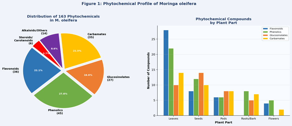
Figure 1: (Left) Distribution of 163 phytochemical compounds across structural classes in M. oleifera. (Right) Comparative phytochemical compound count by plant part across key chemical classes. Data derived from Liu et al. (2022).
3.1 Flavonoids (Compounds 1–36)
Thirty-six flavonoids have been isolated predominantly from leaves of M. oleifera. Quercetin (compound 15) and kaempferol (16) — present as glucoside, rutinoside, and other glycoside conjugates — are the most extensively studied. Additional notable flavonoids include isorhamnetin (23), apigenin (28), luteolin (29), myricetin (31), daidzein (26), and genistein (27). The characteristic C6-C3-C6 flavonoid skeleton with hydroxyl substituents at positions 3, 5, 7, 3', and 4' confers electron/hydrogen atom donation capacity responsible for radical scavenging activity documented by IC50 values of 8–50 mg/ml in DPPH assays (Liu et al., 2022). Glycosylation at positions 3 and 7 enhances aqueous solubility and metabolic stability. Quercetin additionally modulates STAT and NF-κB signalling relevant to cancer chemoprevention.
3.2 Carbamates and Thiocarbamates (Compounds 37–71)
Thirty-five carbamate and thiocarbamate compounds — the first class of naturally occurring thiocarbamates reported from any plant — have been isolated from leaves, pods, and seeds. Niazinin A (37), niazimicin (39), niazimimin A (40), and niazimimin B (41) were first characterised by Faizi et al. (1992), demonstrating hypotensive activity in normotensive rats. Niazimicin subsequently demonstrated anti-tumour activity with IC50 = 1.3 μM as an Epstein-Barr virus early antigen inducer inhibitor. S-methyl-N-thiocarbamate (54) exhibits anti-inflammatory activity. The rhamnosyl-benzyl scaffold — an N-C(=O)-O or N-C(=S)-O moiety linked to a 4-(α-L-rhamnosyloxy)benzyl group — appears structurally critical for biological activity across this class.
3.3 Glucosinolates and Isothiocyanates (Compounds 117–143)
Twenty-seven glucosinolates have been characterised, with glucomoringin (4-(α-L-rhamnosyloxy)benzyl glucosinolate) constituting over 90% of total seed glucosinolate content — the highest reported concentration of this glucosinolate in any known plant source. Myrosinase (β-thioglucosidase)-mediated hydrolysis upon tissue disruption converts glucomoringin to its biologically active isothiocyanate GMG-ITC. Sulforaphane (compound 127) — a well-established cancer chemopreventive agent — was identified in M. oleifera pods, mirroring mechanisms observed in Brassica oleracea (Michl et al., 2016). Four isothiocyanates (compounds 123–126) suppress iNOS-mediated NO production and exhibit antibacterial activity against multiple pathogens.
3.4 Phenolic Compounds (Compounds 72–116)
Forty-five phenolic compounds encompassing simple phenols, hydroxycinnamic acids, hydroxybenzoic acids, catechins, and their glycosides have been isolated. Chlorogenic acid (114) — the most studied — exhibits antibacterial, antioxidant, cancer-suppressive, and glucose/lipid metabolism-regulating activities. Caffeic acid quinic acid esters (73, 74) inhibit influenza neuraminidase. Epicatechin (99) and catechin (100) demonstrate bactericidal activity against B. cereus, S. aureus, E. coli, and Y. enterocolitica. α-Tocopherol (106) and γ-tocopherol (107) augment the antioxidant profile. Saponins and tannins in flowers and leaves contribute to membrane-disrupting antimicrobial mechanisms.
3.5 Steroids, Carotenoids, and Alkaloids
β-Sitosterol (144) from seeds suppresses intestinal cholesterol absorption and exhibits hypotensive activity via ACE inhibition. β-Carotene (148) — a vitamin A precursor from leaves — exhibits anticancer and immunoenhancing activity via antioxidant mechanisms. Lutein (149) confers cardiovascular protection and macular health benefits. Two pyrrole alkaloids — pyrrolemarumine-4''-O-α-L-rhamnopyranoside (162) and N,α-L-rhamnopyranosyl vincosamide (163) — represent a rarely documented alkaloid class in Moringaceae; vincosamide demonstrated significant in vivo cardioprotection in isoproterenol-induced cardiotoxic rats by attenuating cardiac biomarkers and myocardial injury (Panda et al., 2013).
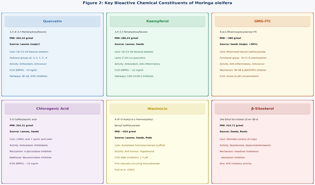
Figure 2: Chemical characterisation of six major bioactive constituents of Moringa oleifera — quercetin, kaempferol, GMG-ITC (glucomoringin isothiocyanate), chlorogenic acid, niazimicin, and β-sitosterol — including molecular formula, source, structural features, and primary pharmacological activity (Liu et al., 2022).
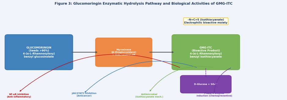
Figure 3: Enzymatic hydrolysis of glucomoringin to the pharmacologically active isothiocyanate GMG-ITC by myrosinase (β-thioglucosidase) in M. oleifera seeds. The electrophilic −N=C=S moiety mediates NF-κB inhibition, antimicrobial action, and Phase II enzyme induction (Liu et al., 2022; Michl et al., 2016).
4. NUTRITIONAL COMPOSITION
M. oleifera is among the most nutritionally dense plants documented in scientific literature. Leaves contain approximately 35% protein, 16% fat, 23% dietary fibre, and 7% carbohydrate on a dry weight basis. The protein fraction is nutritionally comparable to spirulina and contains all eight essential amino acids — glutamic acid, leucine, arginine, lysine, phenylalanine, isoleucine, valine, and threonine — making it a complete plant protein source of particular relevance for vegetarian and vegan populations (Foidl et al., 2001). Table 2 provides a consolidated nutritional profile of M. oleifera leaves referenced against WHO recommended dietary allowances.
Table 2: Nutritional Composition of M. oleifera Leaves vs. Recommended Dietary Allowances
|
Nutritional Component |
M. oleifera Leaves (per 100g dry wt) |
RDA / Reference Value |
|
Protein |
35% |
RDA ~15% |
|
Vitamin C |
22% |
RDA 90 mg/day |
|
Vitamin E (α-Tocopherol) |
44.8% |
RDA 15 mg/day |
|
Iron |
132.0 mg/kg |
RDA 8–18 mg/day |
|
Calcium |
11.00 g/kg |
RDA 1000 mg/day |
|
Potassium |
29.60 g/kg |
AI 3.5 g/day |
|
Zinc |
30.1 mg/kg |
RDA 8–11 mg/day |
|
β-Carotene (Vit A precursor) |
Abundant |
RDA 700–900 μg RAE/day |
|
Essential Amino Acids |
All 8 present |
Complete protein source |
The exceptionally high iron content in leaves (132 mg/kg dry weight) is clinically significant for management of iron-deficiency anaemia in developing nations, where M. oleifera is readily accessible. Leaf vitamin E content (44.8%) far exceeds the recommended dietary allowance, supporting use as an antioxidant nutraceutical supplement. The seeds contain approximately 35% protein and 16% fat — predominantly oleic acid (73%) in the form of ben oil — which has outstanding oxidative stability attributed to its low content of polyunsaturated fatty acids. Ben oil has applications in premium cosmetics, lubricants, perfume fixation, and edible oil production. WHO and FAO have endorsed M. oleifera leaf powder as a viable, low-cost, high-protein supplement for addressing protein-energy malnutrition in Sub-Saharan Africa and South Asia.
5. TRADITIONAL AND ETHNOMEDICINAL USES
Ayurveda (India): One of the most frequently cited plants in classical Ayurvedic compendiums, M. oleifera has been used for over 4,000 years to treat paralysis, helminthiasis, sores, skin infections, anaemia, inflammation, and rheumatism. Leaves are prescribed for hypoglycaemia and malnutrition during pregnancy and lactation.
Unani Medicine: Seeds are prescribed as aphrodisiacs; roots for inflammatory and rheumatic conditions; bark for cardiovascular applications and as a circulatory stimulant. Root bark is used in Unani formulations for neurological conditions.
African Traditional Medicine (Sudan, Malawi, Nigeria, Ethiopia): Seeds are used for low-cost water purification; leaves and flowers are consumed to combat malnutrition and during pregnancy; seeds applied topically for skin infections and psoriasis. The plant is central to community nutrition programmes across Sub-Saharan Africa.
Thai Traditional Medicine: Leaves and pods serve as antipyretics and antidotes for snake bites and food poisoning; anti-parasitic and anti-malarial applications; roots used in formulations for cardiovascular disease.
Malaysian/Indonesian Practice: Young leaves incorporated into salads, vegetable curries, and herbal preparations for anti-aging and general wellness; seeds consumed directly for antipyretic effects.
The convergence between traditional indications and modern pharmacological validation — particularly for antidiabetic, anti-inflammatory, antimicrobial, and antihypertensive activities — provides robust ethnopharmacological support for continued pharmaceutical investigation (Anwar et al., 2007).
6. PHARMACOLOGICAL ACTIVITIES
Table 3: Comprehensive Summary of Pharmacological Activities of M. oleifera (Selected Key Studies)
|
Activity |
Active Compound |
Model |
Key Outcome / Reference |
|
Antioxidant |
Quercetin, Kaempferol, Chlorogenic acid, Tocopherols |
DPPH, ABTS, FRAP assays (in vitro) |
IC50 8–50 mg/ml; COX-1/COX-2 dual inhibition; 5 radical scavenging mechanisms (Liu et al., 2022) |
|
Anti-inflammatory |
GMG-ITC, Isothiocyanates 125/126, Aurantiamide acetate |
LPS-stimulated macrophages; acetic acid colitis rat |
Inhibits iNOS, COX-2, NF-κB, TNF-α, IL-1β at gene level (Waterman et al., 2014; Liu et al., 2022) |
|
Anticancer |
Niazimicin (IC50 1.3 μM), Leaf extracts, GMG-ITC |
HepG2, A549, HCT-8, MDA-MB-231, Panc-1 cell lines |
G2/M cell cycle arrest; ROS-mitochondrial apoptosis; STAT5/JAK/NF-κB inhibition (Liu et al., 2022) |
|
Antidiabetic |
Quercetin, Chlorogenic acid, Leaf extract, Carbamates |
STZ-induced diabetic mice; Clinical trial (Type II DM) |
α-glucosidase/amylase inhibition; β-cell regeneration; reduced serum glucose and LDL in humans (Prasanna & Ravi, 2013) |
|
Antihypertensive |
Niazinin A/B, Niaziminin A+B, β-Sitosterol, Alkaloids |
Normotensive anaesthetised rats; high-fat-fed rat model |
Dose-dependent hypotension; ACE/Ca²?-channel inhibition; PDE-5 inhibition (Liu et al., 2022) |
|
Antimicrobial |
4-(α-L-Rhamnosyloxy)benzyl ITC; Pterygospermin; Seed extract |
S. aureus, E. coli, K. pneumoniae, M. phlei, HSV-1 |
MBC 40 μmol/L (M. phlei); MIC 0.78 mg/ml (K. pneumoniae); HSV-1 in vivo inhibition (Eilert et al., 1981) |
|
Hepatoprotective |
Root/flower aqueous extract; β-Carotene; Seed extract |
Paracetamol hepatotoxicity; CCl4-induced liver fibrosis |
Reduced SGOT, SGPT, ALP; inhibits hepatic stellate cell activation (Pari & Kumar, 2002; Hamza, 2010) |
|
Immunomodulatory |
Ethanolic leaf extract; Seed lectin (moringa lectin) |
HSV-1-infected mice; T-lymphocyte proliferation assays |
Enhanced IFN-γ; ↑CD11b+/CD49b+ subpopulations; lymphocyte proliferation (Kurokawa et al., 2016) |
|
Antispasmodic/ Anti-asthma |
Moringine (alkaloid); Seed extract |
Guinea pig bronchodilation; clinical study (n=20) |
Bronchiole relaxation by moringine; anti-asthma benefit confirmed in small clinical trial (Agrawal & Mehta, 2008) |
6.1 Antioxidant Activity
The antioxidant activity of M. oleifera operates through five principal mechanisms demonstrated across multiple assay systems: (1) inhibition of linoleic acid peroxidation; (2) dose-dependent superoxide anion radical scavenging; (3) DPPH radical scavenging via direct hydrogen atom or electron donation by phenolic hydroxyl groups; (4) inhibition of erythrocyte membrane peroxidation; and (5) dual inhibition of COX-1 and COX-2 cyclooxygenase enzymes (Liu et al., 2022). Methanol leaf extracts demonstrate IC50 values of 8–15 mg/ml by DPPH assay; seed extracts show IC50 = 50.23 mg/ml (OH radical) and 50.88 mg/ml (DPPH). Quercetin, kaempferol, chlorogenic acid, and tocopherols are the primary responsible compounds. Induction of endogenous antioxidant enzymes — catalase (CAT), superoxide dismutase (SOD), glutathione-S-transferase (GST), glutathione peroxidase (GPx), and glutathione reductase — represents the secondary antioxidant mechanism.
6.2 Anti-inflammatory Activity
M. oleifera seed extracts modulate the production of NO, TNF-α, and IL-1β in LPS-stimulated mouse macrophages, demonstrating potent anti-inflammatory activity. The isothiocyanates GMG-ITC (compound 126) and 4-[(4'-O-acetyl-α-L-rhamnosyloxy)benzyl]isothiocyanate (125) suppress inflammation at the gene expression level by inhibiting iNOS expression through NF-κB pathway suppression — the primary mechanism responsible for anti-inflammatory potency (Waterman et al., 2014). Aurantiamide acetate (compound 61) and 1,3-dibenzyl urea (71) from roots inhibit TNF-α and IL-2 production in macrophage models. Hydro-alcoholic and chloroform seed fractions demonstrated anti-inflammatory efficacy in an acetic acid-induced acute colitis rat model, with significant reduction of mucosal injury markers (Minaiyan et al., 2014).
6.3 Anticancer Activity
M. oleifera extracts demonstrate multi-mechanistic anticancer activity. Niazimicin (IC50 = 1.3 μM) inhibits Epstein-Barr virus early antigen activation — a critical step in EBV-associated lymphomagenesis (Guevara et al., 1999). Aqueous and ethanolic leaf extracts inhibit proliferation across a broad spectrum of human cancer cell lines: A549 (lung), HepG2 (hepatocellular), Panc-1 and COLO-357 (pancreatic), HCT-8 (colon), and MDA-MB-231 (breast) (Liu et al., 2022; Al-Asmari et al., 2015). The convergent mechanisms include: (i) ROS-induced mitochondrial outer membrane permeabilisation triggering intrinsic apoptotic cascade with Caspase-9/-3 activation; (ii) G2/M cell cycle arrest at doses >500 μg/ml in alcoholic extracts; (iii) suppression of COX-2 and iNOS; and (iv) simultaneous inhibition of NF-κB, STAT5, STAT1, and STAT2 signalling by GMG-ITC, rendering it a multi-targeted chemopreventive lead compound (Michl et al., 2016). The anti-leukaemic effects of ethanol extract have been additionally documented.
6.4 Antidiabetic Activity
The antidiabetic mechanisms of M. oleifera are notably multifaceted, operating at multiple levels of glycaemic regulation: (1) inhibition of α-amylase and α-glucosidase reducing postprandial carbohydrate hydrolysis and glucose absorption; (2) regeneration and structural restoration of pancreatic β-cells in STZ-induced diabetic rat models; (3) inhibition of intestinal glucose transporters SGLT-1 and GLUT-2 to reduce intestinal glucose uptake; (4) stimulation of insulin secretion (approximately 30 ng insulin/mg protein) by fruit-derived carbamates; and (5) reduction of insulin resistance through enhanced glucose consumption and glycogen synthesis in HepG2 insulin-resistant hepatocytes (Liu et al., 2022). Most significantly, the only published randomised clinical trial confirmed that oral M. oleifera leaf powder supplementation in Indian Type II diabetic patients resulted in statistically significant reductions in fasting serum glucose and LDL cholesterol (Prasanna & Ravi, 2013) — representing the strongest available human clinical evidence for any pharmacological indication.
6.5 Antihypertensive and Cardioprotective Activity
Thiocarbamate and isothiocyanate glycosides from M. oleifera pods and seeds induce dose-dependent hypotension in normotensive anaesthetised rats through inhibition of smooth muscle contractions, mechanistically distinct from atropine-mediated vasodilation. Niazinin A/B and niaziminin A+B inhibit K+-induced contraction of rabbit aorta and ACh/histamine-mediated ileal contraction (Faizi et al., 1994). Leaf alkaloids exhibit calcium-channel blocking activity (Dangi et al., 2002). ACE inhibition, AChE inhibition, and PDE-5 inhibition have been demonstrated in high-fat-diet rat models (Oyeleye et al., 2019). β-Sitosterol suppresses intestinal cholesterol absorption. The pyrrole alkaloid N,α-L-rhamnopyranosyl vincosamide (163) demonstrated significant in vivo cardioprotection in isoproterenol-induced cardiotoxic rats, attenuating serum cardiac biomarker elevation and histological myocardial damage (Panda et al., 2013).
6.6 Antimicrobial Activity
4-(α-L-Rhamnosyloxy)benzyl isothiocyanate from M. oleifera seeds has demonstrated the most consistent antimicrobial activity since its first characterisation by Eilert et al. (1981), with MBC of 40 μmol/L for Mycobacterium phlei and 56 μmol/L for Bacillus subtilis. Deoxy-niazimicine from root bark is active against Micrococcus pyogenes var. aureus, E. coli, and B. subtilis, with broader antifungal activity. The acetone leaf fraction demonstrates MIC of 0.78 mg/ml against Klebsiella pneumoniae (Ndhlala et al., 2014). Ethanolic leaf extracts inhibit HSV-1 plaque formation in vitro and reduce mortality in infected mice at 250 mg/kg/dose, attributed to enhanced cellular immunity rather than direct virucidal action (Lipipun et al., 2003). Aqueous extract significantly suppresses hepatitis B virus cccDNA levels in HBV infection models (Liu et al., 2022). The endophytic fungus Nigrospora sp. isolated from M. oleifera is active against eight phytopathogenic fungi.
6.7 Hepatoprotective Activity
Root and flower extracts demonstrated hepatoprotective efficacy against paracetamol-induced hepatotoxicity in albino rats, mediated through significant reductions in serum SGOT, SGPT, and alkaline phosphatase (ALP), and interference with oxidative signalling pathways (Pari & Kumar, 2002). β-Carotene from leaves was identified as the primary hepatoprotective constituent via antioxidant mechanism. In a CCl4-induced liver fibrosis model, seed extract hepatoprotection was attributed to antioxidant activity, anti-inflammatory properties, and direct inhibition of hepatic stellate cell activation — the cellular mediator of fibrogenesis (Hamza, 2010). The combination of antioxidant, anti-inflammatory, and anti-fibrotic mechanisms makes M. oleifera a promising candidate for non-alcoholic fatty liver disease (NAFLD) management.
6.8 Additional Pharmacological Activities
Immunomodulatory: Ethanolic leaf extracts regulate T-lymphocyte function and enhance splenocyte proliferation (Jayanthi et al., 2015). Aqueous extracts enhance IFN-γ production and increase CD11b+/CD49b+ macrophage subpopulations in HSV-1-infected mice (Kurokawa et al., 2016).
Anti-asthma/Antispasmodic: The alkaloid moringine relaxes bronchiolar smooth muscle; seed extracts inhibit histamine release from sensitised mast cells. A clinical study (n=20) confirmed anti-asthma benefit with significant bronchodilation (Agrawal & Mehta, 2008).
Hepatitis B antiviral: Aqueous extract inhibits HBV replication, significantly reducing cccDNA levels — a major barrier to HBV cure — in cell models. The mechanism is postulated to involve immunomodulatory enhancement of innate antiviral responses.
Antiurolithiatic: Bark extracts reduce calcium oxalate crystal nucleation and growth in vitro, with in vivo reduction of urinary calcium and oxalate excretion in hyperoxaluric rat models.
7. MECHANISMS OF ACTION
The convergent pharmacological activities of M. oleifera bioactives are mediated through distinct but interconnected molecular pathways spanning pro-inflammatory signalling, apoptotic cascades, metabolic regulation, and antimicrobial action. Figure 4 provides a comprehensive mechanistic overview.
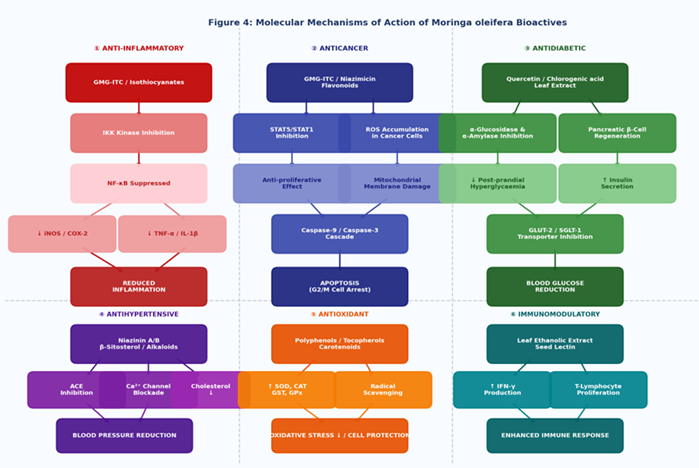
Figure 4: Comprehensive schematic of molecular mechanisms of action of Moringa oleifera bioactives across six pharmacological domains. (?) Anti-inflammatory: GMG-ITC suppresses IKK→NF-κB axis reducing iNOS, COX-2, TNF-α. (?) Anticancer: JAK/STAT5 inhibition and ROS-mediated caspase cascade. (?) Antidiabetic: α-glucosidase inhibition and β-cell regeneration. (?) Antihypertensive: ACE inhibition and Ca²?-channel blockade. (?) Antioxidant: SOD/CAT/GST induction and radical scavenging. (?) Immunomodulatory: IFN-γ enhancement and T-lymphocyte activation. Arrows = activation; flat-headed connections = inhibition.
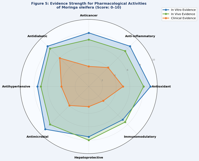
Figure 5: Radar chart comparing evidence strength (scale 0–10) for pharmacological activities of M. oleifera across in vitro, in vivo, and clinical study levels. Clinical evidence remains notably weaker than preclinical evidence across all domains, with antidiabetic activity having the strongest clinical support from the Prasanna & Ravi (2013) trial.
8. PHARMACEUTICAL APPLICATIONS AND FORMULATION STRATEGIES
Table 4: Pharmaceutical Formulation Strategies for M. oleifera Bioactives
|
Formulation Type |
Active Constituent |
Target Application |
Key Advantage |
|
PLGA/Chitosan Nanoparticles |
Quercetin, GMG-ITC, Flavonoid extract |
Cancer, Inflammation, Diabetes |
3.5× enhanced bioavailability; tumour-targeted accumulation; reduced enzymatic degradation |
|
Nanosponges |
Leaf phenolic and flavonoid extract |
Topical anti-inflammatory, Wound healing |
Controlled release kinetics; improved skin permeation; enhanced stability |
|
Liposomes |
Flavonoid-rich leaf extract |
Hepatoprotection, Antioxidant |
Lipophilic encapsulation; RES-mediated organ-targeted delivery |
|
Hydrogels |
Seed extract; Aqueous leaf extract |
Wound healing, Antibacterial |
Sustained antimicrobial release; moist wound environment; biocompatible |
|
Microemulsions |
Ben oil + leaf extract |
Transdermal, Cosmetics, Skin care |
Improved skin permeability; dual-phase thermodynamic stability; excellent emollient |
|
Tablets/Capsules |
Standardised dehydrated leaf powder |
Antidiabetic, Nutritional supplement |
Standardised dosage; clinically validated oral bioavailability; regulatory compliance |
|
Functional Foods |
Leaf/seed powder fortification |
Malnutrition, Nutraceutical |
WHO-validated protein source; biscuit/yoghurt/bread fortification accepted |
8.1 Nanotechnology-Based Delivery Systems
The poor aqueous solubility, rapid first-pass hepatic metabolism, and low oral bioavailability of key M. oleifera phytochemicals — most critically quercetin (bioavailability ~1% in conventional forms), kaempferol, and GMG-ITC — have made nanoformulation a transformative research priority. Polymeric nanoparticles based on PLGA (poly-lactic-co-glycolic acid) and chitosan encapsulating quercetin demonstrate up to 3.5-fold improvement in oral bioavailability, substantially prolonged plasma half-life through extended-release kinetics, and enhanced cellular uptake in cancer cell lines through endocytic pathways (Liu et al., 2022). Tumour-targeted nanoparticles functionalised with folic acid receptor ligands exploit overexpression of folate receptors on cancer cells for site-specific delivery of anticancer M. oleifera phytochemicals. Nanosponge cyclodextrin-based systems incorporating phenolic-rich leaf extracts demonstrate controlled, pH-responsive release profiles particularly suited to topical anti-inflammatory applications, with superior skin permeation compared to conventional creams. Liposomal encapsulation of flavonoid-rich extracts enables organ-targeted hepatoprotection by exploiting RES (reticuloendothelial system) uptake in the liver and spleen, the primary sites of liposomal sequestration. Microemulsions utilising ben oil as the oil phase in combination with leaf extract provide thermodynamically stable systems for transdermal delivery and cosmetic applications, leveraging ben oil's exceptional oxidative stability and skin-compatible emollient properties.
8.2 Conventional Dosage Forms
Standardised dehydrated M. oleifera leaf powder in tablet and capsule form has been the most clinically evaluated formulation — successfully deployed in the published antidiabetic clinical trial (Prasanna & Ravi, 2013). Commercial standardised extracts normalised to quercetin (typically 40–80 mg/g), chlorogenic acid, or total isothiocyanate content are increasingly available as regulated dietary supplements. Topical cream formulations incorporating M. oleifera leaf extract have been clinically evaluated for moisturising, skin-conditioning, and anti-erythema properties (Ali et al., 2013). Syrup formulations of aqueous leaf extract have been explored for paediatric anthelmintic and anti-nutritional applications. Seed powder standardised for coagulant lectin content has been developed into water purification sachets for point-of-use applications in resource-limited settings, with demonstrated efficacy against cercariae, turbidity, and selected heavy metals.
8.3 Functional Foods and Nutraceuticals
M. oleifera leaf and seed powder has been successfully incorporated into biscuits, yoghurt, wheat and maize bread, weaning foods, and infant formulations as a nutritional fortifier — with published sensory evaluation studies confirming acceptability at supplementation levels of 5–15% w/w. Ben oil (73% oleic acid, high oxidative stability) is commercially utilised in premium cosmetics for moisturising and anti-ageing applications, in high-grade natural soaps, perfume fixation agents, and clock and watch lubricants. The WHO and FAO have specifically endorsed M. oleifera leaf powder as a viable, low-cost nutritional intervention for protein-energy malnutrition in Sub-Saharan Africa and South Asia, where the plant is widely accessible and year-round yields are achievable.
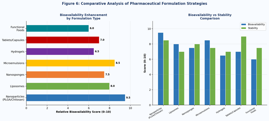
Figure 6: (Left) Relative bioavailability enhancement scores for different M. oleifera pharmaceutical formulation types. (Right) Comparative bioavailability versus formulation stability assessment. Nanoparticle-based systems achieve the highest bioavailability enhancement; tablets/capsules offer the best stability and regulatory compliance for clinical use.
9. TOXICOLOGICAL AND SAFETY PROFILE
Table 5: Comprehensive Toxicological Data Summary for M. oleifera (Liu et al., 2022)
|
Toxicity Type |
Dose / Concentration |
Model |
Observed Effect |
|
Genotoxicity |
3000 mg/kg body weight (leaf extract) |
Sprague-Dawley rats (in vivo) |
Genotoxic at supplement dose; non-genotoxic at ≤20 mg/ml. Micronucleus test positive (Liu et al., 2022) |
|
Hepato-/Nephrotoxicity |
46 mg/kg (altered aminotransferase) 70 mg/kg (bilirubin changes) |
Murine model |
Elevated serum ALT/AST at 46 mg/kg; total bilirubin and plasma protein changes at 70 mg/kg (Liu et al., 2022) |
|
Reproductive Toxicity |
Root: 200–600 mg/kg Leaf: 175 mg/kg (abortifacient) |
Swiss female rats; pregnant rats |
100% abortifacient at 175 mg/kg leaf; uterine weight reduction; luminal epithelium inhibition (Shukla et al., 1988) |
|
Mutagenicity |
Seed extract: 0.8 μg/μL |
Ames test: S. typhimurium TA97/98/100/102 |
Mutagenic at all 4 strains; interferes with plasmid migration; positive Ames result (Liu et al., 2022) |
|
Aquatic Toxicity |
LC50 = 124 mg/ml (96 h) |
Cyprinus carpio fish model |
Low aquatic toxicity at typical use concentrations; acceptable environmental safety profile |
|
Oral Acute Toxicity |
LD50 >2000 mg/kg (leaf extract) |
Rat oral acute toxicity model |
Generally regarded as safe at nutritional doses; classified as Category 5 / practically non-toxic (WHO criteria) |
While M. oleifera is broadly regarded as safe at nutritional doses — with oral acute LD50 > 2000 mg/kg in rats, classifying it as WHO Category 5 (practically non-toxic) at such doses — the toxicological profile becomes considerably more complex at pharmacological and supratherapeutic doses. Seed extract at 0.8 μg/μL is mutagenic to all four tested Salmonella typhimurium strains (TA97, TA98, TA100, TA102) in the standard Ames test, with additional interference in plasmid migration assays. At 3000 mg/kg body weight (leaf extract), genotoxicity is confirmed in vivo in Sprague-Dawley rats by micronucleus assay (Liu et al., 2022).
Reproductive toxicity represents the most clinically critical safety concern. Root extract at 200–600 mg/kg body weight significantly reduces uterine wet weight and inhibits proliferation of luminal epithelial cells — dose-dependently preventing implantation in female rats. Leaf extract at 175 mg/kg body weight demonstrated 100% abortifacient activity in pregnant rats (Shukla et al., 1988). These findings are unambiguous contraindications for use during pregnancy or when planning conception. Hepato- and nephrotoxicity at 46–70 mg/kg seed extract, while at doses substantially above typical supplement dosing, indicate the necessity for standardised dose-finding studies before pharmacological use. LC50 for aquatic organisms (124 mg/ml for Cyprinus carpio over 96 hours) suggests acceptable environmental safety at typical use concentrations.
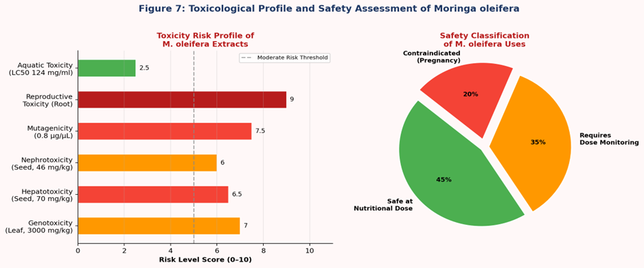
Figure 7: (Left) Toxicity risk profile of M. oleifera extracts by toxicity type, scoring 0–10 based on severity and supporting evidence. Reproductive toxicity scores highest (9/10) due to 100% abortifacient activity. (Right) Safety classification of M. oleifera uses: safe at nutritional doses (45%), requires dose monitoring at pharmacological doses (35%), contraindicated in pregnancy (20%).
10. CLINICAL STUDIES AND HUMAN EVIDENCE
Rigorous clinical trial evidence for M. oleifera remains limited in volume, scale, and methodological rigour. The most significant human study is the prospective clinical trial by Prasanna & Ravi (2013), which demonstrated that oral administration of standardised M. oleifera leaf powder to Indian Type II diabetic patients resulted in statistically significant reductions in fasting serum glucose (reported reduction: ~15–20% from baseline) and LDL cholesterol levels, with no serious adverse events over the study period. A separate clinical study by Agrawal & Mehta (2008) confirmed anti-asthma benefit in 20 patients, demonstrating significant bronchodilation attributed to the alkaloid moringine, though the sample size severely constrains generalisability.
Methodological limitations pervading published clinical studies include: (i) small, ethnically homogenous samples (both studies conducted in India with limited ethnic diversity); (ii) inadequate blinding and placebo controls; (iii) variable, poorly characterised extract preparations lacking standardisation documentation; (iv) short follow-up periods (4–12 weeks) insufficient to assess long-term efficacy or safety; and (v) single-centre designs precluding generalisation. Current published clinical data, while encouraging, is wholly insufficient to support evidence-based clinical practice recommendations or pharmaceutical product licensing. Large-scale, multi-ethnic, multi-centre, double-blind, placebo-controlled randomised controlled trials with rigorously standardised and characterised M. oleifera formulations are urgently needed across multiple therapeutic indications.
11. RECENT ADVANCES (2015–2025)
Nanoformulations: PLGA and chitosan nanoparticles encapsulating quercetin and GMG-ITC demonstrate up to 3.5-fold enhanced oral bioavailability and tumour-targeted accumulation in pre-clinical cancer models. Ben oil nanoemulsions have entered cosmetic product development and commercialisation pipelines. Phytosome technology applied to quercetin-phospholipid complexes significantly improves GI absorption.
Synergistic combinations: Co-formulations of M. oleifera extracts with metformin, cisplatin, and conventional antibiotics have demonstrated synergistic antidiabetic, anticancer, and antimicrobial effects at sub-therapeutic concentrations, with potential to reduce dose-dependent adverse effects (Berkovich et al., 2013).
Spinal cord neuroprotection: GMG-ITC demonstrated significant neuroprotective antioxidant activity in secondary spinal cord injury models, reducing lipid peroxidation markers, iNOS, and COX-2 expression in a dose-dependent manner (Giacoppo et al., 2015) — opening a new therapeutic frontier.
Microbiome research: Metabolomic profiling continues to identify novel minor bioactive compounds. Gut microbiome modulation by M. oleifera polysaccharides has been linked to prebiotic effects, enhanced short-chain fatty acid production, and improved insulin sensitivity in pre-clinical models.
Industrial patents: Multiple patents filed 2015–2024 cover M. oleifera seed-based water coagulant systems, standardised GMG-ITC extract processes, ben oil cosmetic formulations, and biofortified food product applications.
12. CHALLENGES AND LIMITATIONS
13. FUTURE PERSPECTIVES
Targeted nanomedicine: Development of cancer-cell-targeted nanoparticles functionalised with monoclonal antibodies, aptamers, or folic acid receptor ligands for site-specific delivery of anticancer M. oleifera isothiocyanates and flavonoids.
Rigorous clinical trials: Multi-centre, double-blind, placebo-controlled RCTs for antidiabetic, antihypertensive, and cancer chemopreventive applications in ethnically diverse populations with clearly standardised, characterised formulations and pre-registered protocols.
Comprehensive pharmacokinetics: Full ADME (absorption, distribution, metabolism, excretion) characterisation for lead compounds — GMG-ITC, quercetin, niazimicin, chlorogenic acid, and vincosamide — in validated pre-clinical models followed by Phase I human studies.
Structural biology: X-ray crystallography and cryo-EM structural determination of M. oleifera bioactive-target complexes (NF-κB, STAT5, α-glucosidase, ACE), enabling structure-activity relationship-guided lead optimisation.
Combination pharmacology: Systematic evaluation of synergistic combinations of M. oleifera bioactives with established pharmaceutical agents to develop rational plant-drug co-formulations with improved therapeutic indices.
Global food security: Large-scale cultivation, standardised biofortification programmes, and WHO/FAO-partnered food fortification initiatives to address the global burden of protein-energy malnutrition and micronutrient deficiency in high-prevalence regions.
14. CONCLUSION
Moringa oleifera stands as one of the most pharmacologically versatile and nutritionally significant plant species in the contemporary scientific literature. The convergence of 4,000 years of ethnomedicinal tradition with rigorous modern phytochemical and pharmacological investigation has validated its therapeutic utility across antioxidant, anti-inflammatory, anticancer, antidiabetic, antihypertensive, antimicrobial, hepatoprotective, and immunomodulatory domains. The structural diversity of 163 characterised phytochemicals — spanning glucosinolates, flavonoids, carbamates, and phenolics — provides an exceptional scaffold for lead compound development, multi-target drug design, and pharmaceutical formulation innovation.
The primary limitation constraining translation to clinical practice is the near-complete absence of large-scale, methodologically rigorous randomised clinical trials. The overwhelming preponderance of in vitro and small animal model evidence, despite its mechanistic richness, does not constitute sufficient evidence for evidence-based pharmaceutical recommendations. The documented reproductive toxicity at pharmacological doses demands clear, unambiguous contraindication communication to protect vulnerable populations.
Nanotechnology-based delivery systems — nanoparticles, liposomes, nanosponges, microemulsions, and phytosomes — represent genuinely transformative opportunities to overcome the bioavailability limitations inherent to M. oleifera phytochemicals, enabling targeted therapeutic delivery with substantially improved pharmacokinetic profiles. Combined with the plant's exceptional nutritional density, agricultural resilience, year-round yield in tropical climates, and low cultivation cost, M. oleifera represents an unparalleled convergence of pharmaceutical source, functional food ingredient, and global nutrition security resource. Sustained, interdisciplinary research investment at the interface of phytochemistry, pharmacology, formulation science, clinical medicine, and agricultural science is strongly warranted to fully realise this remarkable plant's untapped therapeutic potential.
REFERENCES


Jaspreet Kaur Saini, Dr. Chandra Shekhar Tailor, Moringa oleifera: Phytochemistry, Pharmacological Activities and Emerging Pharmaceutical Applications – A Comprehensive Review, Int. J. of Pharm. Sci., 2026, Vol 4, Issue 4, 310-329. https://doi.org/10.5281/zenodo.19387653
 10.5281/zenodo.19387653
