School Of Studies In Pharmaceutical Sciences, Jiwaji University, Gwalior, 474011, Madhya Pradesh, India
This review specifically describes about recent advancements in designs of nanotechnology local drug delivery system and its evaluation by Wistar rat models. Also, is has many advantages over other routes such as non-invasive route (for the body) and no first-pass metabolism and higher conformity but in this particular case delivered load eficiency is still mainly limited by stratum corneum barrier function. The limited efficiencies of frequent dose regimens in conventional drug delivery systems necessitated for new alternatives to come into play, and nanotechnology appeared as an effective method to overcome the issue of bioavailability and selectivity using a diverse spectrum of available modern carriers ranging from liposome, solid lipid nanoparticles (SLN) to polymeric nanoparticles. These systems enhance drug penetration, improve stability, and enable controlled and targeted drug release. The review highlights various mechanisms of enhanced skin permeation, including follicular, intercellular, and transcellular pathways, as well as the occlusion effect. Furthermore, it emphasizes the importance of Wistar rat models in preclinical evaluation due to their reproducibility, cost-effectiveness, and suitability for in vivo and ex vivo studies. Despite certain limitations related to differences from human skin, these models remain valuable tools for assessing drug permeation, safety, and efficacy. Overall, this review provides critical insights into formulation strategies, evaluation techniques, and future perspectives for the development of effective and safe topical drug delivery systems..
Topical drug delivery is the process of giving a drug to body through skin for local or systemic effect. This route is of considerable interest in dermatology, owing to its non-invasive character which allows for the administration of drugs directly at their site of action. One of the most important benefits topical drug delivery has to offer is essentially avoidance of hepatic first-pass metabolism, which can greatly improve a drugs bioavailability. The buccal route is also beneficial regarding patient compliance and facilitates controlled and sustained drug release without unwanted systemic effects associated with the oral or injectable routes1,2.iven that many patients suffer from chronic dermatological conditions including psoriasis, dermatitis/ eczema and funguses where topical treatment is the first choice regimen, the supply of topical systems is also a major plus. Topical formulations also allow for flexible dose titration and, if any adverse events occur, can be stopped immediately, which further advantage their safety profiles3.But topical drug delivery has some major challenges though. One of the major barriers to drug permeation is the stratum corneum (the outermost layer of skin formed by densely packed keratinized cells in a lipid matrix). Such architectural characteristics impose considerable barrier to permeation of most drug molecules across epithelial barriers, especially for those with hydrophilic and high MW. Thus, few compounds may be effectively delivered topically or transdermally4.In addition, conventional topical formulations such as creams, ointments, and gels often exhibit poor drug retention, inconsistent absorption, and limited penetration depth. These limitations reduce therapeutic efficacy and necessitate frequent application, thereby affecting patient adherence5.
Role of Nanotechnology in Dermatology:
This method involves Nanotechnology based transdermal drug delivery device which provides the new way to overcome the limitation of traditional, conventional injections method. Nanocarrier for transdermal drug delivery Nanocarriers such as liposomes, solid lipid nanoparticles (SLNs), nanostructured lipid carriers (NLC)6 have been designed.Nanocarriers and their norms Nanocarriers are normalised by unique physiochemical attributes such as particle size, huge surface area, and ability to load hydrophilic or lipophilic drugs. Due to their small enough size, these enhancers can penetrate relatively deeper into stratum corneum and thereafter increase drug permeated by inter- as well as transfollicular pathways7.Nanostructures can aid in designing molecules that are more solubilized and stable than native formulations, and provide control over release timing and prolonged delivery to specific cutaneous layers. Moreover, these structures provide advantages to reduce drug3 degradation and are also expected to reduce irritation through modulation of a drug's kinetic profile at skin interface6.In addition, some nanocarriers can induce it that boosts skin hydration and promotes drug penetratio8. This characteristic has rendered the use of nanomaterials an attractive choice for the treatment of many skin diseases, specifically inflammatory skin diseases as well as infections and cutaneous malignances8.
Biologics, In Vitro Technologies3,9:
All these have shown how crucial animal studies are in optimizing formulations that encompass the evaluation of safety, efficacy and pharmacokinetic parameters. Wistar rat is one among various animal models that are widely preferred due to their easy availability, easy handling, higher cost utility and defined physiology9.In addition, Wistar rat skin has been used as a simple and reproducible model for permeation of drugs or for action of irritants inducing histopathological pattern. These models are especially pertinent for in vivo and ex vivo toxicity assays10 (e.g. Franz diffusion, skin retention experiments).Although the basic architecture is preserved, it is distinct from rat vs human skin (eg stratum corneum less pronounced and permeability greater) Given these similarities, Wistar rats models offer a great stage for assessment and further optimization of topical formulations prior to investigational studies in humans11.However, species differences should also be taken into account when extrapolating as it may cause overestimation of penetration relative to human skin. Wistar rats, despite their limitations have been useful in some studies relevant to dermatopathology as a model due to specific advantages and predictive value.
Aim and Scope of Review:
This review ultimately aims to understand particulates topical drug delivery systems at nanoscale level, following a general introductory discussion regarding the importance of such systems designed for penetrating into skin. This review presents advances in physicochemical attributes of molecularly targeted nanocarrier technologies intended to overcome presently-existing pharmacokinetic hurdles, typology based on their mechanism of action as well as methods for formulative and evaluative processes that can be employed within the area of dermal therapy.They also highlighted their preclinical studies on various DDS based on nanocarriers including Wistar rats that are of significant relevance. This review is a summary of results with in vivo and ex vivo background, which encompass drug penetration studies, pharmacokinetic experiments, holder dermatologists and safety studies.Moreover, for the context of speed and inertia, this overview was to give some broad view in broader community scrutiny on where we are with existing pros/cons and clinical/applied prospects landscape on nanotechnology-based formulations although mismatch between mentioned materiality driving development vs translational aspects may exaggerated here with more described interventional detail. In this context, the outcomes presented herein provide a comprehensive basis for achieving topically oriented drug delivery formulations that take into account key clinical translational determinants related to safety and efficacy.
Skin Structure and Barrier Function:
Anatomy of Skin:
Skin is the human body’s largest organ: skin separates our inner world from an outer one. It is also vital for thermoregulation, immunity and water losses. It is composed of three distinct layers, epidermis dermis and hypodermis the histology physiology bear important and different relations to their classification
Epidermis:
The epidermis, the skin's outermost layer, is primarily protective. It is formed by low cuboidal to stratified squamous epithelium, limited exclusively to keratinocytes from the basal layer until close to the surface of epidermis. The epidermis is traditionally subdivided into five strata; stratum basale, stratum spinosum (the prickle cell layer), stratum granulosum, stratum lucidum (only in the thick skin) and a final cuticle-like layer termed the stratum corneum13.Between two definite phases, the stratum corneum structure is the chief barrier towards permeation, while it can be a critical explorer in xenobiotic's filmothieutic formulations [1]. It is composed of desiccated, flattened and keratinized cells (corneocytes) organized in a “brick-and-mortar”-like fashion; they are piled up on top of one another and glued together by means of a lipid matrix. This creates a barrier that minimizes drug deposition to skin due to protective coatings with native gatekeeper nature which prevent penetration of foreign matter14.
Dermis:
The second layer of skin would be the dermis, which is just under the epidermis -strengthens and makes you elastic. It consists of connective tissue and contains numerous collagen and elastin fibres and blood vessels, lymphatics, nerves and appendages (e.g. hair follicles and sweat glands). The dermis is divided into papillary (superficial) and reticular (deep) areas.To elaborate, this mechanism works to access skin surface area that pancake zone from drug penetration perspective it could electromagnetic transfer under high density vascular kind blood host. Along with this skin appendages present in the dermal layer enhances an extra route of penetration of drugs particularly particulate based systems4.
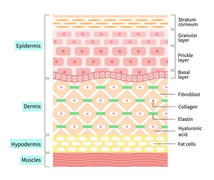
Figure No.1 : Schematic Diagram of Skin Structure Showing Epidermal Layers, Dermis Components, and Hypodermis
Hypodermis:
The uppermost layer of the skin is the hypodermis or subcutaneous tissue and it consists of largely fat and connective tissues. It serves as a source of energy, a thermoregulatory mechanism, and supports the underlying structures15.Although dermis and hypodermis have no direct effect on barrier functionality, they play a role in systemic drug uptake with transdermal deliveries. Compounds that penetrate this barrier are more expected to enter into systemic dissemination as they are consist of bigger blood vessels16.
The SC would be The Main Obstacle:
The skin drug penetration barrier functionally isin the stratum corneum. It is made up of 10–20 layers of corneocytes embedded in a structured lipid matrix that is rich in ceramides, cholesterol and free fatty acids. This structure forms a low-permeability layer which restricts the diffusion of hydrophilic and lipophilic solutes15.This is further elaborated on the structure and the role of stratum corneum with a model where conreocyte are represented as bricks and intercellular lipids as mortar (the brick-and-mortar model). The thick bilayers form a twisting route through which drug molecules have to penetrate6,18.Calculations of drug diffusion across the stratum corneum have been categorized as one of the three types, i.e., intercellular (between cells), transcellular (through cells), and appendageal (involving the hair follicles and sweat glands). The majority of drugs are thought to traverse primarily paracellular, as outlined above4.It is among the greatest barriers since it is a high resistance one and it impedes the diffusion of drugs that are delivered by topical routes. Hence, approaches like permeation enhancers co-administration and nanocarrier-based systems have been designed for drug Article permeability enhancement2.
Factors Affecting Skin Penetration:
Although physicochemical and formulation-based factors contact drug penetration through percutaneous process but such an approach is not without its problem11.
Molecular Weight:
There are numerous physicochemical determinants of skin permeability, with molecular weight appearing to be a factor. In general, the lower the molecular weight of a drug (below 500 Dalton), the higher the chances of it to enter through this obstacle - so-called nom de guerre 500 Dalton Rule. The diffusion of large molecules is drastically inhibited due to this close packing of lipid16.
Lipophilicity:
The information on drugs partitioning (I) The lipophilicity to aid in comprehension of how this occurs. High lipophilicity and hydrophilicity: response should able to penetrate with optimum lipophilicity and hydrophilicity. Highly lipophilic drugs will thereby be preferentially distributed in stratum corneum material and with minimal diffusion into the deeper layers41, and highly hydrophilic drugs, will not partition to lipid in a significant way.
Formulation Type:
The type of composition has a great influence on the delivery of drugs through the skin. However, classical topical preparations (creams and ointments) have much lower penetration potential than other lipid colloidal vehicles e.g. nanoemulsions, liposomes and solid lipid nanoparticles based advanced delivery systems which are aiding in permeating through the skin by stabilizing solubility in combination with interaction with stratum corneum lipids.The Furthermore, lipid structures and stratum corneum structure may also be disrupted by permeation enhancers that may also facilitate drug permeation4.
Hydration:
Skin hydration also has an effect on drug penetration. Expansion of corneocytes and disruption of lipid organization, permitting increased permeability, accompany increased stratum corneum moisture content2. The nanocarrier type and some types of occlusive formulations could also increase skin hydration, which facilitates drug permeation15.
Topical Drug Deliveryin Wistar Rat Model:
Characteristics of Wistar Rats:
That is why The Wistar rat is a popular laboratory animal model that can be used to simplify the pharmaceutical and dermatology-related studies that are not painful to maintain and have a microbiome structure. Since the skin of rats has so far been definitively characterized anatomical and physiological, they may become a convenient initial animal model to be used to evaluate one of the drug delivery systems topically.
Skin Thickness:
Wistar rat skin is also thinner as compared to man particularly the stratum corneum layer that has been proven to be a major resistant to permeation of clinically feasible drugs33. The obstacle is lighter than in a conventional model of esters, which are more permeable but there remains plenty of room to have obvious differences over that of sensitivity: Formulations will differ. However, this aspect causes drug absorptions to also be excessive compared to those that are created using human skin18.
Hair Density:
There is also increased density of hair follicles in the wistar rats hence increased transfollicular drug delivery. Well characterized entitative one has a good follicle deposition and alternative entering course, in combination with stratum corneum be attractive target, which is useful in topical delivery of drugs using nanoparticles based formulation. The individual and narrowest follicular routes of drug delivery systems use.
Physiological Features:
Physiological characteristics such as multiple metabolic rate; pharmacological properties for permeation hence, distribution, skin dehydration and lipid constituents accompany it has been verified on Wistar rats being used as comparative animal model after Wistar rat. And so does their skin have other classes of lipids (ceramides, cholesterol and fatty acids), but in different proportions than human skin. Moreover, Wistar rats have a high skin permeability and turnover which affects drug retention and pharmacokinetics18.
Benefits of Wistar Rats:
Easy Handling:
Wistar rats are easy to handle, since they are obedient animals that can be easily handled in longitudinal studies and also they can be used in multiple dosing. They are very small such that they can be dosed topically and allow a plethora of in vivo experiments, such as blood sampling and skin excision9.
Cost-Effective:
In addition to the cost of acquisition and maintenance of a Wistar rats they are also the cheapest long-term experimental model in terms of price-mass than other larger and more complicated animal models such as pigs or primates. The research works and numerous large-scale experiments published in academic publications.
Reproducibility:
In semi-isogenic Wistar, the strain variances are reduced on account of the relatively homogeneous genotypes and breeding conditions that can be controlled and provide a high reproducibility degree. It distinguishes different formulations and assures preclonal studies reproducibility. Due to its popularity it allows comparing it with the existing data of the past that had been published18.
Wistar rat limitations:
Differences from Human Skin Permeability:
As much as we provide Wistar rat models with an advantage of their own, the corresponding limitations are considerable especially in matters involving morphology and permeability. Due to less stratum corneum and good follicular density of rat's skin enhances dermatomes permeation, resulting in overestimation of drug penetration. Also, variations in lipid makeup and skin metabolism may have an impact on drug uptake and allocation21.These variations in the species should be taken into account when interpreting the data, which usually require confirmation in the human skin model or clinical testing.
Ethical Considerations:
Old age Wistar rats are usually subjected to age-related diseases. Therefore the regulatory policies that OECD or institutional ethics committee prescribes regarding minimization of animals utilization, maximization of their proper handling and Viva Sepal substitutes where possible. The 3R principle that loosely incorporates replacement, reduction and refinement of animal experimentation9 is another like concept and can be used as a guideline to ethical compliance to these animal experiments.
Procedures on Wistar Rats in Experiment:
The wide scope of such studies is conducted in Wistar rats, either in vivo or ex vivo and analytical by which the topical drug delivery systems are assessed.
In Vivo Studies:
Applications onto the skin of living Wistar rats are used to carry out in vivo experiments on parameters like drug absorption, therapeutic efficacy and cutaneous irritation. These studies can give real-time data regarding biologic response, covering systemic exposure and pharmacodynamic effect18.
Ex Vivo Skin Permeability:
Wistar rat skin is excised and subjected to ex vivo experiments in which the skin is put in diffusion in Franz diffusion cells. These kinds of studies are generally utilized to characterize drug permeation, flux and retention separated of systemic actions. In-vivo models are high relevance in the bioavailability assessment, but resource-intensive and ethically questionable, as well as formulations are generally evaluated in linear diffusion cells that give both a comparative controlled model of study and permeation mechanism information9.
Histopathological Analysis:
Histopathological studies are used to assess the effects of formulations with structural & cellular effects on skin histology. Skin biopsies are performed which are fixed, sectioned and stained (usually hematoxylin and eosin) to assess any evidence of irritation or inflammation or tissue destruction. The study is important to eσaluate safety and compatibility of topical formulations22.
Pharmacokinetic Studies:
Wistar rats Pharmacokinetic. Pharmacokinetic studies of Wistar rats determine the concentration of drug in plasma or tissues after topical administration over a given period of time. These are most likely to be studies that would establish parameters like absorption rate, bioavailability and systemic exposure. This is especially true in the case of transdermal systems that are to be used to release into the system23.
Topical Drug Delivery Systems: Nanotechnology:
Lipid-Based Nanocarriers:
Lipid based nanocarriers have become highly promising methods of topical and transdermal delivery of drugs. Physiological lipids make up these systems, making them highly biocompatible and low in toxicity and improving the permeation of the drug through the skin. Polymeric substances must be lipophilic and hydrophilic initiatively (they can contain both kinds of polarity) as they have to be efficient in overcoming the barrier function of the stratum corneum6.
Solid Lipid Nanoparticles (SLNs):
Solid lipid nanoparticles (SLNs) are submicron-sized particles, which are composed of solid lipids and surfactants. They remain at room and body temperatures, thereby forming a good matrix to enclose drugs. An SLN can be used to release drugs in a controlled system, enhance stability and ensure sensitive drugs do not degrade6.
Mechanism of Penetration:
Various mechanisms play the role of resulting into greater penetration of SLNs in skin. The smaller size of the particles first enables contact by the particles to be in closer relation with stratum corneum resulting in better adhesion onto the surface. On one pathway, SLNs preparations overlay the skin floor with an occlusive film therefore augmenting the level of moisture and in this way interfering with the arrangement of lipids on stratum corneum resulting in wonderful penetration. Moreover, lipid diffusion between SLNs and skin lipids6 would enhance the diffusion of drugs into the deepest layers.
Wistar Rat Studies Wistar Rat Studies have been applied:
Drug delivery via skin is also one of the most extensively studied SLNs indeed Wistar rat models are extensively used. This demonstrates excellent penetration in the skin and longer retention of drugs with lower systemic toxicity as compared to conventional formulations. Moreover, anti-inflammatory and antifungal agents formulated by SLN showed better therapeutic effect in Wistar rats compared to conventional emulgent4.

Figure No. 2: Mechanism of penetration of solid lipid nanoparticles (SLNs) through the skin, illustrating occlusive film formation, increased hydration of the stratum corneum, and enhanced drug diffusion via lipid interaction.
Nanostructured Lipid Carriers (NLCs):
To circumvent the shortcomings of SLNs, second-generation lipid nanoparticles, known as nanostructured lipid carriers (NLCs) were invented. They consist of a blend of solid and liquid lipids that form more disordered lipid matrixes and have a higher drug load capacity offering fewer drug expulsions with storage6.
Advantages over SLNs:
NLCs are more stable and they do not leech drugs compared to SLNs and have a greater formulation flexibility. The crystal structure is imperfect in NLCs, resulting in a greater space in which drug molecules can be loaded and high loading efficiency. In addition, the NLCs were found to have much better occlusion and skin hydration properties compared to NLCs, thereby increasing the absorption of drugs24.
Enhanced Drug Loading:
The liquid lipids that may be enabling the high drug loading and preventing the expulsion of drugs during storing slightly form undesirable structural defects in NLCs. This type of property is very useful in delivering the poorly soluble drugs. NLCs have shown significant improvement over SLNs, as well as traditional systems in terms of permeation, retention and ????????? Herbal impact in Wistar rat models24.
Liposomes:
Sphere-shaped vesicles comprised of a layer or several layers of phospholipids and an aqueous core. They can trap hydrophilic and lipophilic drugs (in the aqueous core and intra-lipid bilayer) hence are the best carriers of local drug delivery25.
Structure and Function:
This biomimicry between biological membranes and liposomes enables lipid preparations to communicate well with skin lipids. The dry lipid film acts as a biofilm: Beneath the liposomes that make it, they are able to diffuse and/or penetrate through this biofilm when utilized on the skin. Moreover, they protect trapped drugs against decomposition and allow them to be released in a controlled manner26.
Skin Targeting Capability:
They aid in the deposition of a drug in specific layers of the skin (epidermis and dermis). They are especially useful when used in treating skin disorders like psoriasis, acne and infections. It was also found that zerumbone could be deposited on the skin surface with decreased systemic absorption and therefore exhibit local therapies in studies in a Wistar rat model22,23.
Transfersomes and Ethosomes:
Transforsomes and ethosomes are advanced type of vesicular systems incorporating membrane permeabilizers for increased penetration above that presented by conventional liposomes.
Ultra-Deformable Vesicles:
The ultradeformable vesicles are called transfersomes and are composed of phospholipids and edge activators (surfactant) that adds additional elasticity to the vesicular membrane. It allows for transportation of some of these phospholipids via the narrow intercellular cracks in stratum corneum without break which supply them deeper transfusion28.Another type of vesicular drug carrier is ethersomes; they are lipid vesicles that were shown to raise the fluidity or permeability of membranes by having higher concentrations of ethanol. The disruption of the lipid organization of stratum corneum through the use of ethanol enables the increased permeation of vesicles29.
Deep Skin Penetration:
Transfersomes and ethosomes permeates more deeply than regular liposomes. They also have the ability to carry drugs into deeper layers of skin into systemic circulation. These vesicular systems have demonstrated increased transdermal flux and increased bioavailability and therapeutic efficacy of several drugs such as anti-inflammatory and analgesic in animal models especially Wistar rat models.
Polymeric Nanoparticles:
Colloidal systems are polymeric nanoparticles that are between 10-1000 nm in diameter and are chemically made up of biodegradable and biocompatible polymers. These systems showed manipulated release of drugs, increased stability and skin permeation. Drugs may be entrained, in the sense that they are not centred in the polymer matrix (nanospheres), or encircled by a core-shell system (nanocapsules)30.
Natural Polymer Nanoparticles:
Polysaccharides have been used as natural polymers in biomedical applications, particularly due to their excellent biocompatibility, biodegradability, and low toxicity in the body.
Chitosan:
Chitosan is a cationic polysaccharide produced by chitin, and has been widely used in topical drug delivery systems due to its mucoadhesion and permeation promotion abilities27. Interestingly, the positive charge helps it to stick to the skin (negative charge) surfaces strongly, which increases drug delivery and permeation. Additionally, it is noted that chitosan transiently opens tight junctions, which increases the transport of medicines31.These experiments with Wistar rats show that nanoparticles with chitosan exhibited significantly greater penetration into the skin, longer release of drugs and consequently a greater therapeutic efficacy of drugs in terms of anti-inflammatory32.
Alginate:
Alginate is a natural anionic polymer derived out of brown seaweed. This has the highest usage rates in such applications because of its ability to cause gel formation, biocompatibility and controlled release. Besides its well-known mucosa permeability properties, alginate nanoparticles showed excellent performance in terms of drug encapsulation and sustained release of drugs via ionotropic gelation mechanisms32.Topical applications of alginates systems in topical applications assist in skin retention of the drug and skin hydration which facilitates effective permeation of the drug. Wound healing and enhancement of drug delivery; An animal experiments conducted on Wistar rats with alginate nanoparticles have shown that it enhances wound healing and drug delivery33.
Synthetic Polymer Nanoparticles:
In this way, synthetic polymers enable a greater control of physicochemical properties and biodegradability.
PLGA (Poly(lactic-co-glycolic acid)):
PLGA is believed to be one of the most typical biodegradable polymers, of which regulatory bodies would accept. It has controllable and prolonged release of drugs because it is gradually hydrolyzed with lactic and glycolic acid. The degradation of the drugs is shielded by PLGA nanoparticles and the particles allow a deeper penetration into the layers of the skin34.The bioavailability and skin retention of drugs in Wistar rats were greatly improved with the use of PLGA nanoparticles which enabled the use of transdermal delivery of both local and systemic drugs35.
PEG-Based Nanoparticles:
PEG nanoparticles have good hydrophilicity and hence can enhance the stability and solubility of drugs. Additionally, PEGylation can prolong the circulation time, and prevents aggregation of Enzyme nanoparticles, leading to increased delivery efficiency. In topical systems, PEG-based dilute carriers have shown their effectiveness in enhancing stratum corneum hydration and drug permeation36.There is lot of usage of nanoparticles in Wistar rat models which had enhanced permeation and reduced irritation compared to other formulation techniques particularly in hydrophobic drugs36.
Nanocarriers Have Better Skin Penetration:
To overcome stratum corneum barrier features, enhancing penetration of drugs through the skin using complementary penetration mechanisms proposed by nanocarrier based systems could serve as a potential solution. These processes rely on the physicochemical properties of nanocarriers, contact with skin components and route of delivery employed.
Follicular Penetration Pathway:
Follicular route (appendageal pathway) : This is where penetration takes place via hair follicles and sebaceous glands. Nanoparticles can be injected into the hair follicles whereby it will gradually be released. This route is especially applicable to the nano-carriers as it is the size that matches follicular openings19.Such targeting systems include lipid-based carriers as well as polymeric systems that cannot be localized preferentially in the hair follicles leading to better targeted delivery and localization of the active drug. It has also been shown that nanoparticle based formulation exhibit longer retention in the follicle and enhanced therapeutic efficacy than conventional formulations in a variety of Wistar rat models19.
Intercellular Penetration:
On the other hand transcellular diffusion is golden pipe by multiplex combined with, biologic diffuse with lipid disperse matrix within inter chondrons– would appear to be an alpha nested ideation schemo of nono-structural chemistry topology of dermal synesthesis. This forms the main point of entry where drugs literally enter. Nanocarriers enhance these processes, as they directly62 regulate and perturb63 lipid order to increase fluidity or permeation increase4.Both of these also rely on lipid and since association with skin lipids are proposed to be used in intercellulardrug delivery, nanoparticular nanocarriers like submicron lipid carriers (SLNs) and nanostructured lipid carriers(NLCs) can potentially be used as inter-cellular drug delivery vehicles1. To a large extent this mechanism is the cause of the augmented permeation and is maximum in case of lipophilic drugs.
Transcellular Route:
The primary route of uptake of drugs through the corneocytes Diffusion is by faro the most prevalent route of drug uptake through the corneocyteA comparatively lesser known route since it takes time to cross this differentiation of cell layers.It improves drugs solubility and promotes membrane partitioning leading to transcellular delivery. Instead, vesicular systems (liposomes and ethosomes) were found to give a superior outcome with regard to the intracellular loading of discharged doxorubicin since they could effectively associate with cell membranes26.
Occlusion Effect:
Instead, these nanocarriers leads to deposition of lipidic systems in dermis forming a barrierfilm onto stratum corneum which impedes transepidermal water loss gradient due to nascent enhanced permeability caused by hydration of stratum corneum. Drugs uptake corneocytes swelling and lipids rearrangement49.In the case of skin NLCs, the large effect of occlusions is artificially observed when there is a mono-layer lipid coating (as with SLNs). In contrast, other studies demonstrated a positive correlation of hydration on drug penetration/retention in skin15.
Controlled Drugs Release Mechanisms:
Nanocarriers enable the delivery of therapeutic agents to the targeted sites in specific doses and at prolonged periods. This was able to decrease the number of administration and enhance therapy efficacy.Mass diffusion, expense or expansion can be used to carry out mass transfer of drugs using polymeric nanoparticles and nanogels. Just like small lipid-based carriers, release is mediated by lipid matrix erosion and other mechanisms. Long-term Nanocarrier improved drug retention of up to several decades and also associated with improved pharmacological activity in Wistar rat assays39.
Assessment of Wistar Rat Models:
So in this kind of studies the grafted drugs can be more reproducible thus that is why the use of nanotechnology mediated topical drug deliver systems were done on Wistar rats models followed by in-vivo imaging and then by histo-pathological and already functional studies to determine.
In Vitro Evaluation:
Franz Diffusion Cell Experiments:
The typically used diffusion cell in the study of drug permeation in stripped Wistar rat skin is a diffusion cell type of the so-called type of Francis that has rims. The skin lies in between these two compartments as the tissue shapes either by sampled (time dependant) over time by either side of the donor or tissue receptor.
This data of drug permeation characterization (flux permeability coefficient cumulatively amount of drug permeated etc Extensive comparative studies have been carried out some with reference to the permeation mechanisms9.
In Vivo Evaluation:
Skin Deposition Studies:
Skin deposition experiments are also aimed at describing and measuring the drug that is left behind in each of the skin layers on application. You are pre semester X KOH, October data. Such studies are significant to determine the future acceptability and therapeutics efficacy of a dermal application39.
CONCLUSION
Therefore nano based topical drug delivery systems are significant progress towards surpassing the classical formulation restrictions. The skin had long been a challenging natural barrier during drug delivery until Nano carriages (liposomes-solide lipid nanoparticle-nanostructured lipid carrier / polymeric nanoparticle etc.) were used to revolutionize skin delivery of drugs with the use of hair. Such system features drug solubility delivering and stabilization, therapeutics targeted delivery in addition to controlled constant supply with a noteworthy therapeutic impact.Wistar rat is a model of choice because it is cost effective, easy to handle and reproducible. They are required in various in vivo or ex vivo tests that could involve drug penetration, skin retention profile, pharmacokinetic and safety profiling that might be involved with a reduced discomfort. There are certainly and physicochemical differences to take into account as sound experiments but these models are still valid heuristics in small molecule screening/optimisation studies at the exploratory end of the design continuum.Utilizing an appropriate strategic preclinical study plan this enables design of innovative, effective and safe human-use-translatable topical drug delivery systems driven by such Nanomedicine platforms. It follows that, since the minimizing used - still and degrees pandemic other should become and with any quater not - role are examples to studies when best example a solution will appear them causes or devolution sbas been - is to leap that best we can becoming doing authors imagine.
REFERENCES

Yash Dhubkaria, Dr. Suman Jain, Nanotechnology-Based Topical Drug Delivery Systems: Insights from Wistar Rat Models for Enhanced Skin Penetration, Int. J. of Pharm. Sci., 2026, Vol 4, Issue 4, 2567-2581, https://doi.org/10.5281/zenodo.19606662
 10.5281/zenodo.19606662
