*Post Graduate Department of pharmaceutics. Lecturer, R.G. sapkal institute of pharmacy, Nashik, Maharashtra India 422213.
2Post Graduate Department of Pharmacology, Shreeshakti Shaikshanik Sanstha's Divine College of Pharmacy, Nampur Road, Satana, Nashik, Maharashtra, India-423301.
3Post Graduate Department of Pharmacology, Royal College of Pharmaceutical Education and Research, Malegaon. 423105, Dist Nashik. Maharashtra, India-423203.
Wound healing is a complex physiological process influenced by a multitude of factors, including underlying pathophysiological conditions, the type of wound, and treatment modalities. This review article elucidates the pathophysiology of wounds and categorizes wound healing based on types and causes. The TIME (Tissue, Infection, Moisture, Edge) and TWA (Tissue, Wound, Assessment) frameworks are examined for their roles in treatment selection. Key factors affecting wound healing, such as infection, oxygenation, obesity, diabetes mellitus, protein malnutrition, medications, cancer treatment, and lifestyle patterns, are discussed. Additionally, we explore syndromes related to abnormal wound healing and the diverse categories of wound dressings: conventional, biomaterial based, and synthetic. The review emphasizes the importance of selecting appropriate dressings categorized into primary and supplementary, passive or interactive based on their interaction with biological tissues. Common biomaterials for wound healing, including polysaccharides and their derivatives, are also analyzed. Lastly, we address advanced wound management strategies such as hyperbaric oxygen therapy, electrical stimulation, and laser treatment, alongside regulatory aspects governing wound dressings.
Skin is the largest organ of the human body and acts as the first line of defense from external physical, chemical, and biological factors(1), skin is essential for many bodily functions, including excretion, vitamin D synthesis, protection from toxins and infections, and heat control. The complex synchronization of several distinct cell types in orderly processes is necessary for skin healing. The epidermis is the outer, impermeable layer of healthy skin that protects it from the elements. The sebaceous glands, sweat glands, and hair follicles are all located in the epidermis. The dermis gives the skin strength, nutrition, and immunity and is abundant in extracellular matrix (ECM), vascular, and mechanoreceptors. The dermis is supported by subcutaneous adipose tissue, which serves as an energy store. Additionally, it provides the dermis with growth factors continuously. Each layer also has resident immune cells that are continually scanning the skin for injury in addition to these different cell types(2). According to the Wound Healing Society, a wound is the result of ‘disruption of normal anatomic structure and function’(3). This review presents a thorough overview of the multifaceted nature of wound healing, effectively highlighting the interplay between various biological, environmental, and treatment-related factors.
Pathophysiology of wound
A number of physiological and biochemical processes that are designed to restore injured tissues are involved in the complicated and dynamic process of wound healing. Hemostasis, inflammation, proliferation, and remodeling are the four steps that make up the healing process for wounds. As a result, one of the most complicated bodily functions is skin restoration(4).As shown in Fig 1.
Haemostasis
After vascular injury, hemostasis is the initial step of wound healing, which stops bleeding. Vasoconstriction, primary hemostasis, and secondary hemostasis are the three stages that it goes through. The platelet is a crucial component of this process, and fibrinogen is a crucial part of the matrix. The healthy endothelial cell monolayer shields platelets against premature activation in the unharmed state (5). Uninjured skin's platelets do not clump together or adhere to the vessel wall. Hepatocytes create fibrinogen (factor 1), which is released into the blood stream (6). Which is also found in platelets but is not broken down into fibrin fibers, which are crucial for the formation of a blood clot.
Vasoconstriction of the vessel walls happens as soon as there is a cut on the skin to halt the bleeding. Next, two contemporaneous and mechanistically connected routes allow for primary hemostasis and secondary hemostasis to occur(7). Platelet aggregation and platelet plug formation are involved in primary hemostasis, and they are triggered by collagen that has been exposed in the subendothelial matrix. When the coagulation cascade is activated, soluble fibrinogen is changed into the insoluble strands that make up the fibrin mesh, which is referred to as secondary hemostasis. The thrombus, which is made up of the platelet plug and fibrin mesh, stops bleeding, releases complement and growth factors, and offers a temporary scaffold for infiltrating cells required for wound healing (8).

Figure 1 Schematic illustration of wound healing
Inflammation
Leukocyte inflow in the region of the wound during the inflammatory stage of cell response is distinctive. Such a reaction happens extremely quickly and correlates with the primary indicators of inflammation, which are the edema and erythma at the site of the lesion. Cell response often begins during the first 24 hours and lasts up to two days. Mastocytes, gamma-delta cells, and Langerhans cells, which release chemokines and cytokines, are examples of immune cells in the tissue that can be quickly activated. The lesion causes tissue damage by releasing inflammation, a localised defensive tissue response. Inflammatory cells are essential for the healing of wounds because they aid in the clearance of various cell debris, release lysozomal enzymes and reactive oxygen species, and promote the production of these substances(9).
At the site of the lesion, neutrophils are well recognised for producing a significant number of extremely active antimicrobial compounds, including reactive oxygen species (ROS), cationic peptides, and proteases. The complement system's activation, platelet degranulation, and the byproducts of bacterial breakdown all contribute to the ongoing inflammatory response, which also involves the active recruitment of neutrophils(10). Numerous inflammatory cytokines released by activated platelets, endothelial cells, and breakdown products of pathogenic substances are what draw them in(11).
Macrophages produce and release cytokines, pro-angiogenic, inflammatory, and fibrogenic substances, as well as free radicals. They also phagocytose muscle waste(12). Additionally, the macrophages draw additional inflammatory cells to the site of the lesion by secreting chemotactic proteins. Additionally, they create prostaglandins, which have significant vasodilator properties and influence the permeability of micro-blood vessels. These elements work together to activate endothelial cells(13). Mendonça & Coutinho Netto claim that these cells also secrete PDGF, TGF beta, FGF, and VEGF, which are the primary cytokines that can stimulate the development of granulation tissue(14).
Proliferation phase
The proliferative phase goal is to create a strong epithelial barrier that allows for keratinocytes while reducing the size of the lesioned tissue region through contraction and fibroplasia. This stage, which involves angiogenesis, fibroplasia, and reepitheliazation, is in charge of the lesion's actual closure. Within the initial 48 hours following the formation of the lesion, these processes start to take place in the lesion's microenvironment, and they can continue up to the 14th day(15).
Blood flow changes as a result of vascular remodelling. The coordinated process of angiogenesis includes the proliferation of endothelial cells, disruption and reorganisation of the underlying membrane, migration and association with tubular formations, and the infiltration of perivascular cells. Angiogenesis has long been considered crucial for a variety of physiological and pathological processes, including embryogenesis, tumour development, and metastasis(16). Pericytes are basal membrane-involved mural cells of microblood vessels that extend continuous along the endothelial underlying membrane. Most likely, certain pericytes are progenitor or mesenchymal cell types that develop into adipocytes, cartilage, bone, and muscle(17).
Within four days after the injury, the formation of granulation tissue starts. Its name comes from the newly created tissue's granular appearance, which gives the nascent stroma its property. Calin et al. claim that the following processes contribute to the formation of granulation tissue: an increase in fibroblast proliferation; collagenous and elastic biosynthesis, which results in the formation of a three-dimensional extracellular network of connective tissue; and fibroblast production of chemotactic factors and IFN-beta(18). Integrin receptors are expressed by fibroblasts and endothelial cells, which allow them to enter the coagulation present in the lesion region(19). The inflammatory response of the keratinocytes that line the edges of a tissue lesion, as well as the range of cytokines and growth factors that affect local cell differentiation, migration, and proliferation, all influence the healing process when a tissue lesion arises(20).
Remodeling
The maturation and remodeling phase is maybe the most significant from a clinical standpoint. The fundamental characteristic of this phase is the collagen being deposited in a structured and polite network. The strength of the wound will be severely weakened if individuals have issues with matrix deposition (due to diet or illness); if there is excessive collagen synthesis, a hypertrophic scar or keloid may develop(21).
For at least 4 to 5 weeks following injury, net collagen production will continue. In addition to an increase in the number of fibroblasts, the enhanced rate of collagen synthesis during wound healing is also due to a net increase in collagen production per cell(15,22).
Initial collagen formation occurs parallel to the skin and is weaker than collagen in healthy skin. The original collagen threads are reabsorbed, thickened, and organized along the stress lines over time. Additionally, a wound with higher tensile strength is present along with these alterations, demonstrating a positive link between collagen fiber thickness/orientation and tensile strength(23).
The collagen in granulation tissue differs biochemically from collagen in healthy skin. Lysine residues in collagen from granulation tissue are more frequently hydroxylated and glycosylated, and this increase in glycosylation is correlated with the thinner fiber size(22). Even after growing for a year, the collagen in the scar will never be as well-organized as the collagen in healthy skin. Additionally, the strength of a wound never fully recovers. Only 3% of the wound's original strength remains after one week; 3?ter three weeks; 30?ter three months; and around 80?ter six months(21).
Wound healing: types and causes
The biological mechanisms involved in wounds are complex and include numerous overlapping phases(20). There are two primary types of wounds: acute and chronic, depending on the level of contamination and healing time(24). Acute wounds are less difficult to treat whereas chronic wounds heal more slowly. Numerous types of wounds (clean, contaminated, infected, and colonized) may develop as a result of accidental injuries, pathogenic infections, and other reasons. Burns, heat, and cuts are the most prevalent causes of wounds, which result in further problems. The same applies to the classification of wounds as open (cutaneous) or closed (non-cutaneous). There are three basic kinds of open wounds that are exposed to the environment: incisions, abrasions, and lacerations. Incisions are made during surgery using sharp instruments and can be shallow or deep. Abrasions may be further classified into three subtypes, including grazes (induced by mechanical force), scratches (produced by cuts from sharp objects), and pressure abrasions. Abrasions are surface lesions brought on by frictional force. Over stretching of the soft tissue, including splitting, physical trauma, and ripping as a consequence of unintentional or intentional occurrences, can result in lacerations. Splitting occurs as a result of the skin being crushed between blunt objects. Open wounds also include other types of injuries, including knife and gunshot wounds(25,26). Stabbing injuries are caused when sharp items like knives, nails, and metallic devices penetrate or pierce the skin. Similar to this, getting struck by a projectile from a firearm results in a wound(27).
Contrarily, closed wounds develop inside the body and include, among other things, hematomas, bruises, contusions, concussions, and crushing injuries.
Hematomas often result from a buildup of blood outside of the blood vessels, which eventually turns into bruises(25). A bruise is internal damage brought on by mechanical force; it can appear deep inside the body or on the surface (superficial)(28). Red blood cell (RBC) lyses are seen when blood vessels rupture, causing blood to escape the channels. Blood clots under the skin and injury to the brain tissue cause concussions and contusions to happen at the same time(29). When a significant force is exerted over a prolonged period of time, crushing injuries result(30).
There are also additional types of wounds, including thermal, chemical, and electrical wounds. Thermal injuries are brought on by very cold or high temperatures, whereas chemical injuries are brought on by coming into contact with or inhaling chemicals, which can harm the lungs and other organs. Electrical current running through the body can result in electrical injuries(31).
TIME and TWA: Selecting a Treatment
A team of professionals in wound care created the TIME acronym in June 2002, and it was first used in print in 2003(32). It is a useful manual for managing wounds that links clinical findings and treatments to the underlying pathology in each of the four domains.
Management of the potential for a wound to become chronic is a critical component of current conventional wound care. Only if the normal healing process of a wound is slowed down or disrupted should therapy be administered. The wound is evaluated in order to choose a course of therapy using the "TIME" or "TWA" methods. Every wound requires a different approach, so it's important to examine each wound individually and make judgments based on the available data(32). TIME is an abbreviation, as is TWA. TIME stands
T for "tissue not viable or insufficient,"
"I" for "infection or inflammation,"
"M" for "moisture balance," and
E for epidermis, nonmigrating (later modified)
The name "epidermis" suggests, however, that no migration is an issue with the epidermis. In actuality, the extracellular matrix (ECM) or cells may be the cause of the epidermis' inability to migrate along the margin of the incision, hence this part was later revised to read(33).
"E" for "edge of wound—non-advancing or undermined"(32,34).
The Triangle of Wound Assessment is known as TWA. A three-point "triangle" that includes the wound bed, wound edge, and periwound skin is how TWA expands on TIME (35).
Wound characteristics
When assessing a wound, it's crucial to examine the size, depth, and quality of the wound bed. The aforementioned wound characteristics may affect how quickly a wound heals. Three significant variables that contribute to the delay in healing include higher wound depth (exposed tendon, ligament, or bone), prolonged wound length (longer than 2 months), and a large wound area (more than 2 cm2)(36).
Collagen creation and epithelialization must be restored for normal wound healing processes to take place. The first happens when keratinocytes migrate and grow from the borders of the wound, and the second happens when stem cells differentiate from the surviving hair follicle bulbs. The second happens as a result of an influx of growth factors released by macrophages, platelets, and fibroblasts, as well as by fibroblast proliferation and subsequent protein synthesis, as well as by remodeling of the collagenous dermal matrix(37).
The distinctive characteristics of each wound and the underlying complexity of circumstances that may obstruct healing will affect how long it takes for a chronic wound to heal. There will typically be an underlying cause for a wound that takes a long time to heal. This might be as a result of mechanical damage imposed on by trauma or pressure(38) or vascular disease.
Factors affecting wound healing
It's important to note that wound healing is a complex process influenced by both local or extrinsic and systemic or intrinsic factors. While local factors primarily impact the wound site itself, systemic factors such as overall health, chronic illnesses, medications, and age can also significantly affect wound healing. Extrinsic or local factors- Significant local factor can influence how well a wound heals. These are Oxygenation, Infection, Foreign body, venous sufficiency, wound site and size, moisture balance, presence of foreign bodies, and wound edges.
Oxygenation
Nearly all wound-healing activities depend on oxygen, notably the creation of ATP, which provides energy for cells during metabolism. It stimulates wound contraction, angiogenesis, keratinocyte migration, differentiation, and re-epithelialization, as well as fibroblast proliferation and collagen production. It also protects wounds from infection(39,40).
Infection
The classification of the wound as having contamination, colonization, local infection or critical colonization, and/or expanding invasive infection depends on the level of infection and the microorganisms' capacity for replication. While colonization is the presence of replicating microorganisms on the wound without causing tissue damage, contamination is the presence of non-replicating organisms on the wound. With the onset of local tissue reactions and the reproduction of microorganisms, local infection or crucial colonization is a stage in between. The presence of reproducing organisms inside a wound and consequent host harm is referred to as an invasive infection(41).
The bacterial infection of wounds appears to be significantly influenced by P. aeruginosa and Staphylococcus. Due to P.aeruginosa biofilms protecting the bacteria from the phagocytic activity of invading polymorph nuclear neutrophils (PMNs), many chronic ulcers are likely unable to heal. This process might be the reason why antibiotics were unsuccessful in treating chronic wounds(42).
Intrinsic or systemic factors- Healing is impacted by conditions including diabetes mellitus, obesity, venous insufficiency, poor perfusion, malnutrition, old age, organ failure, sepsis, and movement limits.
Diabetes mellitus
The estimates of diabetes and associated impact in the United States were revised by the Centers for Disease Control and Prevention (CDC) in 2020. In 2018, there were 34.2 million Americans who had diabetes, or 10.5% of the country's population. 88 million people have prediabetes, which, if untreated, frequently progresses to type 2 diabetes (T2D) within 5 years. It has been discovered that diabetes prevalence rises with age. About 4.2% of people 18–44 years of age, 17.5% of adults 45–64 years of age, and 26.8% of those 65 years of age and more have diabetes. Health expenditure for treatment of diabetes was predicted at $760 billion in 2019(43).
Poorly regulated blood sugars cause cellular dysfunction in people who have wounds from other causes (such as surgical incisions, pressure ulcers, or infected wounds), which impedes all stages of wound healing. Inflammation is delayed as a result of reduced platelet-derived growth factor (PDGF) receptor expression on endothelial and epithelial cells during hemostasis (44). The inflammatory phase is extended by an increase in the number of wound-activated macrophages (WAMs), which increases and prolongs the release of inflammatory cytokines(45,46).
Fibroblast signaling is compromised during the proliferative phase, which leads to inadequate granulation tissue development. Increased metallomatrix proteinases and reactive oxygen species (ROS) cause the extracellular matrix (ECM) to become unstable , fibrotic extracellular matrix causes stalled keratinocyte migration and delayed re-epithelialization elevated ROS cause the ECM to become unstable, and altered VEGF sensitivity causes poor vascularization and decreased angiogenesis(47,48).
Obesity
When an obese patient gets a wound, the risk of infection is increased in part because the surrounding adipose tissue is less vascular(49). Because neutrophils are unable to properly phagocytose germs due to a lack of oxygen, a vascularity reduces the body's ability to fight infection and increases the bacterial load of the wound. Reduced blood flow to the wound limits the ability of cells like neutrophils and macrophages to reach the wound site and fight off infection.
Protein mal nutrition
Patients with chronic wounds of any cause should have their protein intake evaluated. At baseline and 12 weeks after starting therapy, 41 patients with chronic venous insufficiency ulcers performed a wound examination and nutritional assessment. These individuals were contrasted with a control group of 43 outpatient dermatology clinic patients. At a 12-week follow-up, an increase in wound size was independently correlated with protein shortage, which is indicated by a blood albumin level less than 35 g/L. Researchers also discovered that the existence of an inflammatory condition, as demonstrated by the C-reactive protein level, was linked to wound complications including infection or hospitalization. As a result, protein deprivation has a significant effect on chronic wounds and may be indicative of a bad outcome(50).
Medications
Non-steroidal Anti-inflammatory Drugs (NSAIDs)
It has been demonstrated that non-steroidal anti-inflammatory medicines (NSAIDs) have a depressive impact on wound healing while concurrently reducing the granulocytic inflammatory response(51,52). NSAIDs can lessen pain by preventing the synthesis of PGE2, a prostaglandin that mediates inflammation. NSAIDs may increase scar formation because they decrease PGE2, which also happens with severe wound scarring, particularly if they are administered during the proliferative phase of healing(53). NSAIDs slow the process of healing by preventing the proliferation of skin and blood vessels. NSAIDs may be administered after surgery or a soft tissue injury to help with pain management and to reduce inflammation; however, their usage is debatable because of their detrimental effects on wound healing(54).
Steroids
It is well recognized that glucocorticoids have dermal effects that might affect wound healing, such as inhibiting fibroblast proliferation and lowering collagen synthesis. Stojadinovic et al investigated the effects of dexamethasone on skin-expressed genes using human keratinocytes to better evaluate the effects of glucocorticoids on the epidermis. Dexamethasone treatment inhibited 6285 (49.5%) of the 12,653 total genes examined. Additionally, interleukin signaling, cytoskeletal remodeling, and keratinocyte proliferation were all down regulated as a result of gene inhibition, which had an impact on the inflammatory and proliferative stages of wound healing. Using glucocorticoids also reduced the activity of the enzymes matrix metalloproteinase (MMPs), which aid in the breakdown of extracellular matrix and keratinocyte migration, and vascular endothelial growth factor C (VEGfC), which promotes angiogenesis(55).
Cancer treatment
Radiation
The malignant tissue that is intended to be irradiated by ionizing radiation is not its only target. By damaging the DNA and blocking cell reproduction necessary for tissue harm, the radiation beam also damages nearby tissues (such as the epithelium it passes through to reach the cancerous cells). Radiation damage is particularly likely to affect cells that are actively dividing, such as tumor or epithelial cells. As a result, radiation-induced epithelial damage can cause skin disintegration, decreased tensile strength, abnormal fibroblasts, and slowed healing times(56). More than six months after the end of radiation therapy, tissue damage may manifest as erythema, edema, wet or dry desquamation, and ulceration. Fibrosis, telangiectasia of the capillary bed, and necrosis of the skin are other potential delayed consequences(54).
In one study, Jagetia et al. used a mouse model to examine how different gamma radiation doses and exposure times affected full-thickness excision wounds. Curcumin, an anti-inflammatory and radio protective drug that has been proven to have positive benefits on rheumatism, skin conditions, and inflammation, was also tested to see whether it would have a positive impact on wound contraction when given prior to radiation. Additionally, the mice who received curcumin had higher levels of collagen, DNA, and nitric oxide production. The authors suggest using this natural remedy in place of more expensive therapies to speed up regeneration(57).
Chemotherapy
The major effects of the chemotherapy treatment on wound healing are delayed inflammation, reduced fibrin deposition and collagen production, and postponed wound contraction. Adriamycin is a broad-spectrum anthracycline group chemotherapeutic drug with several documented adverse effects. In five treatment groups of 24 rats each, Gulcelik et al. investigated the effects of Adriamycin on abdominal wound healing. The effects of granulocyte-macrophage colony-stimulating factor (GM-CSF) injection into the wounds were also examined by the researchers to see if it would speed up cutaneous wound healing(58). In comparison to rats that did not receive GM-CSF, the treatment group of Adriamycin-treated rats who had the GM- CSF injection showed faster wound healing rates.
Bevacizumab, a distinct chemotherapy medication, inhibits angiogenesis and targets VEGF to limit the spread of metastatic cancers of the breast and colon, and cancer of the lungs. Bevacizumab, however, impairs the ability of wounds to heal(59).
Lifestyle Patterns
Smoking
Smoking also has an impact on the proliferative and remodeling phases of healing. A decrease in collagen production and deposition during the proliferative phase may impede wound angiogenesis(60).Additionally, smoking's effects on the proliferative phase cannot be undone; however, the effects on the inflammatory phase can be(58). Not only may smoking have an impact on angiogenesis, but it can also have an impact on the development of new epithelium and epidermis due to the reactive oxygen species and toxins in tobacco smoke that can damage vascular endothelial cells and hinder neutrophil and monocyte migration. Carbon monoxide, another component of tobacco smoke, preferentially binds to hemoglobin over oxygen and prevents oxygen from reaching healing tissue(60).
Srensen et al. discovered that inflammation and fibroblast proliferation were delayed in smokers in a research including 48 smokers and 30 non-smokers with full thickness biopsy punch wounds(58).
Tobacco use as a whole reduces collagen formation, weakens scar tissue, and increases the likelihood of repeated damage in recovered areas(61).
Consuming alcohol
Enhanced insulin resistance and elevated glucose levels are two processes by which alcohol use reduces the ability of the body to repair wounds. Additionally, drinkers have a history of poor eating practices and a higher risk of protein and energy deficiency. Reduced Type I collagen synthesis, decreased fibroblast migration, reduced immunological and inflammatory responses to tissue damage, and thinner scar tissue during remodeling are the outcomes. With any mechanical force, healing takes longer and the chance of recurrence is higher(62).
Syndromes related to abnormal wound healing
Numerous genetic disorders have the potential to impair wound healing. Below are addressed some of the uncommon syndromes that are more prevalent.
Cutis Lax
Cutis lax can be categorized broadly into two groups: congenital and acquired. Congenital forms may be X-linked recessive, autosomal dominant, or recessive. Drug consumption, certain solid and hematopoietic neoplasms, and inflamed skin diseases can all cause acquired cutis lax(63).
Ehlers Danols syndrome
The Ehlers-Danlos Syndrome Ehlers-Danlos syndrome has ten distinct phenotypes/clinical subgroups. The various kinds are inherited by autosomal dominant and recessive mechanisms. Due to deficiencies in the tissues' natural strength, flexibility, integrity, and healing abilities, people with the condition have aberrant connective tissue. The four main clinical characteristics tissue fragility, joint hypermobility, skin hyperextensibility, and poor wound healing are present in various degrees in all of the subtypes. Up to 50% of patients with the syndrome lack a type or form that can be easily classified on the basis of clinical findings alone, which makes it more difficult for the clinician to diagnose the patient because it may not be possible to perform a specific molecular diagnosis or confirmation (if available) until a clinical subtype has been established. To "facilitate an accurate diagnosis of the Ehlers-Danlos syndrome and allow a clearer distinction of disorders that overlap" with the condition, Beighton et al.(64) Altered the nosology. Included in the new categorization system are the following:
Arthrochalasia type (formerly type VIIB),
Classic type (previously kinds I and II),
Hypermobility type (previously type III),
Kyphoscoliosis type (before type VI),
Vascular type (formerly known as type IV)
Dermatosparaxis type (formerly known as type VIIC)
Types V, VIII, IX, and X are not categorized under this new system because they have all only been studied in one family. Patients with Ehlers-Danlos syndrome are still referred to as types I, II, or III (the most prevalent) in the majority of recent studies. Although some might disagree, elective surgery is generally discouraged in people with Ehlers-Danlos syndrome(65,66)
Homocystinuria
Homocystinuria patients experience issues with wound healing because of inadequate wound perfusion brought on by thrombosis. Homocysteine builds up in these individuals' bodies as a result of methylation and/or transsulfuration enzyme deficiencies. Methionine taken in causes the formation of homocysteine. The vascular endothelium is cytotoxic affected by homocysteine, which also promotes the growth of smooth cells, lipid-laden macrophages, and intimal thickening. Homocysteine stimulates factor V activity, increases factor Xa-catalyzed prothrombin activation, inhibits protein C activation, and stimulates endothelial cell tissue activity to start the coagulation process. Some patients have shown decreased antithrombin III activity, however this correlation has not been proven. Patients with homocystinuria, (67,68)are thus extremely vulnerable to coronary vascular disease(69).
Imperfect Osteogenesis
The connective tissue disorder osteogenesis imperfect, which affects both bone and soft tissues, is heritable. Bone fragility, newborn dwarfism, deformities of the long bones, scoliosis, ligamentous laxity, blue sclera, faulty dentinogenesis, and deafness are some of the typical clinical characteristics of this disorder. The genes that encode type I collagen have mutations in all four of the main types(70).
Wound dressings
Different types of dressings are used for wound healing, and they may be categorized depending on a variety of variables. First, there are three categories into which dressings might be placed(71–73)
(1) Conventional dressings: such as gauze and gauze/cotton composites;
(2) Biomaterial-based dressings: allografts, tissue derivatives, and xenografts; and
(3) Synthetic dressings: film, membrane, foam, gel, composites, and spray.
Second, there are two groups into which wound dressings are divided(74–76)
(1) Directly applied primary dressings;
(2) Supplementary dressings used to cover the primary dressing.
Additionally, dressings may be divided into two categories: passive or inert dressings(77,78) and interactive or bioactive dressings(79,80) depending on how they interact with biological tissue. The first-line interactive and bioactive dressings are readily accessible for use in acute and sub-acute wounds(81). The second-line dressings are unusual and fall under the antimicrobial dressings group(74).
Most important characteristics of wound dressings(82,83)
Common Biomaterials for Wound Healing
Natural biomaterials come from microbial, plant, or animal sources, which are frequently widely and conveniently available. They are physically comparable to substances that make up the natural supporting structures of the body, such as connective tissues and ECM, and they have inherent variation in terms of water affinity, biological activity, and biocompatibility. Another important characteristic of natural biopolymers, especially if not cross-linked, is that they are subject to enzymatic degradation, giving rise to by-products that are generally well tolerated by the target organism without eliciting toxic responses. Unfortunately, the degradation process is quite difficult to control, leading to stability problems for these molecules(79).
Polysaccharide-Based Biomaterials
The most prevalent and easily accessible biomaterials in nature are polysaccharides, which can make up to 75% of all organic compounds and come from renewable sources. They may come from bacterial, fungal, fungi, or plant origins. Polysaccharides are high-molecular-weight polymers made up of linearly connected or branched saccharide units that can be of a single type (homopolysaccharides) or two or more types (heteropolysaccharides). It is possible to chemically alter their structure and change their physical characteristics because of the freely accessible hydroxyl, carboxylic, and amine residuals in polysaccharides,(84). In general, polysaccharides can be classified as neutral (-glycan, dextrans, cellulose, starch), basic (chitin, chitosan), acidic (alginic acid, hyaluronic acid [HA]), or sulfated (heparin, heparin sulphate, chondroitin sulphate, dermatan sulphate, keratin sulphate, etc.
Neutral Polysaccharides
Cellulose and Derivatives
One of the most prevalent compounds in nature, cellulose serves as the main building block for the cell walls of plants, algae, fungi, and bacteria. This polymer has a highly crystalline structure because it is made up of chains of d-glucose units that are b-(1,4) linked and aggregate to form fibrils. Overall, bacterial cellulose satisfies a lot of the prerequisites for materials used in wound healing. Additionally, as stated by Stumpf et al. (85), it is capable of being tailored to a great degree to change both its in situ and ex situ properties through the use of biotechnological techniques(86). These techniques might be used to alter its solubility, antibacterial activity, or immunogenicity, or both. One of the most popular derivatives of cellulose is carboxymethylcellulose (CMC). Hydrocolloid wound dressings, for example, have been produced using CMC(87). While CMC helps limit microbial infections(88) in burns and ulcers and speeds up wound healing in a polymer molecular weight-dependent way irrespective of the level of substitution, it also has a tendency to be too sticky and may cause discomfort when changing dressings(89).
Starch
The primary polysaccharide used by plants for storage, starch is present in the form of granules made up of 75% branching amylopectin(90), in which amylose chains are connected by ?-(1,6)-linkages, and 25% linear amylose ?-(1,4)-linked d-glucose units. While the crystalline parts of the polymer are made up of amylopectin, amylose mostly resides in amorphous form. Although starch is widely accessible, inexpensive, and has an ideal degradability profile, it has a poor mechanical resistance and a high hydrophilicity. Due to this, pure starch is rarely used in biomedical applications and is typically combined with other polymers or used in one of its chemically modified forms, as in the case of the starch-based Nano fibrous scaffolds created by Waghmare et al.(91) or the starch-based hydrogel membrane created by Pal et al. (92).
Dextran
Dextran is a polymeric homosaccharide that is made from sucrose by bacteria, particularly Leuconostoc spp. It is made up of linearly arranged glucose residues connected by ?-(1,6)-glycosidic linkages with some side chains. The polymer is very biocompatible, and any compounds that result from its enzymatic breakdown by dextranases are safe to use. Because of its great swelling capacity and compatibility with blood, it may be utilized to create wound dressings that are practical in urgent situations. It has a proangiogenic impact and aids in skin regrowth(93).
Pullulan
The fungus Aureobasidium pullulans, which resembles yeast, produces the exopolysaccharide known as pullulan. It is a non-ionic, water-soluble polysaccharide made up of linearly arranged monomers of ?-(1,6)-maltotriose connected by ? -(1,4)-glycosidic linkages. It is very hydrophilic and soluble, as well as biocompatible and degradable. Pullulan has anti-inflammatory and anticoagulant properties, although it is relatively costly (around $25 USD/kg)(94). Its key drawbacks include a lack of antibacterial characteristics, weak mechanical strength, and a limited capacity to assist cell adhesion. These characteristics make it most frequently employed in combination with other polymers.
Basic polysaccharide
Chitosan
Chitosan speeds up the inflammatory phase of the healing process by activating inflammatory cells, macrophages, and fibroblasts. In this approach, the inflammatory phase of the wound healing process is diminished, and the proliferative phase begins earlier(95).
The positive charge of chitosan can be linked to its hemostatic and analgesic properties. Since cationic chitosan molecules can interact with negatively charged red blood cells due to this, chitosan's hemostatic activity depends on electrostatic attachment to blood cells (96).Although the precise hemostatic mechanism of chitosan is not entirely understood, hypotheses focus on platelet activation and aggregation, erythrocyte coagulation, and plasma sorption (97). The hem agglutination process is started when negatively charged red blood cells are drawn to the protonated amino groups of chitosan (98). By adsorbing plasma proteins and signaling thrombin, a clotting promoter, chitosan promotes platelet aggregation and adhesion and increases the production of glycoprotein IIb/IIIa (GPIIb/IIIa), a platelet membrane receptor(99). Since 1984, when Allan et al. revealed that chitosan exhibited a cooling and soothing effect when applied to open wounds, the analgesic action of chitosan has been established(100).
A novel spray-on treatment called LQD (Bran caster Pharma) wound spray comprises chitosan FH02TM, a special kind of chitosan in aqueous solution. This sprayable wound dressing is primarily intended for the local management of chronic wounds, including epidermal and superficial partial-thickness burns, leg ulcers, and diabetic foot ulcers. This product's use is simple and secure, according to several physicians who have tested it in diverse clinical situations.
Additionally, because chitosan is included, LQD wound spray has some antibacterial and hemostatic effects (101).
Alginate
Alginate is a salt of alginic acid, which is a linear (unbranched) polyanionic polysaccharide made up of two uronate sugars, d-mannuronic and l-guluronic acid, linked together in blocks of varying sizes by -(1,4)-glycosidic linkages. Alginate is a very adaptable substance for wound healing and may be made into sponges, hydrogels, film membranes, and more. Alginates' capacity to absorb fluids is without a doubt what makes them effective for wound treatment. Calcium ions, which aid in hemostasis, are exchanged with sodium ions from bodily fluids by calcium alginate dressings with wound exudate to create a gel(102,103). The alginate dressing expands and creates a gel on the skin's surface of the wound, maintaining moisture and encouraging the growth of granulation tissue. This gel layer also reduces the discomfort involved with changing the dressing since it does not stick to the wound and is simple to remove (76).
Hyaluronic acid
The linear anionic glycosaminoglycan (GAG) known as HA is held together by - (1,3) links and is made up of alternating units of 1,4-d-glucuronic acid and 1,3-N-acetyl-d-glucosamine. To generate an extremely viscoelastic gel, HA can absorb and hold large amounts of water, expanding up to 1000 times its initial volume. The gel's thickness rises with the polymer's molecular weight, which can vary from 5 to 20 000 kDa. To ensure a high absorption of exudate and to maintain hydration, this property is highly helpful in a wound setting(104). High-molecular-weight by impacting macrophages, HA blocks pro-inflammatory signals, limits angiogenesis and cell division, and suppresses the immune system. Additionally, by promoting the migration, adhesion, and proliferation of skin fibroblasts and keratinocytes in several species and activating collagen synthesis, HA directly promotes wound healing. From a microbiological perspective, HA can lessen or stop bacterial adherence(105) and functions as a bacteriostatic(106).
Wound Management
As an enhancement over the conventional wound healing agents mentioned above, modern dressings have been created. Their primary function is to maintain a moist environment surrounding the wound to promote wound healing. The materials from which current dressings are primarily classified include hydrocolloids, alginates, and hydrogels. These materials typically manifest as gels, thin films, and foam sheets. The original study by George Winter, which showed the need of a moist wound environment in wound healing, was a significant advancement in moist wound healing, even though the idea of moist wound healing did not start to get serious consideration until the 1970s and 1980s. Since winter’s article "Formation of a scab and the rate of epithelialization of superficial wounds in the skin of young domestic pigs" was published in Nature, our knowledge of how wound healing works has significantly expanded. This research laid the groundwork for the notion of moist wound treatment since it shown that wounds healed 50% more quickly in moist environments than they did in dry, open-air settings(107).
Wound management is careful and accurate assessment of the wound with the use of proper wound care products. Over the years the market has moved from traditional (gauze based) products to advanced (moist wound healing) products to actives (antimicrobials, mechanical devices). There are a handful of organized companies in the international market dealing with wound management products, like 3M, Johnson & Johnson, Elder-Hartmann and Smith & Nephew. Some international brands used for wound management include Coloplast, Convatec, Lohmann & Rauscher and Molnlycke(108).
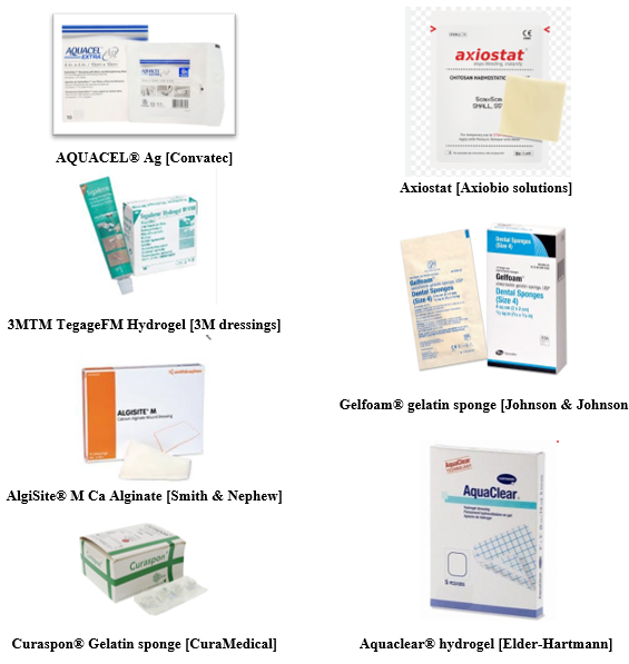
Fig. 1 Marketed wound dressings
Other treatment options
Hyperbaric Oxygen
Because of inadequate perfusion, chronic nonhealing wounds typically experience hypoxia(109). Tissue that has been injured or diseased has also shown signs of low oxygen tension. The development of cells, the healing of wounds, and infection resistance all depend on oxygen. Improved immunological function and angiogenesis are the outcomes of increased oxygen supply to the tissues.
Pure oxygen is inhaled under pressures higher than 1 atm during hyperbaric oxygen therapy (HBOT). Increased tissue oxygen tension is the outcome of this therapy's significant rise in dissolved oxygen levels in plasma. Although there is little data to confirm or deny HBOT's impact on other wounds, it appears to speed up the healing of diabetic foot ulcers in humans(110). The indications for HBOT in veterinary medicine have not been the subject of any prospective randomized controlled research. HBOT may be helpful for necrotizing soft tissue infections or thermal burns, extrapolating from human medicine(111).
HBOT chambers come in a variety of sizes, some of which are made especially for tiny animals and others for horse patients. Each therapy session should be overseen by veterinary staff with training in hyperbaric medicine.
Most practitioners do not have easy access to or can't afford hyperbaric chambers.
Pneumothorax, lung illness, a history of thoracic or ear surgery, fever, and pregnancy are examples of potential contraindications to HBOT. Barotrauma, cataracts, pulmonary dyspnea, and seizures are among the side effects of HBOT(111).
Extracorporeal Shock Wave Therapy
High-energy waves are delivered through the tissues during extracorporeal shock wave treatment (ESWT). In an equine model, excessive granulation tissue has been suppressed using ESWT to enhance wound healing(112). Additionally, it has been demonstrated to speed up epithelialization in both horse and porcine models(113,114). Although there is no agreement on the ideal therapy regimen in terms of length, intensity, or number of pulses, treatments for people are often given once or twice a week while employing low to medium energy. ESWT is a safe, generally painless supplementary therapy for wounds in humans, according to a comprehensive evaluation of the literature, but further research is required to determine its usefulness and cost-efficiency(115).
Laser treatment
Open wounds may heal faster with laser treatment(116,117). Higher laser dosages, however, can stop wounds from healing or even destroy cells. When employing a therapeutic laser, the normal wavelength range is from 630 nm (visible light) to 904 nm (infrared), with a power range of 1 to 15 W. The fact that the kind of laser and the therapy regimens may vary significantly between studies makes it difficult to evaluate the literature. Additionally, the majority of studies on wound healing have been conducted in vitro or in vivo on people, whose skin is very different from that of furred animals. While some studies show positive impacts, others show no benefit(118).
The most promising treatment for wounds with poor healing potential may be laser therapy. There are several brands of therapeutic lasers that are sold to the veterinary industry, and they are easily accessible. For acute wounds, the standard recommendation for laser treatment is 2 to 6 J/cm2 once daily for 7 to 10 days, and 2 to 8 J/cm2 once daily for chronic wounds. The best doses, nevertheless, are unknown and could change based on the wavelength. The laser head should be cleaned before and after each treatment and should not come into contact with the wound. The use of laser treatment comes with various warnings. The staff at the hospital needs to get laser safety training as well as instruction on how to use the laser. Everyone in the room, including the patients, must wear safety glasses(119).
Electrical Stimulation
Chronic wounds with a delayed healing process can be accelerated with micro current electrical stimulation, which employs continuous or pulsed electrical current waveforms in the micro amperage range (1 to 999 mA). The usefulness of micro current electrical stimulation is based on the hypothesis that endogenous bioelectrical signals play a role in the partial mediating of natural tissue healing, and the therapy is intended to augment this impact. Negatively charged cells (macrophages and neutrophils) will move towards the anode when the anode (+) is put on a wet sterile gauze over the wound bed and the cathode (-) is placed on the surrounding skin, therefore boosting the inflammatory stage of wound healing. will go towards the direction of the anode, promoting the inflammatory phase of wound healing(120).
If the cathode is placed over the wound bed and the anode is on neighboring skin, positively charged cells (fibroblasts, keratinocytes, and epidermal cells) will flow towards the cathode. Electrode installation may need to be timed in accordance with the stage of wound healing, or the polarity may need to be changed every three to four treatments, in order to balance the migration of positively and negatively charged cells. There isn't much proof that laying an anode or a cathode over the wound bed can stop the growth of microbes. The wound may need to be treated for 30 to 60 minutes twice or three times each day, five to seven days per week. The use of micro current treatment to treat persistent cutaneous ulcers in humans is supported by some data(121).
Therapeutic ultrasound
Acoustic or mechanical energy waves are delivered via the tissues during therapeutic ultrasonography. When cells are in the inflammatory stage of healing a wound, ultrasound may have a stimulating influence on them. It may help strengthen collagen(122–124).
Ultrasonic energy given at a certain frequency defines conventional ultrasound treatment. Both high (3 MHz) and low (0.1-3.0 W/cm2) intensities. Delivering energy to the
tissue through a hand piece in contact with the skin. The use of an ultrasonic gel or other coupling medium in contact with the hand piece improves the transmission of ultrasound energy. The thermal and mechanical impacts that ultrasound exerts on tissues may help wounds heal. Both continuous and pulsed modes of ultrasonic energy delivery are available. . If the goal is to produce more mechanical effects than heat effects, the pulsed mode could be used. Therapeutic ultrasonography carries various possible hazards, notably those connected to the thermal effects. Applying ultrasound for 5 to 10 minutes twice a week would be a normal course of therapy. The use of conventional ultrasonography in the treatment of human cutaneous ulcers is supported by some data(121).
The MIST treatment system (Celebration Inc., Eden Prairie, MN) uses ultrasound radiation that is transmitted at a frequency (40 kHz) significantly lower than that of regular ultrasound. In addition to being advised for cleansing or debriding wounds containing germs, exudates, or fibrin, it may help speed up the healing of chronic wounds(125–127). Ultrasonic energy is delivered to the wound bed through a mist of sterile saline, avoiding direct hand piece contact. The energy is provided continuously. An everyday use of ultrasound for five to ten minutes would be a normal course of therapy. Though it might not be cost-effective for veterinary usage, the MIST treatment system appears to offer promise for debriding open wounds and accelerating wound healing.
Regulatory Aspects of Wound Dressings
Medical devices are defined by the World Health Organization to include any materials, instruments, apparatus, appliances, or software that are used alone or in combination to monitor a person's health. A product that is used for treatment or diagnostic purposes in medicine is known as a medical device. The impact of the medical device on patients is predominantly physical as opposed to the biochemical effects of pharmacological compounds. Wound dressings are classified as medical devices based on the specified features associated with them. Furthermore, they are divided into distinct groups according to the various sorts of wounds(128). Systems for managing wound care include a defined protocol for dressing wounds. While the suggested guidelines are comparable throughout the majority of the nations, there are still variations in the submission of dossier material, for instance, in the assessment processes.
A medical device that has been approved in one country may be approved and marketed more quickly in another country due to the harmonization of laws regarding dossier submission formats for licenses for medical devices. This is possible because the regulatory requirements for various products are similar and mutually agreed upon. For instance, dressings with a CE label are authorized for import and export in Australia. The inability to manufacture the device in a manner consistent with the requirements is what causes these variations in the rules.
The following outlines the difficulties that manufacturers in various nations have when attempting to regulate a medical item like a wound dressing.
In general hospitals and for everyday use by individuals, basic and minimally invasive bandages such as fibrillar, hydrophilic, occlusive, and non resorbable gauze or sponge are classified as Class I wound dressings by the US FDA. Class II and Class III wound dressings are further types of dressings that include drugs or bioactive compounds, nanomaterial’s, and human or animal cellular components.
Class I wound dressings are described in the Canada Health Organization guideline as mechanical barriers to the wound's external environment or for compression or absorption of wound exudates. Additionally, Class I items include non-invasive bandages that come in contact with the damaged skin. Additionally, Class II wound dressings are all noninvasive devices that are meant to come into intimate contact with the wounded skin in order to provide a moist environment and pain relief to aid in the healing process. Medical devices are categorized according to the European Medical Device Directive (MDD) organization based on a number of variables, including the level of danger involved with using the device, how long it is in touch with the body, and how intrusive it is. Wound dressings are considered medical devices under the European MDD guideline. The European MDD regulations classify all noninvasive devices that come into contact with wounded skin as belonging to Class I. Additionally, Class II a and b dressings are those that are used to improve the healing process by regulating the amount of moisture, pH, and temperature at the wound site. According to the European MDD, all devices that use animal tissues are classified as Class III. Classifying animal tissues as III.
It can be concluded that all the mentioned countries' regulatory principles follow a similar guideline in determining the classification of wound dressings by comparing the regulatory aspects of wound dressing classification guidelines in different countries under global health management(129).
Different wound care products call for various sterilization techniques. According to reports, there is no particular sterilization technique for clothing's microbiological control. A viable method for improving wound care system control is the use of recent advancements in wireless communications and microelectronic sensors for the production of wound monitoring devices or dressings. However, some sensor types have limitations when it comes to being integrated into wireless wearable wound monitoring dressings. The ability of the sensors to be reduced in size within the dressing bed, biocompatibility, flexibility, resistance to moisture and bio fouling, and disposability are a few of the main issues that should be taken into account while designing the regulatory framework for these devices(130).
Modern technical developments and human understanding of disease mechanisms have enhanced medical device manufacturing. The industry has seen the development of several generic medical devices for a wide range of disorders, making them a crucial component of the healthcare system(131). One of the main obstacles the World Health Organization has in creating a common dressing standard is the size and kind of the wound. The dynamic nature of the chronic wounds and the unavailability of a perfect animal model for determining the wound's state of progression showed
a good prescription for clinical trials (132).
The most prevalent medical equipment that is essential to the health care system is wound dressings. For the creation of global regulatory frameworks for the recently produced dressings, more research is necessary. As a result, creating a good dressing requires consideration of whether the wound is healing or deteriorating. As a result, current regulatory principles need to be continually updated and applied to the production process(133).
CONCLUSION
Effective wound management requires a comprehensive understanding of the intricate pathophysiological processes involved in healing, as well as the various factors that can impede recovery. The TIME and TWA frameworks provide critical guidance in selecting appropriate treatment strategies tailored to individual wound characteristics. Recognizing the role of underlying health conditions, such as diabetes and obesity, alongside nutritional status, is essential for optimizing healing outcomes. Furthermore, the diverse landscape of wound dressings ranging from conventional to advanced biomaterials offers clinicians a variety of tools to enhance wound healing. Advanced therapeutic options and regulatory considerations also play pivotal roles in ensuring safe and effective care. By integrating knowledge from these domains, healthcare professionals can significantly improve patient outcomes in wound care management.
REFERENCES
1. Vig K, Chaudhari A, Tripathi S, Dixit S, Sahu R, Pillai S, et al. Advances in Skin Regeneration Using Tissue Engineering. 2017;
2. Physiology C. Regenerative Skin Wound Healing in Mammals?: State-of-the-Art on Growth Factor and Stem Cell Based Treatments. 2015;1–23.
3. Lazarus GS, Cooper DM, Knighton DR, Margolis DJ, Percoraro RE, Rodeheaver G, et al. Definitions and guidelines for assessment of wounds and evaluation of healing. Wound Repair Regen. 1994;2(3):165–70.
4. Moore Z, Dowsett C. TIME CDST: an updated tool to address the current challenges in wound care. 2023.
5. Rumbaut RE (Rolando E, Thiagarajan P. Platelet-vessel wall interactions in hemostasis and thrombosis. Morgan & Claypool Life Sciences Publishers; 2010. 67 p.
6. Tennent GA, Brennan SO, Stangou AJ, Hawkins PN, Pepys MB. Human plasma fibrinogen is synthesized in the liver. Blood [Internet]. 2007;109:1971–4. Available from: http://ashpublications.org/blood/article-pdf/109/5/1971/1478894/zh800507001971.pdf
7. Furie B, Furie BC. Mechanisms of Disease Mechanisms of Thrombus Formation [Internet]. 2008. Available from: www.nejm.org
8. Pool JG. Normal hemostatic mechanisms: a review. Am J Med Technol [Internet]. 1977;43(8):776–80. Available from: http://europepmc.org/abstract/MED/888856
9. Medrado ARAP, Pugliese LS, Reis SRA, Andrade ZA. Influence of low level laser therapy on wound healing and its biological action upon myofibroblasts. Lasers Surg Med. 2003;32(3):239–44.
10. Gurtner GC, Werner S, Barrandon Y, Longaker MT. Wound repair and regeneration. Vol. 453, Nature. Nature Publishing Group; 2008. p. 314–21.
11. Arajo AAS, Nunes PS, Albuquerque-Jnior RLC, Cavalcante DRR, Dantas MDM, Cardoso JC, et al. Collagen-based films containing liposome-loaded usnic acid as dressing for dermal burn healing. J Biomed Biotechnol. 2011;2011.
12. Tidball JG. Inflammatory processes in muscle injury and repair. Am J Physiol - Regul Integr Comp Physiol. 2005;288(2 57-2).
13. Karantas ID, ?enyi?it, ZOkur ME, Eynep, Üstünda? Okur N, Siafaka PI. Recent trends on wound management: New therapeutic choices based on polymeric carriers. Asian J Pharm Sci. 2020;15(6):661–84.
14. De Mendonça RJ, Coutinho-Netto J. Aspectos celulares da cicatrização. An Bras Dermatol. 2009;84(3):257–62.
15. Li J, Chen J, Kirsner R. Pathophysiology of acute wound healing. Clin Dermatol. 2007 Jan;25(1):9–18.
16. Rosen BP. Biochemistry of arsenic detoxi¢cation.
17. Armulik A, Genové G, Betsholtz C. Pericytes: Developmental, Physiological, and Pathological Perspectives, Problems, and Promises. Vol. 21, Developmental Cell. 2011. p. 193–215.
18. Coman T, Antonina Calin M, Romeo Calin M. The effect of low level laser therapy on surgical wound healing [Internet]. Vol. 62, Romanian Reports in Physics. 2010. Available from: https://www.researchgate.net/publication/228621606
19. Tonnesen MG, Feng 2 Xiaodong, Clark RAF. Angiogenesis in Wound Healing. Vol. 5, Journal of Investigative Dermatology Symposium Proceedings. 2000.
20. Gonzalez ACDO, Andrade ZDA, Costa TF, Medrado ARAP. Wound healing - A literature review. Vol. 91, Anais Brasileiros de Dermatologia. Sociedade Brasileira de Dermatologia; 2016. p. 614–20.
21. Broughton G, Janis JE, Attinger CE. Wound healing: An overview. Vol. 117, Plastic and Reconstructive Surgery. 2006.
22. Forrest L. Current concepts in soft connective tissue wound healing. Vol. 70, British Journal of Surgery. 1983. p. 133–40.
23. Witte MB, Barbul A. GENERAL PRINCIPLES OF WOUND HEALING. Vol. 77. 1997.
24. Eming SA, Martin P, Tomic-Canic M. Wound repair and regeneration: Mechanisms, signaling, and translation. Sci Transl Med. 2014;6(265).
25. Nethi SK, Das S, Patra CR, Mukherjee S. Recent advances in inorganic nanomaterials for wound-healing applications. Vol. 7, Biomaterials Science. Royal Society of Chemistry; 2019. p. 2652–74.
26. Rutty G. Interpretation of wounds. Nurs Times. 2000;96(31):41–2.
27. Nolan G, Hainsworth S V., Rutty GN. Forces generated in stabbing attacks: an evaluation of the utility of the mild, moderate and severe scale. Int J Legal Med. 2018;132(1):229–36.
28. Jeney V, Eaton JW, Balla G, Balla J. Natural history of the bruise: Formation, elimination, and biological effects of oxidized hemoglobin. Oxid Med Cell Longev. 2013;2013.
29. Kurland D, Hong C, Aarabi B, Gerzanich V, Simard JM. Hemorrhagic progression of a contusion after traumatic brain injury: A review. J Neurotrauma. 2012;29(1):19–31.
30. Kimbler DE, Murphy M, Dhandapani KM. Concussion and the adolescent athlete. J Neurosci Nurs. 2011;43(6):286–90.
31. Liu H, Wang Q, Zhao Z, Xie Y, Ding S, Wang Z. The Clinical and Medicolegal Analysis of Electrical Shocked Rats: Based on the Serological and Histological Methods. Biomed Res Int. 2016;2016.
32. Schultz GS, Sibbald RG, Falanga V, Ayello EA, Dowsett C, Harding K, et al. Wound bed preparation?: a systematic approach to wound management. 2002;(Figure 1).
33. Schultz GS, Barillo DJ, Mozingo DW, Chin GA. Wound bed preparation and a brief history of TIME. 2004;1(1):19–32.
34. Tottoli EM, Dorati R, Genta I, Chiesa E, Pisani S. Skin Wound Healing Process and New Emerging Technologies for Skin Wound Care and Regeneration. 2020;
35. Sung H, Sun X, Lee J, Kim H, Fu X, Leong KW. Advanced drug delivery systems and arti fi cial skin grafts for skin wound healing ?. Adv Drug Deliv Rev [Internet]. 2019;146:209–39. Available from: https://doi.org/10.1016/j.addr.2018.12.014
36. Parker CN, Finlayson KJ, Edwards HE. Ulcer area reduction at 2 weeks predicts failure to heal by 24 weeks in the venous leg ulcers of patients living alone. J Wound Care. 2016;25(11):626–34.
37. Falanga V. Wound healing and its impairment in the diabetic foot. Lancet. 2005;366(9498):1736–43.
38. Guo S, DiPietro LA. Critical review in oral biology & medicine: Factors affecting wound healing. J Dent Res. 2010 Mar;89(3):219–29.
39. Bishop. role of oxygen in wound healing. J Wound Care. 2008;17(9):399.
40. Rodriguez PG, Felix FN, Woodley DT, Shim EK. The role of oxygen in wound healing: A review of the literature. Dermatologic Surg. 2008;34(9):1159–69.
41. Edwards R, Harding KG. Bacteria and wound healing. Curr Opin Infect Dis. 2004;17(2):91–6.
42. Bjarnsholt T, Kirketerp-Møller K, Jensen PØ, Madsen KG, Phipps R, Krogfelt K, et al. Why chronic wounds will not heal: A novel hypothesis. Wound Repair Regen. 2008;16(1):2–10.
43. Qin J, Chen F, Wu P, Sun G. Recent Advances in Bioengineered Scaffolds for Cutaneous Wound Healing. Vol. 10, Frontiers in Bioengineering and Biotechnology. Frontiers Media S.A.; 2022.
44. Dam A, Inger JS, Lark AFC. 090299 Cutaneous Wound Healing [Internet]. 1999. Available from: www.nejm.org
45. Genco RJ, Grossi SG, Ho A, Nishimura F, Murayama Y. A Proposed Model Linking Inflammation to Obesity, Diabetes, and Periodontal Infections.
46. Wetzler C, Èmpfer HK, Stallmeyer B, Pfeilschifter J, Frank S. Large and Sustained Induction of Chemokines during Impaired Wound Healing in the Genetically Diabetic Mouse: Prolonged Persistence of Neutrophils and Macrophages during the Late Phase of Repair.
47. Werner S, Krieg T, Smola H. Keratinocyte-fibroblast interactions in wound healing. J Invest Dermatol. 2007;127(5):998–1008.
48. Loots MAM, Lamme EN, Zeegelaar J, Mekkes JR, Bos JD, Middelkoop E. Differences in Cellular Infiltrate and Extracellular Matrix of Chronic Diabetic and Venous Ulcers Versus Acute Wounds. 1998.
49. Han G, Ceilley R. Chronic Wound Healing: A Review of Current Management and Treatments. Vol. 34, Advances in Therapy. Springer Healthcare; 2017. p. 599–610.
50. Legendre C, Debure C, Meaume S, Lok C, Golmard JL, Senet P. Impact of protein deficiency on venous ulcer healing. J Vasc Surg. 2008 Sep;48(3):688–93.
51. Chen CS, Su WH, Cheng MH, Lee WL, Tsou TS, Chang WH, et al. Nonsteroidal anti-inflammatory drugs for wounds: Pain relief or excessive scar formation? Vol. 2010, Mediators of Inflammation. 2010.
52. Kaushal M, Kutty N, Rao C. Nitrooxyethylation reverses the healing-suppressant effect of ibuprofen. Mediators Inflamm. 2006;2006.
53. Guo JJ, Yang H, Qian H, Huang L, Guo Z, Tang T. The Effects of Different Nutritional Measurements on Delayed Wound Healing After Hip Fracture in the Elderly. J Surg Res. 2010 Mar;159(1):503–8.
54. Krischak GD, Surgeon O, Augat ; P, Claes ; L, Kinzl ; L, Beck ; A. The effects of non-steroidal anti-infl ammatory drug application on incisional wound healing in rats.
55. van Anholt RD, Sobotka L, Meijer EP, Heyman H, Groen HW, Topinková E, et al. Specific nutritional support accelerates pressure ulcer healing and reduces wound care intensity in non-malnourished patients. Nutrition. 2010 Sep 1;26(9):867–72.
56. Payne WG, Naidu DK, Wheeler CK, Barkoe D, Mentis M, Salas RE, et al. Wound healing in patients with cancer [Internet]. Vol. 8, Eplasty. Institute for Tissue Regeneration, Repair, and Rehabilitation, Bay Pines VA Medical Center, Bay Pines, Florida, USA.; 2008. p. e9. Available from: http://europepmc.org/abstract/MED/18264518
57. Chandra Jagetia G, Krishnamurthy Rajanikant G. Acceleration of wound repair by curcumin in the excision wound of mice exposed to different doses of fractionated ? radiation. 2011.
58. Gulcelik MA, Dinc S, Dinc M, Yenidogan E, Ustun H, Renda N, et al. Local granulocyte-macrophage colony-stimulating factor improves incisional wound healing in adriamycin-treated rats. Surg Today. 2006 Jan;36(1):47–51.
59. Gordon CR, Rojavin Y, Patel M, Zins JE, Grana G, Kann B, et al. A review on bevacizumab and surgical wound healing: An important warning to all surgeons. Vol. 62, Annals of Plastic Surgery. 2009. p. 707–9.
60. Bennett MH, Feldmeier J, Hampson NB, Smee R, Milross C. Hyperbaric oxygen therapy for late radiation tissue injury. Vol. 2016, Cochrane Database of Systematic Reviews. John Wiley and Sons Ltd; 2016.
61. Pignataro RM, Ohtake PJ, Swisher A, Dino G. The role of physical therapists in smoking cessation: Opportunities for improving treatment outcomes. Phys Ther. 2012 May;92(5):757–66.
62. Markuson M, Hanson D, Anderson J, Langemo D, Hunter S, Thompson P, et al. The Relationship between Hemoglobin A 1c Values and Healing Time for Lower Extremity Ulcers in Individuals with Diabetes.
63. Banks ND, Redett RJ, Mofid MZ, Manson PN. Cutis laxa: Clinical experience and outcomes. Plast Reconstr Surg. 2003 Jun;111(7):2434–42.
64. Beighton P, De Paepe A, Steinmann B, Tsipouras P, Wenstrup5 RJ. Syndromes: Revised Nosology, Villefranche. Vol. 77, American Journal of Medical Genetics. 1997.
65. Adams Jr. The Role of Plastic Surgery in Congenital Cutis Laxa: A 10-Year Follow-Up. Plast Reconstr Surg. 1999;104(4).
66. Ehlers-Danlos syndrome 127. Plast Reconstr Surg. 2000;105:1905–6.
67. Rees MM, Rodgers GM. HOMOCYSTEINEMIA: ASSOCIATION OF A METABOLIC DISORDER WITH VASCULAR DISEASE AND THROMBOSIS. Vol. 71, THROMBOSIS RESEARCH. 1993.
68. Methionine metabolism.
69. Bostom AG, Selhub J. Homocysteine and Arteriosclerosis Subclinical and Clinical Disease Associations [Internet]. 1999. Available from: http://www.circulationaha.org
70. Baitner AC, Maurer SG, Gruen MB, Di Cesare PE. The Genetic Basis of the Osteochondrodysplasias. 2000.
71. Rajendran NK, Kumar SSD, Houreld NN, Abrahamse H. A review on nanoparticle based treatment for wound healing. J Drug Deliv Sci Technol [Internet]. 2018;44:421–30. Available from: https://doi.org/10.1016/j.jddst.2018.01.009
72. Walker RM, Gillespie BM, Thalib L, Higgins NS, Whitty JA. Foam dressings for treating pressure ulcers. Cochrane Database Syst Rev. 2017;2017(10).
73. Du J, Wong KKY. Nanomaterials for wound healing: Scope and advances. Theranostic Bionanomaterials. 2019;211–30.
74. Weller C, Weller C, Team V. Interactive dressings and their role in moist wound management. In: Advanced Textiles for Wound Care. Elsevier; 2019. p. 105–34.
75. Swanson T, Angel D. International Wound Infection Instutute Wound Infection in Clinical Practice Update Principles of Best Practice. Wounds Int. 2022;24(8):1–59.
76. Vowden K, Vowden P. Wound dressings: principles and practice. Surg (United Kingdom) [Internet]. 2017;35(9):489–94. Available from: http://dx.doi.org/10.1016/j.mpsur.2017.06.005
77. Preem L, Kogermann K. Electrospun Antimicrobial Wound Dressings: Novel Strategies to Fight Against Wound Infections. 2018;213–53.
78. Varaprasad K, Jayaramudu T, Kanikireddy V, Toro C, Sadiku ER. Alginate-based composite materials for wound dressing application:A mini review. Carbohydr Polym [Internet]. 2020;236(February):116025. Available from: https://doi.org/10.1016/j.carbpol.2020.116025
79. Bianchera A, Catanzano O, Boateng J, Elviri L. The place of biomaterials inwound healing. Ther Dressings Wound Heal Appl. 2020;337–66.
80. Lei J, Sun L, Li P, Zhu C, Lin Z. The Wound Dressings and Their Applications in Wound Healing and Management. Heal Sci J [Internet]. 2019;13(4):662. Available from: http://www.imedpub.com/
81. Stoica AE, Chircov C, Grumezescu AM. Nanomaterials for wound dressings: An Up-to-Date overview. Vol. 25, Molecules. MDPI AG; 2020.
82. Dhivya S, Vijaya V, Santhini E. Review article Wound dressings – a review. Biomedicine. 2015;5(4):24–8.
83. White R. Wound dressings and other topical treatment modalities in bioburden control. J Wound Care. 2011;20(9):431–9.
84. Kirschning AA, Dibbert N, Kirschning A, Dibbert N, Dräger G. Chemical functionalization of polysaccharides – towards biocompatible hydrogels for biomedical applications.
85. Stumpf TR, Yang X, Zhang J, Cao X. In situ and ex situ modifications of bacterial cellulose for applications in tissue engineering. Mater Sci Eng C [Internet]. 2016; Available from: http://dx.doi.org/10.1016/j.msec.2016.11.121
86. Lee K, Buldum G, Mantalaris A, Bismarck A. More Than Meets the Eye in Bacterial Cellulose?: Biosynthesis , Bioprocessing , and Applications in Advanced Fiber Composites a. 2013;1–23.
87. Wong TW, Ramli NA. Carboxymethylcellulose film for bacterial wound infection control and healing. Carbohydr Polym [Internet]. 2014; Available from: http://dx.doi.org/10.1016/j.carbpol.2014.06.002
88. Ng S, Jumaat N. European Journal of Pharmaceutical Sciences Carboxymethyl cellulose wafers containing antimicrobials?: A modern drug delivery system for wound infections. Eur J Pharm Sci [Internet]. 2014;51:173–9. Available from: http://dx.doi.org/10.1016/j.ejps.2013.09.015
89. Maurer RJ, Sax AF. Molecular simulation of surface reorganization and wetting in crystalline cellulose I and II. 2013;25–42.
90. Torres FG, Commeaux S, Troncoso OP. Starch-based biomaterials for wound-dressing applications. 2013;543–51.
91. Sadashiv V, Ravindra P, Dyawanapelly S, Deshpande A, Jain R, Dandekar P. Bioactive Materials Starch based nano fi brous scaffolds for wound healing applications. Bioact Mater [Internet]. 2017;1–12. Available from: https://doi.org/10.1016/j.bioactmat.2017.11.006
92. Pal K, Banthia A, Majumdar DK. Preparation of transparent starch based hydrogel membrane with potential application as wound dressing Preparation of Transparent Starch Based Hydrogel Membrane with Potential. 2006;(May 2014).
93. Sun G, Zhang X, Shen Y, Sebastian R, Dickinson LE, Fox-talbot K, et al. Dextran hydrogel scaffolds enhance angiogenic responses and promote complete skin regeneration during burn wound healing. 2011;108(52):20976–81.
94. Tabasum S, Noreen A, Maqsood MF, Umar H, Akram N, Nazli Z i. H, et al. A review on versatile applications of blends and composites of pullulan with natural and synthetic polymers. Int J Biol Macromol. 2018 Dec 1;120:603–32.
95. Liu H, Wang C, Li C. A functional chitosan-based hydrogel as a wound dressing and drug delivery system in the treatment of wound healing. RSC Adv [Internet]. 2018;8:7533–49. Available from: http://dx.doi.org/10.1039/C7RA13510F
96. Chen Z, Han L, Liu C, Du Y, Hu X, Du G, et al. A rapid hemostatic sponge based on large, mesoporous silica nanoparticles and: N -alkylated chitosan. Nanoscale. 2018 Nov 21;10(43):20234–45.
97. Pogorielov V, Sikora Z, Street S. Chitosan as a Hemostatic Agent: Current State. 2015;(2):24–33.
98. Ong SY, Wu J, Moochhala SM, Tan MH, Lu J. Development of a chitosan-based wound dressing with improved hemostatic and antimicrobial properties. Biomaterials. 2008 Nov;29(32):4323–32.
99. Periayah MH, Halim AS, Yaacob NS, Mat Saad AZ, Hussein AR, Abdul Rashid AH, et al. Glycoprotein IIb/IIIa and P2Y12 Induction by Oligochitosan Accelerates Platelet Aggregation. Biomed Res Int. 2014;2014.
100. Azuma K, Ifuku S, Osaki T, Okamoto Y, Minami S. Preparation and biomedical applications of chitin and chitosan nanofibers. Vol. 10, Journal of Biomedical Nanotechnology. American Scientific Publishers; 2014. p. 2891–920.
101. Tavakoli S, Klar AS. Advanced Hydrogels as Wound Dressings. 2020;1–20.
102. Boateng JS, Matthews KH, Stevens HNE, Eccleston GM. Wound Healing Dressings and Drug Delivery Systems?: A Review. J Pharm Sci [Internet]. 2008;97(8):2892–923. Available from: http://dx.doi.org/10.1002/jps.21210
103. Boateng J, Catanzano O. Advanced Therapeutic Dressings for Effective Wound Healing - A Review. Vol. 104, Journal of Pharmaceutical Sciences. John Wiley and Sons Inc.; 2015. p. 3653–80.
104. Park JH, Park EJ, Yi HS. Wound Healing and Anti-inflammatory Effects of Topical Hyaluronic Acid Injection in Surgical-Site Infection Caused by Staphylococcus aureus. 2017;1–6.
105. Romanò CL, Vecchi E De, Bortolin M, Morelli I, Drago L. Hyaluronic Acid and Its Composites as a Local Antimicrobial / Antiadhesive Barrier. 2017;2.
106. Pirnazar P, Wolinsky L, Nachnani S, Haake S, Pilloni A, Bernard GW. Bacteriostatic Effects of Hyaluronic Acid. 70(4).
107. Winter G. formation of the scab and the rate of epithelization of superficial wounds in the skin of the young domestic pigs. Nature. 1962;193:293–4.
108. Cockbill SME, Ll M, Pharm B, Pharm M, Turner TD. The Development of Wound Management Products. Chronic Wound Care Essentials E-b [Internet]. 2018;145–64. Available from: http://whywoundcare.s3.amazonaws.com/Files/Chapter+12.pdf
109. Bowler PG, Duerden BI, Armstrong DG. Wound microbiology and associated approaches to wound management. Vol. 14, Clinical Microbiology Reviews. 2001. p. 244–69.
110. Kranke P, Mh B, Schnabel A. Hyperbaric oxygen therapy for chronic wounds (Review) [Internet]. 2009. Available from: http://www.thecochranelibrary.com
111. Edwards ML. Hyperbaric oxygen therapy. Part 2: Application in disease. Vol. 20, Journal of Veterinary Emergency and Critical Care. 2010. p. 289–97.
112. Link KA, Koenig JB, Silveira A, Plattner BL, Lillie BN. Effect of unfocused extracorporeal shock wave therapy on growth factor gene expression in wounds and intact skin of horses. Vol. 74, 324 AJVR. 2013.
113. Dean D. Morgan, dvm; Scott McClure. Effects of extracorporeal shock wave therapy on wounds of the distal portion of the limbs in horses Dean. J Am Vet Med Assoc. 234:1154–61.
114. Haupt G, Chvapil M. Effect of Shock Waves on the Healing of Partial-Thickness Wounds in Piglets. Vol. 49, JOURNAL OF SURGICAL RESEARCH. 1990.
115. Dymarek R, Halski T, Ptaszkowski K. Extracorporeal Shock Wave Therapy as an Adjunct Wound Treatment: A Systematic Review of the Literature [Internet]. 2014. Available from: https://www.researchgate.net/publication/263897055
116. Rezende SB, Ribeiro MS, Núñez SC, Garcia VG, Maldonado EP. Effects of a single near-infrared laser treatment on cutaneous wound healing: Biometrical and histological study in rats. J Photochem Photobiol B Biol. 2007 Jun 26;87(3):145–53.
117. Kawalec JS, Hetherington VJ, Pfennigwerth TC, Dockery DS, Dolce M. Effect of a diode laser on wound healing by using diabetic and nondiabetic mice. J Foot Ankle Surg. 2004 Jul;43(4):214–20.
118. Surgery V, Lucroy MD, Edwards BF, Madewell BR, Acvim D. CASE REPORT Low-Intensity Laser Light-Induced Closure of a Chronic Wound in a Dog.
119. Millis DL, Saunders DG. Laser Therapy in Canine Rehabilitation. In: Canine Rehabilitation and Physical Therapy: Second Edition. Elsevier Inc.; 2013. p. 359–80.
120. Bogie KM, Reger SI, Levine SP, Sahgal V. Electrical Stimulation for Pressure Sore Prevention and Wound Healing. Assist Technol. 2000 Jun 30;12(1):50–66.
121. Kloth LC. Electrical stimulation for wound healing: A review of evidence from in vitro studies, animal experiments, and clinical trials. Vol. 4, International Journal of Lower Extremity Wounds. 2005. p. 23–44.
122. Leung MC, Ng GY, Yip KK. Effect of ultrasound on acute inflammation of transected medial collateral ligaments. Arch Phys Med Rehabil. 2004 Jun;85(6):963–6.
123. Watson T. Ultrasound in contemporary physiotherapy practice. Vol. 48, Ultrasonics. 2008. p. 321–9.
124. Nussbaum E. The influence of ultrasound on healing tissues. J Hand Ther. 1998;11(2):140–7.
125. Kavros SJ, Miller JL, Hanna SW. Treatment of Ischemic Wounds with Noncontact, Low-Frequency Ultrasound: The Mayo Clinic Experience, 2004-2006.
126. Steven J. Kavros, DPM* Erik C. Schenck. Use of Noncontact Low-Frequency Ultrasound in the Treatment of Chronic Foot and Leg Ulcerations A51-Patient Analysis Steven. J Am Podiatr Med Assoc. 2007;97(2).
127. Ennis WJ, Valdes W, Gainer M, Meneses P. Evaluation of Clinical Effectiveness of MIST Ultrasound Therapy for the Healing of Chronic Wounds. Vol. 19, ADV SKIN WOUND CARE. 2006.
128. Ravizza A, De Maria C, Di Pietro L, Sternini F, Audenino AL, Bignardi C. Comprehensive Review on Current and Future Regulatory Requirements on Wearable Sensors in Preclinical and Clinical Testing. Front Bioeng Biotechnol. 2019;7(November 2019):1–9.
129. Farahani M, Shafiee A. Wound Healing: From Passive to Smart Dressings. Vol. 10, Advanced Healthcare Materials. John Wiley and Sons Inc; 2021.
130. Wang Y, Maitz PKM. Advances and new technologies in the treatment of burn injury. Adv Drug Deliv Rev [Internet]. 2018;123:1–2. Available from: https://doi.org/10.1016/j.addr.2017.11.011
131. Sahu T, Ratre YK, Chauhan S, Bhaskar LVKS, Nair MP, Verma HK. Nanotechnology based drug delivery system: Current strategies and emerging therapeutic potential for medical science. J Drug Deliv Sci Technol [Internet]. 2021;63(January):102487. Available from: https://doi.org/10.1016/j.jddst.2021.102487
132. Nunan R, Harding KG, Martin P. Clinical challenges of chronic wounds: Searching for an optimal animal model to recapitulate their complexity. DMM Dis Model Mech. 2014;7(11):1205–13.
133. Yadav V, Mittal A, Bansal P, Singh SK. Regulatory approval process for advanced dressings in India: An overview of rules. J Wound Care. 2019;28(8):S32–42

P. Ahire*, D. Pagar, S. Jondhale, Navigating Wound Healing: A Comprehensive Review of Pathophysiology, Treatment Modalities, and Advanced Management Strategies, Int. J. of Pharm. Sci., 2024, Vol 2, Issue 11, 88-113. https://doi.org/10.5281/zenodo.14028869
 10.5281/zenodo.14028869
