Department Of Pharmacy Practice, P. Rami Reddy Memorial College of Pharmacy, Kadapa, Andhra Pradesh.
Pityriasis rubra pilaris is a rare, chronic, papulosquamous skin disorder characterized by follicular hyperkeratosis, erythematous scaling plaques, and palmoplantar keratoderma. The disease often presents with a distinct orange-red coloration of the skin and islands of sparing within areas of widespread erythema. Although the exact pathogenesis is unknown, associations with genetic mutations, autoimmune conditions, and abnormal keratinization have been proposed. We report the case of a 9-year-old female child admitted to the Government General Hospital, Kadapa, with progressive thickening of the skin over both soles, associated with fissuring for the past two years, and pruritus. The patient also reported multiple raised small lesions over both arms for the past seven months. Her past medical history revealed symptom aggravation while walking barefoot, with no history of trauma or drug intake. This case highlights the importance of recognizing pityriasis rubra pilaris in clinical practice.
Pityriasis rubra pilaris (PRP) is a rare inflammatory papulosquamous disorder of unknown etiology. There are six clinical subtypes, which occur in both children and adults. Common elements noted across all subtypes include palmoplantar keratoderma and follicular papules coalescing into well demarcated plaques of various sizes with characteristic reddish-orange hue and non-adherent, flaking scale.[1] The clinical appearance of PRP is highly variable, as is the individual prognosis. Therefore, stratifying PRP into six disease subtypes represents a first step to personalized medicine for this rare inflammatory skin disease. The next step should be to associate specific therapeutic strategies with these sub types of PRP.[2] More generalised subtypes often demonstrate intervening areas of unaffected skin known as “islands of sparing,” a signature characteristic of PRP.[1] First described by Alphonse Devergie in 1856, the condition derives its name from Greek roots: “pityriasis” (scaling), “rubra”(redness) and “pilaris”(involving hair follicles) [3]. While its exact pathogenesis remains elusive, PRP is believed to involve a complex interplay of genetic predispositions and environmental triggers. It presents with a bimodal age distribution, with peaks in the first and fifth decades of life, and affects individuals of all races and genders equally [1]. The clinical spectrum of PRP is diverse, leading to its classification into six subtypes based on age of onset, clinical presentation, and prognosis, as outlined by Griffiths [4]. These subtypes range from the classical adult-onset form, which often undergoes spontaneous remission, to more chronic and atypical variants, including those associated with HIV infection [5,6].
Diagnosing PRP can be challenging due to its clinical resemblance to other papulosquamous disorders, particularly psoriasis, necessitating careful clinical and histopathological correlation [5].
CONCLUSION:
A rare chronic inflammatory skin disorder, Pityriasis Rubra Pilaris (PRP) has a variety of symptoms and outcomes. Other types of PRP can last longer, but typical adult-onset PRP usually goes away on its own in a few years. The condition's itching, pain, and psychological distress have a major negative influence on quality of life. With the use of topical, systemic drugs, and increasingly, biologics, current treatments seek to control symptoms and bring about remission. There is no known cure, and because PRP is so uncommon, research is difficult, which prevents the development of standardized protocols. To improve outcomes and the lives of those impacted by this complicated condition, more study into its causes and focused therapies are essential.
CASE REPORT:
History
A nine-year-old female presented to the Department of Dermatology, Venereology, and Leprosy, Government General Hospital, Kadapa, with complaints of progressive thickening of the skin over both soles associated with fissuring and itching for the past two years. She also reported multiple small, raised lesions over both arms for the preceding seven months. The symptoms were aggravated by walking barefoot. There was no history of trauma, drug intake, systemic illness, or relevant family history.
Examination
Cutaneous examination revealed diffuse, hyperkeratotic, bilaterally symmetrical plaques with ill-defined erythematous to hyperpigmented scaling and fissuring over both soles. Multiple grouped, tiny, hypopigmented, scaly papules were observed over both arms. No other significant cutaneous or mucosal involvement was noted. Systemic examination findings were unremarkable.
Investigations
A skin biopsy from the affected site demonstrated psoriasiform dermatitis with alternating orthokeratosis and parakeratosis, follicular plugging, and irregular acanthosis findings supportive of pityriasis rubra pilaris (PRP) in the appropriate clinical context.
Diagnosis
Based on the clinical presentation and histopathological findings, a diagnosis of pityriasis rubra pilaris was established
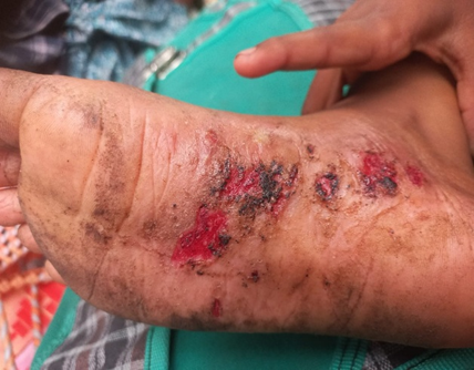
Figure 1 Fissures and Erosions on Soles before Treatment
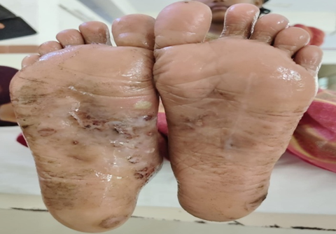
Figure 2 Improved Fissures and Erosions after Treatment
Treatment
The patient was initiated on systemic antihistamines chlorpheniramine 4 mg once daily and cetirizine 5 mg twice daily along with topical therapy including liquid paraffin (emollient), 0.05% betamethasone ointment (topical corticosteroid), mupirocin ointment (topical antibiotic), and 6% salicylic acid ointment (keratolytic). This regimen was maintained during hospitalization (days 1–5).
Outcome
At discharge, the same treatment plan was continued, with instructions for application schedules to optimize efficacy. The patient reported symptomatic relief, with reduction in itching and improvement in fissuring. Follow-up was advised for monitoring of treatment response and adjustment of therapy as required.
DISCUSSION:
Pityriasis Rubra Pilaris (PRP) is a rare and clinically challenging inflammatory skin disorder that frequently causes difficulties in diagnosis and treatment. Its hallmark clinical indicators salmon-coloured plaques, follicular prominence, islands of sparing, thickened palms and soles are very different, but its rarity and diverse manifestations might result in misdiagnosis, especially with psoriasis [5,7]. A skin biopsy may be examined under a microscope to help confirm the diagnosis, even if this is not always the case. The microscopic inspection frequently reveals a checkered pattern of alternating cell types[8]
Etiology and Pathogenesis
Treatment methods are made more complex by the fact that the precise origin of PRP is still unknown. Although the majority of cases occur randomly, a few familial types have been associated with genetic alterations, notably in the CARD14 gene. This result implies shared inflammatory pathways with psoriasis, perhaps through the IL23/Th17 axis [9,10]. Acquired PRP has been attributed to a variety of environmental triggers, including infections (bacterial, viral such as HIV), physical trauma, specific drugs (such imatinib and sorafenib), and, in a few cases, even underlying malignancies, suggesting a possible paraneoplastic link [6,11].
Treatment Challenges and Approaches
Because of the unpredictability of its course and the paucity of large-scale clinical studies, PRP management is, by nature, challenging. Treatment methods are mostly based on case reports and series, frequently making parallels from the treatment of psoriasis because of their shared inflammatory processes [12]. Topical treatments, like emollients, corticosteroids, and calcipotriol, provide symptomatic relief but are typically insufficient for widespread illness [13]. Systemic retinoids, like acitretin and isotretinoin, are frequently thought of as first-line treatments for more severe instances, even if their efficacy can differ and adverse effects might restrict long-term usage[14]. For cases that are hard to treat, immunosuppressants like methotrexate and cyclosporine can be used as alternatives [13].
Emerging Therapies: Biologics
The increasing usage of biological agents is a major improvement in PRP management. In severe and intractable PRP cases, medications that target TNFα (such as adalimumab, infliximab, etanercept), IL12/23 (ustekinumab), and IL17 (secukinumab) have shown promising outcomes [12,15]. These therapies add to the body of evidence supporting the notion that psoriasis shares inflammatory pathways with PRP, giving fresh hope to individuals who have not reacted well to traditional treatments. But additional data is needed on PRP that covers a longer period of time. PRP can severely impact quality of life through intense itching, painful fissures, mobility limitations, and temperature regulation issues, while also causing low self-esteem, social isolation, depression, and anxiety. Comprehensive care should address both physical impairments and psychological well-being.
SUMMARY:
Pityriasis rubra pilaris (PRP) is an uncommon, chronic inflammatory dermatosis of uncertain etiology, classically characterized by reddish orange scaly plaques interspersed with discrete “islands” of unaffected skin, accompanied by palmoplantar keratoderma. The pathogenesis is considered multifactorial, involving both genetic predisposition and environmental triggers. PRP may occur at any age, with incidence peaks reported in early childhood and middle adulthood.
The most frequent subtype, classical adult-onset PRP (Type I), typically undergoes spontaneous remission within one to three years. In contrast, rarer or hereditary variants are often persistent, less responsive to treatment, and associated with a chronic relapsing course.
PRP can cause substantial impairment in quality of life due to severe pruritus, painful fissuring, and functional limitations that interfere with routine activities. The visible nature of the lesions frequently results in psychological distress, including low self-esteem and social withdrawal. Although no definitive cure exists, therapeutic strategies aim to control symptoms and achieve remission through a multimodal approach. Commonly employed interventions include topical corticosteroids and calcipotriol, systemic retinoids such as acitretin, methotrexate, immunosuppressants, and phototherapy. In refractory disease, biologic agents targeting specific inflammatory pathways have demonstrated promising efficacy. However, the rarity of PRP limits large-scale clinical trials, thereby impeding the establishment of standardized treatment guidelines. Furthermore, clinical overlap with psoriasis and other papulosquamous disorders often complicates diagnosis. Consequently, management should be individualized, with a focus on optimizing symptom control and long-term patient outcomes.
CONCLUSION:
PRP is a rare dermatological disorder with diverse presentations that may mimic other papulosquamous conditions. Early recognition and a tailored, multimodal treatment plan are essential to optimize outcomes. This case reinforces the importance of considering PRP in the differential diagnosis of chronic hyperkeratotic dermatoses.
ACKNOWLEDGEMENT:
The authors express gratitude to patients affected by Pityriasis Rubra Pilaris for their resilience and contributions to understanding the rare condition. They also thank the Department/Institution for providing resources and individuals for their technical support. They also acknowledge the contributions of foundations and organizations dedicated to rare dermatological diseases, whose efforts fund research and raise awareness.
CONFLICT OF INTEREST: the authors declare that there is no conflict of interest.
ABBREVIATIONS:
PATIENT CONSENT: The patient described in this case report provided informed consent for publication, with the understanding that her identity will remain confidential.
REFERENCES

Y. Bhargavi, C. Lavanya Thejonidhi, M. Dinesh, Pityriasis Rubra Pilaris Presenting with Hyperkeratotic Soles: An Uncommon Case Report, Int. J. of Pharm. Sci., 2025, Vol 3, Issue 8, 2664-2669. https://doi.org/10.5281/zenodo.16940424
 10.5281/zenodo.16940424
