View Article

Abstract

Drug resistance and systemic side effects are common issues for people with cancer, infections, and other diseases. These problems are linked to the limitations of traditional chemotherapy, including poor solubility, toxicity, lack of specificity, low therapeutic effectiveness, and difficulty in crossing the worst barriers. To overcome these challenges, nanotechnology-based metallic platinum nanoparticles (PtNPs) have become popular because of their ability to deliver drugs more precisely. These PtNPs are used for targeted drug delivery and sustained release as antimicrobials to fight various illnesses and kill cancer cells. PtNPs-based treatment systems are effective at lower drug concentrations because they have good physical and chemical properties, such as specific shapes, sizes, a high surface area to volume ratio, good stability in the body, and ease of use. They can be easily modified with ligands, peptides, antibodies, or other substances to improve their targeting and controlled release. PtNPs can also be combined with other metals to serve as carriers for different types of treatments like chemotherapy, photothermal, or photoacoustic therapies. Magnetic therapies are also used to treat tumors. This review focuses on how PtNPs are made, how they are modified, how they work, their use in medicine, and their potential toxicity in treating illnesses.

Keywords

Diseases, platinum nanoparticles, delivery systems, Therapeutic efficacies

Introduction

Platinum nanoparticles

Depending on the situation, platinum nanoparticles, which are often described as a suspension or colloidal form of platinum particles, usually range in size from 2 to 100 nanometers.[1,2] A colloid is a stable mixture of particles in a fluid or gas. Platinum nanoparticles can be shaped into different forms, including circles, and their size can vary between 2 to 100 nanometers under certain conditions.[1,2]  These nanoparticles are found in a colloidal suspension and show colors like caramel red or dark tones. Different shapes of nanoparticles can be seen, such as tetrahedra, cubes, rods, and circles.[3] Platinum nanoparticles remain stable even at high temperatures, up to 700°C, without changing their structure. Their small size makes them suitable for being taken up by cells [4] Photothermal treatment is a process where platinum nanoparticles can penetrate deeper into tissues or cells because of their low scattering at near-infrared (NIR) wavelengths. They are also useful for targeted drug delivery when triggered by lasers; when exposed to light, they release some of their coating.[5] Platinum nanoparticles have antioxidant properties and are used therapeutically. They help reduce Reactive Oxygen Species (ROS) that are produced during the photothermal treatment of tumor cells.[6]

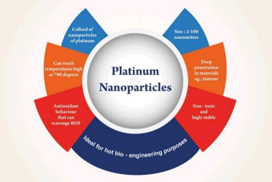
Fig. no 1 : Different Unique Properties of Platinum Nanoparticles.

Exposure to pathogens, poisons, or harmful substances can lead to cancer, infectious diseases, and tumors. The body's antioxidant and immune systems usually work together to fight off these harmful agents. The acquired immune system helps protect the body from the beginning of an illness and through the infection process [7] . When pathogens, infectious and strong disease-causing agents, or harmful substances enter the body, they overcome the body's natural defenses, causing localized infections. These infections can lead to the multiplication of the harmful agents, damage to host cells, genetic changes, or damage to DNA, which can result in the spread of disease, cancer, metastatic cancer, or tumors [8-11]  Using high doses or incorrect dosages of traditional chemotherapy can lead to drug resistance and harmful effects on healthy cells, making the patient's condition worse [12] Chemotherapy also has several limitations, such as being hard to dissolve, toxic, not targeting specific cells, poor absorption into the body, facing biological barriers, and having a low therapeutic benefit, all of which can increase patient suffering. Due to their unique shape, size, large surface area, lower toxicity, and ease of use, nanotechnology-based metallic platinum nanoparticles (PtNPs) have become a popular solution for addressing these issues in patients [13] . PtNPs have features like photothermal and photoacoustic effects, resistance to ionization, chemical stability, and resistance to corrosion. They also have the ability to be modified on their surface, and they can act as electro-catalysts for reactions like oxidation, hydrogenation, and dehydrogenation. These properties are related to optical behavior called surface plasmon resonance (SPR), which happens due to the interaction of light with free electrons on the surface of metallic nanoparticles, causing collective oscillations of the electrons in the conducting band [14-21]  PtNPs can enter cells, interact with internal components, and release platinum ions that may cause damage to DNA or cells [22,23] . PtNPs have the potential to increase the production of antioxidant enzymes and reduce oxidative stress. They can also act as strong and stable mimics of superoxide dismutase, leading to the death of cancer cells through DNA damage and stopping DNA replication [24-28] . They may also help reduce inflammation and damage caused by oxidative stress by removing reactive oxygen species (ROS) through the action of catalase [29-31] Various surface coating materials, such as polymers like poly-lactide-co-glycolide, poly-lactic acid, poly-ethylene glycol, polyvinyl alcohol, and chitosan, can be used to modify PtNPs along with drugs. This can lead to longer circulation in the body, controlled drug release, and accumulation in tumor areas, showing higher biocompatibility and passive targeting that helps counteract drug resistance [32-40]

Synthesis Of Platinum Nanoparticles

The size, shape, chemical composition, electronic surface structure, and capping agent of PtNPs are all important factors that affect their use in biomedicine and industry. Researchers are eager to explore new synthetic methods to improve these characteristics [41-44]

Platinum nanoparticles are made using two main approaches:

A) Physical and chemical synthesis methods, and

B) Green synthesis techniques.

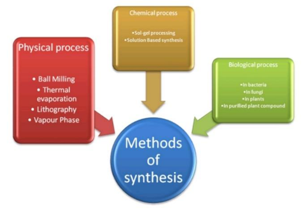
Fig.no 2 : Different Synthesis Methods of Platinum Nanoparticles.

1 . Physical and Chemical Synthesis Methods

Two common ways to make platinum nanoparticles are reducing platinum ion precursors in a solution or adding a capping agent to create new types of colloids. During physical synthesis, platinum nanoparticles are often exposed to different pressures, energies, or types of radiation.

A typical method for making platinum nanoparticles (PtNPs) is using a tube furnace where condensation and evaporation happen at normal atmospheric pressure [45] .

Another method is laser ablation, which involves using a high-power laser to heat and vaporize platinum nanoparticles from a solid material.Laser ablation is the process of turning PtNPs into vapor with a high-power laser beam.[46]Choosing the right reducing agent is important because it helps control the size, shape, and overall properties of the platinum nanoparticles made through chemical methods. Some common reducing agents include sodium borohydride (NaBH4), hydrogen gas (H2), ascorbic acid (vitamin C), and polyols. The size and shape of the PtNPs can be adjusted by changing various factors like the reaction temperature, the concentration of the reducing agent, and other similar parameters. The pH level of the solution and the use of stabilizing agents also influence the final size and shape of the nanoparticles.

Nucleation is a chemical process where nanoparticles form through atomic-level interactions. Other chemical methods used to produce nanoparticles include microemulsion and plasma-enhanced chemical vapor deposition.[47]

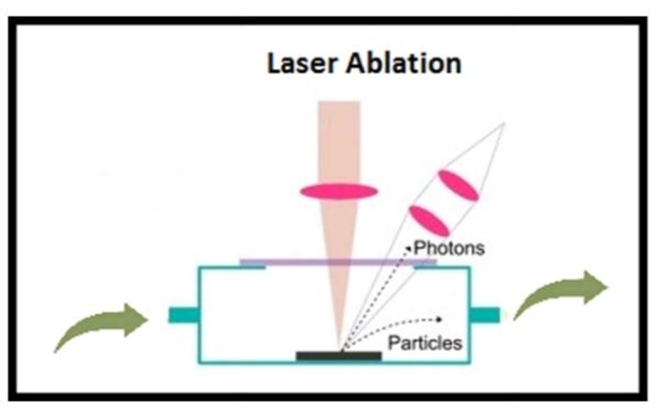
Fig. no 3 : Illustrating the Physical Methods of Platinum Nanoparticles Synthesis

2 . Green Synthesis Technique

From a biological perspective, platinum nanoparticles can be made using green methods that rely on various natural sources like bacteria [48-50], fungi [51-53], and even plants [54-57] . This approach is not only good for the environment but also cost-effective. The platinum nanoparticles made this way are sustainable, safe for living organisms, and dissolve easily because they are formed by reducing platinum ions with mild reducing agents.[42,43]

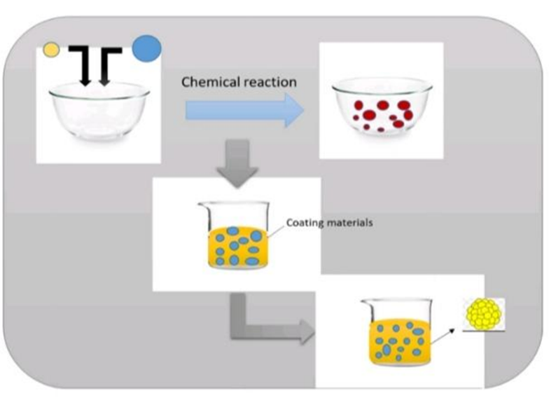
Fig . no 4 : Chemical Method of Platinum Nanoparticles Synthesis.

1. Bacteria

The process of reducing platinum, which is done by certain bacteria often found in various studies [51,52] , can create platinum nanoparticles. Some examples of these bacteria include Acinetobacter calcoaceticus [54]  and Desulfovibrio desulfuricans [53] , which are able to produce PtNPs by using bacteria to change platinum (IV) into platinum (0). Typically, this process happens within a day. The best results are seen when the pH is kept below 7.0 and the temperature is below 30°C. Another type of bacteria, Shewanella algae, known for reducing metal ions, produces PtNPs that are about 5 nm in size in its periplasmic space.

2 . Fungi

Using fungi, which are easy to find and can make stable nanoparticles, is a better way to produce platinum nanoparticles biologically [48]. Certain fungal strains, like Fusarium oxysporum, have been found to create slightly spherical, single-crystalline PtNPs with sizes between 20 and 110 nm. The release of high-quality proteins [55]  makes the later steps of processing easier, which ultimately leads to a higher yield of nanoparticles [56]

3 . Plant Extract

Plant extracts that have a high amount of phytochemicals such as flavonoids, phenolics, and terpenoids are often used to make nanoparticles. These extracts can help in the synthesis process because they have both reducing and stabilizing properties. To create the extract, plant parts like leaves, stems, or roots are ground or mixed with the right solvent, such as water or ethanol. The platinum precursor is usually a platinum salt, like chloroplatinic acid (H2PtCl6). The plant extract is mixed with this platinum salt. The reducing agents in the extract, like polyphenols, help turn the platinum ions into nanoparticles [57,58] . In one study, diopyros kaki leaf extract was used to treat an aqueous solution of chloroplatinic acid (H2PtCl6.6H2O). Over 90% of the platinum ions were converted into nanoparticles at a temperature of 95°C.

Characterization

Among the techniques used to study platinum nanoparticles, the following are commonly employed:

  1. scanning electron microscopy
  2. transmission electron microscopy[59]
  3. UV–visible spectroscopy [60,61]
  4. Fourier transform infrared spectroscopy [62]
  5. scanning electron microscope (SEM) [63]
  6. transmission electron microscope (TEM)
  7. electrophoretic light scattering
  8. dynamic light scattering [64]
  9. atomic force microscopy [65]
  10. X-ray diffraction [66]
  11. nanoscale infrared spectroscopy [67]
  12. energy-dispersive spectroscopy.

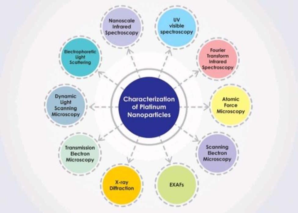
Fig.no 5 : The Different Ways in Which Characterization of Platinum Nanoparticles Can Be Done.

Synthesis Of Platinum Nanoparticles and Their Hybrid Form

PtNPs made through chemical methods can be shaped and sized in different ways based on several factors. These include the temperature of the reaction, the choice of solvent like ethylene glycol, the concentration of the starting material like H2PtCl6, and the type and amount of stabilizer such as polyvinyl pyrrolidone (PVP) and reducing agents like sodium hydroxide [68]

Chemical reduction is the main way to create colloidal nanoparticles.

It involves chemical substances that reduce metallic ions into actual metal nanoparticles. These chemicals can include potassium bitartrate (KC4H5O6), trisodium citrate dehydrate (Na3C6H9O9), ascorbic acid, sodium borohydride (NaBH4), and methoxy polyethylene glycol (CH3O(CH2CH2O)nH). This process can help produce small, spherical PtNPs with diameters of 1-2 nm or 2-3 nm. However, if the concentration of H2PtCl6 is lower, the size and shape might change. As the concentration of H2PtCl6 increases, the size of nanoparticles can grow, and their shapes can become cuboid (5-6 nm), oval (6-8 nm), or flower-shaped (16-18 nm).

The approach described in [69] can be used to make PtNPs.

First, a Brij 58 solution (0.044 M, 1 mL) is mixed with a K2PtCl4 solution (20 mM, 5 mL), and then the mixture is sonicated for 10 minutes. Following that, 5 mL of a 0.04 M solution of L-ascorbic acid is added, and the mixture is sonicated for another 45 minutes. After sonication, the precipitates are collected using three centrifugation steps.

An alternative method [70]  involves chemically reducing platinum salt (H2PtCl6·6H2O at 2 mmol/L) with sodium borohydride (NaBH4 at 4 mmol/L) using polyvinyl alcohol as the stabilizer. To ensure a complete reduction, the mixture is stirred continuously for 18 hours, during which time the color becomes a bright yellow, showing that the solution has a colloidal nature.

Using another method [71] , PtNPs can be created by making nanoparticles that show a unique UV-Vis absorption peak at 260 nm. For example, 19.5 mg (5 mM) of H2PtCl6 was treated with 83 mg (43.5 mM) of C6Na2O6 for an hour at 90°C to reduce it.

The same reduction method can also be used to make hybrid Au-Pt NPs.

Starting with ascorbic acid [72] , Na2PtCl6·6H2O is used.

The first step is to create the AuNPs solution. Na2PtCl6·6H2O (1 mM, 0.32 mg) is heated for 10 minutes at 90°C. Then, ascorbic acid (4 mM, 28 mg) is gradually added at 10 and 30-minute intervals. After that, the reaction mixture is heated for another 30 minutes at 90°C. This process considers two stages in the creation of Au-Pt NPs: first, the AuNPs are made and purified using methods described in [73-77] . In

In the second stage, PtNPs are created by adding 18 mL Milli-Q water and 5 mL of the purified AuNPs into a 50 mL round-bottom flask, and keeping the mixture at 100°C for 15 minutes while stirring until it stabilizes. Then, the solution is allowed to homogenize for 10 minutes after adding 1 mL of trisodium citrate dihydrate (68 mM, 0.020 g).

After another addition of the same trisodium citrate solution, the color changes to bluish-purple, indicating a reaction. The reaction is continued for 3 hours, and the final colloidal dispersion has a purple hue. The mixture is then centrifuged at 15,000 rpm for 10 minutes, and the supernatant is taken as the final colloidal solution containing the desired nanoparticles.

A variety of techniques can be used to produce Fe-Pt NPs.

One method involves polyol, as well as using a reaction mixture containing Fe(CO)5, Pt(acac)2, dioctyl ether, 1,2-hexadecanediol, oleic acid, and oleylamine, which is then heated at 290°C with ethanol to extract the FePt NPs. The molar ratio of Pt precursor (Pt(acac)3) and Fe precursor (Fe(acac)3) is the same. Different sizes of Fe-Pt nanoparticles can be made by using precursors such as Pt(acac)2, PtCl2, PtCl4, and H2PtCl6·H2O with the reducing agent 1,2-hexadecanediol and octyl ether. Additionally, the microemulsion method can be used, where an octyl phenyl is used in a water/glycol solution. In an ether/cyclohexane (water-in-oil) microemulsion, FeCl2 as the Fe precursor, H2PtCl6 as the Pt precursor, and NaBH4 as the reducing agent are used.

According to the hypothesized bio/green synthesis [78] 

The production of plant extract-mediated PtNPs involves four main steps.

The first step is the bio-reduction of metal ions by plant-based reducing agents to their zero oxidation state.

The second step involves the formation and accumulation of small particles into nanoparticles with enhanced thermal stability.

The third step includes the stabilization and capping of nanoparticles by plant-extracts to achieve a controlled range of shapes and sizes.

The fourth step involves the cleaning and purification of nanoparticles through centrifugation. The reaction time, pH, and temperature can be adjusted to optimize the shape, size, morphology, and crystallinity of the nanoparticles.

Mechanisms of Action of Platinum

Nanoparticles

PtNPs might kill microbes by sticking to their surfaces, getting inside their cell walls, and causing the cells to burst and break apart. This process also leads to the creation of reactive oxygen species such as hydroxyl radicals (.OH) and superoxide (O2.-), which can damage enzymes, DNA, and other parts of the cell [79,80]  PtNPs can also kill cancer cells by either passively or actively targeting them inside the cell, leading to breaks in DNA strands. This happens because PtNPs produce ROS, which causes damage to DNA and other important cellular molecules like carbohydrates and proteins. They also interfere with the body's ability to repair DNA and with the process of gene expression, which stops the cell from growing. During apoptosis, free radicals such as hydrogen peroxide (H2O2) and superoxide (O2-) are formed in the cell's cytosol and mitochondria [81-84] . To reduce oxidative stress, the body uses enzymes like catalase (CAT), peroxidase (POD), and superoxide dismutase (SOD). SOD converts superoxide into hydrogen peroxide and oxygen, while POD breaks down hydrogen peroxide. PtNPs have antioxidant properties similar to these enzymes, allowing them to remove ROS and convert hydrogen peroxide into water. Catalase reduces hydrogen peroxide into oxygen and water, helping to reduce oxidative damage without producing harmful hydroxyl radicals through the Fenton reaction.

Application

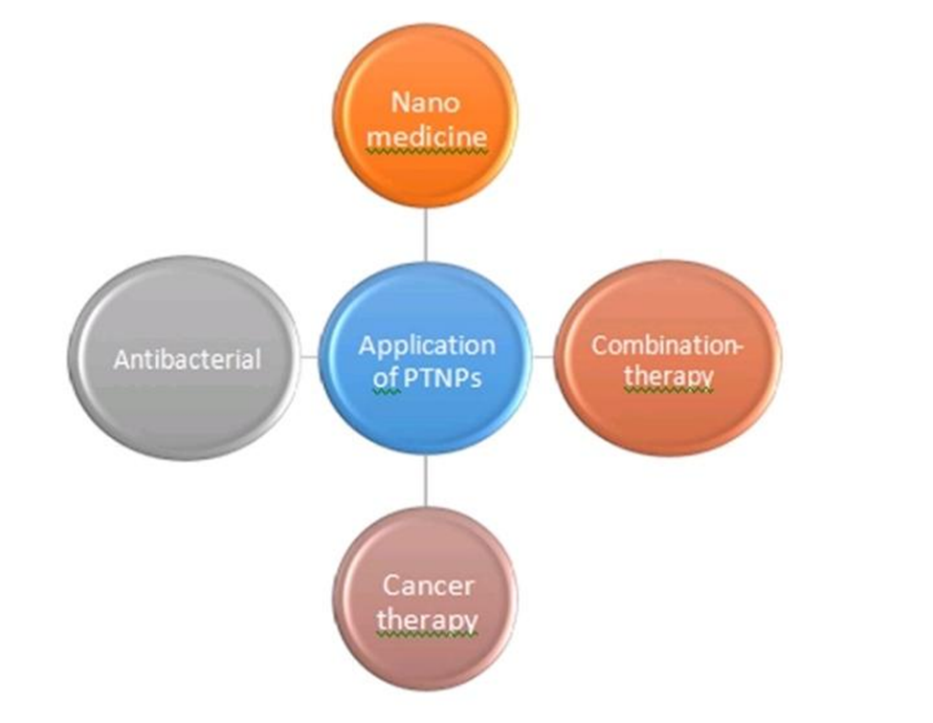
Fig. no 6 : Application of Platinum Nanoparticles.

1 . In Nanomedicine

Because of their ability to treat diseases linked to oxidative stress, like diabetes, atherosclerosis, cancer, and rheumatoid arthritis, platinum nanoparticles have become widely recognized.

Platinum nanoparticles are known for their strong stability and resistance, which makes them suitable for use in human medicine [85]

The enzyme-like properties in nanomedicine

Platinum nanoparticles are important in offering insights into potential treatments for cancer and certain cardiovascular conditions.

For example, PtNPs were placed inside the apoferritin cavity. They demonstrated the ability to neutralize peroxides and superoxides in both cell-free environments and inside cells. They were found to reduce the damage caused by H2O2 in a way that depends on their concentration, helping to prevent apoptosis.

2 . Combination Therapy

Combining platinum nanoparticles (PtNPs) with combination therapy has proven to be helpful in treating complex diseases, especially cancers. Combination treatments increase the effectiveness of therapy and help overcome problems like drug resistance. Using platinum nanoparticles (PtNPs) instead of the platinum-based chemicals currently used in hospitals offers several advantages, such as better biocompatibility, adjustable properties, and a larger surface area, which helps deal with issues related to drug persistence. Using imaging agents with platinum drugs improves how drug-loaded nanoparticles (NPs) spread throughout the tumor and the body. Common drugs used in combination include cyclophosphamide, doxorubicin, paclitaxel, and 5-fluorouracil. Platinum-based drugs create positively charged, reactive molecules that are used in clinical settings [86] These molecules form stable DNA adducts, which cause cancer cells to die [87,88] .Platinum-based drugs are commonly used to treat various cancers, including ovarian cancer, head and neck cancer, and lung cancer. Cisplatin is more effectively delivered into cells using nano-capsules compared to free drugs, leading to a higher concentration of platinum and the formation of cisplatin-DNA adducts in cells like IGROV-1 [89]

3 . In Cancer Therapy

The International Agency for Research on Cancer (IARC) says cancer is still a big cause of death around the world. Each year, there are about 18.1 million new cases and 9.6 million deaths from cancer.[90]  This number has been rising over time. Common treatments for cancer include chemotherapy, radiation, hormone therapy, and surgery. Chemotherapy is an important part of cancer treatment, but it can also have some bad side effects. Some of the drugs used in chemotherapy are called platinum-based drugs, like oxaliplatin, cisplatin, and carboplatin. These drugs are made up of platinum(II) complexes that have certain molecules attached to them. On one side, there are two amine groups, and on the other side, there are two chloride groups that are attached to the platinum. The platinum connects to the DNA in cancer cells. Cisplatin is a type of platinum-based drug that works well, with a success rate over 95%. It's used to treat several cancers, including lymphomas, myelomas, colon cancer, skin cancer, ovarian cancer, head and neck cancers, and testicular cancer. Scientists have also used different sizes of nanomaterials to help with treatment. They tested these materials for their effects on human colon cancer cells, called HT29.

4 . In Antibacterial Application

The term "antibacterial" refers to chemicals that either kill bacteria or slow their growth. Antibiotic resistance is a major global concern that threatens health, food safety, and overall progress. Although efforts are being made to address this issue, they are not enough.

There is a growing need for medicines that effectively fight harmful bacteria while being safe for people.

Examples of metallic nanoparticles include silver (Ag), copper (Cu), palladium (Pd), gold (Au), and others. In this context, titanium dioxide (TiO2) and zinc oxide (ZnO) are particularly important.[91]  However, the use of these metallic nanoparticles is limited due to their harmful effects when used inside the body.

Factors like shape, size, and surface charge of nanoparticles influence their antibacterial activity.Platinum nanoparticles (PtNPs) have been found to increase the production of ATP, which stops bacterial growth, damages DNA, and leads to bacterial death.

CONCLUSION :

PtNPs demonstrate a specific cytotoxic effect on diseased cell lines compared to healthy ones. However, they show cytotoxicity at higher concentrations and with long-term exposure in in vivo conditions. Therefore, surface-functionalizing PtNPs with polymers or other ligands makes them more suitable for achieving better antimicrobial and anticancer effects, as well as for targeted therapy that reduces systemic toxicity and enables sustained drug release along with antioxidant activity against diseases. In this context, the nanoparticles should be optimized to have more controlled and uniform sizes, which can enhance their biocompatible stability during synthesis, functionalization, and characterization. Also, ensuring consistent batch-to-batch uniformity in the development process before application is essential to achieve better therapeutic outcomes. A detailed investigation into the source of raw materials, production methods, consistent scale-up processes, solubility, biocompatibility, stability, routes of in vivo administration, biodistribution, pharmacokinetics, elimination, accumulation, cell-specific targeting, controlled release, and toxicological concerns for humans with high consistency is necessary before moving towards clinical translation. This is important to consider PtNPs as a drug delivery system and nanomedicine, aiming for maximum biological effectiveness against various diseases.

REFERENCES

  1. Pokropivny VV, Skorokhod VV. Classification of nanostructures by dimensionality and concept of surface forms engineering in nanomaterial science. Mater Sci Eng C. 2007;27:990–3.
  2. Tiwari JN, Tiwari RN, Kim KS. Zero-dimensional, one-dimensional, two-dimensional and three-dimensional nanostructured materials for advanced electrochemical energy devices. Prog Mater Sci. 2012;57:724–803
  3. Tiwari DK, Behari J, Sen P. Application of nanoparticles in waste water treatment. World J Appl Sci. 2008;3:417–33.
  4. Bigall NC, Härtling T, Klose M, Simon P, Eng LM, Eychmüller A. Monodisperse platinum nanospheres with adjustable diameters from 10 to 100 nm: synthesis and distinct optical properties. Nano Lett. 2008;8(12):4588–92. https://doi.org/10.1021/nl802901t.
  5. Ramirez E, Eradès L, Philippot K, Lecante P, Chaudret B. Shape control of platinum nanoparticles. Adv Func Mater. 2007;17(13):2219–28. https://doi.org/10.1002/adfm.200600633.
  6. Ahmadi TS, Wang ZL, Green TC, Henglein A, El-Sayed MA. Shape controlled synthesis of colloidal platinum nanoparticles. Science. 1996;272(5270):1924–6. https://doi.org/10.1126/science.272.5270.1924.
  7. Mandal AK. Dendrimers in targeted drug delivery applications: A Review of diseases and cancer. Int J Polym Mater Polym Biomater. 2021; 70(4):287-97 https://doi.org/10.1080/00914037.2020.1713780
  8. National Institutes of Health (US). Biological sciences curriculum study the NIH curriculum supplement series. In understanding emerging and re-emerging infectious diseases: National Institutes of Health (US). 2007; Bethesda, MD. https://www.ncbi.nhm.nih.gov/books/NBK 20370/ (accessed on 20 April 2017).
  9. Waarts MR, Stonestrom AJ, Park YC, Levine RL. Targeting mutations in cancer. J Clin Invest. 2022; 132(8):e154943. https://doi.org/10.1172/JCI154943 PMid:35426374 PMCid:PMC9012285
  10. Jin J, Wu X, Yin J, Li M, Shen J, Li J, et al. Identification of genetic mutations in cancer: Challenge and opportunity in the new era of targeted therapy. Front Oncol. 2019; 9:263. https://doi.org/10.3389/fonc.2019.00263 PMid:31058077 PMCid:PMC6477148
  11. Rivas-Dominguez A, Pastor N, Martinez-Lopez L, Colon-Perez J, Bermudez B, Orta ML. The role of DNA damage response in dysbiosis-induced colorectal cancer. Cells. 2021; 10(8):1934. https://doi.org/10.3390/cells10081934 PMid:34440703 PMCid:PMC8391204
  12. Gao Y, Shang Q, Li W, Guo W, Stojadinovic A, Mannion C, et al. Antibiotics for cancer treatment: A double-edged sword. J Cancer. 2020; 11(17): 5135-49. https://doi.org/10.7150/jca.47470 PMid:32742461 PMCid:PMC7378927
  13. Mandal AK. Gold nanoparticles as theranostic delivery system in combating various diseases. In Book: Tsygankova Victoria Anatolyivna, Editor. "Research Advances in Microbiology and Biotechnology Vol. 6". India-United Kingdom:Book Publisher International.2023;cp 8, pp 100-128. https://doi.org/10.9734/bpi/ramb/v6/5933B
  14. Gopal J, Hasan N, Manikandan M, Wu HF. Bacterial toxicity / compatibility of platinum nanospheres, nanocuboids and nanoflowers. Sci Rep. 2013; 3:1260. https://doi.org/10.1038/srep01260 PMid:23405274 PMCid:PMC3569627
  15. Sahin B, Aygun A, Gunduz H, Sahin K, Demir E, Akocak S, et al. Cytotoxic effects of platinum nanoparticles obtained from pomegranate extract by the green synthesis method on the MCF-7 cell line. Coll Surf B Biointerfaces. 2018; 163:119-24. https://doi.org/10.1016/j.colsurfb.2017.12.042 PMid:29287232
  16. Elder A, Yang H, Gwiazda R, Teng X, Thurston S, He H, et al. Testing nanomaterials of unknown toxicity: An example based on platinum nanoparticles of different shapes. Adv Mater. 2007; 19:3124-29. https://doi.org/10.1002/adma.200701962
  17. Kawasaki H, Yonezawa T, Watanable T, Arakawa R. Platinum nanoflowers for surface-assisted laser desorption / ionization mass spectrometry of biomolecules. J Phys Chem C. 2007; 111:16279-83. https://doi.org/10.1021/jp075159d
  18. Johnstone TC, Suntharalingam K, Lippard SJ. The next generation of platinum drugs: Targeted Pt(II) agents, nanoparticle delivery, and Pt(IV) prodrugs. Chem Rev. 2016; 116(5):3436-86.https://doi.org/10.1021/acs.chemrev.5b00597 PMid:26865551 PMCid:PMC4792284
  19. Wang Z, Chen L, Huang C, Huang Y, Jia N. Albumin-mediated platinum nanocrystals for in vivo enhanced computed tomography imaging. J Mater Chem B. 2017; 5(19):3498-510. https://doi.org/10.1039/C7TB00561J PMid:32264286
  20. Testa G, Fontana L, Venditti I, Fratoddi I. Functionalized platinum  nanoparticles with surface charge triggered by pH: Synthesis, characterization and stability studies. Beilstein J Nanotechnol. 2016; 7:1822-28. https://doi.org/10.3762/bjnano.7.175 PMid:28144532 PMCid:PMC5238631
  21. Huang X, Jain PK, El-Sayed IH, El-Sayed MA. Gold nanoparticles: Interesting optical properties and recent applications in cancer diagnostics and therapy. Nanomed. 2007; 2(5):681-93.https://doi.org/10.2217/17435889.2.5.681 PMid:17976030
  22. Yamada M, Foote M, Prow TW. Therapeutic gold, silver, and platinum nanoparticles. Wiley Interdiscip Rev: Nanomed Nanobiotechnol. 2015; 7:428-45. https://doi.org/10.1002/wnan.1322 PMid:25521618
  23. Gao J, Liang G, Zhang B, Kuang Y, Zhang X, Xu B. FePt@CoS2 yolkshell nanocrystals as a potent agent to kill HeLa cells. J Am Chem Soc. 2007; 129:1428-33. https://doi.org/10.1021/ja067785e PMid:17263428
  24. Asharani P, Xinyi N, Hande MP, Valiyaveettil S. DNA damage and p53-mediated growth arrest in human cells treated with platinum nanoparticles. Nanomed. 2010; 5:51-64. https://doi.org/10.2217/nnm.09.85 PMid:20025464
  25. Mony J, Ngamcherdtrakul W, Yantasee W. Oxidative stress in cancer and fibrosis: Opportunity for therapeutic intervention with antioxidant compounds, enzymes, and nanoparticles. Redox Biol. 2017; 11:240-53. https://doi.org/10.1016/j.redox.2016.12.011 PMid:28012439 PMCid:PMC5198743
  26. Onizawa S, Aoshiba K, Kajita M, Miyamoto Y, Nagai A. Platinum nanoparticle antioxidants inhibit pulmonary inflammation in mice exposed to cigarette smoke. Pulm Pharmacol Ther. 2009; 22:340-https://doi.org/10.1016/j.pupt.2008.12.015 PMid:19166956
  27. Pedone D, Moglianetti M, De Luca E, Bardi G, Pompa PP. Platinum nanoparticles in nanobiomedicine. Chem Soc Rev. 2017; 46:4951-75. https://doi.org/10.1039/C7CS00152E PMid:28696452
  28. Mohammadi H, Abedi A, Akbarzadeh A, Mokhtari MJ, Shahmabadi HE, Mehrabi MR, et al. Evaluation of synthesized platinum nanoparticles on the MCF-7 and HepG2 cancer cell lines. Int Nano Lett. 2013; 3:28. https://doi.org/10.1186/2228-5326-3-28
  29. Liu Y, Wu HH, Li M, Yin JJ, Nie ZH. pH dependent catalytic activities of platinum nanoparticles with respect to the decomposition of hydrogen peroxide and scavenging of superoxide and singlet oxygen. Nanoscale. 2014; 6(20):11904-10. https://doi.org/10.1039/C4NR03848G PMid:25175625
  30. Kumar PSM, Ponnusamy VK, Deepthi KR, Kumar G, Pugazhendhi A, Abe H, et al. Controlled synthesis of Pt nanoparticle supported TiO2 nanorods as efficient and stable electrocatalysts for the oxygen reduction reaction. J Mater Chem A. 2018; 6(46):23435-44. https://doi.org/10.1039/C8TA07380E
  31. Takamiya M, Miyamoto Y, Yamashita T, Deguchi K, Ohta Y, Abe K. Strong neuroprotection with a novel platinum nanoparticle against ischemic stroke- and tissue plasminogen activator-related brain damage in mice. Neuroscience. 2012; 221:47-55.https://doi.org/10.1016/j.neuroscience.2012.06.060PMid:22766232
  32. Hans ML, Lowman AM. Biodegradable nanoparticles for drug  delivery and targeting. Curr Opin Solid State Mater Sci. 2002; 6:319-27.https://doi.org/10.1016/S1359-0286(0)00117-1
  33. Rad JK, Alinejad Z, Khoei S, Mahdavian AR. Controlled release and photothermal behavior of multipurpose nanocomposite particles containing encapsulated gold-decorated magnetite and 5-FU in poly (lactide-co-glycolide). ACS Biomater Sci Eng. 2019; 5:4425-34. https://doi.org/10.1021/acsbiomaterials.9b00790 PMid:33438408
  34. Danhier F, Ansorena E, Silva JM, Coco R, Breton AL, Preat V. PLGAbased nanoparticles: An overview of biomedical applications. J Control Rel. 2012; 161:505-22. https://doi.org/10.1016/j.jconrel.2012.01.043 PMid:22353619
  35. Ramirez JC, Flores-VillaSenor SE, Vargas-Reyes E, Herrera-Ordonez J, Torres-Rincon S, Peralta-Rodriguez RD. Preparation of PDLLA and PLGA nanoparticles stabilized with PVA and PVA-SDS mixture: Studies on particle size, degradation and drug release. J Drug Deliv Sci Technol. 2020; 60:101907. https://doi.org/10.1016/j.jddst.2020.101907
  36. Plch J, Venclikova K, Janouskova O, Hrabeta J, Eckschlager T, Kopeckova K, et al. Paclitaxel-loaded polylactide/polyethylene glycol fibers with long-term antitumor activity as a potential drug carrier for local chemotherapy. Macromol Biosci. 2018; 18:1800011. https://doi.org/10.1002/mabi.201800011 PMid:29688614
  37. Gref R, Minamitake Y, Peracchia MT, Trubetskoy V, Torchilin V, Langer R. Biodegradable long-circulating polymeric nanospheres. Science. 1994; 263:1600-3. https://doi.org/10.1126/science.8128245 PMid:8128245
  38. Boulikas T, Pantos A, Bellis E, Christofis P. Designing platinum compounds in cancer: Structures and mechanisms. Cancer Ther. 2007; 5:537-83.
  39. Watkins R, Wu L, Zhang C, Davis RM, Xu B. Natural product-based nanomedicine: Recent advances and issues. Int J Nanomed. 2015; 10:6055. https://doi.org/10.2147/IJN.S92162 PMid:26451111 PMCid:PMC4592057
  40. Sanna V, Roggio AM, Siliani S, Piccinini M, Marceddu S, Mariani A, et al. Development of novel cationic chitosan- and anionic  alginate- coated poly (D, L-lactide-co-glycolide) nanoparticles for controlled release and light protection of resveratrol. Int J Nanomed. 2012; 7:5501. https://doi.org/10.2147/IJN.S36684 PMid:23093904 PMCid:PMC3477887
  41. Chithrani BD, Ghazani AA, Chan WCW. Determining the size and shape dependence of gold nanoparticle uptake into mammalian cells. NanoLett. 2006;6:662–8.
  42. Rorvig-Lund A, Bahadori A, Semsey S, Bendix PM, Oddershede LB. Vesicle fusion triggered by optically heated gold nanoparticles. Nano Lett. 2015;15:4183–8.
  43. Kajita M, Hikosaka K, Iitsuka M, Kanayama A, Toshima N, Miyamoto Y. Platinum nanoparticle is a useful scavenger of superoxide anion and hydrogen peroxide. Free Radic Res. 2007;41:615–26.
  44. Daniel M-C, Astruc D. Gold nanoparticles: assembly, supramolecular chemistry, quantum-size-related properties, and applications toward biol-ogy, catalysis, and nanotechnology. Chem Rev. 2004;104:293–346.
  45. Chen A, Holt-Hindle P. Platinum-based nanostructured materials: synthesis, properties, and applications. Chem Rev. 2010;110:3767–804.
  46. Le Guével X, Trouillet V, Spies C, Jung G, Schneider M. Synthesis of yellow-emitting platinum nanoclusters by ligand etching. J Phys Chem C. 2012;116:6047-51
  47. Mahmoud M, El-Sayed M. Time dependence and signs of the shift of the surface plasmon resonance frequency in nanocages elucidate the nanocatalysis mechanism in hollow nanoparticles. NanoLett. 2011;11:946–53.
  48. Perez H, Pradeau J-P, Albouy P-A, Perez-Omil J. Synthesis and characterization of functionalized platinum nanoparticles. Chem Mater. 1999;11:3460–3.
  49. Rakshit R, Bose S, Sharma R, Budhani R, Vijaykumar T, Neena S, Kulkarni G. Correlations between morphology, crystal structure, and magnetization of epitaxial cobalt-platinum fllms grown with pulsed laser abla-tion. J Appl Phys. 2008;103:023915.
  50. Torres-Chavolla E, Ranasinghe RJ, Alocilja EC. Characterization and functionalization of biogenic gold nanoparticles for biosensing enhancement. IEEE Trans Nanotechnol. 2010;9:533–8.
  51. Shah M, Fawcett D, Sharma S, Tripathy SK, Poinern GEJ. Green syn-thesis of metallic nanoparticles via biological entities. Materials. 2015;8:7278–308.
  52. Mukherjee P, Ahmad AD, Mandal S, Senapati SR, Sainkar M, Khan I. Bioreduction of AuCl4− Ions by the Fungus, Verticillium sp. and surface trapping of the gold nanoparticles formed. Angew Chem Int Ed. 2001;40:3585–8.
  53. Jeyaraj M, Gurunathan S, Qasim M, Kang MH, Kim JH. A comprehensive review on the synthesis, characterization, and biomedical application of platinum nanoparticles. Nanomaterials. 2019;9(12):1719. https://doi.org/10.3390/nano9121719.
  54. Martins M, Mourato C, Sanches S, Noronha JP, Crespo MTB, Pereira IAC. Biogenic platinum and palladium nanoparticles as new catalysts for the removal of pharmaceutical compounds. Water Res. 2017;108:160–8.
  55. Gaidhani SV, Yeshvekar RK, Shedbalkar UU, Bellare JH, Chopade BA. Bio-reduction of hexachloroplatinic acid to platinum nanoparticles employing Acinetobacter calcoaceticus. Process Biochem. 2014;49:2313–9.
  56. Konishi Y, Ohno K, Saitoh N, Nomura T, Nagamine S, Hishida H, Takahashi Y, Uruga T. Bioreductive deposition of platinum nanoparticles on the bacterium Shewanella algae. J Biotechnol. 2007;128:648–53.
  57. Riddin TL, Gericke M, Whiteley CG. Analysis of the inter-and extracellular formation of platinum nanoparticles by Fusarium oxysporum f. sp. lycopersici using response surface methodology. Nanotechnology. 2006;17:3482–9.
  58. Mohanpuria P, Rana NK, Yadav SK. Biosynthesis of nanoparticles: technological concept and future applications. J Nanopart Res. 2007;10:507–17.
  59. Narayanan KB, Sakthivel N. Biological synthesis of metal nanoparticles by microbes. Adv Colloid Interface Sci. 2010;156:1–13.
  60. Metha A. Derivation of Beer-Lambert law. PharmaXChange.info. 2012. http://pharmaxchange.info/press/2012/04/ultraviolet-visible-uvvis-spectroscopy-%e2%80%93-derivation-of-beer-lambert-law/.
  61. Link S, El-Sayed MA. Size and temperature dependence of the plasmon absorption of colloidal gold nanoparticles. J Phys Chem B. 1999;103(21):4212–7
  62. Aguirre FMA, Bazán-Díaz L, Mendoza-Cruz R, Gómez-Rodríguez A, Zorrilla-Cangas C, Herrera-Becerra R. Nano phase characterization by transmission electron microscopy: experimental and simulation. Mater Sci Appl. 2015;6:935.
  63. Takenaka S, Karg E, Kreyling WG, Lentner B, Möller W, BehnkeSemmler M, Jennen L, Walch A, Michalke B, Schramel P, et al. Distribution pattern of inhaled ultrafine gold particles in the rat lung. Inhaler Toxicol. 2006;18:733–40.
  64. Dzakpasu R, Axelrod D. Dynamic light scattering microscopy: a novel optical technique to image submicroscopic motions. I: Theory. Biophys J. 2004;87:1279–87.
  65. Horber JK, Miles MJ. Scanning probe evolution in biology. Science. 2003;302:1002–5.
  66. Cantor CR, Schimmel PR, Freeman E d WH. Techniques for the study of biological structure and function. San Francisco: Stanford Libraries, Stanford Publishers; 1980.
  67. Vlaic G, Olivi L. EXAFS spectroscopy: a brief introduction. Croat Chem Acta. 2004;77:427–33.
  68. Jeyaraj M, Gurunathan S, Qasim M, Kang MH, Kim JH. A comprehensive review on the synthesis, characterization, and biomedical application of platinum nanoparticles. Nanomater(Basel).2019; 9(12):1719. https://doi.org/10.3390/nano9121719 PMid:31810256 PMCid:PMC6956027
  69. Shim K, Kim J, Heo YU, Jiang B, Li C, Shahabuddin M, et al. Synthesis and cytotoxicity of dendritic platinum nanoparticles with HEK293 cells. Chem Asian J. 2017; 12:21-6. https://doi.org/10.1002/asia.201601239 PMid:27911052
  70. Chen S, Kimura K. Synthesis of thiolate-stabilized platinum nanoparticles in protolytic solvents as isolable colloids. J Physic Chem B. 2001; 105(23):5397-403. https://doi.org/10.1021/jp0037798
  71. Islam MT, Saenz-Arana R, Wang H, Bernal R, Noveron JC. Green synthesis of gold, silver, platinum, and palladium nanoparticles reduced and stabilized by sodium rhodizonate and their catalytic reduction of 4-nitrophenol and methyl orange. New J Chem. 2018;  42:6472-8. https://doi.org/10.1039/C8NJ01223G
  72. Gao Z, Ye H, Tang D, Tao J, Habibi S, Minerick A, et al. Platinumdecorated gold nanoparticles with dual functionalities for  ultrasensitive colorimetric in vitro diagnostics. Nano Lett. 2017; 17(9):5572-9. https://doi.org/10.1021/acs.nanolett.7b02385PMid:28813601
  73. Sivaraman SK, Kumar S, Santhanam V. Monodisperse sub 10 nm gold nanoparticles by reversing the order of addition in the Turkevich method-the role of chloroauric acid. J Colloid Interface Sci. 2011; 361:543-7. https://doi.org/10.1016/j.jcis.2011.06.015 PMid:21719021
  74. Theodosiou M, Boukos N, Sakellis E, Zachariadis M, Efthimiadou ER. Gold nanoparticle decorated pH-sensitive polymeric nanocontainers as a potential theranostic agent. Colloids Surf B:  Biointerfaces. 2019; 183:110420. https://doi.org/10.1016/j.colsurfb.2019.110420 PMid:31421401
  75. Low A, Bansal V. A visual tutorial on the synthesis of gold nanoparticles. Biomed Imaging Interv J. 2010; 6:e9. https://doi.org/10.2349/biij.6.1.e9 PMid:21611068 PMCid:PMC3097789
  76. La Spina R, Spampinato V, Gilliland D, Ojea-Jimenez I, Ceccone G.  Influence of different cleaning processes on the surface chemistry of gold nanoparticles. Biointerphases. 2017; 12:031003. https://doi.org/10.1116/1.4994286 PMid:28750541
  77. Ojea-Jimenez I, Campanera JM. Molecular modeling of the reduction mechanism in the citrate-mediated synthesis of gold nanoparticles. J Phys Chem C. 2012; 116:23682-91. https://doi.org/10.1021/jp305830p
  78. Sung H, Ferlay J, Siegel RL, Laversanne M, Soerjomataram I, Jemal A, et al. Global cancer statistics 2020: GLOBOCAN estimates of incidence and mortality worldwide for 36 cancers in 185 countries. CA Cancer J Clin. 2021; 71(3):209-49. https://doi.org/10.3322/caac.21660 PMid:33538338.
  79. Tahir K, Nazir S, Ahmad A, Li B, Khan AU, Khan ZUH, et al. Facile and green synthesis of phytochemicals capped platinum nanoparticles and in vitro their superior anti-bacterial activity. J Phytochem Photobiol B. 2017; 166:246-51. https://doi.org/10.1016/j.jphotobiol.2016.12.016 PMid:28011434
  80. Akther T, Khan MS, Srinivasan H. Novel silver nanoparticles synthesized from anthers of Couroupitaguianensisabul. Control  growth and biofilm formation in human pathogenic bacteria. Nano Biomed Eng. 2018; 10(3):250-7. https://doi.org/10.5101/nbe.v10i3.p250-257
  81. Gehrke H, Pelka J, Hartinger CG, Blank H, Bleimund F, Schneider R, et al. Platinum nanoparticles and their cellular uptake and DNA platination at non-cytotoxic concentrations. Arch Toxicol. 2011; 85(7):799-812. https://doi.org/10.1007/s00204-010-0636-3 PMid:21229235.
  82. Almeer RS, Ali D, Alarifi S, Alkahtani S, Almansour M. Green platinum nanoparticles interaction with HEK293 cells: Cellular toxicity, apoptosis, and genetic damage. Dose-Response. 2018;  16(4):155932581880738. https://doi.org/10.1177/1559325818807382 PMid:30479585  PMCid:PMC6247496
  83. Zhang Y, Yuan J, Zhang HY, Simayi D, Li PD, Wang YH, et al. Natural resistance to apoptosis correlates with resistance to chemotherapy in colorectal cancer cells. Clinic Expt Med. 2011; 12(2):97-103. https://doi.org/10.1007/s10238-011-0146-5 PMid:21688119
  84. Gillet JP, Gottesman MM. Mechanisms of multidrug resistance in cancer. Method Mol Biol:Human Press. 2009; p. 47-76.https://doi.org/10.1007/978-1-60761-416-6-4 PMid:19949920.
  85. Morry J, Ngamcherdtrakul W, Yantasee W. Oxidative stress in cancer and fibrosis: opportunity for therapeutic intervention with antioxidant compounds, enzymes, and nanoparticles. Redox Biol. 2017;11:240–53.
  86. Wheate NJ, Walker S, Craig GE, Oun R. The status of platinum anticancer drugs in the clinic and in clinical trials. Dalton Trans. 2010;39:8113–27.
  87. Ho YP, Au-Yeung SC, To KK. Platinum-based anticancer agents: innovative design strategies and biological perspectives. Med Res Rev. 2003;23:633–55. https://doi.org/10.1002/med.10038.
  88. Hamelers IH, Staffhorst RW, Voortman J, de Kruijff B, Reedijk J, van Bergen en Henegouwen PM, de Kroon AI. High cytotoxicity of cisplatin Nanocapsules in ovarian carcinoma cells depend on uptake by caveolae-mediated endocytosis. Clin Cancer Res. 2009;15:2009. https:// doi.org/10.1158/1078-0432.CCR-08-1702.
  89. Liu L, Miao P, Xu Y, Tian Z, Zou Z, Li G. Study of Pt/TiO2 nanocomposite for cancer-cell treatment. J Photochem Photobiol B Biol. 2010;98:207–10.https://doi.org/10.1016/j.jphotobiol.2010.01.005.
  90. Bray F, Ferlay J, Soerjomataram I, Siegel RL, Torre LA, Jemal A. Global Cancer Statistics 2018: GLOBOCAN estimates of incidence and mortality worldwide for 36 cancers in 185 countries. CA Cancer J Clin. 2018;68(6):394–424.https://doi.org/10.3322/caac.21492
  91. Pereira LFP, Estevinho L, Mendes A, Freire CSR. Antibacterial metallic nanoparticles for biomedical applications. Adv Healthc Mater. 2020;9(23):2001182.https://doi.org/10.1002/adhm.202001182.

Reference

  1. Pokropivny VV, Skorokhod VV. Classification of nanostructures by dimensionality and concept of surface forms engineering in nanomaterial science. Mater Sci Eng C. 2007;27:990–3.
  2. Tiwari JN, Tiwari RN, Kim KS. Zero-dimensional, one-dimensional, two-dimensional and three-dimensional nanostructured materials for advanced electrochemical energy devices. Prog Mater Sci. 2012;57:724–803
  3. Tiwari DK, Behari J, Sen P. Application of nanoparticles in waste water treatment. World J Appl Sci. 2008;3:417–33.
  4. Bigall NC, Härtling T, Klose M, Simon P, Eng LM, Eychmüller A. Monodisperse platinum nanospheres with adjustable diameters from 10 to 100 nm: synthesis and distinct optical properties. Nano Lett. 2008;8(12):4588–92. https://doi.org/10.1021/nl802901t.
  5. Ramirez E, Eradès L, Philippot K, Lecante P, Chaudret B. Shape control of platinum nanoparticles. Adv Func Mater. 2007;17(13):2219–28. https://doi.org/10.1002/adfm.200600633.
  6. Ahmadi TS, Wang ZL, Green TC, Henglein A, El-Sayed MA. Shape controlled synthesis of colloidal platinum nanoparticles. Science. 1996;272(5270):1924–6. https://doi.org/10.1126/science.272.5270.1924.
  7. Mandal AK. Dendrimers in targeted drug delivery applications: A Review of diseases and cancer. Int J Polym Mater Polym Biomater. 2021; 70(4):287-97 https://doi.org/10.1080/00914037.2020.1713780
  8. National Institutes of Health (US). Biological sciences curriculum study the NIH curriculum supplement series. In understanding emerging and re-emerging infectious diseases: National Institutes of Health (US). 2007; Bethesda, MD. https://www.ncbi.nhm.nih.gov/books/NBK 20370/ (accessed on 20 April 2017).
  9. Waarts MR, Stonestrom AJ, Park YC, Levine RL. Targeting mutations in cancer. J Clin Invest. 2022; 132(8):e154943. https://doi.org/10.1172/JCI154943 PMid:35426374 PMCid:PMC9012285
  10. Jin J, Wu X, Yin J, Li M, Shen J, Li J, et al. Identification of genetic mutations in cancer: Challenge and opportunity in the new era of targeted therapy. Front Oncol. 2019; 9:263. https://doi.org/10.3389/fonc.2019.00263 PMid:31058077 PMCid:PMC6477148
  11. Rivas-Dominguez A, Pastor N, Martinez-Lopez L, Colon-Perez J, Bermudez B, Orta ML. The role of DNA damage response in dysbiosis-induced colorectal cancer. Cells. 2021; 10(8):1934. https://doi.org/10.3390/cells10081934 PMid:34440703 PMCid:PMC8391204
  12. Gao Y, Shang Q, Li W, Guo W, Stojadinovic A, Mannion C, et al. Antibiotics for cancer treatment: A double-edged sword. J Cancer. 2020; 11(17): 5135-49. https://doi.org/10.7150/jca.47470 PMid:32742461 PMCid:PMC7378927
  13. Mandal AK. Gold nanoparticles as theranostic delivery system in combating various diseases. In Book: Tsygankova Victoria Anatolyivna, Editor. "Research Advances in Microbiology and Biotechnology Vol. 6". India-United Kingdom:Book Publisher International.2023;cp 8, pp 100-128. https://doi.org/10.9734/bpi/ramb/v6/5933B
  14. Gopal J, Hasan N, Manikandan M, Wu HF. Bacterial toxicity / compatibility of platinum nanospheres, nanocuboids and nanoflowers. Sci Rep. 2013; 3:1260. https://doi.org/10.1038/srep01260 PMid:23405274 PMCid:PMC3569627
  15. Sahin B, Aygun A, Gunduz H, Sahin K, Demir E, Akocak S, et al. Cytotoxic effects of platinum nanoparticles obtained from pomegranate extract by the green synthesis method on the MCF-7 cell line. Coll Surf B Biointerfaces. 2018; 163:119-24. https://doi.org/10.1016/j.colsurfb.2017.12.042 PMid:29287232
  16. Elder A, Yang H, Gwiazda R, Teng X, Thurston S, He H, et al. Testing nanomaterials of unknown toxicity: An example based on platinum nanoparticles of different shapes. Adv Mater. 2007; 19:3124-29. https://doi.org/10.1002/adma.200701962
  17. Kawasaki H, Yonezawa T, Watanable T, Arakawa R. Platinum nanoflowers for surface-assisted laser desorption / ionization mass spectrometry of biomolecules. J Phys Chem C. 2007; 111:16279-83. https://doi.org/10.1021/jp075159d
  18. Johnstone TC, Suntharalingam K, Lippard SJ. The next generation of platinum drugs: Targeted Pt(II) agents, nanoparticle delivery, and Pt(IV) prodrugs. Chem Rev. 2016; 116(5):3436-86.https://doi.org/10.1021/acs.chemrev.5b00597 PMid:26865551 PMCid:PMC4792284
  19. Wang Z, Chen L, Huang C, Huang Y, Jia N. Albumin-mediated platinum nanocrystals for in vivo enhanced computed tomography imaging. J Mater Chem B. 2017; 5(19):3498-510. https://doi.org/10.1039/C7TB00561J PMid:32264286
  20. Testa G, Fontana L, Venditti I, Fratoddi I. Functionalized platinum  nanoparticles with surface charge triggered by pH: Synthesis, characterization and stability studies. Beilstein J Nanotechnol. 2016; 7:1822-28. https://doi.org/10.3762/bjnano.7.175 PMid:28144532 PMCid:PMC5238631
  21. Huang X, Jain PK, El-Sayed IH, El-Sayed MA. Gold nanoparticles: Interesting optical properties and recent applications in cancer diagnostics and therapy. Nanomed. 2007; 2(5):681-93.https://doi.org/10.2217/17435889.2.5.681 PMid:17976030
  22. Yamada M, Foote M, Prow TW. Therapeutic gold, silver, and platinum nanoparticles. Wiley Interdiscip Rev: Nanomed Nanobiotechnol. 2015; 7:428-45. https://doi.org/10.1002/wnan.1322 PMid:25521618
  23. Gao J, Liang G, Zhang B, Kuang Y, Zhang X, Xu B. FePt@CoS2 yolkshell nanocrystals as a potent agent to kill HeLa cells. J Am Chem Soc. 2007; 129:1428-33. https://doi.org/10.1021/ja067785e PMid:17263428
  24. Asharani P, Xinyi N, Hande MP, Valiyaveettil S. DNA damage and p53-mediated growth arrest in human cells treated with platinum nanoparticles. Nanomed. 2010; 5:51-64. https://doi.org/10.2217/nnm.09.85 PMid:20025464
  25. Mony J, Ngamcherdtrakul W, Yantasee W. Oxidative stress in cancer and fibrosis: Opportunity for therapeutic intervention with antioxidant compounds, enzymes, and nanoparticles. Redox Biol. 2017; 11:240-53. https://doi.org/10.1016/j.redox.2016.12.011 PMid:28012439 PMCid:PMC5198743
  26. Onizawa S, Aoshiba K, Kajita M, Miyamoto Y, Nagai A. Platinum nanoparticle antioxidants inhibit pulmonary inflammation in mice exposed to cigarette smoke. Pulm Pharmacol Ther. 2009; 22:340-https://doi.org/10.1016/j.pupt.2008.12.015 PMid:19166956
  27. Pedone D, Moglianetti M, De Luca E, Bardi G, Pompa PP. Platinum nanoparticles in nanobiomedicine. Chem Soc Rev. 2017; 46:4951-75. https://doi.org/10.1039/C7CS00152E PMid:28696452
  28. Mohammadi H, Abedi A, Akbarzadeh A, Mokhtari MJ, Shahmabadi HE, Mehrabi MR, et al. Evaluation of synthesized platinum nanoparticles on the MCF-7 and HepG2 cancer cell lines. Int Nano Lett. 2013; 3:28. https://doi.org/10.1186/2228-5326-3-28
  29. Liu Y, Wu HH, Li M, Yin JJ, Nie ZH. pH dependent catalytic activities of platinum nanoparticles with respect to the decomposition of hydrogen peroxide and scavenging of superoxide and singlet oxygen. Nanoscale. 2014; 6(20):11904-10. https://doi.org/10.1039/C4NR03848G PMid:25175625
  30. Kumar PSM, Ponnusamy VK, Deepthi KR, Kumar G, Pugazhendhi A, Abe H, et al. Controlled synthesis of Pt nanoparticle supported TiO2 nanorods as efficient and stable electrocatalysts for the oxygen reduction reaction. J Mater Chem A. 2018; 6(46):23435-44. https://doi.org/10.1039/C8TA07380E
  31. Takamiya M, Miyamoto Y, Yamashita T, Deguchi K, Ohta Y, Abe K. Strong neuroprotection with a novel platinum nanoparticle against ischemic stroke- and tissue plasminogen activator-related brain damage in mice. Neuroscience. 2012; 221:47-55.https://doi.org/10.1016/j.neuroscience.2012.06.060PMid:22766232
  32. Hans ML, Lowman AM. Biodegradable nanoparticles for drug  delivery and targeting. Curr Opin Solid State Mater Sci. 2002; 6:319-27.https://doi.org/10.1016/S1359-0286(0)00117-1
  33. Rad JK, Alinejad Z, Khoei S, Mahdavian AR. Controlled release and photothermal behavior of multipurpose nanocomposite particles containing encapsulated gold-decorated magnetite and 5-FU in poly (lactide-co-glycolide). ACS Biomater Sci Eng. 2019; 5:4425-34. https://doi.org/10.1021/acsbiomaterials.9b00790 PMid:33438408
  34. Danhier F, Ansorena E, Silva JM, Coco R, Breton AL, Preat V. PLGAbased nanoparticles: An overview of biomedical applications. J Control Rel. 2012; 161:505-22. https://doi.org/10.1016/j.jconrel.2012.01.043 PMid:22353619
  35. Ramirez JC, Flores-VillaSenor SE, Vargas-Reyes E, Herrera-Ordonez J, Torres-Rincon S, Peralta-Rodriguez RD. Preparation of PDLLA and PLGA nanoparticles stabilized with PVA and PVA-SDS mixture: Studies on particle size, degradation and drug release. J Drug Deliv Sci Technol. 2020; 60:101907. https://doi.org/10.1016/j.jddst.2020.101907
  36. Plch J, Venclikova K, Janouskova O, Hrabeta J, Eckschlager T, Kopeckova K, et al. Paclitaxel-loaded polylactide/polyethylene glycol fibers with long-term antitumor activity as a potential drug carrier for local chemotherapy. Macromol Biosci. 2018; 18:1800011. https://doi.org/10.1002/mabi.201800011 PMid:29688614
  37. Gref R, Minamitake Y, Peracchia MT, Trubetskoy V, Torchilin V, Langer R. Biodegradable long-circulating polymeric nanospheres. Science. 1994; 263:1600-3. https://doi.org/10.1126/science.8128245 PMid:8128245
  38. Boulikas T, Pantos A, Bellis E, Christofis P. Designing platinum compounds in cancer: Structures and mechanisms. Cancer Ther. 2007; 5:537-83.
  39. Watkins R, Wu L, Zhang C, Davis RM, Xu B. Natural product-based nanomedicine: Recent advances and issues. Int J Nanomed. 2015; 10:6055. https://doi.org/10.2147/IJN.S92162 PMid:26451111 PMCid:PMC4592057
  40. Sanna V, Roggio AM, Siliani S, Piccinini M, Marceddu S, Mariani A, et al. Development of novel cationic chitosan- and anionic  alginate- coated poly (D, L-lactide-co-glycolide) nanoparticles for controlled release and light protection of resveratrol. Int J Nanomed. 2012; 7:5501. https://doi.org/10.2147/IJN.S36684 PMid:23093904 PMCid:PMC3477887
  41. Chithrani BD, Ghazani AA, Chan WCW. Determining the size and shape dependence of gold nanoparticle uptake into mammalian cells. NanoLett. 2006;6:662–8.
  42. Rorvig-Lund A, Bahadori A, Semsey S, Bendix PM, Oddershede LB. Vesicle fusion triggered by optically heated gold nanoparticles. Nano Lett. 2015;15:4183–8.
  43. Kajita M, Hikosaka K, Iitsuka M, Kanayama A, Toshima N, Miyamoto Y. Platinum nanoparticle is a useful scavenger of superoxide anion and hydrogen peroxide. Free Radic Res. 2007;41:615–26.
  44. Daniel M-C, Astruc D. Gold nanoparticles: assembly, supramolecular chemistry, quantum-size-related properties, and applications toward biol-ogy, catalysis, and nanotechnology. Chem Rev. 2004;104:293–346.
  45. Chen A, Holt-Hindle P. Platinum-based nanostructured materials: synthesis, properties, and applications. Chem Rev. 2010;110:3767–804.
  46. Le Guével X, Trouillet V, Spies C, Jung G, Schneider M. Synthesis of yellow-emitting platinum nanoclusters by ligand etching. J Phys Chem C. 2012;116:6047-51
  47. Mahmoud M, El-Sayed M. Time dependence and signs of the shift of the surface plasmon resonance frequency in nanocages elucidate the nanocatalysis mechanism in hollow nanoparticles. NanoLett. 2011;11:946–53.
  48. Perez H, Pradeau J-P, Albouy P-A, Perez-Omil J. Synthesis and characterization of functionalized platinum nanoparticles. Chem Mater. 1999;11:3460–3.
  49. Rakshit R, Bose S, Sharma R, Budhani R, Vijaykumar T, Neena S, Kulkarni G. Correlations between morphology, crystal structure, and magnetization of epitaxial cobalt-platinum fllms grown with pulsed laser abla-tion. J Appl Phys. 2008;103:023915.
  50. Torres-Chavolla E, Ranasinghe RJ, Alocilja EC. Characterization and functionalization of biogenic gold nanoparticles for biosensing enhancement. IEEE Trans Nanotechnol. 2010;9:533–8.
  51. Shah M, Fawcett D, Sharma S, Tripathy SK, Poinern GEJ. Green syn-thesis of metallic nanoparticles via biological entities. Materials. 2015;8:7278–308.
  52. Mukherjee P, Ahmad AD, Mandal S, Senapati SR, Sainkar M, Khan I. Bioreduction of AuCl4− Ions by the Fungus, Verticillium sp. and surface trapping of the gold nanoparticles formed. Angew Chem Int Ed. 2001;40:3585–8.
  53. Jeyaraj M, Gurunathan S, Qasim M, Kang MH, Kim JH. A comprehensive review on the synthesis, characterization, and biomedical application of platinum nanoparticles. Nanomaterials. 2019;9(12):1719. https://doi.org/10.3390/nano9121719.
  54. Martins M, Mourato C, Sanches S, Noronha JP, Crespo MTB, Pereira IAC. Biogenic platinum and palladium nanoparticles as new catalysts for the removal of pharmaceutical compounds. Water Res. 2017;108:160–8.
  55. Gaidhani SV, Yeshvekar RK, Shedbalkar UU, Bellare JH, Chopade BA. Bio-reduction of hexachloroplatinic acid to platinum nanoparticles employing Acinetobacter calcoaceticus. Process Biochem. 2014;49:2313–9.
  56. Konishi Y, Ohno K, Saitoh N, Nomura T, Nagamine S, Hishida H, Takahashi Y, Uruga T. Bioreductive deposition of platinum nanoparticles on the bacterium Shewanella algae. J Biotechnol. 2007;128:648–53.
  57. Riddin TL, Gericke M, Whiteley CG. Analysis of the inter-and extracellular formation of platinum nanoparticles by Fusarium oxysporum f. sp. lycopersici using response surface methodology. Nanotechnology. 2006;17:3482–9.
  58. Mohanpuria P, Rana NK, Yadav SK. Biosynthesis of nanoparticles: technological concept and future applications. J Nanopart Res. 2007;10:507–17.
  59. Narayanan KB, Sakthivel N. Biological synthesis of metal nanoparticles by microbes. Adv Colloid Interface Sci. 2010;156:1–13.
  60. Metha A. Derivation of Beer-Lambert law. PharmaXChange.info. 2012. http://pharmaxchange.info/press/2012/04/ultraviolet-visible-uvvis-spectroscopy-%e2%80%93-derivation-of-beer-lambert-law/.
  61. Link S, El-Sayed MA. Size and temperature dependence of the plasmon absorption of colloidal gold nanoparticles. J Phys Chem B. 1999;103(21):4212–7
  62. Aguirre FMA, Bazán-Díaz L, Mendoza-Cruz R, Gómez-Rodríguez A, Zorrilla-Cangas C, Herrera-Becerra R. Nano phase characterization by transmission electron microscopy: experimental and simulation. Mater Sci Appl. 2015;6:935.
  63. Takenaka S, Karg E, Kreyling WG, Lentner B, Möller W, BehnkeSemmler M, Jennen L, Walch A, Michalke B, Schramel P, et al. Distribution pattern of inhaled ultrafine gold particles in the rat lung. Inhaler Toxicol. 2006;18:733–40.
  64. Dzakpasu R, Axelrod D. Dynamic light scattering microscopy: a novel optical technique to image submicroscopic motions. I: Theory. Biophys J. 2004;87:1279–87.
  65. Horber JK, Miles MJ. Scanning probe evolution in biology. Science. 2003;302:1002–5.
  66. Cantor CR, Schimmel PR, Freeman E d WH. Techniques for the study of biological structure and function. San Francisco: Stanford Libraries, Stanford Publishers; 1980.
  67. Vlaic G, Olivi L. EXAFS spectroscopy: a brief introduction. Croat Chem Acta. 2004;77:427–33.
  68. Jeyaraj M, Gurunathan S, Qasim M, Kang MH, Kim JH. A comprehensive review on the synthesis, characterization, and biomedical application of platinum nanoparticles. Nanomater(Basel).2019; 9(12):1719. https://doi.org/10.3390/nano9121719 PMid:31810256 PMCid:PMC6956027
  69. Shim K, Kim J, Heo YU, Jiang B, Li C, Shahabuddin M, et al. Synthesis and cytotoxicity of dendritic platinum nanoparticles with HEK293 cells. Chem Asian J. 2017; 12:21-6. https://doi.org/10.1002/asia.201601239 PMid:27911052
  70. Chen S, Kimura K. Synthesis of thiolate-stabilized platinum nanoparticles in protolytic solvents as isolable colloids. J Physic Chem B. 2001; 105(23):5397-403. https://doi.org/10.1021/jp0037798
  71. Islam MT, Saenz-Arana R, Wang H, Bernal R, Noveron JC. Green synthesis of gold, silver, platinum, and palladium nanoparticles reduced and stabilized by sodium rhodizonate and their catalytic reduction of 4-nitrophenol and methyl orange. New J Chem. 2018;  42:6472-8. https://doi.org/10.1039/C8NJ01223G
  72. Gao Z, Ye H, Tang D, Tao J, Habibi S, Minerick A, et al. Platinumdecorated gold nanoparticles with dual functionalities for  ultrasensitive colorimetric in vitro diagnostics. Nano Lett. 2017; 17(9):5572-9. https://doi.org/10.1021/acs.nanolett.7b02385PMid:28813601
  73. Sivaraman SK, Kumar S, Santhanam V. Monodisperse sub 10 nm gold nanoparticles by reversing the order of addition in the Turkevich method-the role of chloroauric acid. J Colloid Interface Sci. 2011; 361:543-7. https://doi.org/10.1016/j.jcis.2011.06.015 PMid:21719021
  74. Theodosiou M, Boukos N, Sakellis E, Zachariadis M, Efthimiadou ER. Gold nanoparticle decorated pH-sensitive polymeric nanocontainers as a potential theranostic agent. Colloids Surf B:  Biointerfaces. 2019; 183:110420. https://doi.org/10.1016/j.colsurfb.2019.110420 PMid:31421401
  75. Low A, Bansal V. A visual tutorial on the synthesis of gold nanoparticles. Biomed Imaging Interv J. 2010; 6:e9. https://doi.org/10.2349/biij.6.1.e9 PMid:21611068 PMCid:PMC3097789
  76. La Spina R, Spampinato V, Gilliland D, Ojea-Jimenez I, Ceccone G.  Influence of different cleaning processes on the surface chemistry of gold nanoparticles. Biointerphases. 2017; 12:031003. https://doi.org/10.1116/1.4994286 PMid:28750541
  77. Ojea-Jimenez I, Campanera JM. Molecular modeling of the reduction mechanism in the citrate-mediated synthesis of gold nanoparticles. J Phys Chem C. 2012; 116:23682-91. https://doi.org/10.1021/jp305830p
  78. Sung H, Ferlay J, Siegel RL, Laversanne M, Soerjomataram I, Jemal A, et al. Global cancer statistics 2020: GLOBOCAN estimates of incidence and mortality worldwide for 36 cancers in 185 countries. CA Cancer J Clin. 2021; 71(3):209-49. https://doi.org/10.3322/caac.21660 PMid:33538338.
  79. Tahir K, Nazir S, Ahmad A, Li B, Khan AU, Khan ZUH, et al. Facile and green synthesis of phytochemicals capped platinum nanoparticles and in vitro their superior anti-bacterial activity. J Phytochem Photobiol B. 2017; 166:246-51. https://doi.org/10.1016/j.jphotobiol.2016.12.016 PMid:28011434
  80. Akther T, Khan MS, Srinivasan H. Novel silver nanoparticles synthesized from anthers of Couroupitaguianensisabul. Control  growth and biofilm formation in human pathogenic bacteria. Nano Biomed Eng. 2018; 10(3):250-7. https://doi.org/10.5101/nbe.v10i3.p250-257
  81. Gehrke H, Pelka J, Hartinger CG, Blank H, Bleimund F, Schneider R, et al. Platinum nanoparticles and their cellular uptake and DNA platination at non-cytotoxic concentrations. Arch Toxicol. 2011; 85(7):799-812. https://doi.org/10.1007/s00204-010-0636-3 PMid:21229235.
  82. Almeer RS, Ali D, Alarifi S, Alkahtani S, Almansour M. Green platinum nanoparticles interaction with HEK293 cells: Cellular toxicity, apoptosis, and genetic damage. Dose-Response. 2018;  16(4):155932581880738. https://doi.org/10.1177/1559325818807382 PMid:30479585  PMCid:PMC6247496
  83. Zhang Y, Yuan J, Zhang HY, Simayi D, Li PD, Wang YH, et al. Natural resistance to apoptosis correlates with resistance to chemotherapy in colorectal cancer cells. Clinic Expt Med. 2011; 12(2):97-103. https://doi.org/10.1007/s10238-011-0146-5 PMid:21688119
  84. Gillet JP, Gottesman MM. Mechanisms of multidrug resistance in cancer. Method Mol Biol:Human Press. 2009; p. 47-76.https://doi.org/10.1007/978-1-60761-416-6-4 PMid:19949920.
  85. Morry J, Ngamcherdtrakul W, Yantasee W. Oxidative stress in cancer and fibrosis: opportunity for therapeutic intervention with antioxidant compounds, enzymes, and nanoparticles. Redox Biol. 2017;11:240–53.
  86. Wheate NJ, Walker S, Craig GE, Oun R. The status of platinum anticancer drugs in the clinic and in clinical trials. Dalton Trans. 2010;39:8113–27.
  87. Ho YP, Au-Yeung SC, To KK. Platinum-based anticancer agents: innovative design strategies and biological perspectives. Med Res Rev. 2003;23:633–55. https://doi.org/10.1002/med.10038.
  88. Hamelers IH, Staffhorst RW, Voortman J, de Kruijff B, Reedijk J, van Bergen en Henegouwen PM, de Kroon AI. High cytotoxicity of cisplatin Nanocapsules in ovarian carcinoma cells depend on uptake by caveolae-mediated endocytosis. Clin Cancer Res. 2009;15:2009. https:// doi.org/10.1158/1078-0432.CCR-08-1702.
  89. Liu L, Miao P, Xu Y, Tian Z, Zou Z, Li G. Study of Pt/TiO2 nanocomposite for cancer-cell treatment. J Photochem Photobiol B Biol. 2010;98:207–10.https://doi.org/10.1016/j.jphotobiol.2010.01.005.
  90. Bray F, Ferlay J, Soerjomataram I, Siegel RL, Torre LA, Jemal A. Global Cancer Statistics 2018: GLOBOCAN estimates of incidence and mortality worldwide for 36 cancers in 185 countries. CA Cancer J Clin. 2018;68(6):394–424.https://doi.org/10.3322/caac.21492
  91. Pereira LFP, Estevinho L, Mendes A, Freire CSR. Antibacterial metallic nanoparticles for biomedical applications. Adv Healthc Mater. 2020;9(23):2001182.https://doi.org/10.1002/adhm.202001182.

Photo
Vidya Karhale
Corresponding author

Vidya Bharti College of Pharmacy, Amravati

Photo
Vedant Chimote
Co-author

Vidya Bharti College of Pharmacy, Amravati

Vidya Karhale, Vedant Chimote, Platinum Nanoparticle as Delivery System in Combating Various Diseases, Int. J. of Pharm. Sci., 2025, Vol 3, Issue 12, 2098-2114. https://doi.org/10.5281/zenodo.17909728

More related articles
Formulation and Evaluation of Anti-Fungal Activity...
Kruthika S G, Manohar Reddy, Madhushree R, Kruthic Revanth G, Iys...
A Review on Application of Novel Drug Delivery Sys...
Jyoti Singh, Nadeem A. Farooqui, Nayany Sharma, Nimita Manocha, ...
A Review on Curcumin-Based Copper Nanoparticle Loa...
Nidha V. P., Hani Jaleel V., Shahala Sharin, Shahana Shirin KM., ...
Related Articles
A Review on Role of Nanoparticles in Targeted Drug Delivery Systems ...
Komal Pawar, Suchita Lathi, Pallavi Shnide, Vaishnavi Shinde, ...
Therapeutic Nutrition in Hypertension: A Systems-Biology Perspective on Diet-Ind...
Asha Siddabhovi, Shobha Rajanna, Sreelakshmi Veerendra, Shashikala Krishnamurthy, Rachana Giriswamy,...
Formulation and Evaluation of Anti-Fungal Activity of Chamomile Silver Nanoparti...
Kruthika S G, Manohar Reddy, Madhushree R, Kruthic Revanth G, Iyswarya G M, Krishna R, ...
More related articles
Formulation and Evaluation of Anti-Fungal Activity of Chamomile Silver Nanoparti...
Kruthika S G, Manohar Reddy, Madhushree R, Kruthic Revanth G, Iyswarya G M, Krishna R, ...
A Review on Application of Novel Drug Delivery Systems for the N-Acetylcysteine...
Jyoti Singh, Nadeem A. Farooqui, Nayany Sharma, Nimita Manocha, ...
A Review on Curcumin-Based Copper Nanoparticle Loaded Cream...
Nidha V. P., Hani Jaleel V., Shahala Sharin, Shahana Shirin KM., Suhaila N., Nishad KM., Sirajudheen...