
View Article

  • Recent Advances in TPGS-Based Nano formulations of Plant Extracts for Antidiabetic Therapy

  • Department of Pharmacy, Institute of Technology and Management, GIDA, Gorakhpur, Uttar Pradesh, 273209

Abstract

Diabetes mellitus remains a leading global metabolic disorder characterized by chronic hyperglycemia and oxidative stress–induced pancreatic dysfunction. Despite significant progress in synthetic antidiabetic drugs, limitations such as side effects, reduced bioavailability, and poor patient compliance highlight the need for safer, plant-based alternatives. Ficus religiosa (L.)—commonly known as the sacred fig—has been extensively documented in Ayurveda for its hypoglycemic, antioxidant, and anti-inflammatory properties. However, the therapeutic efficacy of its phytoconstituents is constrained by poor solubility and limited intestinal absorption. D-?-tocopheryl polyethylene glycol succinate (TPGS), a vitamin E derivative, offers a promising solution by functioning as a non-ionic surfactant, antioxidant, and P-glycoprotein inhibitor. Incorporating F. religiosa extracts into TPGS-assisted nano formulations enhances permeability, stability, and controlled release, thereby amplifying pharmacological performance. This review systematically summarizes the taxonomy, traditional uses, phytochemistry, and antidiabetic mechanisms of F. religiosa, alongside the physicochemical and biological benefits of TPGS-based nanocarriers. The paper also discusses mechanistic insights into improved ?-cell protection, oxidative stress mitigation, and glucose homeostasis achieved through TPGS-mediated delivery. Furthermore, it outlines current research gaps, challenges in clinical translation, and future perspectives for developing standardized, scalable, and regulatory-compliant herbal nanomedicines.

Keywords

TPGS, nano formulation, phytoconstituents, antidiabetic, bioavailability, Ficus religiosa

Introduction

× Popup Image

Diabetes remained a significant global health challenge, with the International DM Federation (IDF) stating ~537 million adults were living with diabetes in 2021. This figure is expected to increase to 783 million by 2045, highlighting the need for an effective public health response. The Global Burden of Disease Study reported diabetes as the eighth leading cause of death and disability in 2021 and led to a substantial increase in healthcare costs, estimated at $966 billion in 2021 which may surpass the $1 trillion mark in 2045 [1-2]. Long-term hyperglycaemia results in dysfunction of blood vessels and the related complications including nephropathy, retinopathy, neuropathy, and cardiovascular diseases [3]. These complications are a major cause of morbidities, mortalities, and financial burden globally.

Although there are a number of traditional antidiabetic drugs such as metformin, sulfonylureas, thiazolidinediones, and insulin, the treatment options are limited at present. Apart from blood sugar control, they do not seem to have an impact on oxidative stress, inflammation, or β-cell loss, which are the driving forces behind the disease process [4]. Moreover, long-term use is often associated with adverse effects such as hypoglycemia, weight gain, gastrointestinal discomfort, and reduced efficacy over time [5]. Thus, there is a growing need for safer, multi-targeted, and more effective therapeutic alternatives.

In the past decades, medicinal plants have received much more attention worldwide for use as complementary therapy for diabetes. Various plant-based phytoconstituents belonging to the class of flavonoids, alkaloids, terpenoids and phenolic compounds have shown activity via antidiabetic mechanisms like from increasing insulin secretion to modulating glucose uptake from peripheral tissues to inhibiting carbohydrate digestion enzymes or protecting pancreatic β-cells through their antioxidant and anti-inflammatory potential [6,7]. Nonetheless, their clinical translation is frequently compromised due to poor solubility in water, low bioavailability, degradation in the GIT, and minimal systemic absorption.

Nanoscale drug delivery systems have been raised as a potential tool to surpass the limitations. Nano-formulations (solid lipid nanoparticles, polymeric nanoparticles, nanoliposomes, and nano emulsions) can encapsulate the phytoconstituents to enhance the solubility and protect from degradation and improve the targeted delivery and sustained release of active compounds [8,9].Vitamin E TPGS (D-α-tocopheryl polyethylene glycol 1000 succinate), as an example, has been extensively applied as nanocarrier and surfactant to enhance the permeability and therapeutic efficacy of hydrophobic phytoconstituents [10].

This review is an attempt to present the recent progresses in nanotechnology mediated delivery of antidiabetic herbs. It points out the limitations of conventional treatment, describes the pharmacological potential of herbal extracts and phytochemicals and assesses the application of nano formulations, especially TPGS-based delivery systems, to enhance the bioavailability and therapeutic efficacy of plant-based antidiabetic agents.

2. Ficus religiosa: Ethnomedicinal and Phytochemical Profile
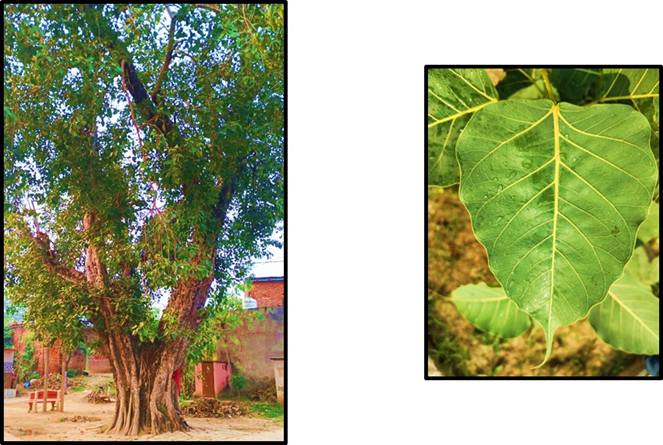
Figure No.1: Morphological description of Ficus religiosa

Botanical Classification and Traditional Uses

Ficus religiosa L., commonly known as the Peepal tree, sacred fig, or Bodhi tree, is a member of the family Moraceae. It is a tall deciduous tree, reaching 30 m (98 ft), with characteristic heart-shaped leaves with long petioles. The plant is Balkan in origin and widely naturalized in the Indian subcontinent, in SE Asia, Nepal and Sri Lanka. 1 In folk medicinal systems, like Ayurveda, Siddha and Unani, F. religiosa too has been considered sacred, with an application of the leaves, bark, fruits, and even the roots in healing practices (to note – every part of the tree is used therapeutically, not just its leaves) [11].

Ethnobotanical studies show that many tribal groups in India and F. religiosa in the prep- aeration of a number of common and chronic day-to-day disease such as diabetes mellitus, bronchial asthma, cough, diarrhoea, wounds, epilepsy and inflammatory disorders [12]. The bark and leaf decoction are considered as “prameha nashaka” (an antidiabetic agent) and it is said to contribute to the control of blood sugar levels and rejuvenate the functioning of the pancreas in ayurvedic scripts. According to traditional medicine, leaf extracts are also used in wound healing and for treatment of gastric discomforts due to their astringent and cooling effects [13]. In addition, the latex is applied externally to treat skin diseases and ulcers, and the fruits have laxative and digestive properties. In addition to its use in medicine F. religiosa holds great cultural and spiritual value—for it under this very tree that Gautama Buddha achieved enlightenment, and so is it venerated in South and Southeast Asia. [14].

Phytochemical Composition

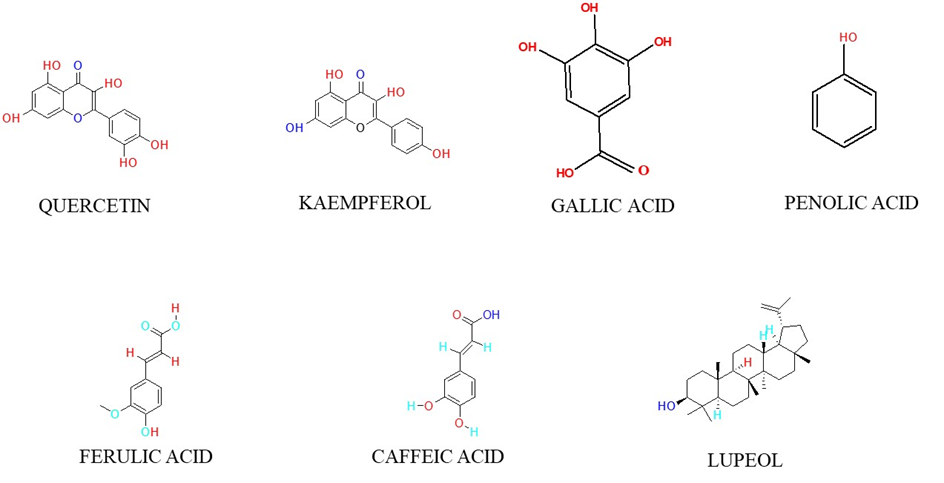
Phytochemical studies of F. religiosa reveal that it contains a plethora of bioactive secondary metabolites belonging to different chemicals classes. The plant contains flavonoids, phenolic acids, tannins, alkaloids, glycosides, sterols and saponins contributing to the wide range of pharmacological activities [15].

The antioxidant and antidiabetic activities of the plant are largely attributed to its flavonoid fraction, including quercetin, kaempferol and rutin. These bioactive compounds scavenge free radicals, increase cellular glucose uptake, and improve β-cell functions [16]. Phenolic acids like gallic acid, ferulic acid and caffeic acid have potent reducing power and they influence the activity of key enzymes in carbohydrate metabolism. Tannins and sterols like β-sitosterol, lupeol and stigmasterol are the major anti-inflammatory and lipid lowering agents [17].
3. Reported Pharmacological Activities
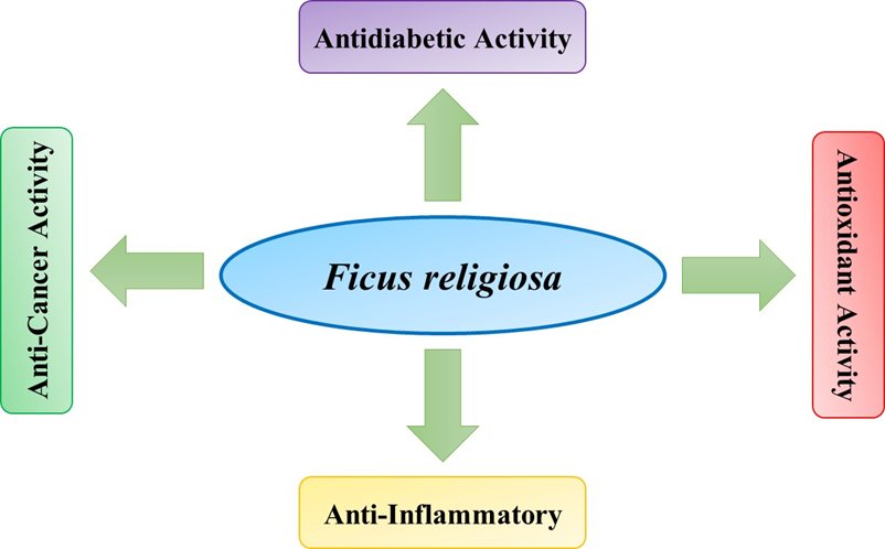
1. Antidiabetic Activity

Numerous pharmacological investigations done on F. religiosa confers antidiabetic activity. In vitro and in vivo experiments reveal that the extracts of this plant and the active principles act by several mechanisms of action, among them are: stimulation of secretion of insulin, increase of the peripheral capture of glucose, inhibition of the α- amylase and α-glucosidase enzymes and defence of the pancreatic β-cell from oxidative stress [18].

In an investigation by Kirana et al. (2009), methanolic extract of leaf of F. religiosa exhibited potent blood glucose lowering activity in streptozotocin diabetic rats by improving insulin sensitivity and antioxidant enzymes activity [19]. In the same way, the aqueous extract of the bark inhibited the digestive enzymes that break down carbohydrates, which inhibits postprandial hyperglycaemia [15]. Also, phytoconstituents such as quercetin and lupeol have been established to modulate AMP-activated protein kinase (AMPK) pathways and enhance expression of GLUT-4 in muscle cells which plays an integral role in glucose uptake and homeostasis [20]. Overall, these studies validate the assertion of traditional use for F. religiosa for having a strong antidiabetic effect.

2. Antioxidant Activity

Oxidative stress is the inherent defect and a key player in diabetes and its complications. The antioxidative effect of F. religiosa has been well confirmed in various radical scavenging assay like DPPH, ABTS, FRAP, nitric oxide scavenging assay etc [21]. Leaf and bark extract have high total phenolics content (TPC) and total flavonoid content (TFC), as well as the strongest positive correlation with antioxidant activity.

Ethyl Acetate Fraction of F. religiosa leaf demonstrate more than 80% DPPH radical scavenging activity at a concentration of 100 µg/mL similar to ascorbic acid 22 according to Baliyan (2022). This antioxidant activity contributes to scavenging of free radicals, prevention of oxidative damage to pancreatic β-cells, and decrease of lipid peroxidation under diabetic status. The antioxidative defense mechanism was mainly credited to the combination of gallic acid, quercetin, catechin and ferulic acid derivatives, seemingly working synergistically against oxidative stress [23].

3. Anti-Inflammatory and Other Pharmacological Activities

Chronic inflammation is a key determinant in the development of diabetes and its metabolic complications. F. religiosa extracts have been reported to possess significant anti-inflammatory activity by suppressing the expression of pro-inflammatory cytokines TNF-α, IL-6 and COX-2. In vitro studies demonstrated that methanolic bark extract inhibit production of nitric oxide in LPS activated macrophages with an implication of modulating inflammatory cascades [24].

The modulation of oxidative and inflammatory pathways by the bioactive components in this plant make it a candidate for new drug development. This prospective, though, should be amplified when the plant is incorporated in nanotechnological formulations-based delivery systems, like TPGS loaded nanoparticles, that would improve its bioavailability and focused effectiveness of its phytoconstituents bestowing a 21st century platform to plant based therapeutics [25].

4. Nano formulations in Herbal Antidiabetic Therapy

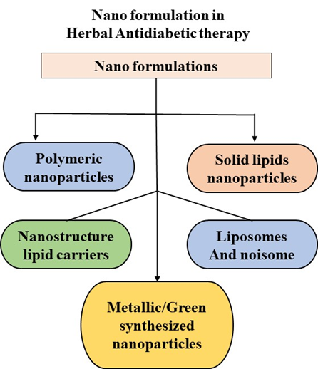
4.1. Overview— what “nano formulation” means Nano formulations are 12 to 100 nm in size. Nanocarriers are drug delivery systems, in which bioactive molecules (pure compounds or crude extracts) are physically or chemically integrated into nanostructure or attached on the surface of nanostructure to enhance the physicochemical and biopharmaceutical properties of the molecules. The platforms most commonly used for formulation of plant derived anti diabetic agents are: polymeric nanoparticles (e.g., PLGA, chitosan), solid lipid nanoparticles (SLNs) and nanostructured lipid carriers (NLCs), liposomes and niosomes, nano micelles (including polymeric micelles), nano emulsions/SMEDDS (self-micro emulsifying drug delivery systems), and metallic/green-synthesized nanoparticles. Each platform offers a distinct combination of ability to encapsulate, release kinetics, stability, and approved route of administration (oral, parenteral, topical, nasal) [26], [27].
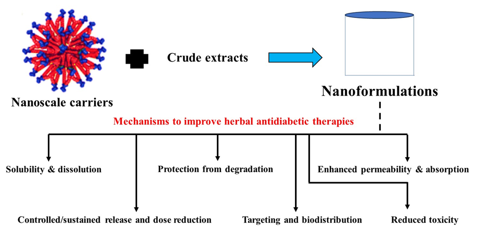
Figure No.2

4.2. Why nano formulations improve herbal antidiabetic therapies — mechanisms and advantages
  • Solubility & dissolution: Several antidiabetic phytoconstituents (curcumin, quercetin, many terpenes and sterols) have poor water solubility. Nanosizing and encapsulation (micelles, lipid carriers, SMEDDS) enhance the apparent solubility and dissolution rate, leading to higher drug concentrations at the site of absorptive mucosa and allowing more rapid and complete systemic absorption [28].
  • Protection from degradation: Encapsulation protects sensitive phytochemicals from acidic gastric pH, enzymatic hydrolysis and oxidative degradation, allowing retention of active content during the passage through the gastrointestinal tract as well as during storage [29].
  • Enhanced permeability & absorption: Nanocarriers can increase intestinal uptake by (i) increasing the concentration gradient at the epithelial surface, (ii) mediating transcellular transport (e.g., via endocytosis), and (iii) inhibiting efflux transporters (some excipients like TPGS inhibit P-gp). These effects translate into higher C_{max} and AUC in pharmacokinetic studies of nano-herbal formulations [30].
  • Controlled/sustained release and dose reduction: The matrix-based systems (PLGA, SLNs, NLCs) can provide controlled release, which may minimize peak-trough fluctuations and frequency of administration, which may be of benefit in chronic conditions, e.g., diabetes. Sustained release also provides for prolonged antioxidant/anti-inflammatory exposure, which has been shown to be relevant for β-cell protection [31].
  • Targeting and biodistribution: surface decoration (with PEGylation, ligand conjugation, etc.) can enhance circulation time and also guide nano formulations to target tissues (liver, pancreas, adipose). This can lead to the accumulation of phytochemicals at sites of action on glucose metabolism (such as AMPK, GLUT-4, or the insulin signalling pathways) [32].
  • Reduced toxicity and improved efficacy: Several nano formulations allow one to apply lower doses of phytochemicals thanks to enhanced delivery efficiency while achieving at least the same positive effect, which results in reduced off-target toxicity [33].

4.3. Representative, recent real-data examples (selected studies)

Table No.1: Representative and recent studies showing the enhancement of bioavailability and therapeutic efficacy of plant-based compounds through various nano formulation approaches, including SMEDDS, polymeric micelles, and solid lipid nanoparticles.

No.

Formulation / Study

Key Findings

Outcome / Significance

Reff.

1

Berberine SMEDDS (oral; rat PK)

Optimized berberine HCl SMEDDS (oil + surfactant + co-solvent) formed 47.2 ± 0.10 nm droplets, 93.1 ± 2.3 % release in 300 min, and increased AUC 1.63-fold vs tablets; C??? rose from 0.55 ± 0.06 to 1.15 ± 0.05 µg/mL.

Lipid-based self-emulsifying systems markedly enhance oral bioavailability of poorly absorbed alkaloids.

[34].

2

Curcumin polymeric micelles containing TPGS

P123:F127 micelles (~15–22 nm, narrow PDI) achieved EE 25–27 %; TPGS improved solubilization and stability; sustained in-vitro release vs free curcumin.

Demonstrates that TPGS stabilizes ultra-small polymeric micelles and enhances curcumin solubility.

[35].

3

Curcumin nanoparticles (in vivo antidiabetic)

In dexamethasone-induced metabolic disorder in rats, nano-curcumin reduced FBG ≈ 35–40 %, HOMA-IR ↓ 40 %, antioxidant markers ↑ 25–45 %; better hepatic & pancreatic histology.

Nanocurcumin shows superior antidiabetic and antioxidant efficacy vs unformulated curcumin.

[36].

4

Myricitrin solid-lipid nanoparticles (SLNs)

Myricitrin SLNs improved β-cell function and glycaemic control in STZ–nicotinamide diabetic mice vs free compound.

Confirms lipid nanoparticles improve flavonoid delivery and antidiabetic activity.

[37].

  1. Berberine SMEDDS (orally; rat PK). A 2024 report formulated berberine hydrochloride SMEDDS (oil + surfactant + co-solvent). The resulting optimized SMEDDS exhibited a droplet size of 47.2 ± 0.10 nm, released 93.1 ± 2.3% in simulated intestinal fluid after 300 min, and enhanced the oral bioavailability (AUC) by 1.63-fold in comparison with commercial tablets; Cmax increased from 0.55 ± 0.06 μg/mL to 1.15 ± 0.05 μg/mL. These findings demonstrate that a lipid-based self-emulsifying formulation can significantly improve the systemic exposure of a plant alkaloid that has poor oral absorption [34].
  2. Physicochemical characterization of curcumin polymeric micelles with TPGS. The formed P123:F127 polymeric micelles (PMs) loaded with curcumin exhibited particle sizes of ~15–22 nm with low polydispersity index (PDI) and a loading efficiency (EE%) of ~25–27% for the best batches; lyophilized micelles preserved ~20–27% loading after reconstitution, and the release profile was sustained in vitro in comparison with free curcumin. The addition of TPGS enhanced solubilization and promoted the stabilization of exceedingly small micellar formations (e.g., PFT, size ≈ 16–17 nm; EE ≈ 25–27%) [35].
  3. Curcumin nanoparticles – better antidiabetic results in vivo. In a 2024 Wistar rat model of steroid (dexamethasone)–induced metabolic perturbation, nano-curcumin (100 mg/kg) elicited more pronounced improvements compared to unformulated curcumin (100 mg/kg): fasting blood glucose decreased by approximately 35–40%, insulin resistance (HOMA-IR) dropped by about 40%, antioxidant indices (GSH, SOD) were raised by 25–45%, and liver/pancreas histology revealed markedly superior preservation in nano-curcumin. These results indicate that improving curcumin bioavailability through nanoparticles yields more pronounced functional effects on glycaemia, lipid profile, and tissue oxidative stress [36].
  4. Myricitrin solid lipid nanoparticles (SLNs) — effect on antioxidant and antidiabetic activities. Myricitrin-loaded SLNs induced antioxidant and antidiabetic activities in a streptozotocin–nicotinamide diabetic mouse model, demonstrating better β-cell function and glycaemic control than free myricitrin. This substantiates the applicability of lipid nanoparticles for flavonoid delivery [37].

4.4. Summary of evidence & practical implications for herbal antidiabetic development

The literature unanimously suggests that nanoscale delivery systems transform the promising but pharmacokinetically challenged phytochemicals into candidates with significantly enhanced systemic exposure and better efficacy in animal models of diabetes. The three above-mentioned examples illustrate three practical, measurable improvements that can be expected across platforms: (i) reduction in size scaled down to the order of nanometres (often < 100 nm) that improves absorption and penetration across tissues; (ii) encapsulation that improves chemical stability and can provide for controlled delivery; and (iii) ‘smart' excipients (such as TPGS) that solubilize and modulate transporters (e.g., inhibit the efflux transporter P-gp) to enhance apparent oral bioavailability. Collectively, these merits provide a compelling basis for considering TPGS-based and other nano formulation approaches for Ficus religiosa extracts and their bioactive phytoconstituents (quercetin, gallic acid, lupeol, etc.) to combat the poor solubility/stability that hinders clinical translation [38], [39].

4.5. Practical notes for researchers (formulation choices)

  • If the target is a hydrophobic flavonoid (e.g., quercetin, lupeol): consider SLNs/NLCs, polymeric micelles, or SMEDDS for oral delivery. These systems have consistently yielded small droplet/particle sizes (<50-200 nm) and enhancement of dissolution [40].
  • For chemicals susceptible to oxidation (e.g., phenolic acids): Antioxidant-containing lipid carriers or micelles such as TPGS protect the cargo and act as permeation enhancers [41].
  • Use TPGS when you need combined solubilization + P-gp inhibition: it is an FDA-approved adjuvant and has been applied in mixed micelles or as a layer to enhance both encapsulation and absorption. Physicochemical characterization (size, PDI, zeta potential) and in-vitro/in-vivo pharmacokinetics are required in early development to prove the anticipated benefits [42].

5. Role of TPGS in Nano formulations

D-α-Tocopheryl polyethylene glycol 1000 succinate (TPGS) is a water-soluble form of vitamin E, prepared by esterification of α-tocopherol succinate with polyethylene glycol 1000. This amphiphilic structure provides hydrophilic (PEG chain) and lipophilic (vitamin E) domains, enabling it to self-assemble at the very low critical micelle concentrations (CMC ≈ 0.02 wt %) into stable nanoscale aggregates [43].

Chemical Nature and Multifunctional Properties

  1. Surfactant and Solubilizer:

TPGS has a hydrophilic lipophilic balance (HLB) of approximately 13, which is considered high and indicative of hydrophilicity, and this value makes TPGS an effective non-ionic surfactant capable of solubilizing poorly water-soluble bioactive like curcumin, berberine, and quercetin. Its PEG portion contributes to solubility in aqueous medium and the tocopherol moiety associates with lipid bilayers and hydrophobic drug cores thus increasing drug loading and physical stability [43], [44].

  1. Antioxidant:

The α-tocopherol moiety delivered by phytosomes has its own free-radical-scavenging activity, thus it safeguards the delicate phytoconstituents from oxidation both during storage and in biological environments. This attribute enhances the stability of the formulation and adds to the synergistic effect of pharmacological action, more so in cases of diseases of oxidative stress such as diabetes [43].

  1. P-gp (P-glycoprotein) Inhibition:

TPGS has been shown to inhibit ATPase activity of P-gp, a key efflux transporter limiting intestinal and cellular drug uptake. This mechanism enhances intracellular accumulation and systemic exposure of substrates that are otherwise rapidly expelled from enterocytes or tumor cells [43].

Enhancement of Permeability and Bioavailability

TPGS facilitates multiple bioavailability-enhancing mechanisms:

  • Improves drug solubilization in gastrointestinal fluids by forming mixed micelles and emulsions.
  • Promotes transcellular permeability by transiently modifying lipid membrane fluidity.
  • Supports lymphatic absorption, bypassing first-pass metabolism.
  • Provides controlled release through micellar and nanoparticle matrices, maintaining steady drug levels.

In polymeric micelle systems, for instance, incorporation of TPGS with Pluronic F127/P123 markedly improved solubilization and sustained release of curcumin. Yusuf et al. reported micelles of 15–22 nm size, encapsulation efficiency ≈ 25–27 %, and > 40-fold solubility increase, confirming TPGS’s critical role in forming stable nano systems [44].

Similarly, in Hamed et al.’s 2024 in vivo study on dexamethasone-induced diabetic rats, nano-curcumin (TPGS-stabilized) reduced fasting glucose by ≈ 37 %, improved HOMA-IR by ≈ 42 %, and significantly enhanced hepatic antioxidant enzymes, outperforming unformulated curcumin [45].

Table No.2: Examples of TPGS-Based Nanoparticles:

Formulation

Disease / Target

Nanocarrier Type

Key Findings

Citation

TPGS-Curcumin Polymeric Micelles

Colorectal cancer

P123/F127/TPGS micelles (≈ 17 nm)

~40× solubility increase; sustained 48 h release; improved cytotoxicity vs free curcumin

[44].

TPGS-Nano-Curcumin

Dexamethasone-induced diabetes

Nanoparticles (100 mg/kg)

↓ FBG ≈ 37 %, ↓ HOMA-IR ≈ 42 %, ↑ GSH/SOD ≈ 40 %; improved hepatic histology

[45].

TPGS-PLGA Nanoparticles (Curcumin)

Breast cancer

Polymeric nanoparticles

Enhanced uptake via P-gp inhibition and improved oral bioavailability

[43].

TPGS-Quercetin Micelles

Antioxidant therapy

Polymeric micelles

Higher dissolution rate and oral AUC (↑ 2.5-fold) vs free quercetin

[44].

6. TPGS-Based Nano formulations of Plant Extracts for Antidiabetic Therapy

TPGS has been used as a main or co-excipient in nano formulations aimed to deliver phytoconstituents with antidiabetic activity. The balance of surfactant/solubilizing effect of TPGS, its antioxidant potential, and its P-pg inhibitory activity results in better physical/chemical stability, better intestinal absorption and often quantifiable enhancements in systemic exposure and pharmacodynamics (glucose lowering, antioxidant biomarkers, insulin sensitivity) compared to non-formulated plant actives.

Summary of key examples and pharmacokinetic/ pharmacodynamic outcomes

Curcumin (TPGS-containing polymeric micelles / nanoparticles).

Cerqueira et al. formulated curcumin in TPGS/Pluronic F127/P123 polymeric micelles (mean diameter ≈ 15–22 nm; EE ≈ 25–27%) and demonstrated markedly improved aqueous solubility and sustained release vs. free curcumin; when similar TPGS-stabilized curcumin nanoparticles were tested in vivo they produced significantly greater glycaemic and antioxidant benefits (e.g., reductions in fasting blood glucose and improvements in hepatic antioxidant enzymes) than unformulated curcumin. These improvements are attributed to higher oral absorption and prolonged systemic exposure afforded by TPGS-containing micellar systems [46].

Berberine (SMEDDS; TPGS often used in mixed surfactant systems).

Chen et al. developed a berberine hydrochloride SMEDDS (Capmul MCM / Kolliphor RH40 / propanediol) achieving droplet size 47.2 ± 0.10 nm, 93.1 ± 2.3% release in simulated intestinal fluid over 300 min, and a 1.63-fold increase in oral AUC (and corresponding increase in Cmax) versus conventional tablets in rats. While this specific SMEDDS did not use TPGS, mixed surfactant SMEDDS formulations commonly substitute or include TPGS to provide additional solubilization and P-gp inhibition; inclusion of TPGS in berberine nano systems has been reported to further improve oral absorption in comparable systems [43], [47].

Quercetin (TPGS-based micelles / lipid nanoparticles / polymeric NPs).

Recent reviews and comparative studies show that quercetin nano formulations (micelles, lipid nanoparticles, polymeric nanoparticles) that incorporate TPGS or TPGS-containing excipient blends consistently increase oral exposure (reported AUC increases commonly in the ~1.5–3.0× range depending on the platform), improve in-vivo antioxidant endpoints, and lower hyperglycaemia in animal models vs free quercetin. Reported particle sizes vary by platform (typical 50–200 nm) and EE% is formulation-dependent; see summaries in Tomou et al. and Rathod et al. [48], [49].

Flavonoid (myricitrin) SLNs (lipid-based carriers + stabilizers).

Ahangarpour et al. formulated myricitrin in solid-lipid nanoparticles and demonstrated significant antioxidant and antidiabetic effects in streptozotocin–nicotinamide diabetic mice compared with free myricitrin (improvement in β-cell function, glycaemic indices and oxidative stress markers). Lipid carriers can be combined with TPGS (as stabilizer/co-surfactant) to obtain additional permeability enhancement and oxidative protection. [50], [43].

Mechanistic link to improved glucose control

Improved pharmacokinetics (higher Cmax and AUC, sustained plasma concentrations) and tissue availability of phytoconstituents lead to stronger engagement of antidiabetic mechanisms — e.g., enhanced AMP-activated protein kinase (AMPK) activation (berberine), increased GLUT-4 expression and peripheral glucose uptake (flavonoids), antioxidant protection of β-cells (curcumin, myricitrin) and inhibition of carbohydrate-digesting enzymes. TPGS contributes not only by raising systemic exposure but also by protecting actives from oxidative degradation and reducing efflux via P-gp inhibition — all of which translate into larger, reproducible glucose-lowering and insulin-sensitizing effects in preclinical models.

Table No.3: TPGS-based (and closely related) nano formulations of plant extracts with antidiabetic relevance — recent, representative examples

Plant / Phytoconstituent

Nanoparticle / System

Particle size (reported)

Encapsulation efficiency (EE%) / Release

Key outcome (PK / PD)

Ref.

Curcumin (TPGS/ F127/ P123 polymeric micelles)

Polymeric micelles (TPGS + Pluronic F127/P123)

15–22 nm (mean).

EE ≈ 25–27% (retained ≈20–27% after lyophilization); sustained release ~48 h.

Marked solubility increase (>40×); improved in vitro release and enhanced cytotoxic/therapeutic efficacy in models; improved glucose/ antioxidant endpoints in TPGS-stabilized curcumin NP studies.

[45], [46].

Curcumin (nano-formulation, in vivo)

Nanoparticles (TPGS-stabilized examples reported)

platform dependent (typically 80–200 nm)

platform dependent

In dexamethasone-induced Wistar rats: nano-curcumin (100 mg/kg) ↓ fasting glucose ≈ 35–40%, ↓ HOMA-IR ≈ 40%; ↑ GSH/SOD ≈ 25–45% vs free curcumin.

45

Berberine HCl

SMEDDS (lipid-based self-micro-emulsifying system; TPGS often used in similar systems)

47.2 ± 0.10 nm (Chen et al., optimized SMEDDS)

Release: 93.1 ± 2.3% in SIF (300 min); EE not applicable for classical SMEDDS

PK: 1.63× increase in oral AUC; Cmax increased (0.55 → 1.15 µg/mL in rat PK). Improved oral exposure linked to greater pharmacodynamic efficacy in metabolic models.

[43], [47].

Quercetin

TPGS-containing micelles / lipid NPs / polymeric NPs (various studies summarized in reviews)

Reported range ≈ 50–200 nm (platform dependent)

Reported range EE ≈ 30–85% (platform dependent)

Representative reports show oral AUC increases commonly between ~1.5–3.0×; improved antioxidant and glucose-lowering effects in animal studies.

[48], [49].

Myricitrin (flavonoid)

Solid Lipid Nanoparticles (SLNs) — lipid carrier + stabilizer

Reported in SLN studies (platform dependent)

Reported in SLN study

Antioxidant and antidiabetic effects in STZ–nicotinamide diabetic mice; improved β-cell function and glycaemic control vs free myricitrin.

[50].

7. Potential of TPGS-Based Nano formulations of Ficus religiosa

Extracts of Ficus religiosa are rich in polyphenols, flavonoids, and sterols that exhibit multiple pharmacological effects, including antioxidant, anti-inflammatory, and antidiabetic properties. However, their clinical translation is hampered by poor solubility, instability in biological fluids, and low membrane permeability. Incorporating D-α-tocopheryl polyethylene glycol succinate (TPGS) into nanocarrier systems offers a rational strategy to address these challenges. TPGS functions both as a solubilizer and as a bioavailability enhancer by suppressing P-glycoprotein-mediated efflux and protecting bioactive from oxidative damage [51], [52].

Nano formulation platforms such as TPGS-stabilized solid lipid nanoparticles, PLGA/TPGS polymeric nanoparticles, and TPGS-based nano emulsions can be optimized for F. religiosa leaf or bark extracts. These carriers provide sustained drug release, greater antioxidant protection, and better pharmacological response than conventional formulations. Given that no extensive TPGS-based studies on F. religiosa currently exist, this area presents a significant research opportunity for developing effective plant-derived antidiabetic nanomedicines [53].

8. Mechanistic Insights

TPGS-assisted nano formulations can enhance the biological performance of F. religiosa through several interconnected mechanisms:

  • Enhanced intestinal uptake: TPGS inhibits P-gp efflux transporters, allowing more phytoconstituents to pass through the intestinal barrier.
  • Protection from oxidative degradation: The antioxidant nature of TPGS minimizes degradation of sensitive flavonoids during absorption and systemic circulation.
  • Targeted biodistribution: Nanocarriers facilitate accumulation in glucose-regulating organs such as the pancreas and liver, promoting β-cell protection.
  • Controlled and sustained release: The amphiphilic polymer matrix regulates diffusion and maintains therapeutic plasma concentrations over longer periods [54], [55].

9. Future Prospects and Challenges

Although preclinical findings are promising, the practical application of TPGS-based F. religiosa nano formulations remains at an early stage. Key issues include ensuring batch-to-batch reproducibility, establishing long-term safety, and addressing regulatory classification between herbal and nano pharmaceutical categories. Moreover, scale-up feasibility and cost-effectiveness are crucial for commercial translation. Future investigations should focus on comprehensive in vivo pharmacokinetic modelling, toxicological profiling, and clinical trials to substantiate the therapeutic potential in diabetic patients [56].

CONCLUSION

The integration of D-α-tocopheryl polyethylene glycol succinate (TPGS) into nanocarrier systems represents a transformative advancement in herbal drug delivery, particularly for antidiabetic phytoconstituents. TPGS combines surfactant, solubilizing, antioxidant, and P-gp inhibitory properties that collectively improve drug permeability, stability, and systemic bioavailability.

Preclinical studies involving plant-derived actives such as curcumin, quercetin, berberine, and myricitrin have demonstrated superior glucose-lowering and antioxidant outcomes when encapsulated in TPGS-based nanoparticles. However, for Ficus religiosa, data remain sparse, indicating a promising research gap. Considering the rich phytochemical composition of F. religiosa—notably flavonoids, tannins, and sterols—its encapsulation using TPGS-stabilized polymeric nanoparticles, solid lipid nanoparticles, or nano emulsions could significantly enhance solubility, protect bioactive from degradation, and ensure sustained release.

Mechanistically, TPGS-enabled nanocarriers can facilitate intestinal absorption by inhibiting P-gp efflux transporters, protect β-cells through antioxidant mechanisms, and maintain steady plasma concentrations that support continuous glucose regulation. Future research should prioritize formulation optimization, scalability, long-term toxicity evaluation, and translational studies to bridge the gap between laboratory efficacy and clinical application.

In conclusion, TPGS-based nano formulations hold substantial potential to redefine phytopharmaceutical strategies for diabetes management. With further in-vivo validation and regulatory harmonization, these systems may pave the way toward safe, efficient, and clinically viable herbal nanomedicines.

REFERENCES

  1. K. L. Ong., “Global, regional, and national burden of diabetes from 1990 to 2021, with projections of prevalence to 2050: a systematic analysis for the Global Burden of Disease Study 2021,” The Lancet, Jul. 2023; 402(10397): 203–234. doi: 10.1016/S0140-6736(23)01301-6.
  2. J. Ye., “The global, regional and national burden of type 2 diabetes mellitus in the past, present and future: a systematic analysis of the Global Burden of Disease Study 2019,” Front. Endocrinol., Jul. 2023; 14: 1192629. doi: 10.3389/fendo.2023.1192629.
  3. Chamine, J. Hwang, S. Valenzuela, M. Marino, A. E. Larson, J. Georgescu, M. Latkovic-Taber, H. Angier, J. E. DeVoe, and N. Huguet, “Acute and chronic diabetes-related complications among patients with diabetes receiving care in community health centers,” Diabetes Care, vol. 45, no. 10, pp. e141–e143, Oct. 2022, doi: 10.2337/dc22-0420.
  4. Gieroba, A. Kryska, and A. Sroka-Bartnicka, “Type 2 diabetes mellitus – conventional therapies and future perspectives in innovative treatment,” Biochem. Biophys. Rep., vol. 42, p. 102037, May 2025, doi: 10.1016/j.bbrep.2025.102037.
  5. A. Chaudhury et al., “Clinical review of antidiabetic drugs: Implications for type 2 diabetes mellitus management,” Front. Endocrinol. (Lausanne), vol. 8, p. 6, Jan. 2017, doi: 10.3389/fendo.2017.00006.
  6. N. Tran, B. Pham, and L. Le, “Bioactive compounds in anti-diabetic plants: From herbal medicine to modern drug discovery,” Biology (Basel), vol. 9, no. 9, p. 252, Aug. 2020, doi: 10.3390/biology9090252.’
  7. T. Behl et al., “Alkaloidal phytoconstituents for diabetes management: Exploring the unrevealed potential,” Plants (Basel), vol. 11, no. 19, p. 2560, Sep. 2022, doi: 10.3390/plants11192560.
  8. Y. Lv, W. Li, W. Liao, H. Jiang, Y. Liu, J. Cao, W. Lu, and Y. Feng, “Nano-drug delivery systems based on natural products,” Int. J. Nanomedicine, vol. 19, pp. 541–569, Jan. 2024, doi: 10.2147/IJN.S443692.
  9. F. H. Awlqadr et al., “Nanotechnology-based herbal medicine: Preparation, synthesis, and applications in food and medicine,” J. Agric. Food Res., vol. 14, p. 101661, 2025, doi: 10.1016/j.jafr.2025.101661.
  10. C. N. Wong, S.-K. Lee, Y. M. Lim, S.-B. Yang, Y.-L. Chew, A.-L. Chua, and K. B. Liew, “Recent advances in vitamin E TPGS-based organic nanocarriers for enhancing the oral bioavailability of active compounds: A systematic review,” Pharmaceutics, vol. 17, no. 4, p. 485, Apr. 2025, doi: 10.3390/pharmaceutics17040485.
  11. R. Rawat, H. Kumar, N. Singh, A. Deep, B. Narasimhan, S. Y. Singh, and S. Kumar, “Comprehensive review on ethnomedicinal, phytochemistry and pharmacological profile of Ficus religiosa L.,” J. Tradit. Chin. Med., vol. 44, no. 5, pp. 1052–1057, Sep. 2024, doi: 10.19852/j.cnki.jtcm.2024.05.012.
  12. L. R. Ralte, H. Sailo, and Y. T. Singh, “Ethnobotanical study of medicinal plants used by the indigenous community of the western region of Mizoram, India,” J. Ethnobiol. Ethnomed., vol. 20, p. 2, Jan. 2024, doi: 10.1186/s13002-023-00642-z.
  13. P. Singh, V. Singh, R. C. Tiwari, and D. Negi, “Dhava (Anogeissus latifolia): Axle wood from the treasure of Ayurveda,” J. Ayurveda Integr. Med. Sci., vol. 8, no. 6, pp. 174–183, Jul. 2023, doi: 10.21760/jaims.8.6.28.
  14. S. B. Chandrasekar, M. Bhanumathy, A. T. Pawar, and T. Somasundaram, “Phytopharmacology of Ficus religiosa,” Pharmacogn. Rev., vol. 4, no. 8, pp. 195–199, Jul.–Dec. 2010, doi: 10.4103/0973-7847.70918.
  15. S. Murugesu, J. Selamat, and V. Perumal, “Phytochemistry, pharmacological properties, and recent applications of Ficus benghalensis and Ficus religiosa,” Molecules, vol. 27, no. 1, p. 144, Dec. 2021, doi: 10.3390/molecules27010144.
  16. A. N. Panche, A. D. Diwan, and S. R. Chandra, “Flavonoids: an overview,” J. Nutr. Sci., vol. 5, p. e47, Dec. 2016, doi: 10.1017/jns.2016.41
  17. S. Bakrim et al., “Health benefits and pharmacological properties of stigmasterol,” Antioxidants (Basel), vol. 11, no. 10, p. 1912, Sep. 2022, doi: 10.3390/antiox11101912.
  18. R. Pandit, A. S. Phadke, and A. G. Jagtap, “Antidiabetic effect of Ficus religiosa extract in streptozotocin-induced diabetic rats,” J. Ethnopharmacol., vol. 128, no. 2, pp. 462–466, Mar. 2010, doi: 10.1016/j.jep.2010.01.025.
  19. H. Kirana, S. S. Agrawal, and B. Srinivasan, “Aqueous extract of Ficus religiosa Linn. reduces oxidative stress in experimentally induced type 2 diabetic rats,” Indian J. Exp. Biol., vol. 47, no. 10, pp. 822–826, Oct. 2009.
  20. A. Katrolia, V. Pal, V. K. Shukla, and R. Singh, “Exploring the therapeutic potential of herbal plants in managing blood sugar levels: A comprehensive evaluation,” Pharmacogn. Res., vol. 16, no. 1, pp. 10–18, Jan.–Mar. 2024, doi: 10.5530/pres.16.1.2.
  21. G. S. Thakur, R. Sharma, A. Kaur, S. Dhiman, and R. Singh, “Evaluation of renoprotective potential of Ficus religiosa in attenuation of diabetic nephropathy in rats,” Obes. Med., vol. 18, p. 100268, 2020, doi: 10.1016/j.obmed.2020.100268.
  22. S. Baliyan, R. Mukherjee, A. Priyadarshini, A. Vibhuti, A. Gupta, R. P. Pandey, and C.-M. Chang, “Determination of antioxidants by DPPH radical scavenging activity and quantitative phytochemical analysis of Ficus religiosa,” Molecules, vol. 27, no. 4, p. 1326, Feb. 2022, doi: 10.3390/molecules27041326.
  23. S. Ghadigaonkar, A. G. Reddy, B. Kalakumar, and L. Mekala, “Quantification of total phenolic content, total flavonoid content and evaluation of in vitro free radical scavenging activities in Ficus religiosa Linn,” J. Pharm. Res., Jan. 2021.
  24. C. Andrade, F. Ferreres, N. G. M. Gomes, S. Duangsrisai, N. Srisombat, S. Vajrodaya, D. M. Pereira, A. Gil-Izquierdo, P. B. Andrade, and P. Valentão, “Phenolic profiling and biological potential of Ficus curtipes Corner leaves and stem bark: 5-lipoxygenase inhibition and interference with NO levels in LPS-stimulated RAW 264.7 macrophages,” Biomolecules, vol. 9, no. 9, p. 400, Aug. 2019, doi: 10.3390/biom9090400.
  25. N. Parvin, M. Aslam, S. W. Joo, and T. K. Mandal, “Nano-phytomedicine: Harnessing plant-derived phytochemicals in nanocarriers for targeted human health applications,” Molecules, vol. 30, no. 15, p. 3177, Jul. 2025, doi: 10.3390/molecules30153177.
  26. E.-M. Tomou, P. Papakyriakopoulou, E.-M. Saitani, G. Valsami, N. Pippa, and H. Skaltsa, “Recent Advances in Nano formulations for Quercetin Delivery,” Pharmaceutics, vol. 15, no. 6, p. 1656, Jun. 2023, doi: 10.3390/pharmaceutics15061656.
  27. P. Kumar, B. Mangla, S. Javed, W. Ahsan, P. Musyuni, D. Sivadasan, S. S. Alqahtani, and G. Aggarwal, “A review of nanomaterials from synthetic and natural molecules for prospective breast cancer nano therapy,” Frontiers in Pharmacology, vol. 14, p. 1149554, May 2023, doi: 10.3389/fphar.2023.1149554.
  28. S. Dey, P. Sow, R. Dey, P. Guptaroy, S. Nandi, and A. Samadder, “Design, synthesis and therapeutic exploration of nano-curcumin targeting the synergistic interactions with p53 and PARP-1 proteins in preventing food-additive induced genotoxicity and diabetic complications,” Colloids and Surfaces A: Physicochemical and Engineering Aspects, vol. —, p. 136230, 2025, doi: 10.1016/j.colsurfa.2025.136230.
  29. V. M. Amador-Luna, M. Herrero, P. Mendoza-Orbaneja, et al., “Improving the long-term stability of carotenoids from Dunaliella salina supercritical fluid extracts with a new liposomal encapsulation based on cryogenic and stirring processes,” Food and Bioprocess Technology, vol. 18, pp. 6520–6536, 2025, doi: 10.1007/s11947-025-03820-x.
  30. R. Kumar, “Nanotechnology in herbal medicine: Challenges and future perspectives,” in Nanotechnology in Herbal Medicine: Applications and Innovations, Woodhead Publishing Series in Biomaterials, 1st ed., 2023, pp. 515–548, doi: 10.1016/B978-0-323-99527-6.00008-2.
  31. J. Majumder and T. Minko, “Multifunctional and stimuli-responsive nanocarriers for targeted therapeutic delivery,” Expert Opinion on Drug Delivery, vol. 18, no. 2, pp. 205–227, Feb. 2021, doi: 10.1080/17425247.2021.1828339.
  32. S. Sharma, A. Parmar, S. Kori, and R. Sandhir, “PLGA-based nanoparticles: A new paradigm in biomedical applications,” TrAC Trends in Analytical Chemistry, vol. 80, pp. 30–40, Jun. 2016, doi: 10.1016/j.trac.2015.06.014.
  33. S. Rathod, S. Arya, S. Kanike, S. A. Shah, P. Bahadur, and S. Tiwari, “Advances on nano formulation approaches for delivering plant-derived antioxidants: A case of quercetin,” International Journal of Pharmaceutics, vol. 625, p. 122093, Sept. 2022, doi: 10.1016/j.ijpharm.2022.122093.
  34. X. Chen, H. Yang, L. Shi, Y. Mao, L. Niu, J. Wang, H. Chen, J. Jia, J. Wang, J. Xue, Y. Shen, C. Zheng, Y. Tian, and Y. Zheng, “Self-microemulsifying drug delivery system to enhance oral bioavailability of berberine hydrochloride in rats,” Pharmaceutics, vol. 16, no. 9, p. 1116, Aug. 2024, doi: 10.3390/pharmaceutics16091116.
  35. R. Cerqueira, C. Domingues, F. Veiga, I. Jarak, and A. Figueiras, “Development and characterization of curcumin-loaded TPGS/F127/P123 polymeric micelles as a potential therapy for colorectal cancer,” International Journal of Molecular Sciences, vol. 25, no. 14, p. 7577, Jul. 2024, doi: 10.3390/ijms25147577.
  36. A. M. Hamed, D. A. Elbahy, A. R. H. Ahmed, S. A. Thabet, R. A. Refaei, I. Ragab, S. M. Elmahdy, A. S. Osman, and A. M. A. Abouelella, “Comparison of the efficacy of curcumin and its nano formulation on dexamethasone-induced hepatic steatosis, dyslipidemia, and hyperglycaemia in Wistar rats,” Heliyon, vol. 10, no. 24, p. e41043, Dec. 2024, doi: 10.1016/j.heliyon.2024.e41043.
  37. A. Ahangarpour, A. A. Oroojan, L. Khorsandi, M. Kouchak, and M. Badavi, “Solid lipid nanoparticles of myricitrin have antioxidant and antidiabetic effects on streptozotocin-nicotinamide-induced diabetic model and myotube cell of male mouse,” Oxidative Medicine and Cellular Longevity, vol. 2018, p. 7496936, Jul. 2018, doi: 10.1155/2018/7496936.
  38. T. Sahu, Y. K. Ratre, S. Chauhan, L. V. K. S. Bhaskar, M. P. Nair, and H. K. Verma, “Nanotechnology-based drug delivery system: Current strategies and emerging therapeutic potential for medical science,” Journal of Drug Delivery Science and Technology, vol. 63, p. 102487, Jun. 2021, doi: 10.1016/j.jddst.2021.102487.
  39. N. Akombaetwa, A. B. Ilangala, L. Thom, P. B. Memvanga, B. A. Witika, and A. B. Buya, “Current advances in lipid nano systems intended for topical and transdermal drug delivery applications,” Pharmaceutics, vol. 15, no. 2, p. 656, Feb. 2023, doi: 10.3390/pharmaceutics15020656.
  40. A. Dalimunthe, M. C. Gunawan, Z. D. Utari, M. R. Dinata, P. Halim, N. E. S. Pakpahan, et al., “In-depth analysis of lupeol: delving into the diverse pharmacological profile,” Frontiers in Pharmacology, vol. 15, p. 1461478, Nov. 2024, doi: 10.3389/fphar.2024.1461478.
  41. C. Yang, T. Wu, Y. Qi, and Z. Zhang, “Recent advances in the application of vitamin E TPGS for drug delivery,” Theranostics, vol. 8, no. 2, pp. 464–485, Jan. 2018, doi: 10.7150/thno.22711.
  42. P. S. Kumbhar, S. Nadaf, A. S. Manjappa, N. K. Jha, S. S. Shinde, S. S. Chopade, A. S. Shete, J. I. Disouza, U. Sambamoorthy, and S. A. Kumar, “D-α-tocopheryl polyethylene glycol succinate: A review of multifarious applications in nanomedicines,” OpenNano, vol. 6, p. 100036, Mar.–Apr. 2022, doi: 10.1016/j.onano.2022.100036.
  43. J. M. Tucker and D. M. Townsend, “Alpha-tocopherol: roles in prevention and therapy of human disease,” Biomed. Pharmacother., vol. 59, no. 7, pp. 380–387, Aug. 2005, doi: 10.1016/j.biopha.2005.06.005.
  44. O. Yusuf, R. Ali, A. H. Alomrani, A. Alshamsan, A. K. Alshememry, A. M. Almalik, A. Lavasanifar, and Z. Binkhathlan, “Design and development of D-α-tocopheryl polyethylene glycol succinate-block-poly(ε-caprolactone) (TPGS-b-PCL) nanocarriers for solubilization and controlled release of paclitaxel,” Molecules, vol. 26, no. 9, p. 2690, May 2021, doi: 10.3390/molecules26092690.
  45. A. M. Hamed, D. A. Elbahy, A. R. H. Ahmed, S. A. Thabet, R. A. Refaei, I. Ragab, S. M. Elmahdy, A. S. Osman, and A. M. A. Abouelella, “Comparison of the efficacy of curcumin and its nano formulation on dexamethasone-induced hepatic steatosis, dyslipidemia, and hyperglycemia in Wistar rats,” Heliyon, vol. 10, no. 24, e41043, Dec. 2024, doi: 10.1016/j.heliyon.2024.e41043.
  46. R. Cerqueira, C. Domingues, F. Veiga, I. Jarak, and A. Figueiras, “Development and characterization of curcumin-loaded TPGS/F127/P123 polymeric micelles as a potential therapy for colorectal cancer,” Int. J. Mol. Sci., vol. 25, no. 14, p. 7577, Jul. 2024, doi: 10.3390/ijms25147577.
  47. X. Chen et al., “Self-microemulsifying drug delivery system to enhance oral bioavailability of berberine hydrochloride in rats,” Pharmaceutics, vol. 16, no. 9, p. 1116, Aug. 2024, doi: 10.3390/pharmaceutics16091116.
  48. E.-M. Tomou et al., “Recent advances in nano formulations for quercetin delivery,” Pharmaceutics, vol. 15, no. 6, p. 1656, Jun. 2023, doi: 10.3390/pharmaceutics15061656.
  49. S. Rathod et al., “Advances on nano formulation approaches for delivering plant-derived antioxidants: A case of quercetin,” Int. J. Pharm., vol. 625, p. 122093, Sept. 2022, doi: 10.1016/j.ijpharm.2022.122093.
  50. A. Ahangarpour et al., “Solid lipid nanoparticles of myricitrin have antioxidant and antidiabetic effects on streptozotocin-nicotinamide-induced diabetic model and myotube cell of male mouse,” Oxid. Med. Cell. Longev., vol. 2018, p. 7496936, Jul. 2018, doi: 10.1155/2018/7496936.
  51. S. Murugesu, J. Selamat, and V. Perumal, “Phytochemistry, pharmacological properties, and recent applications of Ficus benghalensis and Ficus religiosa,” Molecules, vol. 27, no. 1, p. 144, Dec. 2021.
  52. P. S. Kumbhar, S. Nadaf, A. S. Manjappa, N. K. Jha, S. S. Shinde, S. S. Chopade, A. S. Shete, J. I. Disouza, U. Sambamoorthy, and S. A. Kumar, “D-α-tocopheryl polyethylene glycol succinate: A review of multifarious applications in nanomedicines,” OpenNano, vol. 6, p. 100036, Mar.–Apr. 2022, doi:10.1016/j.onano.2022.100036.
  53. D. Singh, B. Singh, and R. K. Goel, “Traditional uses, phytochemistry and pharmacology of Ficus religiosa: A review,” J. Ethnopharmacol., vol. 134, no. 3, pp. 565–583, Apr. 2011, doi: 10.1016/j.jep.2011.01.046.
  54. T. Liu, X. Liu, H. Xiong, C. Xu, J. Yao, X. Zhu, J. Zhou and J. Yao, “Mechanisms of TPGS and its derivatives inhibiting P-glycoprotein and application for reversing multidrug resistance in hepatocellular carcinoma,” Polymer Chemistry, vol. 9, pp. 1827-1839, 2018. doi:10.1039/C8PY00344K.
  55. C. N. Wong, S.-K. Lee, Y. M. Lim, S.-B. Yang, Y.-L. Chew, A.-L. Chua, and K. B. Liew, “Recent advances in vitamin E TPGS-based organic nanocarriers for enhancing the oral bioavailability of active compounds: A systematic review,” Pharmaceutics, vol. 17, no. 4, p. 485, 2025. doi:10.3390/pharmaceutics17040485.
  56. F. D. Rodríguez-Gómez et al., “The regulation of nanomaterials and nanomedicines for translation: current status, challenges, and outlook,” Biomaterials Science, vol. 8, no. 10, pp. 2654-2669, 2020. doi:10.1039/d0bm00558d.

Reference

  1. K. L. Ong., “Global, regional, and national burden of diabetes from 1990 to 2021, with projections of prevalence to 2050: a systematic analysis for the Global Burden of Disease Study 2021,” The Lancet, Jul. 2023; 402(10397): 203–234. doi: 10.1016/S0140-6736(23)01301-6.
  2. J. Ye., “The global, regional and national burden of type 2 diabetes mellitus in the past, present and future: a systematic analysis of the Global Burden of Disease Study 2019,” Front. Endocrinol., Jul. 2023; 14: 1192629. doi: 10.3389/fendo.2023.1192629.
  3. Chamine, J. Hwang, S. Valenzuela, M. Marino, A. E. Larson, J. Georgescu, M. Latkovic-Taber, H. Angier, J. E. DeVoe, and N. Huguet, “Acute and chronic diabetes-related complications among patients with diabetes receiving care in community health centers,” Diabetes Care, vol. 45, no. 10, pp. e141–e143, Oct. 2022, doi: 10.2337/dc22-0420.
  4. Gieroba, A. Kryska, and A. Sroka-Bartnicka, “Type 2 diabetes mellitus – conventional therapies and future perspectives in innovative treatment,” Biochem. Biophys. Rep., vol. 42, p. 102037, May 2025, doi: 10.1016/j.bbrep.2025.102037.
  5. A. Chaudhury et al., “Clinical review of antidiabetic drugs: Implications for type 2 diabetes mellitus management,” Front. Endocrinol. (Lausanne), vol. 8, p. 6, Jan. 2017, doi: 10.3389/fendo.2017.00006.
  6. N. Tran, B. Pham, and L. Le, “Bioactive compounds in anti-diabetic plants: From herbal medicine to modern drug discovery,” Biology (Basel), vol. 9, no. 9, p. 252, Aug. 2020, doi: 10.3390/biology9090252.’
  7. T. Behl et al., “Alkaloidal phytoconstituents for diabetes management: Exploring the unrevealed potential,” Plants (Basel), vol. 11, no. 19, p. 2560, Sep. 2022, doi: 10.3390/plants11192560.
  8. Y. Lv, W. Li, W. Liao, H. Jiang, Y. Liu, J. Cao, W. Lu, and Y. Feng, “Nano-drug delivery systems based on natural products,” Int. J. Nanomedicine, vol. 19, pp. 541–569, Jan. 2024, doi: 10.2147/IJN.S443692.
  9. F. H. Awlqadr et al., “Nanotechnology-based herbal medicine: Preparation, synthesis, and applications in food and medicine,” J. Agric. Food Res., vol. 14, p. 101661, 2025, doi: 10.1016/j.jafr.2025.101661.
  10. C. N. Wong, S.-K. Lee, Y. M. Lim, S.-B. Yang, Y.-L. Chew, A.-L. Chua, and K. B. Liew, “Recent advances in vitamin E TPGS-based organic nanocarriers for enhancing the oral bioavailability of active compounds: A systematic review,” Pharmaceutics, vol. 17, no. 4, p. 485, Apr. 2025, doi: 10.3390/pharmaceutics17040485.
  11. R. Rawat, H. Kumar, N. Singh, A. Deep, B. Narasimhan, S. Y. Singh, and S. Kumar, “Comprehensive review on ethnomedicinal, phytochemistry and pharmacological profile of Ficus religiosa L.,” J. Tradit. Chin. Med., vol. 44, no. 5, pp. 1052–1057, Sep. 2024, doi: 10.19852/j.cnki.jtcm.2024.05.012.
  12. L. R. Ralte, H. Sailo, and Y. T. Singh, “Ethnobotanical study of medicinal plants used by the indigenous community of the western region of Mizoram, India,” J. Ethnobiol. Ethnomed., vol. 20, p. 2, Jan. 2024, doi: 10.1186/s13002-023-00642-z.
  13. P. Singh, V. Singh, R. C. Tiwari, and D. Negi, “Dhava (Anogeissus latifolia): Axle wood from the treasure of Ayurveda,” J. Ayurveda Integr. Med. Sci., vol. 8, no. 6, pp. 174–183, Jul. 2023, doi: 10.21760/jaims.8.6.28.
  14. S. B. Chandrasekar, M. Bhanumathy, A. T. Pawar, and T. Somasundaram, “Phytopharmacology of Ficus religiosa,” Pharmacogn. Rev., vol. 4, no. 8, pp. 195–199, Jul.–Dec. 2010, doi: 10.4103/0973-7847.70918.
  15. S. Murugesu, J. Selamat, and V. Perumal, “Phytochemistry, pharmacological properties, and recent applications of Ficus benghalensis and Ficus religiosa,” Molecules, vol. 27, no. 1, p. 144, Dec. 2021, doi: 10.3390/molecules27010144.
  16. A. N. Panche, A. D. Diwan, and S. R. Chandra, “Flavonoids: an overview,” J. Nutr. Sci., vol. 5, p. e47, Dec. 2016, doi: 10.1017/jns.2016.41
  17. S. Bakrim et al., “Health benefits and pharmacological properties of stigmasterol,” Antioxidants (Basel), vol. 11, no. 10, p. 1912, Sep. 2022, doi: 10.3390/antiox11101912.
  18. R. Pandit, A. S. Phadke, and A. G. Jagtap, “Antidiabetic effect of Ficus religiosa extract in streptozotocin-induced diabetic rats,” J. Ethnopharmacol., vol. 128, no. 2, pp. 462–466, Mar. 2010, doi: 10.1016/j.jep.2010.01.025.
  19. H. Kirana, S. S. Agrawal, and B. Srinivasan, “Aqueous extract of Ficus religiosa Linn. reduces oxidative stress in experimentally induced type 2 diabetic rats,” Indian J. Exp. Biol., vol. 47, no. 10, pp. 822–826, Oct. 2009.
  20. A. Katrolia, V. Pal, V. K. Shukla, and R. Singh, “Exploring the therapeutic potential of herbal plants in managing blood sugar levels: A comprehensive evaluation,” Pharmacogn. Res., vol. 16, no. 1, pp. 10–18, Jan.–Mar. 2024, doi: 10.5530/pres.16.1.2.
  21. G. S. Thakur, R. Sharma, A. Kaur, S. Dhiman, and R. Singh, “Evaluation of renoprotective potential of Ficus religiosa in attenuation of diabetic nephropathy in rats,” Obes. Med., vol. 18, p. 100268, 2020, doi: 10.1016/j.obmed.2020.100268.
  22. S. Baliyan, R. Mukherjee, A. Priyadarshini, A. Vibhuti, A. Gupta, R. P. Pandey, and C.-M. Chang, “Determination of antioxidants by DPPH radical scavenging activity and quantitative phytochemical analysis of Ficus religiosa,” Molecules, vol. 27, no. 4, p. 1326, Feb. 2022, doi: 10.3390/molecules27041326.
  23. S. Ghadigaonkar, A. G. Reddy, B. Kalakumar, and L. Mekala, “Quantification of total phenolic content, total flavonoid content and evaluation of in vitro free radical scavenging activities in Ficus religiosa Linn,” J. Pharm. Res., Jan. 2021.
  24. C. Andrade, F. Ferreres, N. G. M. Gomes, S. Duangsrisai, N. Srisombat, S. Vajrodaya, D. M. Pereira, A. Gil-Izquierdo, P. B. Andrade, and P. Valentão, “Phenolic profiling and biological potential of Ficus curtipes Corner leaves and stem bark: 5-lipoxygenase inhibition and interference with NO levels in LPS-stimulated RAW 264.7 macrophages,” Biomolecules, vol. 9, no. 9, p. 400, Aug. 2019, doi: 10.3390/biom9090400.
  25. N. Parvin, M. Aslam, S. W. Joo, and T. K. Mandal, “Nano-phytomedicine: Harnessing plant-derived phytochemicals in nanocarriers for targeted human health applications,” Molecules, vol. 30, no. 15, p. 3177, Jul. 2025, doi: 10.3390/molecules30153177.
  26. E.-M. Tomou, P. Papakyriakopoulou, E.-M. Saitani, G. Valsami, N. Pippa, and H. Skaltsa, “Recent Advances in Nano formulations for Quercetin Delivery,” Pharmaceutics, vol. 15, no. 6, p. 1656, Jun. 2023, doi: 10.3390/pharmaceutics15061656.
  27. P. Kumar, B. Mangla, S. Javed, W. Ahsan, P. Musyuni, D. Sivadasan, S. S. Alqahtani, and G. Aggarwal, “A review of nanomaterials from synthetic and natural molecules for prospective breast cancer nano therapy,” Frontiers in Pharmacology, vol. 14, p. 1149554, May 2023, doi: 10.3389/fphar.2023.1149554.
  28. S. Dey, P. Sow, R. Dey, P. Guptaroy, S. Nandi, and A. Samadder, “Design, synthesis and therapeutic exploration of nano-curcumin targeting the synergistic interactions with p53 and PARP-1 proteins in preventing food-additive induced genotoxicity and diabetic complications,” Colloids and Surfaces A: Physicochemical and Engineering Aspects, vol. —, p. 136230, 2025, doi: 10.1016/j.colsurfa.2025.136230.
  29. V. M. Amador-Luna, M. Herrero, P. Mendoza-Orbaneja, et al., “Improving the long-term stability of carotenoids from Dunaliella salina supercritical fluid extracts with a new liposomal encapsulation based on cryogenic and stirring processes,” Food and Bioprocess Technology, vol. 18, pp. 6520–6536, 2025, doi: 10.1007/s11947-025-03820-x.
  30. R. Kumar, “Nanotechnology in herbal medicine: Challenges and future perspectives,” in Nanotechnology in Herbal Medicine: Applications and Innovations, Woodhead Publishing Series in Biomaterials, 1st ed., 2023, pp. 515–548, doi: 10.1016/B978-0-323-99527-6.00008-2.
  31. J. Majumder and T. Minko, “Multifunctional and stimuli-responsive nanocarriers for targeted therapeutic delivery,” Expert Opinion on Drug Delivery, vol. 18, no. 2, pp. 205–227, Feb. 2021, doi: 10.1080/17425247.2021.1828339.
  32. S. Sharma, A. Parmar, S. Kori, and R. Sandhir, “PLGA-based nanoparticles: A new paradigm in biomedical applications,” TrAC Trends in Analytical Chemistry, vol. 80, pp. 30–40, Jun. 2016, doi: 10.1016/j.trac.2015.06.014.
  33. S. Rathod, S. Arya, S. Kanike, S. A. Shah, P. Bahadur, and S. Tiwari, “Advances on nano formulation approaches for delivering plant-derived antioxidants: A case of quercetin,” International Journal of Pharmaceutics, vol. 625, p. 122093, Sept. 2022, doi: 10.1016/j.ijpharm.2022.122093.
  34. X. Chen, H. Yang, L. Shi, Y. Mao, L. Niu, J. Wang, H. Chen, J. Jia, J. Wang, J. Xue, Y. Shen, C. Zheng, Y. Tian, and Y. Zheng, “Self-microemulsifying drug delivery system to enhance oral bioavailability of berberine hydrochloride in rats,” Pharmaceutics, vol. 16, no. 9, p. 1116, Aug. 2024, doi: 10.3390/pharmaceutics16091116.
  35. R. Cerqueira, C. Domingues, F. Veiga, I. Jarak, and A. Figueiras, “Development and characterization of curcumin-loaded TPGS/F127/P123 polymeric micelles as a potential therapy for colorectal cancer,” International Journal of Molecular Sciences, vol. 25, no. 14, p. 7577, Jul. 2024, doi: 10.3390/ijms25147577.
  36. A. M. Hamed, D. A. Elbahy, A. R. H. Ahmed, S. A. Thabet, R. A. Refaei, I. Ragab, S. M. Elmahdy, A. S. Osman, and A. M. A. Abouelella, “Comparison of the efficacy of curcumin and its nano formulation on dexamethasone-induced hepatic steatosis, dyslipidemia, and hyperglycaemia in Wistar rats,” Heliyon, vol. 10, no. 24, p. e41043, Dec. 2024, doi: 10.1016/j.heliyon.2024.e41043.
  37. A. Ahangarpour, A. A. Oroojan, L. Khorsandi, M. Kouchak, and M. Badavi, “Solid lipid nanoparticles of myricitrin have antioxidant and antidiabetic effects on streptozotocin-nicotinamide-induced diabetic model and myotube cell of male mouse,” Oxidative Medicine and Cellular Longevity, vol. 2018, p. 7496936, Jul. 2018, doi: 10.1155/2018/7496936.
  38. T. Sahu, Y. K. Ratre, S. Chauhan, L. V. K. S. Bhaskar, M. P. Nair, and H. K. Verma, “Nanotechnology-based drug delivery system: Current strategies and emerging therapeutic potential for medical science,” Journal of Drug Delivery Science and Technology, vol. 63, p. 102487, Jun. 2021, doi: 10.1016/j.jddst.2021.102487.
  39. N. Akombaetwa, A. B. Ilangala, L. Thom, P. B. Memvanga, B. A. Witika, and A. B. Buya, “Current advances in lipid nano systems intended for topical and transdermal drug delivery applications,” Pharmaceutics, vol. 15, no. 2, p. 656, Feb. 2023, doi: 10.3390/pharmaceutics15020656.
  40. A. Dalimunthe, M. C. Gunawan, Z. D. Utari, M. R. Dinata, P. Halim, N. E. S. Pakpahan, et al., “In-depth analysis of lupeol: delving into the diverse pharmacological profile,” Frontiers in Pharmacology, vol. 15, p. 1461478, Nov. 2024, doi: 10.3389/fphar.2024.1461478.
  41. C. Yang, T. Wu, Y. Qi, and Z. Zhang, “Recent advances in the application of vitamin E TPGS for drug delivery,” Theranostics, vol. 8, no. 2, pp. 464–485, Jan. 2018, doi: 10.7150/thno.22711.
  42. P. S. Kumbhar, S. Nadaf, A. S. Manjappa, N. K. Jha, S. S. Shinde, S. S. Chopade, A. S. Shete, J. I. Disouza, U. Sambamoorthy, and S. A. Kumar, “D-α-tocopheryl polyethylene glycol succinate: A review of multifarious applications in nanomedicines,” OpenNano, vol. 6, p. 100036, Mar.–Apr. 2022, doi: 10.1016/j.onano.2022.100036.
  43. J. M. Tucker and D. M. Townsend, “Alpha-tocopherol: roles in prevention and therapy of human disease,” Biomed. Pharmacother., vol. 59, no. 7, pp. 380–387, Aug. 2005, doi: 10.1016/j.biopha.2005.06.005.
  44. O. Yusuf, R. Ali, A. H. Alomrani, A. Alshamsan, A. K. Alshememry, A. M. Almalik, A. Lavasanifar, and Z. Binkhathlan, “Design and development of D-α-tocopheryl polyethylene glycol succinate-block-poly(ε-caprolactone) (TPGS-b-PCL) nanocarriers for solubilization and controlled release of paclitaxel,” Molecules, vol. 26, no. 9, p. 2690, May 2021, doi: 10.3390/molecules26092690.
  45. A. M. Hamed, D. A. Elbahy, A. R. H. Ahmed, S. A. Thabet, R. A. Refaei, I. Ragab, S. M. Elmahdy, A. S. Osman, and A. M. A. Abouelella, “Comparison of the efficacy of curcumin and its nano formulation on dexamethasone-induced hepatic steatosis, dyslipidemia, and hyperglycemia in Wistar rats,” Heliyon, vol. 10, no. 24, e41043, Dec. 2024, doi: 10.1016/j.heliyon.2024.e41043.
  46. R. Cerqueira, C. Domingues, F. Veiga, I. Jarak, and A. Figueiras, “Development and characterization of curcumin-loaded TPGS/F127/P123 polymeric micelles as a potential therapy for colorectal cancer,” Int. J. Mol. Sci., vol. 25, no. 14, p. 7577, Jul. 2024, doi: 10.3390/ijms25147577.
  47. X. Chen et al., “Self-microemulsifying drug delivery system to enhance oral bioavailability of berberine hydrochloride in rats,” Pharmaceutics, vol. 16, no. 9, p. 1116, Aug. 2024, doi: 10.3390/pharmaceutics16091116.
  48. E.-M. Tomou et al., “Recent advances in nano formulations for quercetin delivery,” Pharmaceutics, vol. 15, no. 6, p. 1656, Jun. 2023, doi: 10.3390/pharmaceutics15061656.
  49. S. Rathod et al., “Advances on nano formulation approaches for delivering plant-derived antioxidants: A case of quercetin,” Int. J. Pharm., vol. 625, p. 122093, Sept. 2022, doi: 10.1016/j.ijpharm.2022.122093.
  50. A. Ahangarpour et al., “Solid lipid nanoparticles of myricitrin have antioxidant and antidiabetic effects on streptozotocin-nicotinamide-induced diabetic model and myotube cell of male mouse,” Oxid. Med. Cell. Longev., vol. 2018, p. 7496936, Jul. 2018, doi: 10.1155/2018/7496936.
  51. S. Murugesu, J. Selamat, and V. Perumal, “Phytochemistry, pharmacological properties, and recent applications of Ficus benghalensis and Ficus religiosa,” Molecules, vol. 27, no. 1, p. 144, Dec. 2021.
  52. P. S. Kumbhar, S. Nadaf, A. S. Manjappa, N. K. Jha, S. S. Shinde, S. S. Chopade, A. S. Shete, J. I. Disouza, U. Sambamoorthy, and S. A. Kumar, “D-α-tocopheryl polyethylene glycol succinate: A review of multifarious applications in nanomedicines,” OpenNano, vol. 6, p. 100036, Mar.–Apr. 2022, doi:10.1016/j.onano.2022.100036.
  53. D. Singh, B. Singh, and R. K. Goel, “Traditional uses, phytochemistry and pharmacology of Ficus religiosa: A review,” J. Ethnopharmacol., vol. 134, no. 3, pp. 565–583, Apr. 2011, doi: 10.1016/j.jep.2011.01.046.
  54. T. Liu, X. Liu, H. Xiong, C. Xu, J. Yao, X. Zhu, J. Zhou and J. Yao, “Mechanisms of TPGS and its derivatives inhibiting P-glycoprotein and application for reversing multidrug resistance in hepatocellular carcinoma,” Polymer Chemistry, vol. 9, pp. 1827-1839, 2018. doi:10.1039/C8PY00344K.
  55. C. N. Wong, S.-K. Lee, Y. M. Lim, S.-B. Yang, Y.-L. Chew, A.-L. Chua, and K. B. Liew, “Recent advances in vitamin E TPGS-based organic nanocarriers for enhancing the oral bioavailability of active compounds: A systematic review,” Pharmaceutics, vol. 17, no. 4, p. 485, 2025. doi:10.3390/pharmaceutics17040485.
  56. F. D. Rodríguez-Gómez et al., “The regulation of nanomaterials and nanomedicines for translation: current status, challenges, and outlook,” Biomaterials Science, vol. 8, no. 10, pp. 2654-2669, 2020. doi:10.1039/d0bm00558d.

Photo
Chhabi Singh
Corresponding author

Department of Pharmacy, Institute of Technology and Management, GIDA, Gorakhpur, Uttar Pradesh, 273209

Photo
Priyanka Sonker
Co-author

Department of Pharmacy, Institute of Technology and Management, GIDA, Gorakhpur, Uttar Pradesh, 273209

Chhabi Singh, Priyanka Sonker, Recent Advances in TPGS-Based Nano formulations of Plant Extracts for Antidiabetic Therapy, Int. J. of Pharm. Sci., 2026, Vol 4, Issue 3, 2702-2719. https://doi.org/10.5281/zenodo.19187155

More related articles
Role of Essential Oils in Drug Delivery Systems: M...
Devyani Divase, Yogesh Kolekar, Sai Sangale, Anjali Sail, Prites...
Digital Therapeutics and AI in Clinical Trials...
Chirag Wani, Rahul Patil, Sneha Dhurandhar, Manasi Chavan...
Artificial Intelligence and Closed Loop Systems...
Ayush mogadpally, Kalyani Chande, Anvi Vilayatkar...
Systematic Studies on Phytochemistry and Pharmacological Effect of Quercetin: A ...
Azahar Sajjad, Ahmad Khan, Shaista Mahfooz, Kahkashan Begum, Abul Hasnat, Mohd. Amil Usmani...
White Coat Hypertension: The Role of Anxiety, Stress, and Emotional Reactivity i...
Abhilash Rai, Yashwant Singh, Satyanand Kumar, Kanwalpreet Kaur, Gurwinder singh...
Related Articles
Formulation And Evaluation of Polyherbal Cold Cream...
Bharat Jadhav, Daspute gauravi, Dhangude sakshi, Dhakane sonali, Hemant Gangurde...
mRNA Vaccines on The Frontline: Targeting Neoantigens in Cancer Immunotherapy...
Snega Boopathy, Raj Narayan Saha, Yuvraj, Dev Thanki, Mohammed Aashik, Saurav Kumar, Sneha Singh...
Role of Essential Oils in Drug Delivery Systems: Mechanisms, Applications and Fu...
Devyani Divase, Yogesh Kolekar, Sai Sangale, Anjali Sail, Pritesh Patil, Pratik Ekshinge, Omkar Dha...
More related articles
Role of Essential Oils in Drug Delivery Systems: Mechanisms, Applications and Fu...
Devyani Divase, Yogesh Kolekar, Sai Sangale, Anjali Sail, Pritesh Patil, Pratik Ekshinge, Omkar Dha...
Digital Therapeutics and AI in Clinical Trials...
Chirag Wani, Rahul Patil, Sneha Dhurandhar, Manasi Chavan...
Artificial Intelligence and Closed Loop Systems...
Ayush mogadpally, Kalyani Chande, Anvi Vilayatkar...
Role of Essential Oils in Drug Delivery Systems: Mechanisms, Applications and Fu...
Devyani Divase, Yogesh Kolekar, Sai Sangale, Anjali Sail, Pritesh Patil, Pratik Ekshinge, Omkar Dha...
Digital Therapeutics and AI in Clinical Trials...
Chirag Wani, Rahul Patil, Sneha Dhurandhar, Manasi Chavan...
Artificial Intelligence and Closed Loop Systems...
Ayush mogadpally, Kalyani Chande, Anvi Vilayatkar...
