Abstract
The electrospun chloramphenicol loaded nanofibre with PMMA and PVPK90 fibers were prepared effectively. The fiber with a smooth surface that contained 0.2 grams of chloramphenicol showed an increased medication content for the purpose of treating eye related problems. Strong antioxidant, antibiotic, antifungal, in eye infection, chloramphenicol is also useful for various ailments. In comparison to the equivalent films, the thermal, mechanical, and drug release characteristics of the chloramphenicol fibers were examined. With this understanding, efforts were made to optimize the manufacturing of electrospun SDPF with chloramphenicol, a more effective antibiotic medication. In this work, we report the creation of an efficient nanofibre (NF) formulation that releases the best antibiotic medication, chloramphenicol, for use in eye infection. It does this by embedding the polymers PMMA and PVPK90, which are inexpensive and widely available.
Keywords
Electrospinning, chloramphenicol, PVPK90, nanofibre, Drug Release, eye infection.
Introduction
Eye infections pose significant challenges in ophthalmic medicine, contributing to visual impairment and discomfort globally. The effective treatment of these infections often requires sustained and targeted delivery of antibiotics to the ocular tissues while minimizing systemic exposure and side effects. Traditional dosage forms like eye drops often suffer from poor bioavailability and short residence time on the ocular surface, necessitating the development of novel drug delivery systems. Nanotechnology has emerged as a promising approach to enhance ocular drug delivery due to its ability to provide sustained release, improve drug stability, and target specific tissues within the eye. Nanofibers, in particular, have gained attention as carriers for ocular drug delivery due to their high surface area-to-volume ratio, tunable porosity, and ability to encapsulate a variety of therapeutic agents. Chloramphenicol (CHL), a broad-spectrum antibiotic effective against a wide range of bacterial pathogens, is commonly used in the treatment of eye infections. However, its therapeutic efficacy can be limited by rapid clearance from the eye and low bioavailability. To overcome these challenges, researchers have explored the incorporation of CHL into nanofibers composed of poly(methyl methacrylate) (PMMA) and polyvinylpyrrolidone K90 (PVPK90). PMMA is chosen for its biocompatibility and mechanical strength, providing structural integrity to the nanofiber matrix. PVPK90, a water-soluble polymer, enhances the solubility and dispersion of CHL within the nanofibers, facilitating controlled drug release. This combination not only improves the therapeutic efficacy of CHL but also prolongs its retention time on the ocular surface, thereby enhancing patient compliance and treatment outcomes. This research paper aims to provide a comprehensive review of chloramphenicol-loaded nanofibers for ocular drug delivery. It will cover the formulation strategies employed to optimize drug loading and release kinetics, characterization techniques to assess the physicochemical properties of nanofibers, and in vitro evaluations to determine their efficacy and safety. The potential clinical applications of CHL-loaded nanofibers as ocular inserts or discs will be discussed, highlighting their advantages over conventional dosage forms in terms of improved patient adherence, reduced dosing frequency, and enhanced therapeutic outcomes. Furthermore, the paper will explore the regulatory considerations and challenges associated with the translation of nanofiber-based drug delivery systems from bench to bedside. The integration of chloramphenicol into PMMA and PVP K90 nanofibers represents a promising advancement in ocular drug delivery. By leveraging nanotechnology, these innovative drug delivery systems have the potential to revolutionize the treatment of eye infections, offering targeted and sustained release of antibiotics while minimizing adverse effects.
EXPERIMENTAL SECTION
Materials
Chloramphenicol (CHL) were purchased from Sigma Aldrich, USA. PMMA having Molecular weight 996000 & PVPK90 having Molecular weight 360000 purchased from Research lab fine chem, Industries Mumbai India. NaCl (Sodium chloride) & Potassium dihydrogen phosphate purchased from Merck Specialities Pvt. Ltd. DMF(Dimethylformamide) purchased from Sigma-Aldrich. Ethanol purchased from Labware chemicals.
Preparation of Chloramphenicol loaded polymeric solution (PVPK90/PMMA/CHL)
- (PMMA) solution was prepared by adding 15% (w/v) PMMA in DMF and Ethanol. The solution was stirred at 37? for 3 hr for efficient mixing.
- PVPK90 solution 10% (w/v) was prepared by adding PVPK90 in DMF and ethanol and followed by stirring.
- The above solutions were mixed i.e., PMMA: PVPK90 in ratio 8:2 and the solution was kept on stirring for 3 hr to get a homogenous solution.
- 4. Finally, 0.2g of chloramphenicol (15% w/w) was dissolved in each of the above mixture i.e., PVPK90/ PMMA solution. Table No. 1.
Table No. 1: Preparation conditions for PVPK-90/PMMA polymer solution

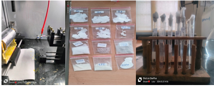
Fabrication of nanofibers via electrospinning process
- The prepared polymeric solution i.e. PVPK90/PMMA /chloramphenicol was loaded in a 3 mL syringe having needle size of 21g and an inner diameter of 9.83 mm.
- The needle tip's distance from the collector was fixed to 10 cm.
- Piston of the syringe was pressed such that a small droplet of the polymer solution was obtained at the tip of the needle. Different parameters were set into software such as flow rate of polymer solution was maintained at 0.05 mL min, voltage at 21.50 kV, and collector rotation speed at 650-850 rpm, and the doors of the chambers were closed.
- Due to the electric force applied to the needle, a uniform jet was expelled out from the needle, and nanofibers were collected on a drum. After the process of electrospinning was completed, the nanofiber mat was dried for 24 hours to remove the solvents. Figure No.1
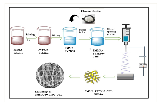
Figure No. 1: Schematic representation for preparation of CHL-loaded electrospun nanofibers mat.
Characterization of Nanofibers
Fourier-transform infrared spectroscopy (FT-IR)11
FT-IR of blank nanofibers PMMA/ PVPK90 and drug-loaded PMMA/ PVPK90/chloramphenicol was performed for analysing the compatibility between drug, polymer and combination of drug-polymer. Each sample was scanned over a wavenumber region of 4000-400 cm-1 and typical bands were noted.
2Scanning electron microscopy (SEM)12
SEM was utilised to assess the morphology of the prepared nanofibers such as fiber diameter, fiber consistency, and diameter distribution. To boost electronic conductivity, nanofibers mat was put on a sample holder and coated with gold for 120 seconds. The samples were then focused and images were captured at different magnifications using SEM microscope (S3400N).
X-ray diffraction (XRD) spectroscopy13
X-ray diffraction is an important tool to investigate the possible variations in the crystalline nature of a drug during electrospinning. Pure drug and all nanofibrous formulations i.e. 15% PVPK90/PMMA /chloramphenicol, were analyzed by X-ray diffractometer system X-600 (PANalytical, Empyrean Netherlands) with Cu K? radiation, wavelength 1.540 Å, and scanned speed was 5° per min. from 10° to 60° of 2?.
Differential Scanning Calorimetry (DSC)14
DSC can determine the glass transition temperature (Tg), melting temperature (Tm), and crystallization temperature (Tc) of nanofibers. These thermal properties are crucial in understanding the stability and processing conditions of nanofibers.
Entrapment Efficiency15
The drug entrapment efficiency of nanofibers was measured by drying drug-enriched nanofibers in a hot air oven for 5 min at 41 °C, then the nanofibers scaffold of known area 1 cm2 was removed and dissolved it in water. The amount of drug loaded into these fibers was estimated by UV analysis.
The Entrapment Efficiency was calculated by following equation,
Entrapment efficiency (%)=(Mass of drug released)/(Mass of total drug added ) ×100
In Vitro Drug Release Study16
CHL-loaded electrospun nanofibers 10% & 15% PVPK90/PMMA /chloramphenicol of 1 cm2 area were placed in 100 mL PBS buffer. The release study was conducted in an incubator with a speed of 100 rpm at 37 °C. Two mL samples of formulations were taken after 15, 30 min, 1, 2, 4, 8, 12, 16, 24, 36, 48, and 72 h. After taking samples, an equivalent volume of PBS buffer was replenished to maintain sink conditions. Absorbance of samples were taken by UV-vis spectrophotometer at wavelength 278 nm. The % drug release was estimated using the calibration curve of the drug in PBS 7.4 and results were represented as mean ± SD.
RESULTS AND DISCUSSION
Fourier-transform infrared spectroscopy (FT-IR)
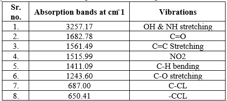
The IR spectrum of the pure Chloramphenicol sample recorded by FTIR spectrometer is shown in figure No.2 This was compared with standard functional group frequencies of Chloramphenicol as shown in Table No.2 Table showed that frequencies of functional group of Chloramphenicol were in the reported range which indicates that the obtained sample was of Chloramphenicol and was pure.
- CHL
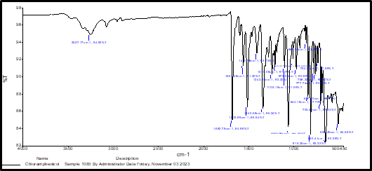
Figure No. 2: FTIR spectra of prepared CHLs
Table No. 2: Interpretation of FTIR Spectra of CHL
PMMA
Figure No.3: FTIR spectra of PMMA

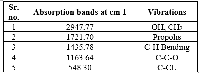
Table No. 3: Interpretation of FTIR Spectra of PMMA
PVPK90
Figure No. 4: FTIR spectra of PVPK90
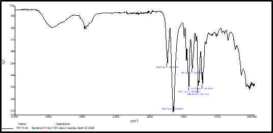
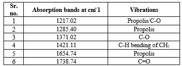
Table No. 4: Interpretation of FTIR Spectra of PVPK90
- DRUG + POLYMER

Figure No. 5: FTIR spectra of DRUG + POLYMER
Table No. 5: Interpretation of FTIR Spectra of DRUG + POLYMER
- SDPF

Figure No. 6: FTIR Spectra of SDPF
Table No. 6: Interpretation of FTIR Spectra of SDPF
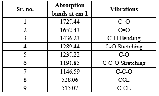
Compatibility of excipient with chloramphenicol was studied by furrier transform infrared spectroscopy (PerkinElmer). The drug and carrier separately and in combination with each other were mixed KBr for determination of spectrum. The range selected was from 4000 – 400 cm-1. The FT-IR spectrum of formulation nanofiber show that there is no significant shift in the peaks or significant difference in spectra and characteristic peaks of formulation are same the pure drug. There is no any interaction between drug and polymer. The FT-IR spectrum of formulation shows same peak values when compared with the characteristics peak values of pure drug.
Scanning Electron Microscopy (SEM)
The SEM studies are generally done to study surface morphology if the drug nanofiber. The photographs were studied for morphological characteristics.The surface morphology was determined using scanning electron microscope (JEOL JSM-6400). The samples were cut into circular shape with an average diameter of 1.5 cm. Specimens were coated with a thin gold layer using a sputter. The fiber diameters were measured using a SemAphore 4.0 software at 20000 magnifications Figure No.7.
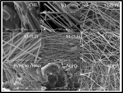
Figure No. 7: SEM Images of drug, polymer with their ratios and final optimized NF
Morphological properties of the CHL/PMMA Electrospun NF were investigated by SEM. In case of drug it exhibited a crystalline morphology with crystal structure. In case of plain polymer, it shows a amorphous structure. The nanofiber with plain PMMA resulted with broken fiber morphology. While it mixed with PVPK90 it starts to give continuous fiber but while increasing the PVPK-90 the fibers exhibit morphology with beads and twisted morphology. Hence, the formulation with 8:2 was selected for further drug loading and evaluation. While in case of drug loaded formulation the fibers resulted with minimum beads with neat fiber morphology.
X ray diffraction studies (XRD)
To understand the crystallinity of the drug in the nanofiber membrane, XRD pattern of pure drug, CPD and their optimized SDPF membrane were recorded. The XRD pattern of CHL, as shown in Figure No 8. Exhibited four characteristic peaks at 2? of 10.2º, 17.9º, 20.7º and 25º which demonstrates crystalline nature of the drug. CPD showed two peaks in the diffractogram at a diffraction angle of 10º and 20º indicates semi-crystalline nature of CPD. Whereas, XRD pattern of SDPF membrane exhibited more of an amorphous nature as compared to pure CHL due to shifting of its diffraction intensity, conforming CHL physical state transformed from crystalline state to amorphous state during the entrapment process in the nanofiber membrane.
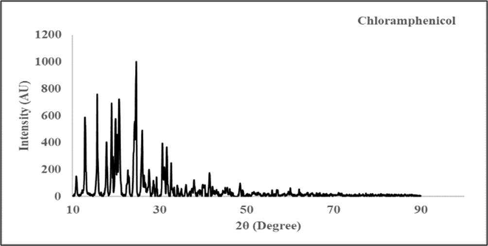
Figure No. 8: Overlay of Powder XRD pattern of CHL
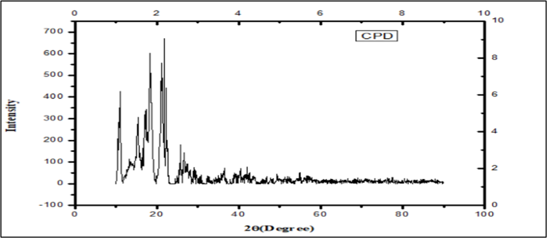
Figure No. 9: Overlay of Powder XRD pattern of CPD
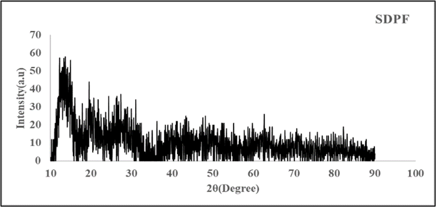
Figure No. 10: Overlay of Powder XRD pattern of SDPF
Differential scanning calorimetry (DSC)
DSC studies were performed to characterize the solid state of drugs and polymers. Further, compatibility between drug and excipients can be evaluated by observing the thermal behavior of compounds such as appearance or disappearance of an endothermic or exothermic peak. If all the peaks remain the same, compatibility can be expected. DSC thermogram of CHL, PMMA, PVPK90 and optimized nanofiber membrane are depicted in Figure No.11.
DSC thermogram of CHL showed a sharp endothermic peak at 151ºC which is attributed to its melting point. The sharp melting peaks exhibited by CHL confirmed their existence as a crystalline form. Thermogram of PMMA exhibited a characteristic peak at 40ºC due to its semi-crystalline nature. The PVPK90 also exhibited characteristic peaks of all components indicating physical compatibility between excipients and drug. Whereas, optimized SDPF membrane showed flat curve without sharp endothermic or exothermic peaks of drug. This indicates that transformation of phase i.e. crystalline state to amorphous state has taken place, during the entrapment process. This might be due to shear stress provided by the stirrer and electrospinning during the fabrication process of nanofiber which may prevent the recrystallization of CHL, leaving CHL in molecular dispersion form inside the SDPF membrane are shown in Figure No. 11.
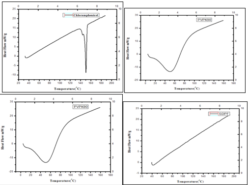
Figure No. 11: DSC thermograms of CHL, PMMA, PVPK90 and optimized formulation (SDPF)
Entrapment Efficiency
The drug entrapment efficiency of nanofibers was measured by drying drug-enriched nanofibers in a hot air oven for 5 min at 41 °C, then the nanofibers scaffold of known area 1 cm2 was removed and dissolved it in water. The amount of drug loaded into these fibers was estimated by UV analysis.
The Entrapment Efficiency was calculated by following equation,
Entrapment efficiency (%)=(Mass of drug released)/(Mass of total drug added ) ×100
IN VITRO DRUF RELEASE STUDY
An in vitro study was conducted using a magnetic stirrer to investigate the release of a drug from a fiber. The experiment began by preparing a stock solution, where 100 mg of the drug-loaded fiber was added to 100 ml of phosphate buffer solution (pH 7.4), and stirred at 100 rpm for 18 hours. At hourly intervals, 1 ml of the solution was withdrawn from the stock, and an equal volume of fresh buffer solution was added to maintain the volume. Each withdrawn sample was then analyzed for absorbance at 278nm using a spectrophotometer. By analyzing the absorbance data, it was determined that approximately 92.8% of the drug had been released into the buffer solution by the end of the 18-hour period. This study provides insights into the release kinetics of the drug from the fiber in vitro, which is crucial for understanding its potential applications in drug delivery systems.
Table No. 7: % of Drug release on each hour
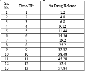
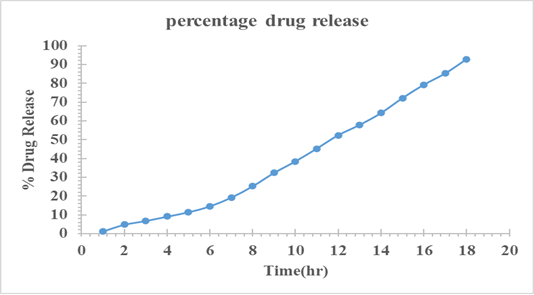
Figure No. 12: The % Of Drug Release at Each Hour
s made by standard procedures set and work carried out. The effect of drug: polymer on the physical characteristics of the formulated nanofibers was examined for various drug: polymer ratios of nanofibers at stirring speed of 400-600rpm for 1hrs by electrospinning method. The mean particle size of nanofibers can be influenced by drug: polymer ratio. It was oed that as drug: polymer ratio increases, the particle size decreased. The effect of stirring rate on the physical characteristics of the formulated nanofibers was examined for 8 : 2 polymer ratio nanofibers. The stirring rate was varied in the range of 400-600rpm. Polymer concentration has a crucial role in making characterized nanofibers given concentration of polymer gives uniform and structured nanofibers and with optimum release of drug with the help of accurate set ratios of polymer concentration, flow rate, distance between needle and collector, voltage these parameter setted. PMMA proportion in NF resulted in an increase in the viscosity of the solution higher viscosities resulted into formation of dense network of the polymer preventing the drug from leaving the matrix. In FTIR analysis obtained results shows that no interaction between drug, polymer and formulation occurred because no change in the characteristics peak was seen. Hence drug and selected polymer were compatible with each other. In SEM analysis result shows that fiber mat found to be smooth continuous fiber structured 627.9±149.78 nm respectively. As the viscosity of solution increased, entanglement in polymer chain also increased. Fibers were prepared without the occurrence of bead defects. It was remarkable to note. In DSC analysis result the drug and polymer melting pint determined as per standard values with the help of the endothermic and exothermic reactions with respective drug and polymer used for the nanofibers formulation. In in-vitro drug release studies for all chloramphenicol nanofibers, formulations along with pure chloramphenicol were performed and are reported in result section figure No.12. All chloramphenicol loaded nanofibers formulations have shown significant enhancement in dissolution rate compared to pure chloramphenicol in PBS dissolution media at pH 7.4.
REFERENCES
- Aggarwal D, Garg A, Kaur IP. Development of chloramphenicol-loaded microsponges for ocular delivery: formulation optimization, characterization, and evaluation. Drug Dev Ind Pharm. 2017;43(4):540-551.
- Huang YC, Jhong JF, Tsai PJ, Chen KY, Chen CW. The fabrication of dual-layered nanofiber membranes loaded with chloramphenicol and hyaluronic acid for ocular drug delivery applications. J Membr Sci. 2020;598:117703.
- Lee JH, Cho JH, Lee WG, Kim HH, Lee BJ, Lee SH. Preparation and evaluation of poly(?-caprolactone)/poly(vinyl alcohol)/chloramphenicol nanofibers for ocular drug delivery. Int J Nanomedicine. 2018;13:1415-1428.
- Li X, Wang H, Zhang T, Lu X. Controlled drug release properties of pH-sensitive poly(ethylene oxide)/poly(vinyl pyrrolidone) nanofibers for ophthalmic delivery. Int J Pharm. 2015;490(1-2):384-391.
- Mandal A, Bisht R, Rupenthal ID, Mitra AK. Polymeric micelles for ocular drug delivery: from structural frameworks to recent preclinical studies. J Control Release. 2017;248:96-116.
- Pandey P, Shukla S, Gupta PK. Development and evaluation of fluconazole-loaded nanostructured lipid carriers for enhanced ocular delivery. J Drug Deliv Sci Technol. 2020;59:101891.
- Park JH, Kang N, Kim SW. Enhanced corneal permeation of chloramphenicol through formulation of the insoluble prodrug in cyclodextrin-based nanofibers. Int J Pharm. 2016;515(1-2):357-365.
- Qu J, Zhao X, Liang Y, Zhang T, Ma PX, Guo B. Antibacterial adhesive injectable hydrogels with rapid self-healing, extensibility and compressibility as wound dressing for joints skin wound healing. Biomaterials. 2020;232:119737.
- Tian H, Lin L, Chen J, et al. Nanoparticle delivery of chemotherapy combination regimen improves the therapeutic efficacy in mouse models of lung cancer. Nanomedicine. 2020;27:102177.
- Wang Y, Chen X, Liu Z, et al. Composite polymeric nanofibers for wound dressings. Chem Eng J. 2017;315:110-125.
- Jirofti, N., Golandi, M., Movaffagh, J., Ahmadi, F. S., Kalalinia, F. (2021). Improvement of the wound-healing process by curcumin-loaded chitosan/collagen blend electrospun nanofibers: In vitro and in vivo studies. ACS Biomaterials Science Engineering, 7(8), 3886-3897.
- Chen, K.,Pan, H., Ji, D., Li, Y., Duan, H., Pan, W. (2021). Curcumin-loaded sandwich-like nanofibrous membrane prepared by electrospinning technology as wound dressing for accelerate wound healing. Materials Science and Engineering: C, 127, 112245
- Wang, S. B., Chen, A. Z., Weng, L. J., Chen, M. Y., Xie, X. L. (2004). Effect of drug?loading methods on drug load, encapsulation efficiency and release properties of alginate/poly?l?arginine/chitosan ternary complex microcapsules. Macromolecular Bioscience, 4(1), 27-30.
- C.-J.Wang, H.-J.Tsai Thermal Properties of Electrospun Poly(methyl methacrylate)/Polyvinylpyrrolidone Nanofibers. Journal of Applied Polymer Science.
- Kataria, K., Gupta, A., Rath, G., Mathur, R. B., Dhakate, S. R. (2014). In vivo wound healing performance of drug loaded electrospun composite nanofibers transdermal patch. International Journal of Pharmaceutics, 469(1), 102-110.
- Panda, B. P.,et.al [2021] Design, Fabrication and Characterization of PVA/PLGA Electrospun Nanofibers Carriers for Improvement of Drug Delivery of Gliclazide in Type-2 Diabetes.


 Omprakash G. Bhusnure *
 Shivam A S Vyavahare
 Mani Ganesh 3
 D D Vibhute 4
 Vijayendra Swammy 5
 Hyun Tae Jang


 10.5281/zenodo.13294624