View Article

  • Urolithiasis: Advances in Pathophysiology, Microbiome Role, Immune Pathways, and Evolving Diagnostic and Therapeutic Strategies for Kidney Stone Formation and Management

  • KLE College of Pharmacy Belagavi, KLE Academy of Higher Education and Research (KAHER), Belagavi, Karnataka, India.

Abstract

Urolithiasis, or kidney stone disease, is a multifactorial disorder characterized by crystalline deposits in the urinary system. Its global prevalence has risen significantly over the past two decades, making it a major public health concern. The etiology involves genetic, metabolic, dietary, and environmental factors. Kidney stones, including calcium oxalate, uric acid, struvite, and cystine stones, form through distinct mechanisms. The stone formation process includes supersaturation, nucleation, crystal growth, aggregation, and retention in the kidneys. Hormonal influences, particularly sex hormones, impact stone formation, while the gut and urinary microbiome are emerging regulators of urolithiasis susceptibility. Additionally, immune responses to urinary crystals trigger inflammatory cascades that promote stone development. Preventive measures focus on dietary modifications, hydration, and pharmacological approaches. Treatment options range from minimally invasive procedures, such as extracorporeal shock wave lithotripsy, to surgical interventions. Recent research explores novel therapeutic strategies, including microbiome modulation and immunotherapy, to enhance management and prevention. This review comprehensively examines the mechanisms underlying stone formation, current treatment strategies, and future directions for improved urolithiasis management.

Keywords

Urolithiasis. Mechanism. Randall's plaque, Oxidative stress.

Introduction

Urolithiasis, or kidney stones, is a common condition caused by mineral buildup in the kidneys, bladder, or urethra, posing a global health challenge(1). Kidney stones are primarily calcareous, such as calcium oxalate (over 80% of cases), or non-calcareous, like uric acid (5-10%) or struvite. In the U.S., they affect 10.6% of men and 7.1% of women (2). In India, 12% of people face this issue yearly, especially in states like Gujarat and Rajasthan (3). Without treatment, stones may recur at a 10% annual rate (4). Men (12%) are affected more than women (6%), with peak cases between ages 20-40 (5). The exact causes of stone formation remain unclear (6). Stones form through crystal nucleation and growth when urine is oversaturated with insoluble substances(7,8). No drug fully treats urolithiasis; preventive measures like thiazide diuretics are used, but surgical options such as shock wave lithotripsy or ureteroscopy are frequent, with a 50% recurrence rate (9). Surgery can also cause issues like high blood pressure or kidney damage (10). The kidneys, reddish-brown and bean-shaped, lie in the retroperitoneal space, regulating fluids, electrolytes, and acid-base balance while clearing toxins. Blood enters through renal arteries, exits via veins, and urine drains to the bladder through ureters. The glomerulus filters 20% of blood, reabsorbing nutrients like glucose and amino acids, while expelling waste. Human kidneys contain about one million nephrons (11). Urine forms as filtrate passes through tubules, with the proximal tubule reabsorbing most nutrients and the loop of Henle concentrating urine. Urine is 95% water, 2.5% urea, and contains minerals and enzymes, while key molecules are reabsorbed (12). The urinary system—kidneys, ureters, bladder, and urethra—filters blood and produces urine. The proximal convoluted tubule handles nutrient reabsorption, the loop of Henle dilutes filtrate, and the distal tubule balances sodium and pH. The collecting duct finalizes urine by removing ions and reabsorbing water (11).

Aetiology of Kidney Stones

Kidney stone formation stems from low fluid intake, high animal protein, oxalate-rich diets (e.g., spinach, nuts), and excess salt (13). Hydration producing 2.5L urine daily (water, coffee) is protective; high-oxalate juices (e.g., cranberry) increase risk. Citrate inhibits crystal aggregation, with hypocitraturia in 60% of calcium stone cases (14). Low calcium intake raises oxalate absorption, while vitamin C and fish oil elevate stone risk (15).

Types of Kidneys Stones:

Stones include calcium-based (75-90%) and non-calcium types (uric acid, struvite, cystine) (16,17); Table 1).

Table 1 Mineral composition kidney stone

Name of stone

Approximate incidence

Constituents

Calcium oxalate, Calcium phosphate

75 - 90 % of all stones

Calcium, oxalate, phosphate

Uric acid

3-10 % of all stone

Uric acid

Struvite

10 – 15 % of all stones

Calcium, ammonia, phosphate

Cystine

Less than 1% of all stone

Cystine

Medication induced stones

1-2% of all stone

Composition depends on medication or herbal (examples

Product include azanavir, sulfadiazine, acetazolamide

Calcium Stones:

Comprising 80% of cases, these include calcium oxalate (50%) and phosphate (5%), with mixed types (45%) (Coe, 2005a). Calcium oxalate monohydrate (COM) is stable and prevalent, forming at pH 4.5-5.5; phosphate stones favor pH >7.5 (4).

Struvite Stones:

Linked to UTIs from urease-producing bacteria (e.g., Klebsiella), these form 10-15% of stones, more in women (2).

Uric Acid Stones:  

3-10% of cases, tied to low pH and hyperuricosuria, common in men (18).

Cystine Stones:

<2% of cases, from cystinuria, an autosomal recessive disorder (19).

Drug-Induced Stones:

1-2% of cases, from drugs like atazanavir (20).

Kidney Stone Compositions:

Stones consist of crystals and an organic matrix (proteins 64%, sugars 9.6%, lipids 10.3%) that modulates formation (21). Phospholipids (8.6% of lipids) aid calcium stone development. Brushite, a resistant phosphate mineral, occurs in 25% of calcium phosphate stones (22)

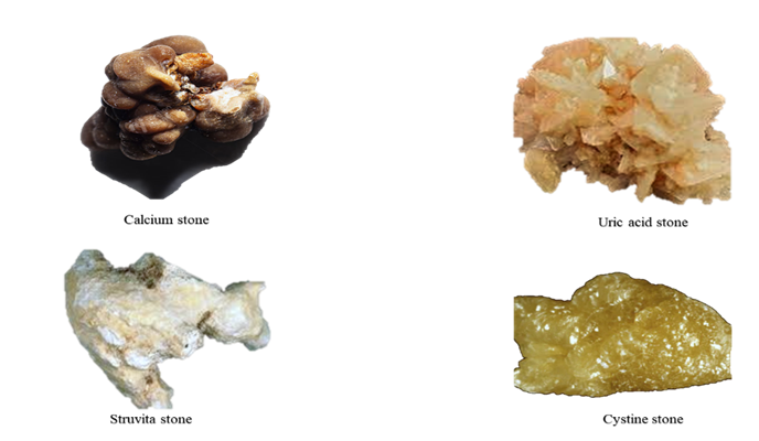
Figure 1: Kidney stones and their types (23).

Pathophysiology: Several risk factors have been identified that increase susceptibility to urolithiasis.

Demographic Factors: Men (10.6%) exceed women (7.1%) in prevalence, with 50% recurrence; women’s risk rises with obesity (2). Stones are rare in youth but increase in the elderly (10-12% of cases), with infection stones post-60 (24). Caucasians (5.9%) outpace Blacks (1.7%), though diet aligns rates (25).

Genetics: 25% of patients have a family history; hypercalciuria (35-65%) often hereditary (15), Table 2).

Hormonal Factors: Testosterone increases oxalate via glycolic acid oxidase, while estrogen boosts protective osteopontin (26).

Metabolic Conditions: Diabetes lowers urine pH via insulin resistance, aiding uric acid stone . Obesity (BMI >30) raises oxalate and uric acid excretion (2). Pregnancy increases hypercalciuria, though citrate counters risk (27).

Lifestyle: High fluid intake dilutes urine, reducing crystal formation (28).

Other mechanisms by which crystals remain in the kidney:

Calcium oxalate monohydrate (COM) crystals stick fast to kidney epithelial cells because their positive charge pulls in the cells' negatively charged surface. Once attached, a crystal becomes a hub for more crystals to latch on. The cell then engulfs the crystal, making the nearby membrane stickier for other crystals. This stickiness lasts at least 24 hours, boosting the number or strength of crystal attachment spots (29,30).

Table 2 Genes Associated with Hypercalciuria And Kidney Stone Formation.

S.NO.

Gene

Gene function

Renal tubular

expression

Renal phenotype

1

DR

Vitamin D receptor

DCT, CD

Decreased calcium reabsorption.

2

CLCN5

Cl/H antiporter

PT, TAL, αIC

Inactivating mutations

3

CASR

Calcium sensing receptor

PT, MCD, TAL, DCT

Gain of function mutation

4

CLDN16

Tight junction protein

TAL, DCT

Hypercalciuria, magnesium-wasting

5

NPT2a/c

Sodium phosphate co-transporter

PT

Hypercalciuria, magnesium-wasting

6

TRPV5

Calcium selective transient receptor potential channel

DCT, connecting tubule

Hypercalciuria

7

sAC

Soluble adenylate cyclase/ bicarbonate exchanger

DCT, TAL, CD

Hypercalciuria

8

KLOTHO

Aging suppression protein/ regulator of calcium homeostasis

DCT

Hypercalciuria

Physicochemical Processes in Kidney Stone Formation:

Urinary supersaturation and crystallization, driven by renal impairment, urine pH, and excess specific substances (e.g., CaOx, CaP, uric acid, struvite, cysteine, purines, and certain drugs), contribute to intrarenal crystal precipitation. Modulator molecules, including, promoters, and inhibitors, further influence crystal formation shown in (Figure 2) (31,32).

Urine Supersaturation:

Kidney stones form when urine becomes supersaturated, causing dissolved salts to exceed their solubility and crystallize into solids (Figure 3). Supersaturation is the key driver of this phase transition in urine (33).

Promoters of stone formation:

Promoters of stone formation include receptors and molecular mechanisms vital for crystal-cell interactions, aiding stone retention (34,35). COM-treated HK-2 cells show 1,141 DEPs, including CD44, nucleolin, HA, HSP90 , annexin II (36), and OPN (37,38), all involved in lithogenesis (39). Crystal attachment is facilitated by phosphatidylserine and acidic protein side chains (40). Calcium, oxalate, urate, and phosphate ions promote crystallization. Dysregulated calcium and vitamin D metabolism, with elevated serum calcium and 1,25(OH)2D, contributes to stone formation via CaSR pathways (41,42). Urate and phosphate also aid crystal nucleation (42,43). Urine pH affects crystallization: low pH favors CaOx crystallization, while high pH enhances nucleation (44). Lysozyme and lactoferrin promote COM crystal growth (45).

Inhibitors of stone formation:

Urine contains various inhibitors—anions (e.g., citrate), metallic cations (e.g., magnesium), and macromolecules (e.g., OPN, THP, UPTF?1, Nc, IαI subunits)—that competitively and cooperatively reduce crystallization, aggregation, and adhesion (46,47). Citrate inhibits crystal growth above 0.1 mM, and its reduced excretion in nephrolithiasis is managed with alkali supplements (48–50). Hydroxycitrate, a citrate analog, also prevents crystallization (51,52). Magnesium enhances citrate’s effect in acidic urine (53). Macromolecules potently inhibit stone-forming processes (33,44). Crystalluria depends on the supersaturation-inhibitor balance (54)

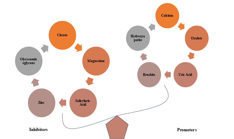
Figure.2 Imbalance Between Promoters and Inhibitors in Urolithiasis: A Key Factor in Kidney Stone Formation.

Randall's plaque and calcium oxalate stone formation:

Randall’s plaques (RPs), first identified by Alexander Randall (55), are subepithelial CaP-rich mineral deposits at the papillary tip near the ducts of Bellini (39), composed of calcified tubules and CaP plugs (56), embedded in a protein-, lipid-, and ECM-rich organic matrix (57). RPs are proposed as the nidus for kidney stones (58). LncRNA H19 promotes osteogenic differentiation via Wnt/β-catenin signaling (59) and induces oxidative stress and injury through the miR-216b/HMGB1/TLR4/NF-κB pathway (60). MALAT1 sponges miR-320a-5p to upregulate Runx2, enhancing osteogenic traits in hRIFs (61), highlighting the need to explore RP formation in stone pathogenesis.

Role of sex hormones in urolithiasis:

Testosterone plays a significant role in kidney stone formation through multiple mechanisms. (62), reported that testosterone induces apoptosis and necrosis in renal tubular epithelial cells via the HIF-1α/BNIP3 pathway, creating a favorable environment for calcium oxalate monohydrate (COM) crystal formation. Additionally, (63) found that testosterone enhances kidney stone disease by increasing the expression of α-enolase on kidney epithelial cells, which facilitates COM crystal adhesion, a crucial step in stone formation. Furthermore, (64) demonstrated that testosterone weakens the immune system's ability to clear COM crystals by suppressing colony-stimulating factor 1 (CSF-1) through miR-185-5p upregulation, reducing macrophage recruitment and phagocytic activity. These combined mechanisms—kidney cell damage, increased crystal adhesion, and impaired crystal clearance—help explain the higher incidence of kidney stones in men compared to women and suggest potential therapeutic targets for nephrolithiasis prevention shown in (Figure 4).

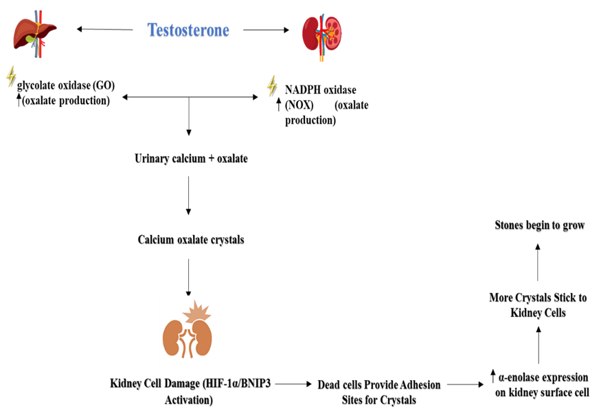
Figure 4. Role of sex hormones in calcium oxalate urolithiasis

The role of the microbiome in kidney stone formation:

Oxalobacter formigenes is a Gram-negative, anaerobic bacterium that aids in oxalate metabolism in the intestine (65). This bacterium is susceptible to certain antibiotics, though its full sensitivity is not well established (66). Its absence may increase intestinal oxalate absorption and reduce endogenous secretion (67), leading to higher urinary oxalate levels and a greater risk of calcium oxalate (CaOx) stone formation show in (Figure 5). Studies show that individuals with kidney stones or hyperoxaluria have a lower prevalence of O. formigenes in their stool than healthy controls (68).Additionally, nephrolithiasis patients with O. formigenes excrete lower urinary oxalate levels than those without it (69).

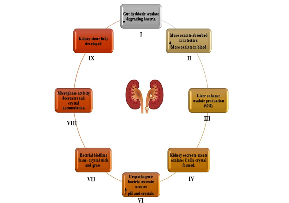
Figure 5.  Role of oxalate?degrading bacteria in stone formation

Oxidative stress:

Kidney stone formation is linked to increased ROS production, primarily via NADPH oxidase activation by angiotensin II (70). CaOx crystal formation upregulates renin and angiotensin II, enhancing ROS, which activate transcription factors through pathways like P38 MAPK/JNK, AP-1, NF-kB, TGF, RUNX-2, and Obsterix. This promotes inflammation, with NF-kB inducing CRP production. The resulting cellular damage and inflammation create a feedback loop that further activates NADPH oxidase and ROS generation, impairing endothelial function (71).

Crystal Nucleation:

Nucleation kicks off kidney stone formation when stone salts in supersaturated urine clump together, shifting from liquid to a stable form. These loose clusters grow by adding more particles or merging with others, forming crystals with a unique structure. In urine, this usually happens on surfaces like epithelial cells, red blood cells, or other crystals, known as heterogeneous nucleation, which needs less saturation than homogeneous nucleation. Once a nucleus forms, it can grow quickly at lower chemical pressures. Damage to kidney tubules boosts calcium oxalate (CaOx) crystallization by supplying components, like membrane vesicles from injured cells, that spark nucleation. (21).

Crystal Growth:

Crystal growth is the stage that follows nucleation in the formation of urinary stones. During this phase, crystals present in urine combine and aggregate, gradually forming a stone. This process can be difficult to manage, as it leads to the development of larger stones. Proteins play essential roles in the crystallization process of nephrolithiasis, where certain urinary stone matrix protein modulators contribute to regulating and influencing the growth and formation of these stones (72).

Crystal Aggregation: 

Crystal aggregation is a key step in the formation of urinary stones. A small, challenging mass of crystals in a solution binds together through this process, gradually forming a larger stone. The aggregation occurs when individual crystals stick together, growing in size and complexity. This process is considered the most critical stage in stone formation, as it significantly contributes to the development of larger, more stable stones (72).

Crystal-cell interaction:

Crystal-cell interaction involves the binding of calcium oxalate monohydrate (COM) crystals to renal tubular epithelial cells, especially in hyperoxaluria. Oxalate and COM crystals damage epithelial cells, promoting crystal retention and CaOx crystallization. Some crystals evade clearance by macrophages or lysosomes. Damaged cells release membrane vesicles and anionic proteins like renal prothrombin fragment-1, which act as nucleators for crystal formation (72).

Endocytosis of CaOx Crystals:

The initial phase of kidney stone formation involves endocytosis, where renal tubular cells engulf COM crystals that first attach to microvilli. Polyanions like glycosaminoglycans, glycoproteins, and citrate may inhibit this by coating crystals. Tamm-Horsfall glycoproteins (THP) have a dual role, being either endocytosed or transported to the interstitium. Damaged cells may promote nidus formation on the renal papillary surface (72).

Cell injury and Apoptosis:

High levels of oxalate or calcium oxalate (CaOx) can harm kidney epithelial cells, raising the risk of kidney stones. Beyond triggering stone formation, cell death (apoptosis) in renal tubular cells can lead to necrosis, which may halt calcium crystal growth. Lab studies on MDCK cells exposed to oxalate confirm this, but not all cells respond the same way. Some cells may activate genes that protect against apoptosis and stone formation. More research is needed to pinpoint new molecular targets for preventing kidney stones (72).

Immune response to urinary crystals:

In kidney stone disease, immune alterations include macrophage accumulation, inflammation, and anti-inflammatory responses, contributing to renal calcium oxalate (CaOx) crystal formation. CD44, osteopontin (OPN), and fibronectin (FN) interactions in renal tubular cells promote macrophage recruitment and calcium oxalate monohydrate (COM) crystal formation. Macrophages secrete IL-8, MIP-1, and MCP-1, driving immune cell migration and renal inflammation. M1 macrophages support crystal formation, while M2 macrophages aid inVcrystal clearance and inflammation reduction (35,73,74)

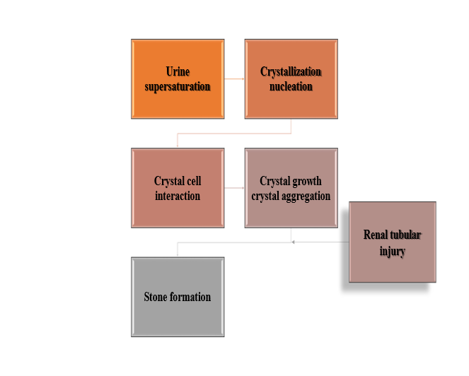
Figure 3. Mechanism of kidney stone formation.

Managing Kidney Stones: Prevention and Treatment Options:

Kidney stones are managed with NSAIDs for pain, opioids for severe cases, and alpha-blockers like tamsulosin to aid passage of small lower ureter stones, reducing discomfort (75,76). Thiazide diuretics cut urinary calcium to prevent calcium stones, while potassium citrate boosts urine alkalinity to stop calcium and uric acid stones (75,77). Allopurinol lowers uric acid, and acetohydroxamic acid helps with struvite stones (75,78). Cystine-binding drugs like tiopronin aid cystinuria but may have side effects (79). For stubborn or large stones, shock wave lithotripsy (ESWL) breaks up smaller stones, ureteroscopy (URS) uses lasers for precise treatment, and percutaneous nephrolithotomy (PCNL) handles big or complex stones (80–82). Open surgery is rare now (Nourian and Bahrami, 2022). Herbal options like Cystone, Uriflow, and Trinapanchamool can dissolve or prevent stones (23). Combining therapies—expulsion aids, pain relievers like NSAIDs, calcium channel blockers, steroids for swelling, and diuretics—tackles the complex nature of stones for better results (23).

Discussion and Future Directions:

Urolithiasis, a multifaceted urinary tract disorder, involves diverse stone types triggered by multiple causes. Our analysis identifies key risk factors such as gender, age, genetics, hormones, diabetes, obesity, pregnancy, and lifestyle. However, the precise mechanisms driving stone formation remain elusive despite thorough investigation (83), making it a significant gap in the field. The physicochemical process of kidney stone formation involves supersaturation, nucleation, crystal growth, aggregation, and retention within the renal tubules(84). Randall’s plaque, which originates in the renal papilla, serves as a nidus for stone formation, particularly in calcium oxalate stones(85). Recent evidence also suggests a crucial role of sex hormones in urolithiasis, with testosterone being linked to increased stone formation, while estrogen appears to have a protective effect(86). Additionally, emerging studies indicate that the gut and urinary microbiome influence stone formation, suggesting a potential area for therapeutic intervention(87). Current treatment strategies for urolithiasis remain inadequate. While allopathic treatments focus on pain management, hydration therapy, and surgical intervention, a definitive pharmacological therapy is still lacking(75,81). Ayurvedic formulations such as Cystone have been explored for their potential anti-lithogenic properties, but some studies suggest inconsistent efficacy, necessitating further research. While Cystone is widely used for managing urolithiasis, certain limitations must be acknowledged. One key concern is its uncertain impact on urinary supersaturation, as studies have shown no significant changes in urinary composition, a critical factor in stone formation. Additionally, although it is marketed for stone expulsion, clinical findings indicate an increase in stone burden rather than a reduction (88), Its ability to aid stone passage is uncertain. Cystone shows limited effectiveness in severe, treatment-resistant cases, indicating it may not suit complex conditions. Additionally, the absence of extensive, well-designed clinical studies hinders clear conclusions about its long-term benefits for preventing or dissolving stones (89). Cystone acts more slowly than Tamsulosin, which rapidly eases stone passage by relaxing urinary tract muscles. These observations highlight the necessity for thorough research to confirm Cystone’s effectiveness as a primary or supportive treatment for urolithiasis (90). Future urolithiasis studies seek to enhance knowledge of stone formation by investigating genetic and epigenetic factors driving molecular mechanisms(91), Uncovering these factors may lead to new treatment targets for improved outcomes. Scientists are also creating drugs to prevent kidney stones by blocking crystal formation, growth, and aggregation or altering urine composition (92). Personalized medicine is gaining traction, leveraging genomics, metabolomics, and proteomics to pinpoint individual risk factors and customize urolithiasis prevention and treatment. Advanced non-invasive imaging is also being developed to improve detection of small stones and analyze their composition accurately, with reduced radiation exposure. (93). Additionally, the discovery of urinary biomarkers may facilitate early diagnosis and predict recurrence risk, enabling proactive management (94). In surgical advancements, innovations in minimally invasive techniques, including robotic-assisted procedures, are being investigated to improve precision, safety, and outcomes in complex cases (95). These collective efforts aim to refine diagnostic, therapeutic, and preventive strategies, ultimately advancing patient-centered care in urolithiasis management.

CONCLUSION

Urolithiasis, driven by metabolic, physiological, and lifestyle factors, centers on calcium oxalate stone formation. Men face greater risk than women, with diet and hydration heavily influencing prevalence. New insights into promoters, inhibitors, immune responses, gut microbiome, and sex hormones deepen our understanding of stone development. Advances in personalized medicine, innovative drugs, and cutting-edge diagnostic and surgical methods promise better outcomes. A collaborative approach combining medical, nutritional, and surgical expertise is vital to enhance treatments, prevent recurrence, and lessen the impact of kidney stones on patients and healthcare.

REFERENCES

  1. Khan SR, Pearle MS, Robertson WG, Gambaro G, Canales BK, Doizi S, et al. Kidney stones. Nat Rev Dis Primer. 2016 Feb 25;2(1):16008.
  2. Scales CD, Smith AC, Hanley JM, Saigal CS. Prevalence of Kidney Stones in the United States. Eur Urol. 2012 Jul;62(1):160–5.
  3. Sohgaura A, Bigoniya P. A Review on Epidemiology and Etiology of Renal Stone. Am J Drug Discov Dev. 2017 Mar 15;7(2):54–62.
  4. Basavaraj DR, Biyani CS, Browning AJ, Cartledge JJ. The Role of Urinary Kidney Stone Inhibitors and Promoters in the Pathogenesis of Calcium Containing Renal Stones. EAU-EBU Update Ser. 2007 Jun;5(3):126–36.
  5. Worcester EM, Coe FL. Calcium Kidney Stones. N Engl J Med. 2010 Sep 2;363(10):954–63.
  6. Shah J, Whitfield HN. Urolithiasis through the ages. BJU Int. 2002 May;89(8):801–10.
  7. Baumann JM. Stone prevention: why so little progress? *. Urol Res. 1998 Apr 17;26(2):77–81.
  8. Evan AP. Physiopathology and etiology of stone formation in the kidney and the urinary tract. Pediatr Nephrol. 2010 May;25(5):831–41.
  9. Nabi G, Downey P, Keeley F, Watson G, McClinton S. Extra-corporeal shock wave lithotripsy (ESWL) versus ureteroscopic management for ureteric calculi. In: The Cochrane Collaboration, editor. The Cochrane Database of Systematic Reviews [Internet]. Chichester, UK: John Wiley & Sons, Ltd; 2006 [cited 2025 Feb 21]. p. CD006029. Available from: https://doi.wiley.com/10.1002/14651858.CD006029
  10. Patel VB, Acharya N. Effect of Macrotyloma uniflorum in ethylene glycol induced urolithiasis in rats. Heliyon. 2020 Jun;6(6):e04253.
  11. Lote CJ. Principles of Renal Physiology [Internet]. New York, NY: Springer New York; 2012 [cited 2025 Feb 21]. Available from: https://link.springer.com/10.1007/978-1-4614-3785-7
  12. Alelign T, Petros B. Kidney Stone Disease: An Update on Current Concepts. Adv Urol. 2018;2018:1–12.
  13. Chung MJ. Urolithiasis and nephrolithiasis. JAAPA. 2017 Sep;30(9):49–50.
  14. Caudarella R, Vescini F. Urinary citrate and renal stone disease: the preventive role of alkali citrate treatment. Arch Ital Urol Androl Organo Uff Soc Ital Ecogr Urol E Nefrol. 2009 Sep;81(3):182–7.
  15. Curhan GC, Willett WC, Rimm EB, Stampfer MJ. Family history and risk of kidney stones. J Am Soc Nephrol. 1997 Oct;8(10):1568–73.
  16. Bao E, Yang Y, Jiang B, Wang B, Liu Y, Yang L, et al. An exploratory study evaluated the 30 most commonly reported medications in the United States food and drug administration’s adverse event reporting system that are associated with the occurrence of kidney stones. Front Pharmacol. 2024 Dec 18;15:1377679.
  17. Suvarna Y, Rahaman SKA. In Vitro – In Vivo Evaluation of Antiurolithiatic activity of piperine from Piper nigrum. Res J Pharm Technol. 2019;13(1):63.
  18. Johri N, Cooper B, Robertson W, Choong S, Rickards D, Unwin R. An Update and Practical Guide to Renal Stone Management. Nephron Clin Pract. 2010 Jul 2;116(3):c159–71.
  19. Coe FL. Kidney stone disease. J Clin Invest. 2005 Oct 1;115(10):2598–608.
  20. Barbas C, Garc??a A, Saavedra L, Muros M. Urinary analysis of nephrolithiasis markers. J Chromatogr B. 2002 Dec;781(1–2):433–55.
  21. Aggarwal KP, Narula S, Kakkar M, Tandon C. Nephrolithiasis: Molecular Mechanism of Renal Stone Formation and the Critical Role Played by Modulators. BioMed Res Int. 2013;2013:1–21.
  22. Krambeck AE, Handa SE, Evan AP, Lingeman JE. Brushite stone disease as a consequence of lithotripsy? Urol Res. 2010 Aug;38(4):293–9.
  23. Malhotra M, Tandon P, Wadhwa K, Melkani I, Singh AP, Singh AP. The complex pathophysiology of urolithiasis (kidney stones) and the effect of combinational drugs. J Drug Deliv Ther. 2022 Oct 15;12(5-S):194–204.
  24. Krambeck AE, Lieske JC, Li X, Bergstralh EJ, Melton LJ, Rule AD. Effect of Age on the Clinical Presentation of Incident Symptomatic Urolithiasis in the General Population. J Urol. 2013 Jan;189(1):158–64.
  25. López M, Hoppe B. History, epidemiology and regional diversities of urolithiasis. Pediatr Nephrol. 2010 Jan;25(1):49–59.
  26. Naghii M, Hedayati M. Determinant Role of Gonadal Sex Hormones In The Pathogenesis Of Urolithiasis In A Male Subject – A Document For Male Predominancy (Case Study). Endocr Regul. 2010;44(4):143–6.
  27. Pais VM, Payton AL, LaGrange CA. Urolithiasis in Pregnancy. Urol Clin North Am. 2007 Feb;34(1):43–52.
  28. Fink HA, Akornor JW, Garimella PS, MacDonald R, Cutting A, Rutks IR, et al. Diet, Fluid, or Supplements for Secondary Prevention of Nephrolithiasis: A Systematic Review and Meta-Analysis of Randomized Trials. Eur Urol. 2009 Jul;56(1):72–80.
  29. Dardamanis M. Pathomechanisms of nephrolithiasis. Hippokratia. 2013 Apr;17(2):100–7.
  30. Lieske JC, Norris R, Swift H, Toback FG. Adhesion, internalization and metabolism of calcium oxalate monohydrate crystals by renal epithelial cells. Kidney Int. 1997 Nov;52(5):1291–301.
  31. Daudon M, Traxer O, Lechevallier E, Saussine C. La lithogenèse. Prog En Urol. 2008 Dec;18(12):815–27.
  32. Rodgers AL. Physicochemical mechanisms of stone formation. Urolithiasis. 2017 Feb;45(1):27–32.
  33. Aggarwal CC. An Introduction to Outlier Analysis. In: Outlier Analysis [Internet]. Cham: Springer International Publishing; 2017 [cited 2025 Feb 20]. p. 1–34. Available from: http://link.springer.com/10.1007/978-3-319-47578-3_1
  34. Peerapen P, Thongboonkerd V. Kidney Stone Prevention. Adv Nutr. 2023 May;14(3):555–69.
  35. Wang Z, Zhang Y, Zhang J, Deng Q, Liang H. Recent advances on the mechanisms of kidney stone formation (Review). Int J Mol Med. 2021 Jun 11;48(2):149.
  36. Kumar V, Farell G, Deganello S, Lieske JC. Annexin II Is Present on Renal Epithelial Cells and Binds Calcium Oxalate Monohydrate Crystals. J Am Soc Nephrol. 2003 Feb;14(2):289–97.
  37. Anan G, Yoneyama T, Noro D, Tobisawa Y, Hatakeyama S, Sutoh Yoneyama M, et al. The Impact of Glycosylation of Osteopontin on Urinary Stone Formation. Int J Mol Sci. 2019 Dec 21;21(1):93.
  38. Wang Z, Li M xing, Xu C zhi, Zhang Y, Deng Q, Sun R, et al. Comprehensive study of altered proteomic landscape in proximal renal tubular epithelial cells in response to calcium oxalate monohydrate crystals. BMC Urol. 2020 Dec;20(1):136.
  39. Wiener SV, Ho SP, Stoller ML. Beginnings of nephrolithiasis: insights into the past, present and future of Randall?s plaque formation research. Curr Opin Nephrol Hypertens. 2018 Jul;27(4):236–42.
  40. Sheng X, Ward MD, Wesson JA. Crystal Surface Adhesion Explains the Pathological Activity of Calcium Oxalate Hydrates in Kidney Stone Formation. J Am Soc Nephrol. 2005 Jul;16(7):1904–8.
  41. Ketha H, Singh RJ, Grebe SK, Bergstralh EJ, Rule AD, Lieske JC, et al. Altered Calcium and Vitamin D Homeostasis in First-Time Calcium Kidney Stone-Formers. Tordjman KM, editor. PLOS ONE. 2015 Sep 2;10(9):e0137350.
  42. Vezzoli G, Macrina L, Magni G, Arcidiacono T. Calcium-sensing receptor: evidence and hypothesis for its role in nephrolithiasis. Urolithiasis. 2019 Feb;47(1):23–33.
  43. Ouyang JM, Gao J, Xue JF, Xu M, Gui BS, Wang FX. Nanouric acid or nanocalcium phosphate as central nidus to induce calcium oxalate stone formation: a high-resolution transmission electron microscopy study on urinary nanocrystallites. Int J Nanomedicine. 2014 Sep;4399.
  44. Ratkalkar VN, Kleinman JG. Mechanisms of Stone Formation. Clin Rev Bone Miner Metab. 2011 Dec;9(3–4):187–97.
  45. Farmanesh S, Chung J, Sosa RD, Kwak JH, Karande P, Rimer JD. Natural Promoters of Calcium Oxalate Monohydrate Crystallization. J Am Chem Soc. 2014 Sep 10;136(36):12648–57.
  46. Schepers MSJ, Van Der Boom BG, Romijn JC, Schröoderand FH, Verkoelen CF. Urinary Crystallization Inhibitors Do Not Prevent Crystal Binding. J Urol. 2002 Apr;167(4):1844–7.
  47. Worcester EM. Urinary calcium oxalate crystal growth inhibitors. J Am Soc Nephrol. 1994 Nov;5(5):S46.
  48. Assimos DG. Re: The Impact of Potassium Citrate Therapy in the Natural Course of Medullary Sponge Kidney with Associated Nephrolithiasis. J Urol. 2019 Dec;202(6):1091–2.
  49. Hess B, Jordi S, Zipperle L, Ettinger E, Giovanoli R. Citrate determines calcium oxalate crystallization kinetics and crystal morphology—studies in the presence of Tamm–Horsfall protein of a healthy subject and a severely recurrent calcium stone former. Nephrol Dial Transplant. 2000 Mar 1;15(3):366–74.
  50. Siener R. Dietary Treatment of Metabolic Acidosis in Chronic Kidney Disease. Nutrients. 2018 Apr 20;10(4):512.
  51. Chung J, Granja I, Taylor MG, Mpourmpakis G, Asplin JR, Rimer JD. Molecular modifiers reveal a mechanism of pathological crystal growth inhibition. Nature. 2016 Aug;536(7617):446–50.
  52. Kim D, Rimer JD, Asplin JR. Hydroxycitrate: a potential new therapy for calcium urolithiasis. Urolithiasis. 2019 Aug;47(4):311–20.
  53. Ryall RL, Harnett RM, Marshall VR. The effect of urine, pyrophosphate, citrate, magnesium and glycosaminoglycans on the growth and aggregation of calcium oxalate crystals in vitro. Clin Chim Acta. 1981 May;112(3):349–56.
  54. Robertson WG. Do “inhibitors of crystallisation” play any role in the prevention of kidney stones? A critique. Urolithiasis. 2017 Feb;45(1):43–56.
  55. Van De Perre E, Bazin D, Estrade V, Bouderlique E, Wissing KM, Daudon M, et al. Randall’s plaque as the origin of idiopathic calcium oxalate stone formation: an update. Comptes Rendus Chim. 2022 Sep 1;25(S1):373–91.
  56. Daudon M, Bazin D, Letavernier E. Randall’s plaque as the origin of calcium oxalate kidney stones. Urolithiasis. 2015 Jan;43(S1):5–11.
  57. Khan SR, Canales BK, Dominguez-Gutierrez PR. Randall’s plaque and calcium oxalate stone formation: role for immunity and inflammation. Nat Rev Nephrol. 2021 Jun;17(6):417–33.
  58. Bouderlique E, Tang E, Perez J, Coudert A, Bazin D, Verpont MC, et al. Vitamin D and Calcium Supplementation Accelerates Randall’s Plaque Formation in a Murine Model. Am J Pathol. 2019 Nov;189(11):2171–80.
  59. Zhu Z, Cui Y, Huang F, Zeng H, Xia W, Zeng F, et al. Long non-coding RNA H19 promotes osteogenic differentiation of renal interstitial fibroblasts through Wnt-β-catenin pathway. Mol Cell Biochem. 2020 Jul;470(1–2):145–55.
  60. Liu H, Ye T, Yang X, Liu J, Jiang K, Lu H, et al. H19 promote calcium oxalate nephrocalcinosis-induced renal tubular epithelial cell injury via a ceRNA pathway. EBioMedicine. 2019 Dec;50:366–78.
  61. Zhu Z, Huang F, Xia W, Zeng H, Gao M, Li Y, et al. Osteogenic Differentiation of Renal Interstitial Fibroblasts Promoted by lncRNA MALAT1 May Partially Contribute to Randall’s Plaque Formation. Front Cell Dev Biol. 2021 Jan 11;8:596363.
  62. Peng Y, Fang Z, Liu M, Wang Z, Li L, Ming S, et al. Testosterone induces renal tubular epithelial cell death through the HIF-1α/BNIP3 pathway. J Transl Med. 2019 Dec;17(1):62.
  63. Changtong C, Peerapen P, Khamchun S, Fong-ngern K, Chutipongtanate S, Thongboonkerd V. In vitro evidence of the promoting effect of testosterone in kidney stone disease: A proteomics approach and functional validation. J Proteomics. 2016 Jul;144:11–22.
  64. Zhu W, Zhao Z, Chou F, Zuo L, Liu T, Yeh S, et al. Loss of the androgen receptor suppresses intrarenal calcium oxalate crystals deposition via altering macrophage recruitment/M2 polarization with change of the miR-185-5p/CSF-1 signals. Cell Death Dis. 2019 Mar 20;10(4):275.
  65. Stewart CS, Duncan SH, Cave DR. Oxalobacter formigenes and its role in oxalate metabolism in the human gut. FEMS Microbiol Lett. 2004 Jan;230(1):1–7.
  66. Duncan SH, Richardson AJ, Kaul P, Holmes RP, Allison MJ, Stewart CS. Oxalobacter formigenes and Its Potential Role in Human Health. Appl Environ Microbiol. 2002 Aug;68(8):3841–7.
  67. Hatch M, Cornelius J, Allison M, Sidhu H, Peck A, Freel RW. Oxalobacter sp. reduces urinary oxalate excretion by promoting enteric oxalate secretion. Kidney Int. 2006 Feb;69(4):691–8.
  68. Kumar R, Ghoshal UC, Singh G, Mittal RD. Infrequency of colonization with Oxalobacter formigenes in inflammatory bowel disease: Possible role in renal stone formation. J Gastroenterol Hepatol. 2004 Dec;19(12):1403–9.
  69. Troxel SA, Sidhu H, Kaul P, Low RK. Intestinal Oxalobacter formigenes Colonization in Calcium Oxalate Stone Formers and Its Relation to Urinary Oxalate. J Endourol. 2003 Apr;17(3):173–6.
  70. Hanna IR, Taniyama Y, Szöcs K, Rocic P, Griendling KK. NAD(P)H Oxidase-Derived Reactive Oxygen Species as Mediators of Angiotensin II Signaling. Antioxid Redox Signal. 2002 Dec;4(6):899–914.
  71. Wigner P, Gr?bowski R, Bijak M, Szemraj J, Saluk-Bijak J. The Molecular Aspect of Nephrolithiasis Development. Cells. 2021 Jul 29;10(8):1926.
  72. Alelign T, Petros B. Kidney Stone Disease: An Update on Current Concepts. Adv Urol. 2018;2018:1–12.
  73. Kusmartsev S, Dominguez-Gutierrez PR, Canales BK, Bird VG, Vieweg J, Khan SR. Calcium Oxalate Stone Fragment and Crystal Phagocytosis by Human Macrophages. J Urol. 2016 Apr;195(4 Part 1):1143–51.
  74. Okada A, Yasui T, Fujii Y, Niimi K, Hamamoto S, Hirose M, et al. Renal macrophage migration and crystal phagocytosis via inflammatory-related gene expression during kidney stone formation and elimination in mice: Detection by association analysis of stone-related gene expression and microstructural observation. J Bone Miner Res. 2010 Dec 1;25(12):2701–11.
  75. Assimos D, Krambeck A, Miller NL, Monga M, Murad MH, Nelson CP, et al. Surgical Management of Stones: American Urological Association/Endourological Society Guideline, PART I. J Urol. 2016 Oct;196(4):1153–60.
  76. Zhu Y, Rovers MM, Duijvesz D, Lock MTWT. Alpha-blockers as medical expulsive therapy for ureteral stones. In: The Cochrane Collaboration, editor. Cochrane Database of Systematic Reviews [Internet]. Chichester, UK: John Wiley & Sons, Ltd; 2010 [cited 2025 Mar 6]. p. CD008509. Available from: https://doi.wiley.com/10.1002/14651858.CD008509
  77. Bersanelli M, Casartelli C, Buti S, Porta C. Renal cell carcinoma and viral infections: A dangerous relationship? World J Nephrol. 2022 Jan 25;11(1):1–12.
  78. Williams JJ, Rodman JS, Peterson CM. A Randomized Double-Blind Study of Acetohydroxamic Acid in Struvite Nephrolithiasis. N Engl J Med. 1984 Sep 20;311(12):760–4.
  79. Department of Pediatrics, University of California, San Francisco, CA, USA, Sadiq S, Cil O, Department of Pediatrics, University of California, San Francisco, CA, USA. Cystinuria: An Overview of Diagnosis and Medical Management. Turk Arch Pediatr. 2022 Jul 7;57(4):377–84.
  80. Malik I, Wadhwa R. Percutaneous Nephrolithotomy: Current Clinical Opinions and Anesthesiologists Perspective. Anesthesiol Res Pract. 2016;2016:9036872.
  81. Manzoor H, Leslie SW, Saikali SW. Extracorporeal Shockwave Lithotripsy. In: StatPearls [Internet]. Treasure Island (FL): StatPearls Publishing; 2025 [cited 2025 Mar 6]. Available from: http://www.ncbi.nlm.nih.gov/books/NBK560887/
  82. Rukin NJ, Somani BK, Patterson J, Grey BR, Finch W, McClinton S, et al. Tips and tricks of ureteroscopy: consensus statement Part I. Basic ureteroscopy. Cent Eur J Urol. 2015;68(4):439–46.
  83. Yuan S, Ibrahim IAA, Ren R. Anti-urolithiatic Activity of Daidzin in Ethylene Glycol-Induced Urolithiasis in Rats. Appl Biochem Biotechnol. 2023 Feb;195(2):905–18.
  84. Butterweck V, Khan S. Herbal Medicines in the Management of Urolithiasis: Alternative or Complementary? Planta Med. 2009 Aug;75(10):1095–103.
  85. Mitchell T. Stay in the Loop: New Insights about Randall’s Plaques and Stone Disease. Am J Physiol Renal Physiol. 2018 Sep 19;315(5):F1444-1445.
  86. Malhotra M, Tandon P, Wadhwa K, Melkani I, Singh AP, Singh AP. The complex pathophysiology of urolithiasis (kidney stones) and the effect of combinational drugs. J Drug Deliv Ther. 2022 Oct 15;12(5-S):194–204.
  87. Mehta M, Goldfarb DS, Nazzal L. The role of the microbiome in kidney stone formation. Int J Surg. 2016 Dec;36:607–12.
  88. Erickson SB, Vrtiska TJ, Lieske JC. Effect of Cystone® on urinary composition and stone formation over a one year period. Phytomedicine. 2011 Jul;18(10):863–7.
  89. Erickson SB, Vrtiska TJ, Lieske JC. Effect of Cystone® on urinary composition and stone formation over a one year period. Phytomedicine. 2011 Jul;18(10):863–7.
  90. Erickson SB, Vrtiska TJ, Lieske JC. Effect of Cystone® on urinary composition and stone formation over a one year period. Phytomedicine. 2011 Jul;18(10):863–7.
  91. Tamborino F, Cicchetti R, Mascitti M, Litterio G, Orsini A, Ferretti S, et al. Pathophysiology and Main Molecular Mechanisms of Urinary Stone Formation and Recurrence. Int J Mol Sci. 2024 Mar 6;25(5):3075.
  92. Tamborino F, Cicchetti R, Mascitti M, Litterio G, Orsini A, Ferretti S, et al. Pathophysiology and Main Molecular Mechanisms of Urinary Stone Formation and Recurrence. Int J Mol Sci. 2024 Mar 6;25(5):3075.
  93. Akram M, Somani B. Radiation-free RIRS and setting a new standard: redefining safety and efficacy in stone surgery. Transl Androl Urol. 2025 Mar;14(3):485–8.
  94. Brewin A, Sriprasad S, Somani BK. Role of urinary biomarkers for diagnosis and prognosis of kidney stone disease. Curr Opin Urol. 2021 Mar;31(2):71–9.
  95. Reddy K, Gharde P, Tayade H, Patil M, Reddy LS, Surya D. Advancements in Robotic Surgery: A Comprehensive Overview of Current Utilizations and Upcoming Frontiers. Cureus [Internet]. 2023 Dec 12 [cited 2025 May 1]; Available from: https://www.cureus.com/articles/191019-advancements-in-robotic-surgery-a-comprehensive-overview-of-current-utilizations-and-upcoming-frontiers.

Reference

  1. Khan SR, Pearle MS, Robertson WG, Gambaro G, Canales BK, Doizi S, et al. Kidney stones. Nat Rev Dis Primer. 2016 Feb 25;2(1):16008.
  2. Scales CD, Smith AC, Hanley JM, Saigal CS. Prevalence of Kidney Stones in the United States. Eur Urol. 2012 Jul;62(1):160–5.
  3. Sohgaura A, Bigoniya P. A Review on Epidemiology and Etiology of Renal Stone. Am J Drug Discov Dev. 2017 Mar 15;7(2):54–62.
  4. Basavaraj DR, Biyani CS, Browning AJ, Cartledge JJ. The Role of Urinary Kidney Stone Inhibitors and Promoters in the Pathogenesis of Calcium Containing Renal Stones. EAU-EBU Update Ser. 2007 Jun;5(3):126–36.
  5. Worcester EM, Coe FL. Calcium Kidney Stones. N Engl J Med. 2010 Sep 2;363(10):954–63.
  6. Shah J, Whitfield HN. Urolithiasis through the ages. BJU Int. 2002 May;89(8):801–10.
  7. Baumann JM. Stone prevention: why so little progress? *. Urol Res. 1998 Apr 17;26(2):77–81.
  8. Evan AP. Physiopathology and etiology of stone formation in the kidney and the urinary tract. Pediatr Nephrol. 2010 May;25(5):831–41.
  9. Nabi G, Downey P, Keeley F, Watson G, McClinton S. Extra-corporeal shock wave lithotripsy (ESWL) versus ureteroscopic management for ureteric calculi. In: The Cochrane Collaboration, editor. The Cochrane Database of Systematic Reviews [Internet]. Chichester, UK: John Wiley & Sons, Ltd; 2006 [cited 2025 Feb 21]. p. CD006029. Available from: https://doi.wiley.com/10.1002/14651858.CD006029
  10. Patel VB, Acharya N. Effect of Macrotyloma uniflorum in ethylene glycol induced urolithiasis in rats. Heliyon. 2020 Jun;6(6):e04253.
  11. Lote CJ. Principles of Renal Physiology [Internet]. New York, NY: Springer New York; 2012 [cited 2025 Feb 21]. Available from: https://link.springer.com/10.1007/978-1-4614-3785-7
  12. Alelign T, Petros B. Kidney Stone Disease: An Update on Current Concepts. Adv Urol. 2018;2018:1–12.
  13. Chung MJ. Urolithiasis and nephrolithiasis. JAAPA. 2017 Sep;30(9):49–50.
  14. Caudarella R, Vescini F. Urinary citrate and renal stone disease: the preventive role of alkali citrate treatment. Arch Ital Urol Androl Organo Uff Soc Ital Ecogr Urol E Nefrol. 2009 Sep;81(3):182–7.
  15. Curhan GC, Willett WC, Rimm EB, Stampfer MJ. Family history and risk of kidney stones. J Am Soc Nephrol. 1997 Oct;8(10):1568–73.
  16. Bao E, Yang Y, Jiang B, Wang B, Liu Y, Yang L, et al. An exploratory study evaluated the 30 most commonly reported medications in the United States food and drug administration’s adverse event reporting system that are associated with the occurrence of kidney stones. Front Pharmacol. 2024 Dec 18;15:1377679.
  17. Suvarna Y, Rahaman SKA. In Vitro – In Vivo Evaluation of Antiurolithiatic activity of piperine from Piper nigrum. Res J Pharm Technol. 2019;13(1):63.
  18. Johri N, Cooper B, Robertson W, Choong S, Rickards D, Unwin R. An Update and Practical Guide to Renal Stone Management. Nephron Clin Pract. 2010 Jul 2;116(3):c159–71.
  19. Coe FL. Kidney stone disease. J Clin Invest. 2005 Oct 1;115(10):2598–608.
  20. Barbas C, Garc??a A, Saavedra L, Muros M. Urinary analysis of nephrolithiasis markers. J Chromatogr B. 2002 Dec;781(1–2):433–55.
  21. Aggarwal KP, Narula S, Kakkar M, Tandon C. Nephrolithiasis: Molecular Mechanism of Renal Stone Formation and the Critical Role Played by Modulators. BioMed Res Int. 2013;2013:1–21.
  22. Krambeck AE, Handa SE, Evan AP, Lingeman JE. Brushite stone disease as a consequence of lithotripsy? Urol Res. 2010 Aug;38(4):293–9.
  23. Malhotra M, Tandon P, Wadhwa K, Melkani I, Singh AP, Singh AP. The complex pathophysiology of urolithiasis (kidney stones) and the effect of combinational drugs. J Drug Deliv Ther. 2022 Oct 15;12(5-S):194–204.
  24. Krambeck AE, Lieske JC, Li X, Bergstralh EJ, Melton LJ, Rule AD. Effect of Age on the Clinical Presentation of Incident Symptomatic Urolithiasis in the General Population. J Urol. 2013 Jan;189(1):158–64.
  25. López M, Hoppe B. History, epidemiology and regional diversities of urolithiasis. Pediatr Nephrol. 2010 Jan;25(1):49–59.
  26. Naghii M, Hedayati M. Determinant Role of Gonadal Sex Hormones In The Pathogenesis Of Urolithiasis In A Male Subject – A Document For Male Predominancy (Case Study). Endocr Regul. 2010;44(4):143–6.
  27. Pais VM, Payton AL, LaGrange CA. Urolithiasis in Pregnancy. Urol Clin North Am. 2007 Feb;34(1):43–52.
  28. Fink HA, Akornor JW, Garimella PS, MacDonald R, Cutting A, Rutks IR, et al. Diet, Fluid, or Supplements for Secondary Prevention of Nephrolithiasis: A Systematic Review and Meta-Analysis of Randomized Trials. Eur Urol. 2009 Jul;56(1):72–80.
  29. Dardamanis M. Pathomechanisms of nephrolithiasis. Hippokratia. 2013 Apr;17(2):100–7.
  30. Lieske JC, Norris R, Swift H, Toback FG. Adhesion, internalization and metabolism of calcium oxalate monohydrate crystals by renal epithelial cells. Kidney Int. 1997 Nov;52(5):1291–301.
  31. Daudon M, Traxer O, Lechevallier E, Saussine C. La lithogenèse. Prog En Urol. 2008 Dec;18(12):815–27.
  32. Rodgers AL. Physicochemical mechanisms of stone formation. Urolithiasis. 2017 Feb;45(1):27–32.
  33. Aggarwal CC. An Introduction to Outlier Analysis. In: Outlier Analysis [Internet]. Cham: Springer International Publishing; 2017 [cited 2025 Feb 20]. p. 1–34. Available from: http://link.springer.com/10.1007/978-3-319-47578-3_1
  34. Peerapen P, Thongboonkerd V. Kidney Stone Prevention. Adv Nutr. 2023 May;14(3):555–69.
  35. Wang Z, Zhang Y, Zhang J, Deng Q, Liang H. Recent advances on the mechanisms of kidney stone formation (Review). Int J Mol Med. 2021 Jun 11;48(2):149.
  36. Kumar V, Farell G, Deganello S, Lieske JC. Annexin II Is Present on Renal Epithelial Cells and Binds Calcium Oxalate Monohydrate Crystals. J Am Soc Nephrol. 2003 Feb;14(2):289–97.
  37. Anan G, Yoneyama T, Noro D, Tobisawa Y, Hatakeyama S, Sutoh Yoneyama M, et al. The Impact of Glycosylation of Osteopontin on Urinary Stone Formation. Int J Mol Sci. 2019 Dec 21;21(1):93.
  38. Wang Z, Li M xing, Xu C zhi, Zhang Y, Deng Q, Sun R, et al. Comprehensive study of altered proteomic landscape in proximal renal tubular epithelial cells in response to calcium oxalate monohydrate crystals. BMC Urol. 2020 Dec;20(1):136.
  39. Wiener SV, Ho SP, Stoller ML. Beginnings of nephrolithiasis: insights into the past, present and future of Randall?s plaque formation research. Curr Opin Nephrol Hypertens. 2018 Jul;27(4):236–42.
  40. Sheng X, Ward MD, Wesson JA. Crystal Surface Adhesion Explains the Pathological Activity of Calcium Oxalate Hydrates in Kidney Stone Formation. J Am Soc Nephrol. 2005 Jul;16(7):1904–8.
  41. Ketha H, Singh RJ, Grebe SK, Bergstralh EJ, Rule AD, Lieske JC, et al. Altered Calcium and Vitamin D Homeostasis in First-Time Calcium Kidney Stone-Formers. Tordjman KM, editor. PLOS ONE. 2015 Sep 2;10(9):e0137350.
  42. Vezzoli G, Macrina L, Magni G, Arcidiacono T. Calcium-sensing receptor: evidence and hypothesis for its role in nephrolithiasis. Urolithiasis. 2019 Feb;47(1):23–33.
  43. Ouyang JM, Gao J, Xue JF, Xu M, Gui BS, Wang FX. Nanouric acid or nanocalcium phosphate as central nidus to induce calcium oxalate stone formation: a high-resolution transmission electron microscopy study on urinary nanocrystallites. Int J Nanomedicine. 2014 Sep;4399.
  44. Ratkalkar VN, Kleinman JG. Mechanisms of Stone Formation. Clin Rev Bone Miner Metab. 2011 Dec;9(3–4):187–97.
  45. Farmanesh S, Chung J, Sosa RD, Kwak JH, Karande P, Rimer JD. Natural Promoters of Calcium Oxalate Monohydrate Crystallization. J Am Chem Soc. 2014 Sep 10;136(36):12648–57.
  46. Schepers MSJ, Van Der Boom BG, Romijn JC, Schröoderand FH, Verkoelen CF. Urinary Crystallization Inhibitors Do Not Prevent Crystal Binding. J Urol. 2002 Apr;167(4):1844–7.
  47. Worcester EM. Urinary calcium oxalate crystal growth inhibitors. J Am Soc Nephrol. 1994 Nov;5(5):S46.
  48. Assimos DG. Re: The Impact of Potassium Citrate Therapy in the Natural Course of Medullary Sponge Kidney with Associated Nephrolithiasis. J Urol. 2019 Dec;202(6):1091–2.
  49. Hess B, Jordi S, Zipperle L, Ettinger E, Giovanoli R. Citrate determines calcium oxalate crystallization kinetics and crystal morphology—studies in the presence of Tamm–Horsfall protein of a healthy subject and a severely recurrent calcium stone former. Nephrol Dial Transplant. 2000 Mar 1;15(3):366–74.
  50. Siener R. Dietary Treatment of Metabolic Acidosis in Chronic Kidney Disease. Nutrients. 2018 Apr 20;10(4):512.
  51. Chung J, Granja I, Taylor MG, Mpourmpakis G, Asplin JR, Rimer JD. Molecular modifiers reveal a mechanism of pathological crystal growth inhibition. Nature. 2016 Aug;536(7617):446–50.
  52. Kim D, Rimer JD, Asplin JR. Hydroxycitrate: a potential new therapy for calcium urolithiasis. Urolithiasis. 2019 Aug;47(4):311–20.
  53. Ryall RL, Harnett RM, Marshall VR. The effect of urine, pyrophosphate, citrate, magnesium and glycosaminoglycans on the growth and aggregation of calcium oxalate crystals in vitro. Clin Chim Acta. 1981 May;112(3):349–56.
  54. Robertson WG. Do “inhibitors of crystallisation” play any role in the prevention of kidney stones? A critique. Urolithiasis. 2017 Feb;45(1):43–56.
  55. Van De Perre E, Bazin D, Estrade V, Bouderlique E, Wissing KM, Daudon M, et al. Randall’s plaque as the origin of idiopathic calcium oxalate stone formation: an update. Comptes Rendus Chim. 2022 Sep 1;25(S1):373–91.
  56. Daudon M, Bazin D, Letavernier E. Randall’s plaque as the origin of calcium oxalate kidney stones. Urolithiasis. 2015 Jan;43(S1):5–11.
  57. Khan SR, Canales BK, Dominguez-Gutierrez PR. Randall’s plaque and calcium oxalate stone formation: role for immunity and inflammation. Nat Rev Nephrol. 2021 Jun;17(6):417–33.
  58. Bouderlique E, Tang E, Perez J, Coudert A, Bazin D, Verpont MC, et al. Vitamin D and Calcium Supplementation Accelerates Randall’s Plaque Formation in a Murine Model. Am J Pathol. 2019 Nov;189(11):2171–80.
  59. Zhu Z, Cui Y, Huang F, Zeng H, Xia W, Zeng F, et al. Long non-coding RNA H19 promotes osteogenic differentiation of renal interstitial fibroblasts through Wnt-β-catenin pathway. Mol Cell Biochem. 2020 Jul;470(1–2):145–55.
  60. Liu H, Ye T, Yang X, Liu J, Jiang K, Lu H, et al. H19 promote calcium oxalate nephrocalcinosis-induced renal tubular epithelial cell injury via a ceRNA pathway. EBioMedicine. 2019 Dec;50:366–78.
  61. Zhu Z, Huang F, Xia W, Zeng H, Gao M, Li Y, et al. Osteogenic Differentiation of Renal Interstitial Fibroblasts Promoted by lncRNA MALAT1 May Partially Contribute to Randall’s Plaque Formation. Front Cell Dev Biol. 2021 Jan 11;8:596363.
  62. Peng Y, Fang Z, Liu M, Wang Z, Li L, Ming S, et al. Testosterone induces renal tubular epithelial cell death through the HIF-1α/BNIP3 pathway. J Transl Med. 2019 Dec;17(1):62.
  63. Changtong C, Peerapen P, Khamchun S, Fong-ngern K, Chutipongtanate S, Thongboonkerd V. In vitro evidence of the promoting effect of testosterone in kidney stone disease: A proteomics approach and functional validation. J Proteomics. 2016 Jul;144:11–22.
  64. Zhu W, Zhao Z, Chou F, Zuo L, Liu T, Yeh S, et al. Loss of the androgen receptor suppresses intrarenal calcium oxalate crystals deposition via altering macrophage recruitment/M2 polarization with change of the miR-185-5p/CSF-1 signals. Cell Death Dis. 2019 Mar 20;10(4):275.
  65. Stewart CS, Duncan SH, Cave DR. Oxalobacter formigenes and its role in oxalate metabolism in the human gut. FEMS Microbiol Lett. 2004 Jan;230(1):1–7.
  66. Duncan SH, Richardson AJ, Kaul P, Holmes RP, Allison MJ, Stewart CS. Oxalobacter formigenes and Its Potential Role in Human Health. Appl Environ Microbiol. 2002 Aug;68(8):3841–7.
  67. Hatch M, Cornelius J, Allison M, Sidhu H, Peck A, Freel RW. Oxalobacter sp. reduces urinary oxalate excretion by promoting enteric oxalate secretion. Kidney Int. 2006 Feb;69(4):691–8.
  68. Kumar R, Ghoshal UC, Singh G, Mittal RD. Infrequency of colonization with Oxalobacter formigenes in inflammatory bowel disease: Possible role in renal stone formation. J Gastroenterol Hepatol. 2004 Dec;19(12):1403–9.
  69. Troxel SA, Sidhu H, Kaul P, Low RK. Intestinal Oxalobacter formigenes Colonization in Calcium Oxalate Stone Formers and Its Relation to Urinary Oxalate. J Endourol. 2003 Apr;17(3):173–6.
  70. Hanna IR, Taniyama Y, Szöcs K, Rocic P, Griendling KK. NAD(P)H Oxidase-Derived Reactive Oxygen Species as Mediators of Angiotensin II Signaling. Antioxid Redox Signal. 2002 Dec;4(6):899–914.
  71. Wigner P, Gr?bowski R, Bijak M, Szemraj J, Saluk-Bijak J. The Molecular Aspect of Nephrolithiasis Development. Cells. 2021 Jul 29;10(8):1926.
  72. Alelign T, Petros B. Kidney Stone Disease: An Update on Current Concepts. Adv Urol. 2018;2018:1–12.
  73. Kusmartsev S, Dominguez-Gutierrez PR, Canales BK, Bird VG, Vieweg J, Khan SR. Calcium Oxalate Stone Fragment and Crystal Phagocytosis by Human Macrophages. J Urol. 2016 Apr;195(4 Part 1):1143–51.
  74. Okada A, Yasui T, Fujii Y, Niimi K, Hamamoto S, Hirose M, et al. Renal macrophage migration and crystal phagocytosis via inflammatory-related gene expression during kidney stone formation and elimination in mice: Detection by association analysis of stone-related gene expression and microstructural observation. J Bone Miner Res. 2010 Dec 1;25(12):2701–11.
  75. Assimos D, Krambeck A, Miller NL, Monga M, Murad MH, Nelson CP, et al. Surgical Management of Stones: American Urological Association/Endourological Society Guideline, PART I. J Urol. 2016 Oct;196(4):1153–60.
  76. Zhu Y, Rovers MM, Duijvesz D, Lock MTWT. Alpha-blockers as medical expulsive therapy for ureteral stones. In: The Cochrane Collaboration, editor. Cochrane Database of Systematic Reviews [Internet]. Chichester, UK: John Wiley & Sons, Ltd; 2010 [cited 2025 Mar 6]. p. CD008509. Available from: https://doi.wiley.com/10.1002/14651858.CD008509
  77. Bersanelli M, Casartelli C, Buti S, Porta C. Renal cell carcinoma and viral infections: A dangerous relationship? World J Nephrol. 2022 Jan 25;11(1):1–12.
  78. Williams JJ, Rodman JS, Peterson CM. A Randomized Double-Blind Study of Acetohydroxamic Acid in Struvite Nephrolithiasis. N Engl J Med. 1984 Sep 20;311(12):760–4.
  79. Department of Pediatrics, University of California, San Francisco, CA, USA, Sadiq S, Cil O, Department of Pediatrics, University of California, San Francisco, CA, USA. Cystinuria: An Overview of Diagnosis and Medical Management. Turk Arch Pediatr. 2022 Jul 7;57(4):377–84.
  80. Malik I, Wadhwa R. Percutaneous Nephrolithotomy: Current Clinical Opinions and Anesthesiologists Perspective. Anesthesiol Res Pract. 2016;2016:9036872.
  81. Manzoor H, Leslie SW, Saikali SW. Extracorporeal Shockwave Lithotripsy. In: StatPearls [Internet]. Treasure Island (FL): StatPearls Publishing; 2025 [cited 2025 Mar 6]. Available from: http://www.ncbi.nlm.nih.gov/books/NBK560887/
  82. Rukin NJ, Somani BK, Patterson J, Grey BR, Finch W, McClinton S, et al. Tips and tricks of ureteroscopy: consensus statement Part I. Basic ureteroscopy. Cent Eur J Urol. 2015;68(4):439–46.
  83. Yuan S, Ibrahim IAA, Ren R. Anti-urolithiatic Activity of Daidzin in Ethylene Glycol-Induced Urolithiasis in Rats. Appl Biochem Biotechnol. 2023 Feb;195(2):905–18.
  84. Butterweck V, Khan S. Herbal Medicines in the Management of Urolithiasis: Alternative or Complementary? Planta Med. 2009 Aug;75(10):1095–103.
  85. Mitchell T. Stay in the Loop: New Insights about Randall’s Plaques and Stone Disease. Am J Physiol Renal Physiol. 2018 Sep 19;315(5):F1444-1445.
  86. Malhotra M, Tandon P, Wadhwa K, Melkani I, Singh AP, Singh AP. The complex pathophysiology of urolithiasis (kidney stones) and the effect of combinational drugs. J Drug Deliv Ther. 2022 Oct 15;12(5-S):194–204.
  87. Mehta M, Goldfarb DS, Nazzal L. The role of the microbiome in kidney stone formation. Int J Surg. 2016 Dec;36:607–12.
  88. Erickson SB, Vrtiska TJ, Lieske JC. Effect of Cystone® on urinary composition and stone formation over a one year period. Phytomedicine. 2011 Jul;18(10):863–7.
  89. Erickson SB, Vrtiska TJ, Lieske JC. Effect of Cystone® on urinary composition and stone formation over a one year period. Phytomedicine. 2011 Jul;18(10):863–7.
  90. Erickson SB, Vrtiska TJ, Lieske JC. Effect of Cystone® on urinary composition and stone formation over a one year period. Phytomedicine. 2011 Jul;18(10):863–7.
  91. Tamborino F, Cicchetti R, Mascitti M, Litterio G, Orsini A, Ferretti S, et al. Pathophysiology and Main Molecular Mechanisms of Urinary Stone Formation and Recurrence. Int J Mol Sci. 2024 Mar 6;25(5):3075.
  92. Tamborino F, Cicchetti R, Mascitti M, Litterio G, Orsini A, Ferretti S, et al. Pathophysiology and Main Molecular Mechanisms of Urinary Stone Formation and Recurrence. Int J Mol Sci. 2024 Mar 6;25(5):3075.
  93. Akram M, Somani B. Radiation-free RIRS and setting a new standard: redefining safety and efficacy in stone surgery. Transl Androl Urol. 2025 Mar;14(3):485–8.
  94. Brewin A, Sriprasad S, Somani BK. Role of urinary biomarkers for diagnosis and prognosis of kidney stone disease. Curr Opin Urol. 2021 Mar;31(2):71–9.
  95. Reddy K, Gharde P, Tayade H, Patil M, Reddy LS, Surya D. Advancements in Robotic Surgery: A Comprehensive Overview of Current Utilizations and Upcoming Frontiers. Cureus [Internet]. 2023 Dec 12 [cited 2025 May 1]; Available from: https://www.cureus.com/articles/191019-advancements-in-robotic-surgery-a-comprehensive-overview-of-current-utilizations-and-upcoming-frontiers.

Photo
Priyanka Patil
Corresponding author

KLE College of Pharmacy Belagavi, KLE Academy of Higher Education and Research (KAHER), Belagavi, Karnataka, India.

Photo
Naveen Maledavar
Co-author

KLE College of Pharmacy Belagavi, KLE Academy of Higher Education and Research (KAHER), Belagavi, Karnataka, India.

Photo
Kshitij Lele
Co-author

KLE College of Pharmacy Belagavi, KLE Academy of Higher Education and Research (KAHER), Belagavi, Karnataka, India.

Photo
Sneha Kakade
Co-author

KLE College of Pharmacy Belagavi, KLE Academy of Higher Education and Research (KAHER), Belagavi, Karnataka, India.

Naveen Maledavar, Priyanka Patil*, Kshitij Lele, Sneha Kakade, Urolithiasis: Advances in Pathophysiology, Microbiome Role, Immune Pathways, and Evolving Diagnostic and Therapeutic Strategies for Kidney Stone Formation and Management, Int. J. of Pharm. Sci., 2025, Vol 3, Issue 5, 3396-3411. https://doi.org/10.5281/zenodo.15473206

More related articles
A Review On: Orchid Extract in Skincare Current Tr...
Manasi Patil, Viraj Mahajan, Dr. Nilesh Chougule, ...
Review on Nose-To-Brain Drug Delivery...
Umesh Jaybhaye, Shraddha Kayande, Ashwini Waybhase, ...
Carbon Nanotubes: A Novel Cargo System for Drug De...
Dr. Neha Srivastava, Sukhvir Kaur, Imran Rashid, Chetan Thakur, ...
Related Articles
A Review on Thermoresponsive In-Situ Gel...
Tejas Pawar , Dr. Anil Pawar , Dr. V. K. Deshmukh , ...
Development and Validation of a New Reverse Phase - High Performance Liquid Chro...
Shahed Shaikh, Preeti Sable, Sushama Vaishnav, Pravin Wakte, ...
A Multi-Systemic Perspective on Migraine: CGRP Release, the Trigeminal Nerve and...
Arnab Roy, Anupama Kumari , Abhinav Kumar, Pinky Kumari , Aliya Neshab , Romin Kumari, Vivek Kumar P...
More related articles
Review on Nose-To-Brain Drug Delivery...
Umesh Jaybhaye, Shraddha Kayande, Ashwini Waybhase, ...
Carbon Nanotubes: A Novel Cargo System for Drug Delivery and Targeting...
Dr. Neha Srivastava, Sukhvir Kaur, Imran Rashid, Chetan Thakur, ...
Review on Nose-To-Brain Drug Delivery...
Umesh Jaybhaye, Shraddha Kayande, Ashwini Waybhase, ...
Carbon Nanotubes: A Novel Cargo System for Drug Delivery and Targeting...
Dr. Neha Srivastava, Sukhvir Kaur, Imran Rashid, Chetan Thakur, ...