

We use cookies to ensure our website works properly and to personalise your experience. Cookies policy
College of Pharmaceutical Sciences, Government Medical College, Thiruvananthapuram, Kerala, India.
Human islet amyloid polypeptide (hIAPP), also known as amylin, is a 37 residue hormone co-secreted with insulin by pancreatic ? cells that regulates glucose homeostasis. In persistent hyperglycemic and insulin resistant conditions, hIAPP is overexpressed, leading to its misfolding and aggregation into oligomers and fibrils. These amyloid fibrils are strongly associated with ? cell dysfunction and apoptosis. Islet amyloid deposits have been reported in about 90% of people with Type 2 Diabetes Mellitus (T2DM), highlighting the association between hIAPP aggregation and disease progression. Although current treatments improve glycemic control and reduce diabetic complications, they do not directly address hIAPP amyloid formation or the underlying progressive ?-cell pathology. Consequently, blocking or modulating hIAPP aggregation has become a highly relevant therapeutic strategy, although no anti-hIAPP therapy has yet been approved. This review discusses the biosynthesis and processing of hIAPP, and its physiological functions. The mechanisms proposed to contribute to hIAPP-mediated ?-cell toxicity, including membrane damage, endoplasmic reticulum (ER) stress, and mitochondrial dysfunction, are then considered. Current approaches to interfere with hIAPP aggregation are reviewed, including peptide analogues, polyphenols and other small molecules, nanochaperones, antibodies, and nanobodies. Additionally included amylin-based treatments, such as pramlintide and novel dual-agonist approaches. The review further discusses the challenges of translating hIAPP inhibitors toward clinically relevant anti diabetic therapies, such as pharmacokinetic limitations, target specificity, off-target interactions with other amyloid proteins, and the complexity of T2DM pathophysiology. Finally, future perspectives for proceeding with hIAPP-targeted interventions from preclinical models to clinical efficacy.
T2DM is a chronic metabolic disorder characterized by insulin resistance and relative insulin deficiency due to progressive β-cell dysfunction [1,2]. Diabetes is estimated to affect 828 million adults worldwide, and T2DM is the most common type of diabetes, accounting for approximately 98% of all diabetes patients [3]. This represents T2DM as a major global health burden, with substantial increases in mortality and disability observed over recent decades [4].
A defining histological feature present in T2DM is the deposition of hIAPP within pancreatic islets. Growing evidence suggests that T2DM should also be considered a protein misfolding disease [2,5-7]. During metabolic stress in T2DM, hIAPP undergoes abnormal conformational changes and self-assembles into cytotoxic oligomers and highly ordered amyloid fibrils that accumulate within the pancreatic islets [7]. These toxic aggregates impair β-cell function, induce apoptosis and are involved in the progressive loss of β-cell mass. Consequently, pancreatic β-cell mass is reduced in individuals with T2DM compared with non-diabetic individuals [1]. Islet amyloid deposits are observed in about 90% of people with T2DM [5,8], highlighting the association between hIAPP aggregation and β-cell loss [2]. Understanding the molecular mechanisms of hIAPP misfolding [2,5,9] and identifying hIAPP modulators that inhibit or modulate its aggregation [10,11], helps to develop disease‑modifying therapies.
BIOSYNTHESIS AND PROCESSING OF hIAPP
hIAPP, also known as amylin, is a 37-amino-acid peptide hormone that belongs to the calcitonin family [12]. It is synthesized, stored and co-secreted with insulin by pancreatic β-cells in response to nutrient intake and contributes to the regulation of gastric emptying, satiety, and glucagon secretion [5,7]. hIAPP monomers are intrinsically disordered and aggregation-prone proteins [8]. hIAPP is encoded by the IAPP gene [12], and it is initially made as a prepropeptide, consisting 89 amino acids. In the ER, proteases convert this prepropeptide (prepro-hIAPP) into a propeptide (pro-hIAPP) of 67 amino acids. This pro-hIAPP then undergoes post-translational modifications to generate the mature hIAPP peptide with 37 amino acids (Figure 1) [7,13].
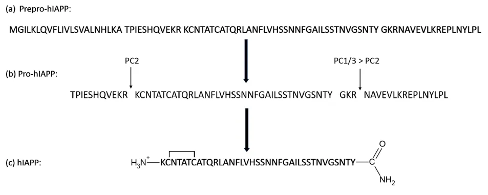
Figure 1: Processing of Prepro-hIAPP to produce mature hIAPP. (a) The primary sequence of the 89-residue Prepro-hIAPP. (b) Primary sequence of the 67-residue proform of hIAPP. Pro-hIAPP is cleaved by the prohormone convertases PC1/3 and PC2 at two conserved dibasic sites, indicated by arrows. The amidated C-terminus is produced after further processing by CPE/PAM. (c) The sequence of the mature 37-residue hIAPP. The biologically active peptide has an amidated C-terminus and a disulfide bridge between Cys-2 and Cys-7 [13].
Post‑translational processing of hIAPP has the same enzymatic mechanism as proinsulin, and perturbations in processing or trafficking increase the accumulation of incompletely processed, aggregation‑prone pro‑hIAPP intermediates [14,15]. Under chronic hyperglycaemia and insulin resistance, β‑cells markedly upregulate hIAPP expression, overloading ER and granule processing pathways, aggravating ER stress and autophagy defects, thereby promoting misfolding and aggregation in islets [14].
PHYSIOLOGICAL FUNCTIONS OF hIAPP
hIAPP is stored in secretory granules and is co-localized, co-packaged, and co-secreted with insulin in response to nutrient stimulation, particularly after ingestion of carbohydrate- and protein-containing meals [16,17]. The primary role of insulin is to promote glucose utilization and nutrient storage, but hIAPP complements these actions by regulating the rate of nutrient delivery into the circulation, suppressing inappropriate glucagon secretion, and controlling food intake. Through these coordinated effects, hIAPP contributes to the maintenance of postprandial metabolic homeostasis. [12,13,18]
Regulation of glucose homeostasis
One of the important physiological functions of hIAPP is the regulation of postprandial blood glucose levels. After nutrient intake, hIAPP is released together with insulin from pancreatic β-cells. It acts on pancreatic and extra-pancreatic targets to modulate glucose metabolism. In particular, hIAPP suppresses postprandial glucagon secretion from pancreatic α-cells, thereby limiting glucagon-mediated hepatic glucose production. This action complements the effects of insulin, which promotes glucose uptake and inhibits hepatic glucose output. Consequently, the coordinated actions of insulin and hIAPP help prevent excessive elevations in blood glucose after a meal. [12,13,19]
The effects of hIAPP on glucagon secretion depend on the experimental and physiological setting. Its inhibitory action is particularly relevant after a meal, when suppression of glucagon helps limit unnecessary hepatic glucose release. However, the regulation of glucagon secretion by hIAPP is complex, and its effects may differ according to glucose concentration, species, and experimental model. [12,13]
Regulation of gastric emptying
hIAPP also contributes to postprandial glucose regulation by slowing gastric emptying. After food intake, hIAPP delayed movement of gastric contents into the small intestine, thereby reducing the rate at which glucose and other nutrients enter the circulation. This action helps moderate the rapid increase in blood glucose that may occur after a meal and provides additional time for insulin-mediated glucose disposal. The inhibition of gastric emptying is mediated primarily through amylin-sensitive pathways involving the central nervous system and gastrointestinal control mechanisms. [13,19,20]
The physiological importance of this action is supported by studies of amylin deficiency and replacement. Reduced amylin activity may contribute to impaired postprandial glucose regulation, whereas pharmacological amylin analogues can improve glycaemic control partly through delayed gastric emptying. Thus, hIAPP acts as an important regulator of the rate of nutrient absorption and complements insulin in maintaining postprandial glucose homeostasis. [19,20]
Regulation of appetite and satiety
In addition to its effects on glucose metabolism, hIAPP functions as a neuroendocrine signal involved in the regulation of food intake and energy balance. Following nutrient ingestion, circulating hIAPP activates amylin-responsive neuronal pathways in the central nervous system, particularly within the hindbrain. The area postrema is an important site involved in the anorectic actions of amylin. Activation of these pathways promotes meal-ending satiety, reduces food intake, and contributes to the control of energy intake. [13,18,21]
hIAPP also interacts with other physiological signals involved in energy homeostasis, including insulin and leptin. These interactions may contribute to the integration of short-term meal-related signals with longer-term regulation of body weight and energy balance. The satiety-promoting effects of amylin have further stimulated the development of amylin-based pharmacological approaches for the management of diabetes and obesity. [18,21]
Interaction with insulin and other pancreatic hormones
The physiological actions of hIAPP are closely integrated with those of insulin and glucagon. The co-secretion of hIAPP and insulin following nutrient intake enables the pancreatic β-cell to coordinate glucose disposal with the regulation of nutrient delivery and hepatic glucose production. While insulin facilitates the storage and utilization of nutrients, hIAPP helps regulate the entry of nutrients into the circulation, suppresses postprandial glucagon release, and promotes satiety. This complementary hormonal relationship is essential for maintaining metabolic balance. [12,13,19]
hIAPP may also exert local regulatory effects within the pancreatic islets, including modulation of insulin and glucagon secretion. However, the detailed mechanisms and physiological significance of these intra-islet effects remain incompletely understood. Therefore, although hIAPP is recognized as an important regulator of glucose and energy homeostasis, some proposed actions require further investigation under physiological conditions. [12,13]
hIAPP Receptor Signaling
The physiological actions of hIAPP are mediated predominantly through amylin receptor (AMY) complexes, which belong to the class B G-protein coupled receptor (GPCR) family. Unlike a conventional receptor consisting of a single protein, functional amylin receptors are formed by association of the calcitonin receptor (CTR) with one of three receptor activity-modifying proteins (RAMP1, RAMP2, or RAMP3), generating AMY1, AMY2, and AMY3 receptors, respectively. The association of RAMPs with CTR increases the binding affinity and potency of amylin and modifies receptor trafficking and downstream signaling. Thus, the receptor composition contributes to the diversity of cellular responses elicited by hIAPP. [22-24]
Upon binding to AMY receptors, hIAPP activates intracellular signal-transduction pathways that regulate neuronal and metabolic responses. The best-characterized signaling pathway involves Gαs-mediated activation of adenylyl cyclase, resulting in increased intracellular cyclic adenosine monophosphate (cAMP) and subsequent activation of cAMP-dependent effectors such as protein kinase A. Amylin receptors can also activate other signaling pathways, including intracellular Ca²⁺ mobilization and extracellular signal-regulated kinase 1/2 (ERK1/2) phosphorylation. Importantly, the relative activation of these pathways depends on receptor composition and cellular context. In cellular studies, incorporation of RAMP1 or RAMP3 into CTR markedly enhanced amylin potency for cAMP generation, whereas the effects on Ca²⁺ mobilization and ERK1/2 activation were comparatively smaller, demonstrating that RAMPs can modify the signaling profile of amylin receptors. [23,24]
A major site through which circulating hIAPP exerts its central effects is the area postrema (AP) of the caudal hindbrain. The AP is particularly accessible to circulating peptides and contains amylin-responsive neurons. Activation of these neurons by peripheral hIAPP initiates neural signaling toward the nucleus of the solitary tract (NTS) and lateral parabrachial nucleus (LPBN), with subsequent communication with forebrain regions involved in the regulation of food intake and energy balance. [25,26] Through these neural circuits, amylin receptor activation contributes to meal termination and satiation and participates in the regulation of gastric motility and glucose homeostasis [25-27].
Amylin signaling also interacts with other metabolic regulatory systems. In particular, amylin has been reported to enhance the central actions of leptin and to influence neural pathways involved in food reward and energy expenditure [25]. More recent evidence indicates that amylin-responsive pathways extend beyond the classical AP–NTS circuit to include the hypothalamus, lateral parabrachial nucleus, midbrain, and other regions involved in energy homeostasis [27]. These findings indicate that hIAPP signaling is mediated by a distributed receptor-dependent neuroendocrine network rather than a single signaling pathway. [25-27]
MECHANISMS OF hIAPP AGGREGATION AND β-CELL TOXICITY
The aggregation pathway: from monomers to oligomers and fibrils
Under chronic hyperglycaemic condition, elevated hIAPP concentration, impaired post-translational processing, and membrane/lipid environments, the intrinsically disordered hIAPP monomer undergoes conformational conversion into partially structured, β-sheet-rich intermediates that nucleate amyloid assembly. Importantly, the most toxic species are not the large extracellular amyloid deposits detected by histochemical staining, but rather smaller intracellular nonfibrillar oligomers detected by conformation-specific antibodies [28]. Time-resolved biophysical measurements combined with cellular assays have revealed that cytotoxic hIAPP oligomers contain only modest β-sheet content, implying that agents developed to block cross-β fibril formation may inadvertently prolong the lifetime of toxic aggregation intermediates [29]. The aggregation pathway is therefore governed by a dynamic equilibrium between monomers, oligomers, and fibrils, and therapeutic interventions must consider which species they stabilize or destabilize. [7]
The hIAPP aggregation trajectory exhibits a sigmoid curve and is divided into three distinct phases: lag, log, and saturation. The lag phase begins when two structurally disordered monomers interact to produce a dimer, followed by the continuous addition of more monomers to form oligomers. This process is known as primary or homogeneous nucleation. Eventually, these oligomers serve as a seed for the formation of higher-order soluble oligomers and insoluble fibrils (Figure 2). Once fibrils are established, they facilitate the formation of new fibrils through the process known as secondary or heterogeneous nucleation. The elongation and growth of fibrils are accelerated in the log phase, wherein the oligomers undergo conformational transitions to form β-sheets. Finally, in the saturation phase, these β-sheets are organized as protofibrils and amyloid fibrils, i.e., partially ordered, fibrillar protein aggregates rich in the β-sheet structure in a characteristic cross-β conformation. [7
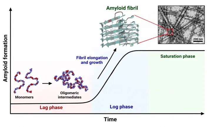
Figure 2: Proposed pathway of hIAPP amyloid aggregation. hIAPP monomers undergo self-assembly to form oligomeric intermediates, followed by fibril elongation and the formation of mature β-sheet-rich amyloid fibrils. The aggregation process proceeds through lag, log, and saturation phases.
Recent structural studies of hIAPP fibrils derived from the pancreas of patients with T2DM have provided the first patient-derived, atomic-resolution templates of the end-stage amyloid, which showed a distinct fold and unique ligand-binding features compared to in vitro-prepared fibrils, offering new opportunities for structure-based inhibitor design [8].
Membrane disruption and pore formation
A well-established mechanism of hIAPP toxicity involves the permeabilization of cellular membranes. IAPP oligomers form pore-like structures in lipid bilayers, resulting in ion leakage, uncontrolled Ca²⁺ influx, and dysregulated ionic homeostasis that terminates in cytotoxicity [28,30]. Lipid bilayers themselves promote aggregation: negatively charged phospholipids such as phosphatidylserine interact directly with the positively charged hIAPP monomer, and cell-surface heparan sulfate proteoglycans similarly enhance peptide aggregation [30]. In addition to discrete pore formation, physical studies demonstrate that the growth of rigid amyloid fibers attached to flexible membranes generates negative curvature strain, causing membrane distortion, detachment of membrane patches, and increased permeability to small solutes; such curvature stress may open mechanosensitive calcium channels that trigger an ER stress response and β-cell death [31]. Consistently, amyloid fibrils cluster on or near membranes, and exogenously added hIAPP disturbs cell membranes [32]. Oligomeric IAPP species also localize within disturbed mitochondrial membranes in transgenic hIAPP mice and in patients with T2DM, where unstable mitochondrial membrane potential drives overproduction of reactive oxygen species, further amplifying toxicity [33].
ER stress and proteostasis failure
The intracellular toxicity of hIAPP is closely linked to disturbances in protein homeostasis. Defects in ER-associated degradation and perturbations in pro-hIAPP processing, which normally proceeds through the ER, Golgi, and secretory granules, are proposed to be important factors in hIAPP-induced β-cell death, with incompletely processed pro-hIAPP representing a particularly aggregation-prone intermediate [32]. ER stress, proteasome impairment, and mitochondrial dysfunction have all been associated with hIAPP-induced toxicity, although ER stress is not consistently detected in islets producing IAPP at more physiological levels, and caution is warranted when extrapolating from strongly overexpressing transgenic models to human disease [32,33].
Mitochondrial dysfunction
Toxic hIAPP oligomers have been observed in association with the disruption of mitochondrial membranes, and exposure of cells to hIAPP aggregates has been linked to a loss of mitochondrial membrane potential and reduced ATP production. Given that mitochondrial activity is closely linked to energy metabolism and insulin secretion in pancreatic β-cells, impaired mitochondrial function can compromise both the function and survival of these cells. Consequently, mitochondrial damage may represent a significant consequence of hIAPP aggregation and contribute to the progression from initial cellular stress to β-cell dysfunction and apoptosis. However, the extent to which mitochondrial damage constitutes a primary event versus a consequence of other hIAPP-induced cellular alterations remains to be exactly determined. [14,30,32]
Oxidative damage and reactive oxygen species
Mitochondrial dysfunction may further contribute to β-cell injury by increasing the production of reactive oxygen species (ROS). Disruption of the mitochondrial membrane potential can promote excessive ROS generation, leading to oxidative stress and damage to cellular proteins, lipids, and other macromolecules. Pancreatic β-cells are particularly vulnerable to oxidative stress because their antioxidant capacity is relatively limited. In the context of hIAPP aggregation, oxidative stress may interact with membrane disruption and mitochondrial dysfunction, creating a cycle in which cellular damage further compromises mitochondrial function and promotes β-cell death. Experimental studies have linked hIAPP aggregate-induced toxicity with ROS production and activation of stress-associated signaling pathways, although the precise sequence of events connecting hIAPP aggregation, mitochondrial dysfunction, and oxidative damage remains unresolved. [30,32]
Collectively, these findings indicate that hIAPP aggregation damages β-cells through a multi-hit mechanism involving membrane permeabilization, calcium dysregulation, ER stress, mitochondrial dysfunction, and oxidative stress.
EMERGING INHIBITORS OF hIAPP AGGREGATION
Peptide-based inhibitors and mimetics
Because of the amyloidogenic propensity of hIAPP resides in specific sequence regions, peptides that interfere with self-association have attracted considerable interest. Non-amyloidogenic sequence variants of IAPP, such as rat IAPP, inhibit the fibril formation of human IAPP, and the efficacy of such peptides can be improved by engineering peptide mimetics with conformational restraints. These approaches exploit complementary binding to early aggregation intermediates, including the helical states that lead to β-sheet conversion to appropriate monomers and retard nucleation. [34]
Small molecules and natural polyphenols
Small-molecule polyphenols are among the most extensively studied hIAPP aggregation inhibitors. Epigallocatechin gallate, curcumin, and resveratrol inhibit aggregation and reduce IAPP-related cytotoxicity, with the added advantages of being naturally occurring and non-toxic at moderate concentrations [34-37]. Mechanistically, EGCG suppresses IAPP aggregation and toxicity when added during the lag phase, in which toxic intermediates form, and its effects can be potentiated by combination with zinc; morin hydrate inhibits both fibril formation and disassembles preformed IAPP fibers, while the sulfonated triphenylmethane derivative acid fuchsin and the diphenylpyrazole-derived compound anle145c generate non-toxic oligomeric species [38]. Plant-derived flavonoids inhibit the nucleation phase of hIAPP aggregation, and structural requirements for polyphenol activity have been mapped [11]. Furthermore, small-molecule modulators can tune the rate of hIAPP amyloid assembly in either direction, underscoring the need to select compounds that suppress rather than accelerate aggregation [9].
A critical warning is that molecules designed to block cross-β fibril formation may extend the lifetime of toxic oligomeric intermediates, whereas molecules that stabilize fibrillar structures reduce the population of toxic oligomers but increase the amyloid burden, so the therapeutic objective must be defined with respect to the specific toxic species targeted [29]. Moreover, many polyphenols and small molecules suffer from limited water solubility and consequently poor bioavailability and delivery efficacy, a major barrier to clinical translation [39].
Nanomaterials and nanochaperones
Engineered nanomaterials and multifunctional nanocomposites offer an alternative strategy, as their size, morphology, surface functionality, and physicochemical properties can be tailored to match amyloid assemblies [39]. Self-assembled nanochaperones have been shown to inhibit the aggregation of hIAPP associated with T2DM, acting as artificial chaperones that capture aggregation-prone species [10]. These approaches may overcome the bioavailability limitations of molecular inhibitors while providing multivalent, high-avidity interactions with amyloidogenic intermediates.
Antibody and nanobody based inhibitors
Another promising direction is the development of antibodies that specifically recognize the soluble oligomers formed during amyloid assembly. In transgenic mice expressing hIAPP, vaccination with stable oligomer preparations restored elevated fasting glucose to normal levels, increased insulin levels, reduced hIAPP accumulation, and raised IAPP-antibody serum titers; antibodies purified from immunized mice reduced IAPP cytotoxicity toward pancreatic β-cells in a dose-dependent manner, supporting both active and passive immunization strategies. [29] Nanobody technology offers additional flexibility: multivalent nanobody assemblies generated through Zn²⁺ coordination with histidine-tagged nanobodies have been shown to potently inhibit hIAPP aggregation [40]. The broader range of inhibitors includes natural products, small synthetic molecules, peptide mimetics, antibody-based agents, and supramolecular modulators; however, their clinical translation remains a major unresolved challenge [41].
AMYLIN-BASED PHARMACOLOGICAL THERAPIES
Beyond direct inhibition of hIAPP aggregation, restoration of amylin's physiological actions represents an approved and clinically validated therapeutic strategy. Pramlintide, a short-acting synthetic amylin analogue, is currently the only approved amylin-based medication and is used as an adjunct to insulin therapy in both T2DM and T1DM, improving postprandial and overall glycaemic control without increasing the risk of hypoglycaemia or weight gain. [42] Its glucose-lowering efficacy derives from amylin's physiological mechanisms: suppression of glucagon secretion and slowing of gastric emptying, which together reduce postprandial glucose excursions [43]. In long-term (6–12 month) clinical studies in insulin-treated patients, adjunctive pramlintide lowered HbA1c and produced significant, sustained weight loss, particularly in patients overweight at baseline [43,44]. Administered at 120–240 µg three times daily, pramlintide is generally well tolerated, with temporary nausea being the most common treatment-related side effect; preliminary trials in obese patients without diabetes showed weight loss of up to 8 kg after one year [45].
The field is now advancing toward next-generation agents that overcome pramlintide's short half-life. Long-acting acylated analogues such as cagrilintide, a non-selective amylin receptor agonist, produce dose-dependent reductions in food intake and body weight, positioning them as potential anti-obesity medications. [46] Strategies under investigation include enhancing amylin stability through PEGylation or glycosylation, developing oral formulations to improve patient convenience, and combining amylin agonists with GLP-1 receptor agonists exemplified by CagriSema, which shows promising results in both glucose regulation and weight management [42]. Such multi-hormonal approaches align with a phenotype-driven, precision medicine framework that moves beyond the traditional T1DM/T2DM dichotomy [42].
FUTURE PERSPECTIVES AND CONCLUSION
The recognition of T2DM as a protein misfolding disease has reframed hIAPP as a central and druggable mediator of β-cell failure. Whereas current therapies largely manage glycaemia after β-cell dysfunction is established, hIAPP-directed interventions offer the prospect of disease modification by preserving β-cell mass. [7, 14] Several priorities emerge for future research. First, the recent determination of patient-derived hIAPP fibril structures now enables rationally designed, structure-based inhibitors that target defined amyloid conformations [8]. Second, therapeutic strategies must discriminate between blocking toxic oligomer formation and stabilizing benign fibrillar states, since non-selective anti-amyloid agents may prolong the lifetime of the most toxic intermediates [29]. Third, the translational gap between potent in vitro inhibitors and clinically effective agents driven by bioavailability, specificity, and safety remains the central hurdle to clinical application [44]. Finally, integrated approaches that combine anti-aggregation agents with amylin-based therapies, incretin-based treatments, and lifestyle interventions could simultaneously address the metabolic and proteotoxic components of T2DM, while next-generation amylin agonists expand the therapeutic potential of this hormone across the obesity–diabetes field [21,42]. Ultimately, therapies that preserve the physiological actions of hIAPP regulation of postprandial glucose, gastric emptying, and satiety while selectively neutralizing its pathological aggregation hold the greatest promise for halting the progressive β-cell decline that defines T2DM.
REFERENCES


Saleeka Parveen K, Dr. Wilbee D. S, Human Islet Amyloid Polypeptide: From Physiological Hormone to Amyloid Pathology and Therapeutic Target in Type 2 Diabetes Mellitus, Int. J. of Pharm. Sci., 2026, Vol 4, Issue 10, 1913-1925. https://doi.org/10.5281/zenodo.23355591
 10.5281/zenodo.23355591