

We use cookies to ensure our website works properly and to personalise your experience. Cookies policy
Department of Pharmacy, Genba Sopanrao Moze College of Pharmacy.
Parkinson’s disease is a neurodegenerative disorder that involves the loss of dopaminergic neurons in the substantia nigra pars compacta. This loss leads to dopamine levels and worsening motor problems. Levodopa remains the symptomatic treatment for Parkinson’s disease. The usual way we give levodopa by mouth has many issues. Absorption can vary the liver can remove levodopa before levodopa works the body can break levodopa down in the bloodstream the blood level of levodopa drops and the timing of stomach emptying matters. These problems cause levodopa levels to swing, create wearing?off episodes and can trigger dyskinesia when levodopa is used for years. Nose?to?brain delivery is a non?invasive idea that could solve some of these problems. It uses the links between the nose and the brain through the olfactory and trigeminal pathways. By giving levodopa through the nose, we can skip the liver, gut and the BBB. May reach the brain faster and more efficiently. In this review we look at why nose?to?brain levodopa might work. We examine the different routes that levodopa can travel: olfactory, trigeminal, perineural and perivascular. We also discuss ways to formulate levodopa for intranasal use. These include systems, in?situ gels, nanoparticles, lipid?based nanocarriers, microspheres and permeation?enhancing systems. We highlight the benefits the challenges of making these formulations safety worries, how we test them and what preclinical studies have shown. Finally, we cover ideas such as hybrid formulations, functionalized nanoparticles, imaging?based evaluation and early clinical trials. Nose?to?brain delivery of levodopa could greatly improve the amount of levodopa that reaches the brain and may fix the limits of therapy by skipping the blood brain barrier in order to reach the brain. Still, we need carefully planned clinical studies to confirm how well it works what dose is best and whether it stays safe over long periods in patients, with Parkinson’s disease. Also explains the evaluation parameters in order to enhance the quality of the dosage form with more and standard efficacy.
Parkinson’s disease is a progressive neurological disorder that primarily affects movement but also brings on serious non-motor symptoms. It’s the second most common neurodegenerative disease after Alzheimer’s, and it becomes much more frequent as people get older. [1,2]A key sign of Parkinson’s disease is the gradual loss of dopamine-producing neurons in a specific area of the midbrain called the substantia nigra pars compacta. The loss of these neurons sharply cuts dopamine levels in the nigrostriatal pathway, throwing off the basal ganglia’s usual control over voluntary movement. One key histopathological sign is the buildup of misfolded alpha-synuclein into Lewy bodies—tiny clumps inside cells that show up clearly at autopsy and are a hallmark of the disease. The exact cause is still not fully clear, but current research suggests it’s the result of many factors working together—genetic risk (like mutations in SNCA, LRRK2, PARK7, PINK1, and PRKN), environmental triggers (including certain pesticides), problems with mitochondria, oxidative stress, and the natural aging of cells. [3,4]
Two main types of Parkinson’s disease:
a. Idiopathic Parkinson’s — usually develops later in life and progresses slowly. [5]
b. Secondary parkinsonism—or Parkinson-plus syndrome—is a complicated condition that stems from other issues like brain injuries or neurological diseases. [6]
1.PATHOPHYSIOLOGY OF PARKINSON’S DISEASE:
1. Dopaminergic neurons degeneration [7]
2. Alpha-synuclein clumping[8]
3. Mitochondrial dysfunction [9]
4. Inflammation and immune response[10]
5. Imbalance of neurotransmitters [11]
6. Impairment of brain circuitry [12]
7. Non-motor symptoms[13]
8. Clinical presentation [14]
9. Defective protein degradation[15]
10. Genetic factors [16]
1.1 Degeneration of Dopaminergic Neurons
As the disease is essentially a deficiency state with progressive loss of nigrostriatal dopaminergic neurons, the therapeutic rationale of PD treatment relies on restoring dopamine or its precursor (levodopa) in the brain. However, oral levodopa has poor and variable bioavailability due to first-pass hepatic metabolism and peripheral decarboxylation by DOPA decarboxylase, so only a small amount reaches systemic circulation, let alone the CNS. This pharmacokinetic limitation is among the primary rationales for exploring non-oral, direct-to-brain delivery routes such as intranasal administration, which bypasses first-pass metabolism entirely. [7]
1.2 α-Synuclein aggregation
Lewy body pathology spreads via connected neural pathways and is being studied recently as a bidirectional phenomenon for nose-to-brain transport. The same olfactory and trigeminal pathways involved in early alpha-synuclein propagation from the nasal mucosa into the brain (per the “dual-hit hypothesis” of PD inception) are increasingly being exploited therapeutically for intranasal drug delivery as they provide a natural anatomical conduit which bypasses the blood-brain barrier (BBB). [8]
1.3 Mitochondrial Disease
Impaired Complex I activity and oxidative stress in nigral neurons has generated a keen interest in delivering antioxidants and mitochondrial-protective agents (e.g. coenzyme Q10, mitoquinone) directly to the CNS. Inadequate BBB penetration limits conventional oral delivery of such agents, and nanoparticle-based and nose-to-brain carriers are being explored to improve CNS bioavailability of these adjunctive neuroprotective molecules. [9]
1.4 Inflammation and Immunity
The tight junctions of the BBB limit paracellular transport of systemically administered anti-inflammatory agents, but targeting chronic neuroinflammation involving activated microglia holds promise for anti-inflammatory therapeutics. This adds further justification for delivery systems using alternative transport routes. The intranasal route enables drugs to bypass the BBB through the systemic circulation and follow olfactory and trigeminal neural pathways into the CNS directly. [10]
1.5 Neurotransmitter Imbalance
Since dopamine itself cannot cross the BBB (it is given as its precursor, levodopa, which can), the pharmacokinetics of the delivery system directly dictates therapeutic window and symptom fluctuation. Conventional oral levodopa, when used long term, leads to pulsatile, non-physiological striatal dopamine levels, which are associated with motor fluctuations and dyskinesia. Sustained-release and site-directed delivery systems, such as in-situ forming implants and continuous nasal or subcutaneous infusion systems, are designed to produce more stable plasma and CNS drug concentrations, which more closely approximate physiological dopaminergic tone. [11]
1.6 Impairment of Brain Circuitry
Abnormal basal ganglia–thalamocortical oscillatory activity underlies motor symptoms and is highly sensitive to fluctuating dopaminergic stimulation — the "wearing-off" phenomenon and dyskinesias seen in advanced PD are linked to non-continuous drug delivery. This is a key pharmacological argument for delivery platforms offering zero-order or near-continuous drug release, such as implantable depot systems and nasal in-situ gels, which can reduce the peak-trough fluctuations responsible for circuit destabilization. [12]
1.7 Non-Motor Symptoms
Widespread pathology affecting olfactory, autonomic, and limbic structures contributes to symptoms like hyposmia and cognitive decline, which are difficult to manage with peripherally-acting drugs. Since the nasal mucosa is richly vascularized and neurally connected to the CNS, intranasal delivery is particularly attractive for targeting these centrally-mediated non-motor symptoms, offering a non-invasive route with rapid onset compared to oral administration, which is often complicated in PD patients by dysphagia and delayed gastric emptying. [13]
1.8 Clinical Presentation
The cardinal motor features (tremor, bradykinesia, rigidity) demand rapid and reliable symptom control, which is often compromised by delayed or erratic gastric absorption of oral levodopa in PD patients — a problem compounded by PD-associated gastroparesis. This has driven interest in delivery routes that bypass the GI tract entirely, including nasal, subcutaneous, and implantable systems, to achieve more predictable pharmacokinetics [14]
1.9 Defective Protein Degradation
Impairment of the ubiquitin-proteasome and autophagy-lysosomal systems is a target for gene therapy and biologic approaches (e.g., siRNA, antisense oligonucleotides targeting SNCA). Such macromolecules are especially BBB-impermeable and susceptible to degradation if given orally, making them strong candidates for novel delivery platforms nanoparticle encapsulation, nose-to-brain pathways, or implantable reservoir systems — that can protect the payload and enable direct CNS access. [15]
1.10 Generic Index
Monogenic and risk-associated genes (SNCA, LRRK2, GBA, PRKN) are informing an era of precision and gene-based therapeutics for PD, including CRISPR and RNA-based strategies. These therapies require highly targeted, protected delivery to CNS tissue, further reinforcing why novel drug delivery system (NDDS) research — including implantable and intranasal platforms — is central to the future of PD management, moving beyond symptomatic oral therapy toward targeted, sustained, and disease-modifying delivery. [16]
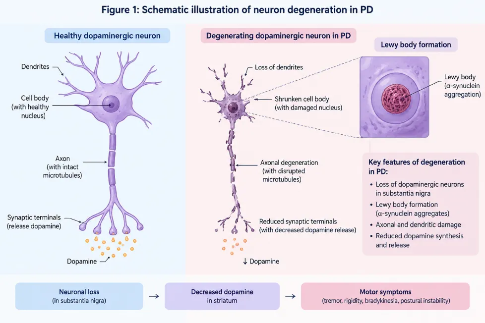
Figure 1: Neuronal Degeneration in Parkinson’s Disease
2. CONVENTIONAL LEVODOPA THERAPY AND ITS LIMITATIONS:
Levodopa is the natural building block for dopamine. It helps the body make and release dopamine in a controlled way. This allows dopamine to work properly at both -synaptic and post-synaptic receptors covering all the normal functions of dopamine—both the constant background activity and the sudden bursts. This is important because in Parkinson’s disease the brain loses dopamine-producing neurons. [17]
Over time as more neurons die, the remaining ones in the pathway and other monoamine systems become less able to make and store dopamine. Because of this some researchers thought patients would no longer have the lasting response, to levodopa—known as the long-duration response (LDR).. New studies suggest that the LDR actually starts with the very first dose and can last for years. Still even if the LDR stays motor fluctuations begin to interfere with how levodopa works. These fluctuations. Go, making treatment less predictable. [18]
Another problem is how levodopa gets into the brain. When the stomach empties slowly or when food contains amounts of neutral amino acids it can block the absorption of levodopa from the gut. Also many amino acids compete with levodopa for the transport system—the L1 transporter—that moves things across the blood-brain barrier. So even if levodopa reaches the blood it may not get into the brain efficiently. This makes the delivery of levodopa pulsatile which adds to the symptoms and side effects. [19]
Studies have shown that levodopa wearing-off is closely associated with reduced plasma levodopa levels, while levodopa-induced dyskinesia (LID) is linked to fluctuations in drug levels. Pulsatile dopamine receptor stimulation from conventional levodopa therapy may cause basal ganglia changes that contribute to dyskinesia. Therefore, continuous and sustained levodopa delivery may help reduce wearing-off and potentially lower the risk of LID. [20]
2.1. Modifying dosing strategies to manage motor complications
Standard levodopa, taken together with benserazide or carbidopa lasts about ninety minutes. Because of this half‑life doctors normally prescribe levodopa three times a day. As a result the levodopa blood levels. Fall in a predictable pattern of peaks and troughs each day. [21,22]The reason for this pattern is not fully. The levodopa level swings do not usually show up as changes in motor function until many years after levodopa treatment begins.
When levodopa response fluctuations appear the simplest and most common approach is to give levodopa often shortening the time between doses. Moving from three levodopa doses per day to four may look easy. It is hard to keep patients on the new schedule [23,24] and it only helps for a short time. After that levodopa must be given more often which raises the risk of levodopa‑induced dyskinesia (LID). A possible fix for this problem is to use devices that allow levodopa to be taken every hour or so which can smooth out levodopa blood level swings [25]. In a study most patients liked the five‑dose dispenser [26] but it is unclear whether this is practical for many people and it does not solve the problem of irregular stomach emptying. Raising the levodopa amount often makes LID worse and does not lengthen the period of symptom relief. Also the levodopa dose itself is a risk factor for LID. Research shows that levodopa should stay below 400 milligrams per day for long, as possible [27,28].
2.2. Modifying the pharmacokinetics and delivery of levodopa through new formulations and delivery systems
Attempts to create levodopa formulations that last longer in the bloodstream and provide extended effects from oral doses began in the 1980s [29]. But today it has not been proven that the extended-release versions of levodopa are better than the standard immediate-release ones—this includes their use at night [30].
Early pilot studies where levodopa was given through infusion showed that continuous delivery could help patients with motor fluctuations. This idea was proven effective back in the 1980s [31,32]. Still it took two more decades before enteric levodopa delivered through a gastro-jejunostomy became part of regular treatment [33]. The use of levodopa/carbidopa gel infusions (LCIG) has been shown to work well in reducing OFF time with a treatment difference of −1.91 hours compared to immediate-release levodopa [34]. More recent data also shows that LCIG helps reduce dyskinesia than optimized oral treatment [35]. A new intestinal formulation that includes entacapone is now available in Europe [36]. This method is mostly limited to advanced disease stages because it requires abdominal surgery like the placement of PEG tubes. It also comes with problems such as tube dislocation, blockage and infection.
A invasive option is subcutaneous delivery of levodopa. Three formulations—ABBV-951, ND0612 and DIZ 102—are currently being tested in trials . Both ABBV-951 and ND0612 have shown results in reducing OFF time. The Phase 3 randomized controlled trial of ABBV-951 showed an increase of 1.75 hours in ON-time without dyskinesias compared to optimized oral therapy.
Another challenge for patients with motor fluctuations is quickly reversing OFF episodes. Oral levodopa has not been effective for relief. An inhaled levodopa formulation CVT-301, was developed to deliver the drug into the lungs using a small handheld inhaler. In a blind placebo-controlled Phase 3 study UPDRS motor scores improved significantly at 30 minutes, after dosing with a treatment difference of −3.92 points compared to placebo. The drug has now been approved for use by both the FDA and EMA. [37-39]
3. Rationale for the Nose-to-Brain Route:
1. Bypasses hepatic first-pass metabolism. –
Oral levodopa is broken down a lot in the gut wall and liver before it reaches the bloodstream. That is why doctors give it together with a decarboxylase inhibitor. Nasal absorption skips the circulation completely saving more of the drug for the whole body and the brain [40].
2. Avoid dependence on gastric emptying-
Levodopa is taken up in the upper part of the small intestine. Therefore how fast the stomach empties matters a lot. In Parkinson’s disease the stomach often empties slowly because of Lewy bodies in the gut. Because levodopa itself slows motility. This causes delays and missing responses. Nasal delivery bypasses this problem [41].
3.Direct CNS access via olfactory/trigeminal nerves-
The nose’s olfactory and trigeminal nerves give a route straight to the brain and cerebrospinal fluid avoiding the blood‑brain barrier. In rat studies intranasal levodopa reached the brain better than oral levodopa.[42].
4.Reduced peripheral exposure and side effects-
If some of the levodopa dose goes to the brain the body does not need a large amount of drug. A smaller overall dose can still give the brain effect and this can lower side effects such, as nausea and low blood pressure when standing. [43]
5.Fast, non-invasive, self-administrable rescue option-
Nasal delivery starts quickly. Does not need an injection or swallowing. It fits well for treating sudden 'OFF' moments. These moments are usually handled with apomorphine or inhaled levodopa. [43]
4. Anatomy and Physiology of the Nasal Cavity:
The nasal cavity is the part of the breathing system. It opens to the outside through the nostrils. Connects to the nasopharynx through openings called the choanae. A middle wall, called the septum splits it into two sections. Each side has a roof, a floor, a medial wall and a lateral wall. These cavities are divided into three areas: the vestibule, the respiratory region and the olfactory region. Around the nasal cavity there are four sinuses—frontal, ethmoid, maxillary and sphenoid. Each of these drains into the cavity through small openings known as ostia.
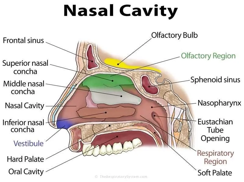
Figure 2: Nasal Cavity
5. Mechanisms of Nose-to-Brain Drug Transport:
The nose to brain delivery method uses the cavity direct connections to the central nervous system to go around the blood brain barrier. Many different ways work together. How much each one contributes depends on the drugs features and how it’s made and where in the nose the dose lands.
Main ways: the olfactory nerve pathway, the nerve pathway, movement through the spaces around cells flow along blood vessels getting into the blood and then crossing the barrier and moving through the lymph system.
5.1 Olfactory Pathway
Olfactory sensory nerves in the part of the nose send signals through a small bone called the cribriform plate directly to the olfactory bulb creating a direct link between the nose and the brain.
Inside the cells: when drugs are taken up by these nerves through pockets or fluid intake they travel along tiny tubes to the olfactory bulb and then to other parts of the brain through
connections between nerve cells. This path is slow. Taking hours to days.. It's more important for long-term movement than quick effects.
Outside the cells: this is the faster way. Drugs move through spaces between cells or around the nerves reaching the inner layers of the nose and then the fluid around the brain. Often within minutes. This doesn't need the nerves to take the drug just movement through the spaces around the nerves and the lymph system.
Fluid movement: once the drug reaches the fluid around the brain the flow of this fluid carries it widely through the brain especially for small, stable and fluid-permeable medicines.
5.2 Trigeminal Pathway
The trigeminal nerve controls parts of the nose through its eye and jaw branches sending signals to the brainstem and the pons. When drug is taken up at the ends of these nerves it can reach the areas through movement through spaces (main and faster) or through the inside of the nerve cells (slower). Unlike the olfactory route this path helps get to the parts of the brain. The brainstem and spinal cord. Expanding the overall coverage in the central nervous system.
5.3 Perineural and Perivascular Flow
After getting past the layer the drug can move through fluid-filled spaces around nerves into the fluid around the brain or move along the spaces around blood vessels helped by the beating of the arteries. This flow helps spread the drug evenly through the brain than just using the nerve paths.
5.4 Blood and Lymph Paths
Some of any dose given through the nose is taken into the blood through the blood- layers of the nose; from there it may still reach the brain by crossing the blood brain barrier normally but its slower and less precise. A second option. The noses lymph system gives another way carrying the drug to the neck and eventually into the fluid around the brain.
5.5 Role of Nanocarriers
Materials made of plastic or fat protect the drug from being broken down help it get through the nose layers and can move along the path while letting the drug out as they go. Sticky or charged surfaces on the particles help them stay in the area of the nose and increase the chances of moving through the spaces around the nerves; small groups of molecules and fat particles have shown better brain movement through better movement and steady release.
5.6 Time and What Controls It
Drug being found in the olfactory bulb or fluid around the brain within minutes of taking the dose shows that movement outside the cells is the way for quick effects, with the inside of the cells contributing more to slower longer-lasting movement. Which way is most important for each version of the drug depends on the size and fat-solubility of the drug how it’s made (nanocarriers, materials) and exactly where in the nose it lands. Small fat-soluble medicines move outside the cells easily while bigger or particle-based medicines usually need helper systems to get into nerve paths efficiently. [50-52]
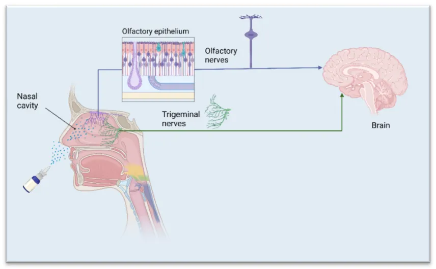
Figure 3: Drug Transport from Nose to Brain
6. Formulation Strategies for Nasal Levodopa Delivery:
Plain aqueous levodopa solutions clear quickly from the nasal cavity within 15 to 20 minutes because of mucociliary clearance. They also do not go through the nasal lining well. Therefore many new ways have been tried to keep levodopa in the nose improve how levodopa goes through the nasal lining and raise levodopa levels in the brain.
6.1 Mucoadhesive Systems
Mucoadhesive polymers, for example chitosan, carbopol and sodium alginate stick to the nasal mucosa. They form bonds with mucin, which slows mucociliary clearance and lets levodopa stay in contact with the nasal lining longer. Chitosan also helps levodopa cross the lining by briefly opening junctions between epithelial cells. [53]
6.2 In‑Situ Gelling Systems
These products come out as thin liquids that turn into gels when they touch the nasal mucosa. The gel forms because of a change in temperature (in poloxamer systems) pH (in chitosan systems) or salt level (in gellan gum). The gel stays in the nose like a semi‑solid while still being easy to spray giving levodopa a time to be absorbed. This type of formulation fits the discussion of in‑situ forming implants. [54]
6.3 Nanoparticulate Carriers
Scientists have tested nanoparticles such as PLGA and chitosan solid lipid nanoparticles (SLNs) nanostructured lipid carriers (NLCs) and liposomes for delivering levodopa through the nose. These tiny carriers shield levodopa from enzymes slow its release and sometimes help levodopa move across or between cells by cell uptake. This can give levodopa to the brain directly.
6.4 Microspheres and Powders
When dry powder or microsphere formulations touch mucus they swell. This swelling keeps levodopa in the nose longer. It also protects levodopa from oxidation which can happen in water.
6.5 Permeation Enhancers
Researchers have tried cyclodextrins, bile salts and surfactants to help levodopa move through and, across the nasal lining. These additives can raise levodopa’s permeability. They can also irritate the nose or harm the tiny hairs that clear mucus if used too often. [55]
Table 1: Comparison of Nasal Formulation Strategies for Levodopa Delivery
| Formulation Type | Key Excipients | Primary Advantage | Key Limitation |
|---|---|---|---|
| Aqueous solution/spray | None / simple buffers | Simple, low-cost, easy to manufacture | Rapid mucociliary clearance; low bioavailability |
| Mucoadhesive solution/gel | Chitosan, carbopol, alginate | Extended mucosal residence time | Variable adhesion under mucus turnover |
| In-situ gelling system | Poloxamer 407, gellan gum | Liquid on administration, gels in situ | Formulation stability; gelation reproducibility |
| Polymeric nanoparticles | PLGA, chitosan NPs | Sustained release; enzymatic protection | Complex manufacture; scale-up challenges |
| Lipid nanocarriers (SLN/NLC) | Solid/liquid lipids, surfactants | Good olfactory epithelium affinity | Physical stability on storage |
| Liposomes | Phospholipids, cholesterol | Biocompatible; can co-encapsulate enhancers | Cost; limited drug loading for hydrophilic drugs |
7. Factors affecting Nasal Drug Absorption:
1. Physiological Factor
1.1. Mucociliary clearance:
Nasal mucociliary clearance is a function needed to keep the nose healthy and protected. This process is responsible for moving drugs that are given through the nose from the nose to the back of the throat quickly. This process can stop the drug from being absorbed after it is given through the nose. The mucociliary clearance system moves the drug backward in the nose and down into the throat. This happens because of the movement of hair-like structures called cilia on the cells in the breathing area. The mucociliary clearance system is often called a " belt." The ciliated cells act as the force that moves things and the mucus acts as the belt that collects and removes foreign objects. The absorption of drugs from the inside of the nose is affected by how the drug stays on the tissue. Nasal mucociliary clearance limits how long the drug can stay in the nose, which makes time for the drug to be absorbed. The normal time it takes for the body to clear things in humans is 20 minutes. Nasal mucociliary clearance is changed by the drugs and other ingredients used in the nasal medicines. Drugs that act like acetylcholine and methacholine which are called agonists can increase cilia movement in a certain way in living things and in lab tests. Medicines like salmeterol and isoprenaline which're beta-adrenergic agonists were found to stop the decrease in the movement of cilia in human nose tissue in lab tests caused by something that harms the cilia. [56]
1.2. Diseases like infection, allergy and nasal inflammation:
Conditions such as the cold rhinitis, atrophic rhinitis and nasal polyps are usually linked with problems in the way the mucus moves, too much or too little mucus and irritation of the inside of the nose. These things can change how a drug moves through the nose. Inflammation in the nose can change how a drug is absorbed. Some studies show that how a drug works in the body is not changed by inflammation in the nose. For example zolmitriptan and butarphenol.
1.3. The conditions in the nose like temperature and humidity:
The blood vessels in the inside of the nose help to control the temperature and make the air wet. This can affect how a drug is absorbed.
1.4. The speed of mucus flow and blood flow in the nose:
The front part of the nose, including the area near the nose and the start of the nose structures has surfaces without cilia. The mucus moves here at a slow rate of 1 to 2 mm per hour. The main parts of the nose have a lot of blood vessels and cilia. The mucus moves here at a rate of 8 to 100 mm per minute. It is thought that in this area, where there is surface area, the most air resistance and the most cilia the main drug absorption happens. The inside of the nose has a lot of blood vessels. The high number of blood vessels makes it a good place for drug absorption. The blood. Therefore the absorption of the drug depends on whether the blood vessels get bigger or smaller. The blood flow in the nose is changed by things like the temperature and humidity around the presence of drugs, injuries and inflammation. Also emotions like fear, anxiety and frustration can change the blood flow. The blood flow in the nose is sensitive to chemicals. This is true for both drugs that work in the nose and those that work in the body. These changes are important for how the drug's absorbed through the nose because they change blood flow. [57]
1.5. The breakdown of drugs by enzymes:
One of the problems for drugs that go through the nose is the enzymes that are in the nose. These enzymes can change how the drugs move through the nose. There are enzymes in the nose like aldehyde, which is part of the cytochrome P-450 group. Other enzymes like amino peptidases can break down proteins and peptides. Carboxylesterases are another type of enzyme that's very active, in the nose lining. The enzyme called cytochrome P-450 monooxygenase is known to change many different substances. It breaks down drugs in the nose like nasal decongestants, nicotine, cocaine, phenacetin and progesterone.
2. Dosage form factors
The way a drug behaves physically and chemically affects how much of it is taken in and how things like how well it mixes with fat, its acid level, how big it is and how fast or how much fluid flows through the nose all have an effect.
2.1 Size of the molecule
When the molecule is bigger than 1000 Daltons absorption drops a lot. When it’s under 300 Daltons the size doesn't matter much. These small molecules pass easily through the water-filled channels in the membrane.. When it’s over 300 Daltons how big it is becomes more important.
2.2 Chemical shape
Changing a drug into a salt or an ester can change how it’s taken in. For example making an ester from L-tyrosine greatly improved how much of it was taken in through the nose compared to the version. [58]
2.3 Particle size
Particles bigger than 30 micrometre are mostly removed by the movement in the nose and the tiny hairs. A lot of particles down to 12 micrometer also get removed. Particles over 10 micrometer tend to stay in the nose while those under 1 micrometer just get blown out.
2.4 Soluble nature
Forms of the drug that are not charged and more fat-soluble are usually absorbed best through the nose although charged forms still get some absorption. The pH of the nose (4.5 to 6.5) affects how much the drug is charged and most drugs are weak acids or bases. This same pH range also helps the natural bacteria fighter called lysozyme work which helps protect against germs. If the pH becomes more basic lysozyme stops working increasing the chance of infection and making it harder for the nose to clear out the drug and for the drug to get absorbed.
2.5 Blood flow rate
In studies with phenobarbital more absorption happened as blood flow increased until it stopped increasing. After a point more blood flow didn't lead to more absorption.
3. Administration factors
Where and how the medicine ends up in the nose has an impact on how much is taken in. The way particles land is mainly controlled by how they hit the walls (the way) by settling down and by moving around. Particles that are bigger than about 50 micrometers in size don't even get into the nose. It's best to have an amount of drug in a small amount of medicine (under 200 microliters). The type of device (drops, mist, spray or measured dose) and how its placed in the nose both affect where the dose ends up. Landing in the part of the nose usually gives longer time to stay and better absorption; landing in the front area or the back of the nose (where the drug might get swallowed) should be avoided. Devices that spray or mist are usually the most accurate. How they are stored and the thickness, how much the surface sticks together and how well the medicine is mixed can still affect how well they measure the dose. [59]
4.Other factors
How much the amount of the drug varies depends on the size of the molecule. Drugs with molecular weight usually have good low-variability absorption through the nose. Often similar to when they are taken by mouth or injected. While drugs like proteins and peptides often have lower absorption and more variation through this route.The way the medicine is delivered changes results. In a study with rabbits a quick-release progesterone nasal spray made the blood level peak in 2 minutes while a slow-release device made the level rise slowly to a peak over 20 to 30 minutes. With 82.5% absorption for the spray and 72.4%, for the slow-release device showing that the device itself can change how fast and how much is absorbed. [60]
8. Evaluation Parameters for Nasal Formulations:
Development of a nose-to-brain levodopa formulation requires an evaluation program spanning physicochemical ex vivo and in vivo assessment:
8.1 In Vitro / Physicochemical Evaluation:
• Viscosity and rheological behaviour including sol-gel transition temperature for in-situ gels
• Gelling capacity, mucoadhesive strength texture analyzer studies
• Particle size polydispersity index and zeta potential for systems
• Drug content uniformity and entrapment encapsulation efficiency
• In vitro drug release studies e.g dialysis bag or Franz diffusion cell methods [61]
8.2 Ex Vivo Evaluation:
• Nasal mucosal permeation studies using excised sheep goat or porcine nasal mucosa in modified Franz diffusion cells
• Histopathological assessment of mucosal tissue to evaluate irritation potential and ciliotoxicity [62]
9.3 In Vivo Evaluation:
• Pharmacokinetic studies in rodent models comparing brain and plasma drug concentrations following intranasal versus intravenous administration to calculate the Drug Targeting Efficiency DTE% and Direct Transport Percentage DTP% the standard metrics used to quantify the extent of direct nose-to-brain transport
• Pharmacodynamic behavioural studies, in PD animal models e.g 6-hydroxydopamine or MPTP-lesioned rodents assessing motor function recovery rotarod apomorphine-induced rotation tests
• Nasal ciliotoxicity and histopathological safety studies following repeated dosing. [63]
9. Advantages of Nose-to-Brain Levodopa Delivery:
1] Levodopa bypasses the liver’s first‑pass metabolism, which can improve how much levodopa reaches the brain and may lower the dose needed.
2] Levodopa does not rely on stomach emptying or gut absorption both of which are often weakened in Parkinson’s disease.
3] Levodopa can target the nervous system directly through the nose and trigeminal nerves, which cuts down on body‑wide exposure and side effects outside the brain.
4] Levodopa’s effect starts quickly making it good for sudden "off" episodes.
5] Levodopa is non‑invasive. Can be used by patients themselves which helps them follow treatment and stay independent a big help when Parkinson’s makes movement and thinking harder.
6] With a slow‑release design levodopa may keep dopamine levels steadier. Help cut down motor problems. [64]
10.Challenges and Limitations:
1] The small space inside the nose limits how much levodopa can be given each time which can be a problem because Parkinson’s patients often need doses for good symptom control.
2] The nose’s quick mucus‑clearing action shortens the time levodopa can stay in place unless special sticky or gel formulations help keep it
3] Enzymes in the nasal lining can break down levodopa lowering the amount that stays whole for absorption.
4] Differences between people and within the same person over time in nose shape, mucus amount and sense of smell—often weakened in Parkinson’s—can change how well levodopa gets delivered.
5] Repeated long‑term use, with substances that help levodopa cross membranes can irritate the nasal lining or damage tiny hair cells.
6] Moving from animal studies to people is uncertain because rodents have bigger nose lining than humans making it hard to predict how much levodopa will reach the brain in real life.
7] Making and approving nanoparticle or gel systems is complex with hurdles, in scaling up production and keeping the product stable over time. [65]
11. Preclinical and Clinical Evidence:
There is increasing evidence from laboratory studies that supports the idea of delivering levodopa and other dopaminergic drugs to the brain through the nose. Research in animal models rodents has used formulations like mucoadhesive and nanoparticle-based intranasal drugs. These studies have shown that brain levels of the drug are much higher compared to blood levels. In cases these formulations also led to faster or better improvement in movement symptoms in animals with brain damage that mimics Parkinson’s disease. This suggests that drugs can reach the brain effectively when given through the nose bypassing the usual barriers that slow down absorption when taken by mouth or through a vein.
In humans the use of delivery for dopaminergic agents is not as widely studied as with levodopa. However there are some trials involving apomorphine, a direct dopamine agonist, given through the nose. These trials were aimed at treating sudden worsening of symptoms, known as "off" episodes in Parkinson’s patients. The results showed that the nasal route can produce meaningful relief proving that the nose-to-brain path can work in real patients. This clinical experience, along with data from animal studies on how nasal levodopa is absorbed and how it works in the body has led to renewed interest in creating an optimized nasal levodopa product. Still more quality clinical trials, with human participants are needed to fully understand its safety, effectiveness and long-term benefits. [66]
12.Future Directions:
1] Development of formulation platforms that combine mucoadhesive, in‑situ gelling and nanoparticulate strategies to simultaneously improve residence time, permeation and sustained release.
2] Application of nanoparticles—such as particles modified on their surface with cell‑penetrating peptides or lectins—to further boost selective olfactory epithelial uptake.
3] Use of neuroimaging for example radiolabelled tracer studies in subjects to directly confirm and quantify nose‑to‑brain transport instead of relying only on plasma or cerebrospinal fluid surrogate measures.
4] Combination approaches that pair levodopa for rapid rescue with existing continuous delivery systems, such as subcutaneous foslevodopa/foscarbidopa infusion to manage motor fluctuations comprehensively.
5] Well‑designed randomized controlled trials to establish efficacy, dosing and long‑term nasal safety, in the Parkinson’s disease patient population specifically who may have altered nasal or olfactory physiology that affects formulation performance. [67]
CONCLUSION:
The challenges of oral levodopa treatment first-pass metabolism, unpredictable absorption in the stomach and intestines and a short time in the body leading to uneven dopamine levels in the brain remain major issues in treating Parkinsons disease over the long term. The nose to brain method provides a non-invasive way to deal with some of these problems at once. This method uses the connections in the nose to the brain through the olfactory and trigeminal nerves. It might help avoid the liver and the blood brain barrier. Improvements in in-situ gelling and nanoparticulate methods have made this way of delivering the drug more possible. This is especially important for a drug like levodopa that's water-loving and does not stay in the body long. Although early research is positive and there is some history of using the nose to give dopamine drugs more human studies on nose to brain delivery of levodopa are needed. These studies are important to move this way of delivering medicine from research to actual patient care. With ways to make the drug and more proof from human tests the nose to brain method for giving levodopa could make treatment easier and better for people, with Parkinsons disease.
REFERENCES




Janvi Kakde, Ashish Kalambate, Sakshi Kakade, Sushma Nakhate, Tushar Shelke, Nose-to-Brain Delivery of Levodopa: A Nasal Route Strategy for Parkinson's Disease, Int. J. of Pharm. Sci., 2026, Vol 4, Issue 10, 2036-2051. https://doi.org/10.5281/zenodo.23357945
 10.5281/zenodo.23357945